Embed Size (px)
Citation preview

POSEIDO. 2014;2(2) Leukocytes populations in L-‐PRP solutions
117
ISSN 2307-5295, Published by the POSEIDO Organization & Foundation
under a Creative Commons Attribution-NonCommercial-NoDerivatives 4.0 International (CC BY-NC-ND 4.0) License
Research article Analysis of the Leukocytes in peripheral blood and Leukocyte- and Platelet-Rich Plasma (L-PRP) in rats: A flow cytometry study Agata Cieslik-Bielecka,1,* Piotr Paczek,2 Lukasz Sedek,3 Aleksandra Szantyr,4 Rafał Skowroński,5 Hom-Lay Wang,6 and David M. Dohan Ehrenfest.7 1 Department of Maxillofacial Surgery, Trauma Center Hospital, Sosnowiec, Poland 2 Department of Cardiology, Trauma Center Hospital, Sosnowiec, Poland 3 Department and Clinic of Pediatric Hematology and Oncology, Medical University of Silesia, Zabrze, Poland 4 Department and Clinic of Orthopaedics, Medical University of Silesia, Sosnowiec, Poland 5 Orthopedic Surgery and Traumatology Department, Medical University of Białystok, Białystok, Poland 6 Department of Periodontics and Oral Medicine, The University of Michigan, School of Dentistry, Ann Arbor, Michigan, USA 7 LoB5 research unit, School of Dentistry & Research Center for Biomineralization Disorders, Chonnam National University, Gwangju, South Korea *Corresponding author: Agata Cieslik-Bielecka, [email protected] Submitted April 26th, 2014; accepted after minor corrections on May 13th, 2014.
Abstract Background and objectives. Platelet concentrates for surgical use were often tested as surgical adjuvants in the literature, as a source of platelet growth factors to stimulate healing. Many products are often regrouped under the generic and inaccurate term of Platelet-Rich Plasma (PRP). However, what is tested in many studies is usually a combination of platelets and leukocytes (accurately termed Leukocyte- and Platelet-Rich Plasma – L-PRP). The quantity and impact of leukocytes in these preparations were not yet accurately investigated. In this article, the characteristics of white blood cells in a L-PRP obtained from rats were investigated, in order to point out the main actors and some of the mechanisms that may influence the properties of the platelet concentrates. Materials and Methods. Blood and platelet concentrate samples were obtained from 64 healthy Wistar rats and leukocyte phenotypes were identified using flow cytometry after labeling leukocytes for CD3, CD4, CD8, CD11bc, CD18, CD25, CD27, CD28, CD45R, CD45RA, CD80, CD90, CD106 (VCAM-1), CD161a and TCRab, TCRgd, RT1B with fluorochrome-conjugated antibodies. Results. The results have shown that the tested L-PRP contained substantial amounts of leukocytes of many different kinds, particularly T lymphocytes, B lymphocytes, NK cells, monocytes, granulocytes and eosinophils. Discussion and Conclusion. To highlight the various ways in which these cells can influence their environment will help to better understand the complex interactions of the PRPs with the tissues. This identification of the exact cell content and the understanding of this complex cell equation are important steps towards using these blood concentrates in the best possible way, as a reliable therapeutic option to promote better healing, particularly in infected surgical or wound sites. Keywords. Blood platelets, infection, leukocytes, platelet-rich plasma, wound healing.

118 Research article: Cieslik-‐Bielecka A, et al. (2014)
ISSN 2307-5295, Published by the POSEIDO Organization & Foundation
under a Creative Commons Attribution-NonCommercial-NoDerivatives 4.0 International (CC BY-NC-ND 4.0) License
1. Introduction Platelet concentrates for surgical use represent a relatively novel inductive therapy that could be valuable to accelerate and improve healing processes [1-3]. The general concept of these technologies is to concentrate the platelets and their many growth factors, to inject them to stimulate healing and hopefully to promote tissue regeneration. All these techniques are using centrifugation of whole blood, in order to reach high concentrations of platelets and growth factors [2,4]. Many products are often regrouped under the generic and inaccurate term of Platelet-Rich Plasma (PRP)[5]. Another family is termed PRF (Platelet-Rich Fibrin), when the platelet concentrate was designed and only exists under a strongly polymerized fibrin gel form [6,7]. However, in the vast literature on these technologies, it is often neglected that many platelet concentrate technologies collect also a significant amount of leukocytes [8,9]. These cells have a strong direct impact on healing and also produce many molecules including large amounts of growth factors [10]. For this reason, a more accurate terminology was proposed and 4 families of products were suggested [5,6,11,12]. Two of these 4 main families of platelet concentrates contain higher concentration of leukocytes compared to the amounts of these cells found in peripheral blood: these are termed Leukocyte and Platelet-Rich Plasma (L-PRP) and Leukocyte and Platelet-Rich Fibrin (L-PRF) [6]. In fact, these two families with leukocytes are the most frequently used platelet concentrates in many fields of medicine [3,6,13-15]. There is still a limited number of studies concerning the detailed composition of leukocytes and their role in these products, and these investigations were mostly done on L-PRF [7,16]. Despite a wide spectrum of available diagnostic techniques, we have not found animal and clinical studies identifying extensively the characteristics of white blood cells (WBC) in the various types of L-PRP. Therefore, the objective of this study was to investigate the detailed characteristics of white blood cells in a L-PRP obtained from rats, in order to point out the main actors and some of the mechanisms that may influence the properties of the platelet concentrates.
2. Materials and methods 2.1. Preparation of L-PRP The study group consisted of 64 healthy male Wistar rats. The Silesian Medical
University Bioethics Committee approval was obtained. The rats were anaesthetized with Ketamin (10 mg/kg) after Diazepam (0.1 mg/kg) premedication. For the study, 3.5±0.1 ml of whole blood were collected directly from the heart into a syringe containing 0.7 ml of sodium citrate 105 mmol/l. 4.0±0.1 ml were drawn into a sterile tube and centrifuged for 10 minutes at 1000 RPM (Janetzki K23, Berlin, Germany). This resulted in blood separation into its three basic components: red blood cells, L-PRP sometimes referred to as “buffy coat”, and leukocyte- platelet-poor plasma (L-PPP). Subsequently, L-PPP and L-PRP were removed into 5 ml syringes and centrifuged for 10 minutes at 3000 RPM. After centrifugation, supernatant was removed and 600±50µl L-PRP was obtained. Next, samples with whole blood and L-PRP were examined.
2.2. Cell preparation and flow-cytometric analysis Blood and L-PRP samples were processed under standardized and optimized
conditions within less than 4 hours after collection. The antibody set was designed to identify all major leukocyte populations as well as different lymphocyte subpopulations. For this
POSEIDO. 2014;2(2) Leukocytes populations in L-‐PRP solutions
119
ISSN 2307-5295, Published by the POSEIDO Organization & Foundation
under a Creative Commons Attribution-NonCommercial-NoDerivatives 4.0 International (CC BY-NC-ND 4.0) License
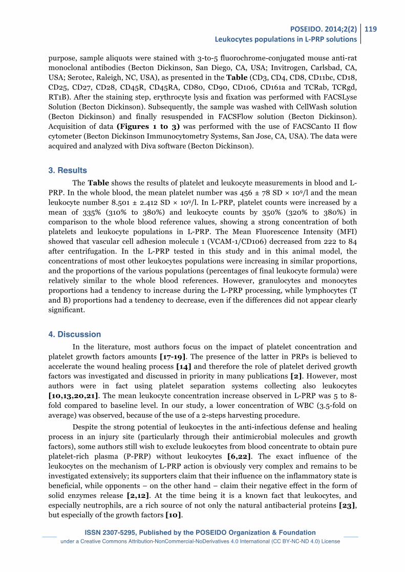
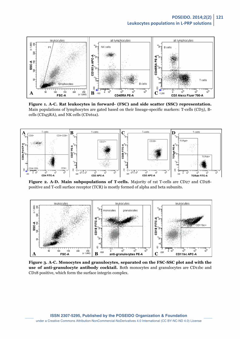
purpose, sample aliquots were stained with 3-to-5 fluorochrome-conjugated mouse anti-rat monoclonal antibodies (Becton Dickinson, San Diego, CA, USA; Invitrogen, Carlsbad, CA, USA; Serotec, Raleigh, NC, USA), as presented in the Table (CD3, CD4, CD8, CD11bc, CD18, CD25, CD27, CD28, CD45R, CD45RA, CD80, CD90, CD106, CD161a and TCRab, TCRgd, RT1B). After the staining step, erythrocyte lysis and fixation was performed with FACSLyse Solution (Becton Dickinson). Subsequently, the sample was washed with CellWash solution (Becton Dickinson) and finally resuspended in FACSFlow solution (Becton Dickinson). Acquisition of data (Figures 1 to 3) was performed with the use of FACSCanto II flow cytometer (Becton Dickinson Immunocytometry Systems, San Jose, CA, USA). The data were acquired and analyzed with Diva software (Becton Dickinson).
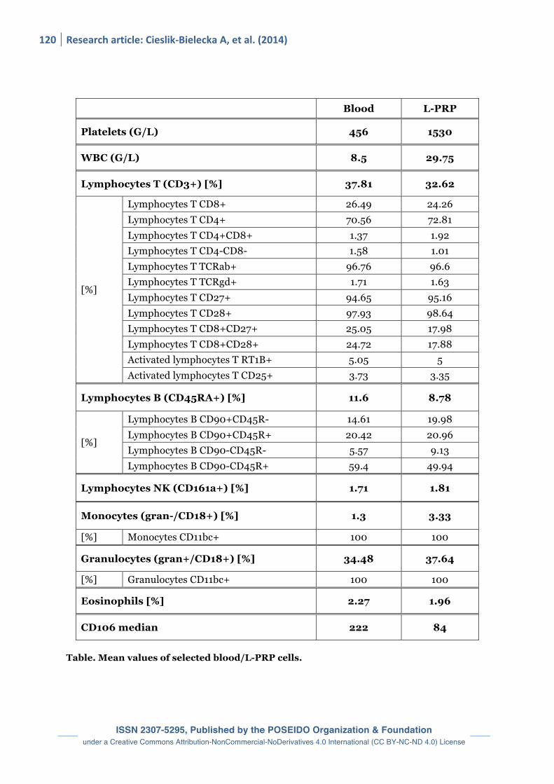
3. Results The Table shows the results of platelet and leukocyte measurements in blood and L-
PRP. In the whole blood, the mean platelet number was 456 ± 78 SD × 109/l and the mean leukocyte number 8.501 ± 2.412 SD × 109/l. In L-PRP, platelet counts were increased by a mean of 335% (310% to 380%) and leukocyte counts by 350% (320% to 380%) in comparison to the whole blood reference values, showing a strong concentration of both platelets and leukocyte populations in L-PRP. The Mean Fluorescence Intensity (MFI) showed that vascular cell adhesion molecule 1 (VCAM-1/CD106) decreased from 222 to 84 after centrifugation. In the L-PRP tested in this study and in this animal model, the concentrations of most other leukocytes populations were increasing in similar proportions, and the proportions of the various populations (percentages of final leukocyte formula) were relatively similar to the whole blood references. However, granulocytes and monocytes proportions had a tendency to increase during the L-PRP processing, while lymphocytes (T and B) proportions had a tendency to decrease, even if the differences did not appear clearly significant.
4. Discussion In the literature, most authors focus on the impact of platelet concentration and
platelet growth factors amounts [17-19]. The presence of the latter in PRPs is believed to accelerate the wound healing process [14] and therefore the role of platelet derived growth factors was investigated and discussed in priority in many publications [2]. However, most authors were in fact using platelet separation systems collecting also leukocytes [10,13,20,21]. The mean leukocyte concentration increase observed in L-PRP was 5 to 8-fold compared to baseline level. In our study, a lower concentration of WBC (3.5-fold on average) was observed, because of the use of a 2-steps harvesting procedure.
Despite the strong potential of leukocytes in the anti-infectious defense and healing process in an injury site (particularly through their antimicrobial molecules and growth factors), some authors still wish to exclude leukocytes from blood concentrate to obtain pure platelet-rich plasma (P-PRP) without leukocytes [6,22]. The exact influence of the leukocytes on the mechanism of L-PRP action is obviously very complex and remains to be investigated extensively; its supporters claim that their influence on the inflammatory state is beneficial, while opponents – on the other hand – claim their negative effect in the form of solid enzymes release [2,12]. At the time being it is a known fact that leukocytes, and especially neutrophils, are a rich source of not only the natural antibacterial proteins [23], but especially of the growth factors [10].
120 Research article: Cieslik-‐Bielecka A, et al. (2014)
ISSN 2307-5295, Published by the POSEIDO Organization & Foundation
under a Creative Commons Attribution-NonCommercial-NoDerivatives 4.0 International (CC BY-NC-ND 4.0) License
Blood L-PRP
Platelets (G/L) 456 1530
WBC (G/L) 8.5 29.75
Lymphocytes T (CD3+) [%] 37.81 32.62
[%]
Lymphocytes T CD8+ 26.49 24.26 Lymphocytes T CD4+ 70.56 72.81 Lymphocytes T CD4+CD8+ 1.37 1.92 Lymphocytes T CD4-CD8- 1.58 1.01 Lymphocytes T TCRab+ 96.76 96.6 Lymphocytes T TCRgd+ 1.71 1.63 Lymphocytes T CD27+ 94.65 95.16 Lymphocytes T CD28+ 97.93 98.64 Lymphocytes T CD8+CD27+ 25.05 17.98 Lymphocytes T CD8+CD28+ 24.72 17.88 Activated lymphocytes T RT1B+ 5.05 5 Activated lymphocytes T CD25+ 3.73 3.35
Lymphocytes B (CD45RA+) [%] 11.6 8.78
[%]
Lymphocytes B CD90+CD45R- 14.61 19.98 Lymphocytes B CD90+CD45R+ 20.42 20.96 Lymphocytes B CD90-CD45R- 5.57 9.13 Lymphocytes B CD90-CD45R+ 59.4 49.94
Lymphocytes NK (CD161a+) [%] 1.71 1.81
Monocytes (gran-/CD18+) [%] 1.3 3.33
[%] Monocytes CD11bc+ 100 100
Granulocytes (gran+/CD18+) [%] 34.48 37.64
[%] Granulocytes CD11bc+ 100 100
Eosinophils [%] 2.27 1.96
CD106 median 222 84
Table. Mean values of selected blood/L-PRP cells.
POSEIDO. 2014;2(2) Leukocytes populations in L-‐PRP solutions
121
ISSN 2307-5295, Published by the POSEIDO Organization & Foundation
under a Creative Commons Attribution-NonCommercial-NoDerivatives 4.0 International (CC BY-NC-ND 4.0) License
Figure 1. A-C. Rat leukocytes in forward- (FSC) and side scatter (SSC) representation. Main populations of lymphocytes are gated based on their lineage-specific markers: T-cells (CD3), B-cells (CD45RA), and NK cells (CD161a). Figure 2. A-D. Main subpopulations of T-cells. Majority of rat T-cells are CD27 and CD28-positive and T-cell surface receptor (TCR) is mostly formed of alpha and beta subunits. Figure 3. A-C. Monocytes and granulocytes, separated on the FSC-SSC plot and with the use of anti-granulocyte antibody cocktail. Both monocytes and granulocytes are CD11bc and CD18 positive, which form the surface integrin complex.

122 Research article: Cieslik-‐Bielecka A, et al. (2014)
ISSN 2307-5295, Published by the POSEIDO Organization & Foundation
under a Creative Commons Attribution-NonCommercial-NoDerivatives 4.0 International (CC BY-NC-ND 4.0) License
To reach plasma with a minimal leukocyte count, the g-force and time of centrifugation need to be decreased. As a consequence, the platelet concentration is lower in comparison to baseline levels and so are, in consequence, the growth factors levels [24]. Only in the studies by Aspenberg and Virchenko, where L-PRP was irradiated with 25 Gy to inactivate the WBCs, truly pure platelet concentrate could be obtained [25]. However such experimental method is clearly not practical in daily use. Consequently, most PRPs used in the literature have in fact a significant quantity of leukocytes. However, most authors do not mention leukocyte concentration even though they use standard separation systems, and they focus mostly on platelets and growth factors [6]. This situation is a major source of confusion and bias in the PRP literature and may explain the many controversies and mixed clinical results obtained with these products in the literature [5,14,15]. To clarify this confusion, a terminology was proposed in 2009 [6], and then reinforced in following consensus articles [5,11,12], in order to separate the many different products following at least 2 parameters, the fibrin density and the leukocyte content. For the PRPs, 2 main families were defined: the L-PRPs have a significant quantity of leukocytes, while the Pure PRP (P-PRP) have no or only traces of leukocytes. This first classification was designed to highlight the issue, and it is expected to be extended in the future, when the exact function and impact of the various possible concentrations and proportions of leukocytes will be clarified [12]. Leukocytes are the cells of the immune system defending the body against both infectious diseases and foreign materials [10]. According to their appearance under light microscope, there are two broad categories of lymphocytes, namely the large granular lymphocytes and the small lymphocytes. Functionally distinct subsets of lymphocytes correlate with their appearance. Most, but not all large granular lymphocytes are more commonly known as the Natural Killer cells (NK cells). The small lymphocytes are the T-cells and B-cells. Lymphocytes play an important and integral role in the body's defenses. T-cells and B-cells are the major cellular components of the adaptive immune response. T-cells are involved in cell-mediated immunity whereas B-cells are primarily responsible for humoral immunity (relating to antibodies). The function of T-cells and B-cells is to recognize specific “non-self” antigens, during a process known as antigen presentation. Once they have identified an invader, the cells generate specific responses that are tailored to maximally eliminate specific pathogens or pathogen infected cells. B-cells respond to pathogens by producing large quantities of antibodies which then neutralize foreign objects like bacteria and viruses. In response to pathogens, some T-cells, called helper T-cells produce cytokines that direct the immune response whilst other T-cells, called cytotoxic T-cells, produce toxic granules that induce the death of pathogen infected cells. Following activation, B-cells and T-cells leave a lasting legacy of the antigens they have encountered, in the form of memory cells [10,23,26]. Finally, B and T lymphocytes have very strong regulatory functions during the inflammatory process, and investigating their role in these complex mechanisms is a very intensive path of research [27-30]. NK cells are a part of innate immune system and play a major role in defending the host from both tumors and virally infected cells. NK cells distinguish infected cells and tumors from normal and uninfected cells by recognizing alterations in levels of a surface molecule called MHC (major histocompatibility complex) class I. NK cells are activated in response to a family of cytokines called interferons. Activated NK cells release cytotoxic (cell-killing) granules, which then destroy the altered cells [10,23]. They were named "natural killers" because of the initial notion that they do not require prior activation in order to kill cells which are missing MHC class I.

POSEIDO. 2014;2(2) Leukocytes populations in L-‐PRP solutions
123
ISSN 2307-5295, Published by the POSEIDO Organization & Foundation
under a Creative Commons Attribution-NonCommercial-NoDerivatives 4.0 International (CC BY-NC-ND 4.0) License
Antibacterial properties of L-PRP were investigated in the literature [31,32]. However, until now no one can explain the exact mechanism that inhibits bacteria growth, despite several authors have tried. Cieslik-Bielecka et al. described various mechanisms, which can cause microbicidal effects [23]. They focused on antibacterial peptides (HDP – Host Defense Peptides), which are produced by the macrophages, epithelial cells, as well as neutrophils and thrombocytes, and are one of the important elements shaping the human natural immunity. Neutrophils are the most common cells with strong phagocytic properties. They constitute the first line of antibacterial defense. The cytoplasm of the neutrophil granulocytes contains numerous granules. The most important ones are primary (azurophil) granules connected to the process of intracellular bacteria destruction and containing numerous bactericidal factors, including defensins, cathelicidins, serprocidins, Bactericidal/permeability-increasing protein (BPI) of gram-negative bacteria, myeloperoxidase and cytoplasmic calprotectin. The secondary (specific) granules are rich in antibacterial proteins such as lysozyme, collagenase, gelatinase, lactoferrin, phospholipase A2, transcobalamin-1 and membrane proteins [26]. Neutrophils release enzymes which destroy some specific components of the damaged tissues (to allow cell migration, tissue cleaning and finally tissue reconstruction), and first of all damage microbes, especially bacteria. Neutrophils circulating in the blood become activated by chemotactic factors and this constitutes a signal for the merging of the secretory granules with the superficial membrane. The process of phagocytosis starts by surrounding the organism by pseudopodia, and then closing it inside a phagosome, which undergoes a merge with granules, mainly the primary. The granules release their content exposing the microbe to the activity of a strong mixture of antibacterial proteins. The destruction of the phagocytosed bacteria takes place with the participation of oxygen species, or without oxygen with the use of lactoferrin or lysozyme [23].
Moojen et al. confirmed L-PRP antimicrobial activity against Staphylococcus Aureus [32]. Bielecki et al. also used L-PRP in infected bone non unions with good outcomes [31,33]. It was justifiable to conclude that L-PRP gel is an inductive biomaterial, which might possess local antimicrobial activity. Some authors have also reported decrease of infections after L-PRP usage in orthopaedic and cardiac surgery [34]. Yuan et al. reported a case of infection after intramedullary nailing, which has been a serious problem in orthopaedic surgery [35]. In this case, many kinds of treatments had been previously applied, but were not effective. During the operation, they observed a great deal of canal or sinus in most of the callus, and dead bone could be seen from the radiographs. However, it was difficult to remove all of these dead tissues, because if these were removed, the femur would fracture again. Furthermore, in the latter period, the patient refused any open operation under anesthesia. That is why they attempted to use L-PRP to treat the patient, as there were no alternatives to be chosen. To their surprise, the wound healed after L-PRP application.
In Khalafi’s study, the L-PRP group had one incidence of sternal infection (0.18%) compared to 11 cases (1.98%) in the control group [34]. There were 3 cases (0.53%) of notable drainage from the sternum in the L-PRP compared to 30 cases (5.39%) in the control group. For the leg vein harvest site, the L-PRP group had no reported infections and 61 (10.89%) incidences of excessive drainage, compared to 3 (0.66%) surgical site infections and 212 (48.4%) cases of excessive leg drainage in the control group. Following propensity scoring, they concluded that L-PRP application reduced the odds of chest wound infection by 93%, chest drainage by 96%, and leg wound drainage by 88%.
124 Research article: Cieslik-‐Bielecka A, et al. (2014)
ISSN 2307-5295, Published by the POSEIDO Organization & Foundation
under a Creative Commons Attribution-NonCommercial-NoDerivatives 4.0 International (CC BY-NC-ND 4.0) License
VCAM-1 with others adhesion molecules have been found to be significantly increased during viral and bacterial infection [36]. However, in our study there was a slight decrease in percentage of CD106-positive leukocytes in L-PRP samples as compared to peripheral blood samples. This may suggest that these rare cells are getting lost during additional centrifugation stages. For other cells, it is interesting to point out that the proportions of the various leukocyte types remain quite stable between blood and final concentrate in the specific L-PRP tested in this study. In another family of platelet concentrates termed L-PRF (Leukocyte- and Platelet-Rich Fibrin), which only exists under a strongly polymerized activated form [37-39], the leukocyte formula of the L-PRF clot is very different from the normal blood composition, with a higher proportion of lymphocytes and a lower proportion of monocytes and granulocytes [7]. This composition was advocated to explain the very strong effects in vitro of L-PRF on the bone cell proliferation and differentiation [40,41], and this parameter needs to be evaluated carefully in all PRP and PRF available on the market. The clinical studies with L-PRP mentioned above illustrate that the associated antibacterial effects of L-PRP (and of L-PRF) play an important role in positive clinical outcomes in many clinical applications. What is also shown is that the characteristics of different L-PRPs are not identical, because their leukocyte content and formula can vary. This aspect is rarely discussed in the literature and more studies are needed in this area. The presence of leukocytes in L-PRP may also influence growth factors levels, as it was already well shown with L-PRF [4,16,42,43]. The relative influence of platelets and leukocytes on growth factor levels in PRPs requires further investigation. As it was proven with L-PRF, leukocytes also influence the proliferation and differentiation pathways of many cell types in culture, not only with mediators but also directly [10,40,41]. The role of WBC as regulation turntables is essential to fully understand the complex biology of the L-PRP/L-PRF.
5. Conclusion Until now, the detailed characteristics of white blood cells in L-PRP in rats have never
been published in the international literature, and very little is known about the leukocytes populations in most L-PRP available on the market and tested in the literature. Considering the many possible ways in which various leukocyte populations can influence the properties of L-PRP, this study confirms the need to analyze the pattern of white blood cells in blood and L-PRP, before starting to investigate the effects of a platelet concentrate.
Leukocytes have obviously a major impact on the properties and biological activity of platelet concentrates. The ways in which these cells influence the intrinsic biology of L-PRP/L-PRF include their immune and antimicrobial potential as well as their key-role in wound healing processes. That is why the presence of leukocytes in PRPs cannot be neglected and requires further investigation, as this field of research opens new perspectives and possibilities in many clinical situations. Disclosure of interests
The authors have no conflict of interest to report. Acknowledgements
This research work on new biotechnologies and therapeutic strategies in regenerative medicine was supported by the PACT (Platelet & Advanced Cell Therapies) Forum Civitatis of the
POSEIDO. 2014;2(2) Leukocytes populations in L-‐PRP solutions
125
ISSN 2307-5295, Published by the POSEIDO Organization & Foundation
under a Creative Commons Attribution-NonCommercial-NoDerivatives 4.0 International (CC BY-NC-ND 4.0) License
POSEIDO Academic Consortium (Periodontology, Oral Surgery, Esthetic & Implant Dentistry Organization), by a grant from the National Research Foundation of Korea (NRF) funded by the Korean government-MEST (No. 2011-0030121) and by the LoB5 Foundation for Research, France. Author Contributions
All authors participated to the technical design and organization of the study, the treatment of data and to the elaboration of the manuscript. ACB, PP, LS and AS were in charge of the collection of the samples and raw data.
References [1] Bielecki T, Dohan Ehrenfest DM. Leukocyte- and platelet-rich Plasma (L-PRP)/fibrin (L-PRF) in medicine - past, present, future. Curr Pharm Biotechnol. 2012;13(7):i-ii. [2] Bielecki T, Dohan Ehrenfest DM. Platelet-rich plasma (PRP) and Platelet-Rich Fibrin (PRF): surgical adjuvants, preparations for in situ regenerative medicine and tools for tissue engineering. Curr Pharm Biotechnol. 2012;13(7):1121-30. [3] Cieslik-Bielecka A, Choukroun J, Odin G, Dohan Ehrenfest DM. L-PRP/L-PRF in esthetic plastic surgery, regenerative medicine of the skin and chronic wounds. Curr Pharm Biotechnol. 2012;13(7):1266-77. [4] Dohan Ehrenfest DM, de Peppo GM, Doglioli P, Sammartino G. Slow release of growth factors and thrombospondin-1 in Choukroun's platelet-rich fibrin (PRF): a gold standard to achieve for all surgical platelet concentrates technologies. Growth Factors. 2009;27(1):63-9. [5] Dohan Ehrenfest DM, Bielecki T, Mishra A, Borzini P, Inchingolo F, Sammartino G, Rasmusson L, Evert PA. In search of a consensus terminology in the field of platelet concentrates for surgical use: platelet-rich plasma (PRP), platelet-rich fibrin (PRF), fibrin gel polymerization and leukocytes. Curr Pharm Biotechnol. 2012;13(7):1131-7. [6] Dohan Ehrenfest DM, Rasmusson L, Albrektsson T. Classification of platelet concentrates: from pure platelet-rich plasma (P-PRP) to leucocyte- and platelet-rich fibrin (L-PRF). Trends Biotechnol. 2009;27(3):158-67. [7] Dohan Ehrenfest DM, Del Corso M, Diss A, Mouhyi J, Charrier JB. Three-dimensional architecture and cell composition of a Choukroun's platelet-rich fibrin clot and membrane. J Periodontol. 2010;81(4):546-55. [8] Cieslik-Bielecka A, Gazdzik TS, Bielecki TM, Cieslik T. Why the platelet-rich gel has antimicrobial activity? Oral Surg Oral Med Oral Pathol Oral Radiol Endod. 2007;103(3):303-5; author reply 5-6. [9] Everts PA, van Zundert A, Schonberger JP, Devilee RJ, Knape JT. What do we use: platelet-rich plasma or platelet-leukocyte gel? J Biomed Mater Res A. 2008;85(4):1135-6. [10] Bielecki T, Dohan Ehrenfest DM, Everts PA, Wiczkowski A. The role of leukocytes from L-PRP/L-PRF in wound healing and immune defense: new perspectives. Curr Pharm Biotechnol. 2012;13(7):1153-62. [11] Dohan Ehrenfest DM, Sammartino G, Shibli JA, Wang HL, Zou DR, Bernard JP. Guidelines for the publication of articles related to platelet concentrates (Platelet-Rich Plasma - PRP, or Platelet-Rich Fibrin - PRF): the international classification of the POSEIDO. POSEIDO. 2013;1(1):17-27. [12] Dohan Ehrenfest DM, Andia I, Zumstein MA, Zhang CQ, Pinto NR, Bielecki T. Classification of platelet concentrates (Platelet-Rich Plasma-PRP, Platelet-Rich Fibrin-PRF) for topical and infiltrative use in orthopedic and sports medicine: current consensus, clinical implications and perspectives. Muscles Ligaments Tendons J. 2014;4(1):3-9. [13] Yuan T, Guo SC, Han P, Zhang CQ, Zeng BF. Applications of leukocyte- and platelet-rich plasma (L-PRP) in trauma surgery. Curr Pharm Biotechnol. 2012;13(7):1173-84. [14] Del Corso M, Vervelle A, Simonpieri A, Jimbo R, Inchingolo F, Sammartino G, Dohan Ehrenfest DM. Current knowledge and perspectives for the use of platelet-rich plasma (PRP) and platelet-rich fibrin (PRF) in oral and maxillofacial surgery part 1: Periodontal and dentoalveolar surgery. Curr Pharm Biotechnol. 2012;13(7):1207-30. [15] Simonpieri A, Del Corso M, Vervelle A, Jimbo R, Inchingolo F, Sammartino G, Dohan Ehrenfest DM. Current knowledge and perspectives for the use of platelet-rich plasma (PRP) and platelet-rich fibrin (PRF) in oral and maxillofacial surgery part 2: Bone graft, implant and reconstructive surgery. Curr Pharm Biotechnol. 2012;13(7):1231-56. [16] Dohan Ehrenfest DM, Bielecki T, Jimbo R, Barbe G, Del Corso M, Inchingolo F, Sammartino G. Do the fibrin architecture and leukocyte content influence the growth factor release of platelet concentrates? An evidence-based answer comparing a pure platelet-rich plasma (P-PRP) gel and a leukocyte- and platelet-rich fibrin (L-PRF). Curr Pharm Biotechnol. 2012;13(7):1145-52.

126 Research article: Cieslik-‐Bielecka A, et al. (2014)
ISSN 2307-5295, Published by the POSEIDO Organization & Foundation
under a Creative Commons Attribution-NonCommercial-NoDerivatives 4.0 International (CC BY-NC-ND 4.0) License
[17] Weibrich G, Kleis WK, Hafner G. Growth factor levels in the platelet-rich plasma produced by 2 different methods: curasan-type PRP kit versus PCCS PRP system. Int J Oral Maxillofac Implants. 2002;17(2):184-90. [18] Weibrich G, Kleis WK, Hafner G, Hitzler WE. Growth factor levels in platelet-rich plasma and correlations with donor age, sex, and platelet count. J Craniomaxillofac Surg. 2002;30(2):97-102. [19] Weibrich G, Kleis WK, Kunz-Kostomanolakis M, Loos AH, Wagner W. Correlation of platelet concentration in platelet-rich plasma to the extraction method, age, sex, and platelet count of the donor. Int J Oral Maxillofac Implants. 2001;16(5):693-9. [20] Bielecki T, Cieslik-Bielecka A, Zelawski M, Mikusek W. A side-effect induced by the combination of a demineralized freeze-dried bone allograft and leucocyte and platelet-rich plasma during treatment for large bone cysts: a 4-year follow-up clinical study. Transfus Apher Sci. 2012;47(2):133-8. [21] Mishra A, Harmon K, Woodall J, Vieira A. Sports medicine applications of platelet rich plasma. Curr Pharm Biotechnol. 2012;13(7):1185-95. [22] Dohan Ehrenfest DM, Bielecki T, Del Corso M, Inchingolo F, Sammartino G. Shedding light in the controversial terminology for platelet-rich products: platelet-rich plasma (PRP), platelet-rich fibrin (PRF), platelet-leukocyte gel (PLG), preparation rich in growth factors (PRGF), classification and commercialism. J Biomed Mater Res A. 2010;95(4):1280-2. [23] Cieslik-Bielecka A, Dohan Ehrenfest DM, Lubkowska A, Bielecki T. Microbicidal properties of Leukocyte- and Platelet-Rich Plasma/Fibrin (L-PRP/L-PRF): new perspectives. J Biol Regul Homeost Agents. 2012;26(2 Suppl 1):43S-52S. [24] Weibrich G, Kleis WK, Hitzler WE, Hafner G. Comparison of the platelet concentrate collection system with the plasma-rich-in-growth-factors kit to produce platelet-rich plasma: a technical report. Int J Oral Maxillofac Implants. 2005;20(1):118-23. [25] Aspenberg P, Virchenko O. Platelet concentrate injection improves Achilles tendon repair in rats. Acta Orthop Scand. 2004;75(1):93-9. [26] Levy O, Sisson RB, Fryer HE, Goldmann D, Valore E, Ganz T, White ML, Carroll SF, Lehmann L, Guinan EC. Neutrophil defense in patients undergoing bone marrow transplantation: bactericidal/permeability-increasing protein (BPI) and defensins in graft-derived neutrophils. Transplantation. 2002;73(9):1522-6. [27] Dang VD, Hilgenberg E, Ries S, Shen P, Fillatreau S. From the regulatory functions of B cells to the identification of cytokine-producing plasma cell subsets. Curr Opin Immunol. 2014;28:77-83. [28] Inoue M, Arikawa T, Chen YH, Moriwaki Y, Price M, Brown M, Perfect JR, Shinohara ML. T cells down-regulate macrophage TNF production by IRAK1-mediated IL-10 expression and control innate hyperinflammation. Proc Natl Acad Sci U S A. 2014;111(14):5295-300. [29] Rosser EC, Blair PA, Mauri C. Cellular targets of regulatory B cell-mediated suppression. Mol Immunol. 2014;62(2):296-304. [30] Shen P, Roch T, Lampropoulou V, O'Connor RA, Stervbo U, Hilgenberg E, Ries S, Dang VD, Jaimes Y, Daridon C, Li R, Jouneau L, Boudinot P, Wilantri S, Sakwa I, Miyazaki Y, Leech MD, McPherson RC, Wirtz S, Neurath M, Hoehlig K, Meinl E, Grutzkau A, Grun JR, Horn K, Kuhl AA, Dorner T, Bar-Or A, Kaufmann SH, Anderton SM, Fillatreau S. IL-35-producing B cells are critical regulators of immunity during autoimmune and infectious diseases. Nature. 2014;507(7492):366-70. [31] Bielecki TM, Gazdzik TS, Arendt J, Szczepanski T, Krol W, Wielkoszynski T. Antibacterial effect of autologous platelet gel enriched with growth factors and other active substances: an in vitro study. J Bone Joint Surg Br. 2007;89(3):417-20. [32] Moojen DJ, Everts PA, Schure RM, Overdevest EP, van Zundert A, Knape JT, Castelein RM, Creemers LB, Dhert WJ. Antimicrobial activity of platelet-leukocyte gel against Staphylococcus aureus. J Orthop Res. 2008;26(3):404-10. [33] Bielecki T, Gazdzik TS, Szczepanski T. Benefit of percutaneous injection of autologous platelet-leukocyte-rich gel in patients with delayed union and nonunion. Eur Surg Res. 2008;40(3):289-96. [34] Khalafi RS, Bradford DW, Wilson MG. Topical application of autologous blood products during surgical closure following a coronary artery bypass graft. Eur J Cardiothorac Surg. 2008;34(2):360-4. [35] Yuan T, Zhang C, Zeng B. Treatment of chronic femoral osteomyelitis with platelet-rich plasma (PRP): a case report. Transfus Apher Sci. 2008;38(2):167-73. [36] Warfel JM, D'Agnillo F. Anthrax lethal toxin enhances TNF-induced endothelial VCAM-1 expression via an IFN regulatory factor-1-dependent mechanism. J Immunol. 2008;180(11):7516-24. [37] Del Corso M, Dohan Ehrenfest DM. Immediate implantation and peri-implant Natural Bone Regeneration (NBR) in the severely resorbed posterior mandible using Leukocyte- and Platelet-Rich Fibrin (L-PRF): a 4-year follow-up. POSEIDO. 2013;1(2):109-16.

POSEIDO. 2014;2(2) Leukocytes populations in L-‐PRP solutions
127
ISSN 2307-5295, Published by the POSEIDO Organization & Foundation
under a Creative Commons Attribution-NonCommercial-NoDerivatives 4.0 International (CC BY-NC-ND 4.0) License
[38] Toeroek R, Dohan Ehrenfest DM. The concept of Screw-Guided Bone Regeneration (S-GBR). Part 2: S-GBR in the severely resorbed preimplant posterior mandible using bone xenograft and Leukocyte- and Platelet-Rich Fibrin (L-PRF): a 5-year follow-up. POSEIDO. 2013;1(2):85-92. [39] Toeroek R, Dohan Ehrenfest DM. The concept of Screw-Guided Bone Regeneration (S-GBR). Part 3: Fast Screw-Guided Bone Regeneration (FS-GBR) in the severely resorbed preimplant posterior mandible using allograft and Leukocyte- and Platelet-Rich Fibrin (L-PRF): a 4-year follow-up. POSEIDO. 2013;1(2):93-100. [40] Dohan Ehrenfest DM, Diss A, Odin G, Doglioli P, Hippolyte MP, Charrier JB. In vitro effects of Choukroun's PRF (platelet-rich fibrin) on human gingival fibroblasts, dermal prekeratinocytes, preadipocytes, and maxillofacial osteoblasts in primary cultures. Oral Surg Oral Med Oral Pathol Oral Radiol Endod. 2009;108(3):341-52. [41] Dohan Ehrenfest DM, Doglioli P, de Peppo GM, Del Corso M, Charrier JB. Choukroun's platelet-rich fibrin (PRF) stimulates in vitro proliferation and differentiation of human oral bone mesenchymal stem cell in a dose-dependent way. Arch Oral Biol. 2010;55(3):185-94. [42] Dohan Ehrenfest DM. How to optimize the preparation of leukocyte- and platelet-rich fibrin (L-PRF, Choukroun's technique) clots and membranes: introducing the PRF Box. Oral Surg Oral Med Oral Pathol Oral Radiol Endod. 2010;110(3):275-8; author reply 8-80. [43] Zumstein MA, Bielecki T, Dohan Ehrenfest DM. The Future of Platelet Concentrates in Sports Medicine: Platelet-Rich Plasma, Platelet-Rich Fibrin, and the Impact of Scaffolds and Cells on the Long-term Delivery of Growth Factors. Operative Techniques in Sports Medicine. 2011;19(3):190-7. This article can be cited as: Cieslik-Bielecka A, Paczek P, Sedek L, Szantyr A, Skowroński R, Wang HL, Dohan Ehrenfest DM. Analysis of the Leukocytes in peripheral blood and Leukocyte- and Platelet-Rich Plasma (L-PRP) in rats: A flow cytometry study. POSEIDO. 2014;2(2):117-27.