Embed Size (px)
Citation preview
Review ArticleVolume 3 Issue 5 - November 2016DOI: 10.19080/OROAJ.2016.03.555622
Ortho & Rheum Open Access Copyright © All rights are reserved by Daniel Elgut
Analysis of Fusion Rates in Patients Undergoing 1st Metatarsal Phalangeal Arthrodesis Utilizing Crossed
Compression Screws with Dorsal Neutralization Plate and Early Weight Bearing
*Daniel Elgut, Zachary Beth, Paul Stone and John S Levin Podiatry Department, USA
Submission: November 17, 2016; Published: November 28, 2016
*Corresponding author: Dan Elgut, JFK Medical Center 5301 S Congress Ave, Atlantis, FL 33462, Patients’ who signed up for study at: Orthopedic Center of Palm Beach County 180 John F Kennedy Dr, Atlantis, FL 33462, Podiatry Department, United States of America.
IntroductionOsteoarthritis is the most common form of arthritis,
affecting more than 50 million people in the United States. By 70 years of age, most people in the United States have evidence of osteoarthritis on radiographs or MRI in at least one joint [1]. Osteoarthritis of the first metatarsal phalangeal joint, known as Hallux limitus/ rigidus, is associated with pain and loss of motion [2]. Limitation of motion of the first MTPJ can lead to pain on ambulation, inability to perform activities of daily living and morbidity. Additionally, altered gait mechanics, due to loss of the first MTPJ motion can lead to other dysfunction of the forefoot. Capsulitis of the second MTPJ, painful plantar callosities and second ray deformities such as hammertoe and pre-dislocation syndrome are not uncommon [2].
Minimum range of motion of dorsiflexion of first MTPJ for normal gait is 65-75 degrees [3]. Failure to produce at least 65 degrees of MTPJ dorsiflexion, alters normal weight-bearing patterns under the foot, resulting in lateral weight transfer and forefoot dysfunction [4]. Etiologies of hallux limitus/rigidus include the following: hyper mobility of the first ray, long first metatarsal, metatarsus primus elevatus and trauma to first ray. Coughlin and Shurnas’ classification is the most comprehensive, and takes account radiographic findings, objective (range of
motion) and subjective (intensity and frequency of stiffness and pain) criteria [3]. Coughlin and Shurnas’ classification scheme consists of six stages. In Grade 0, patient’s have no pain, passive motion limitation of 10% to 20% and radiographic changes are either minimal or absent. In Grade I, pain is mild and present only at the end ranges of motion. A dorsal osteophyte is radiographically visible, and motion loss of 20% to 50% as compared to the normal side. In Grade II, pain is more severe and can be constant; radiographic sclerosis and narrowing is observed.
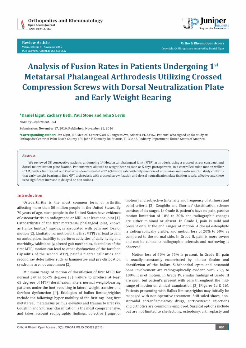
Motion loss of 50% to 75% is present. In Grade III, pain is usually constantly exacerbated by plantar flexion and dorsiflexion of the hallux. Subchondral cysts and sesamoid bone involvement are radiographically evident, with 75% to 100% loss of motion. In Grade IV, similar findings of Grade III are seen, but patient’s present with pain throughout the mid-range of motion on clinical examination [3] (Figures 1a & 1b). Patients presenting with Hallux limitus/rigidus may initially be managed with non-operative treatment. Stiff-soled shoes, non-steroidal anti-inflammatory drugs, corticosteroid injections and orthotics are commonly employed. Surgical options include but are not limited to cheilectomy, osteotomy, arthroplasty and
Ortho & Rheum Open Access J 3(5): OROAJ.MS.ID.555622 (2016)
Abstract
We reviewed 38 consecutive patients undergoing 1st Metatarsal phalangeal joint (MTP) arthrodesis using a crossed screw construct and dorsal neutralization plate fixation. Patients were allowed to weight bear as soon as 5 days postoperative, in a controlled ankle motion walker (CAM) with a first ray cut out. Our series demonstrated a 97.4% fusion rate with only one case of non-union and hardware. Our study confirms that early weight bearing in first MPT arthrodesis with crossed screw fixation and dorsal neutralization plate fixation is safe, effective and there is no significant increase in delayed or non-unions.
001
Orthopedics and RheumatologyOpen Access Journal ISSN: 2471-6804
How to cite this article: Daniel E, Zachary B, Paul St, John S L . Analysis of Fusion Rates in Patients Undergoing 1st Metatarsal Phalangeal Arthrodesis Utilizing Crossed Compression Screws with Dorsal Neutralization Plate and Early Weight Bearing. Ortho & Rheum Open Access J. 2016; 3(5): 555622. DOI: 10.19080/OROAJ.2016.03.555622
002
Orthopedics and Rheumatology Open Access Journal
arthrodesis. This study looked at first metatarsal phalangeal joint fusions with cannulated crossing screws and a dorsal, low profile neutralization plate (Flower Orthopedics, Horsham PA) in association in association with early weight bearing.
Figure 1a: AP view, Stage IV Hallux Rigidus.
Figure 1b: Lateral view, stage IV Hallux Rigidus.
Methods and Materials In this retrospective study 38 consecutive patients, affected
by a debilitating disease in the metatarsal phalangeal joint. All patient’s elected to undergo first MPTJ fusion following the failure of conservative management. The mean age of the patients in this study was 64, (range of 45 to 80 years). The etiologies of first MPTJ pain in our study included Rheumatoid arthritis, Hallux rigidus, Hallux limitus, Hallux varus and geriatric Hallux valgus. The study focused on the effects of early weight bearing on fusion rates of first MTPJ arthrodesis utilizing cannulated crossed screw fixation, along with application of a low profile, dorsal neutralization plate. The end point of the study included, time to fusion, hardware complication and fusion rates. We were interested in determining if early weight bearing five days post operative had any effect on union rates, or if early weight bearing caused any complications with the fixation construct which would result in delayed or non union.
The procedure
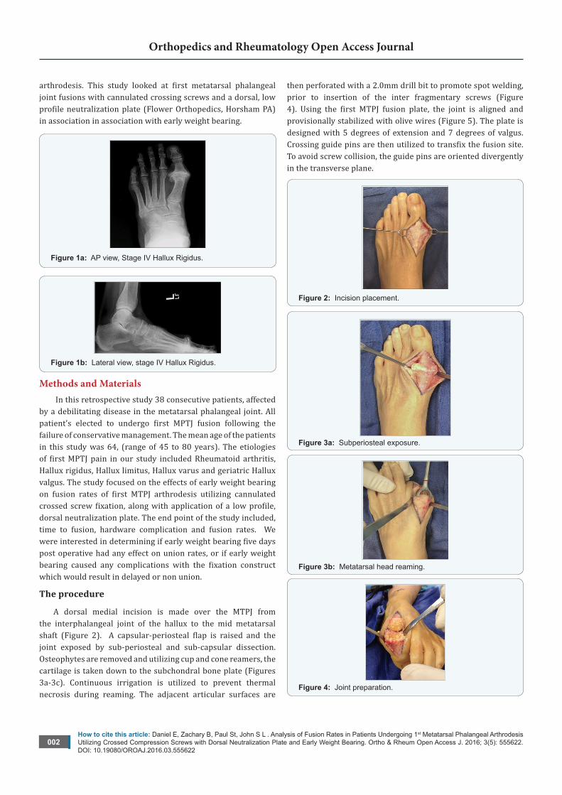
A dorsal medial incision is made over the MTPJ from the interphalangeal joint of the hallux to the mid metatarsal shaft (Figure 2). A capsular-periosteal flap is raised and the joint exposed by sub-periosteal and sub-capsular dissection. Osteophytes are removed and utilizing cup and cone reamers, the cartilage is taken down to the subchondral bone plate (Figures 3a-3c). Continuous irrigation is utilized to prevent thermal necrosis during reaming. The adjacent articular surfaces are
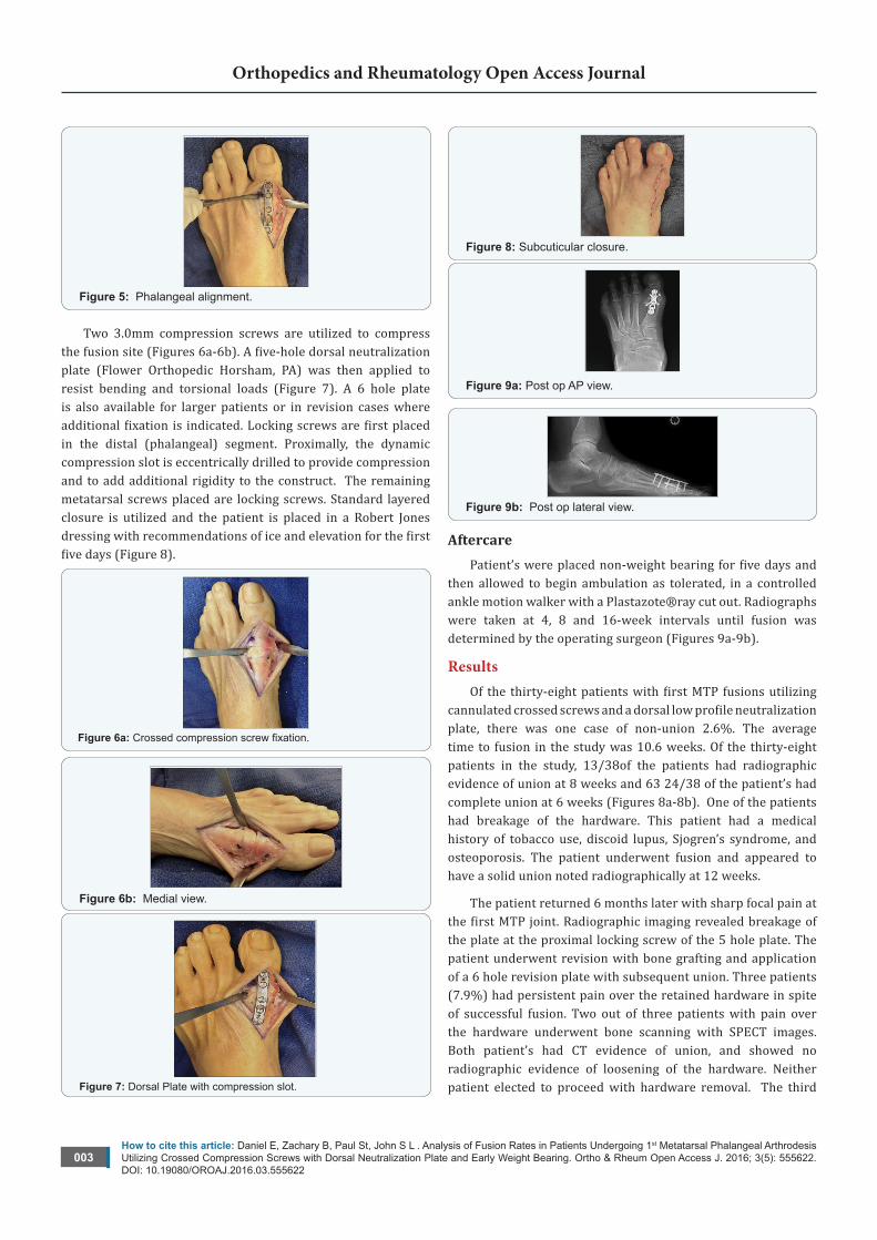
then perforated with a 2.0mm drill bit to promote spot welding, prior to insertion of the inter fragmentary screws (Figure 4). Using the first MTPJ fusion plate, the joint is aligned and provisionally stabilized with olive wires (Figure 5). The plate is designed with 5 degrees of extension and 7 degrees of valgus. Crossing guide pins are then utilized to transfix the fusion site. To avoid screw collision, the guide pins are oriented divergently in the transverse plane.
Figure 2: Incision placement.
Figure 3a: Subperiosteal exposure.
Figure 3b: Metatarsal head reaming.
Figure 4: Joint preparation.
How to cite this article: Daniel E, Zachary B, Paul St, John S L . Analysis of Fusion Rates in Patients Undergoing 1st Metatarsal Phalangeal Arthrodesis Utilizing Crossed Compression Screws with Dorsal Neutralization Plate and Early Weight Bearing. Ortho & Rheum Open Access J. 2016; 3(5): 555622. DOI: 10.19080/OROAJ.2016.03.555622
003
Orthopedics and Rheumatology Open Access Journal
Figure 5: Phalangeal alignment.
Two 3.0mm compression screws are utilized to compress the fusion site (Figures 6a-6b). A five-hole dorsal neutralization plate (Flower Orthopedic Horsham, PA) was then applied to resist bending and torsional loads (Figure 7). A 6 hole plate is also available for larger patients or in revision cases where additional fixation is indicated. Locking screws are first placed in the distal (phalangeal) segment. Proximally, the dynamic compression slot is eccentrically drilled to provide compression and to add additional rigidity to the construct. The remaining metatarsal screws placed are locking screws. Standard layered closure is utilized and the patient is placed in a Robert Jones dressing with recommendations of ice and elevation for the first five days (Figure 8).
Figure 6a: Crossed compression screw fixation.
Figure 6b: Medial view.
Figure 7: Dorsal Plate with compression slot.
Figure 8: Subcuticular closure.
Figure 9a: Post op AP view.
Figure 9b: Post op lateral view.
Aftercare Patient’s were placed non-weight bearing for five days and
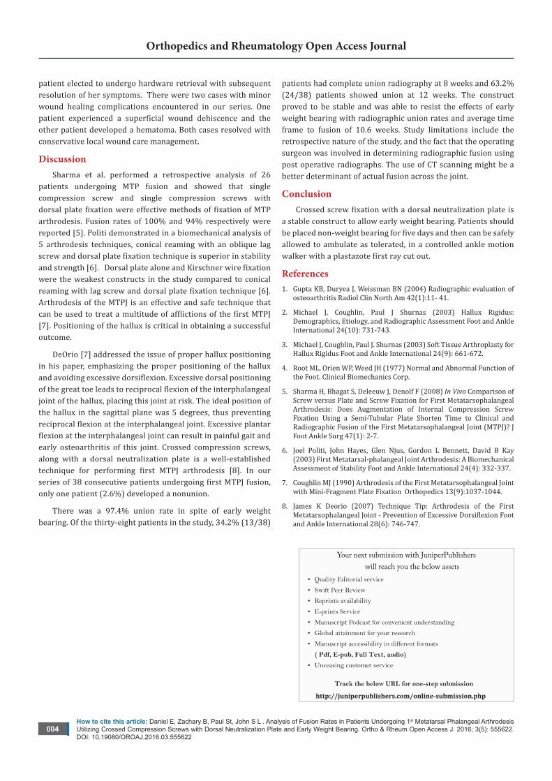
then allowed to begin ambulation as tolerated, in a controlled ankle motion walker with a Plastazote®ray cut out. Radiographs were taken at 4, 8 and 16-week intervals until fusion was determined by the operating surgeon (Figures 9a-9b).
Results Of the thirty-eight patients with first MTP fusions utilizing
cannulated crossed screws and a dorsal low profile neutralization plate, there was one case of non-union 2.6%. The average time to fusion in the study was 10.6 weeks. Of the thirty-eight patients in the study, 13/38of the patients had radiographic evidence of union at 8 weeks and 63 24/38 of the patient’s had complete union at 6 weeks (Figures 8a-8b). One of the patients had breakage of the hardware. This patient had a medical history of tobacco use, discoid lupus, Sjogren’s syndrome, and osteoporosis. The patient underwent fusion and appeared to have a solid union noted radiographically at 12 weeks.
The patient returned 6 months later with sharp focal pain at the first MTP joint. Radiographic imaging revealed breakage of the plate at the proximal locking screw of the 5 hole plate. The patient underwent revision with bone grafting and application of a 6 hole revision plate with subsequent union. Three patients (7.9%) had persistent pain over the retained hardware in spite of successful fusion. Two out of three patients with pain over the hardware underwent bone scanning with SPECT images. Both patient’s had CT evidence of union, and showed no radiographic evidence of loosening of the hardware. Neither patient elected to proceed with hardware removal. The third
How to cite this article: Daniel E, Zachary B, Paul St, John S L . Analysis of Fusion Rates in Patients Undergoing 1st Metatarsal Phalangeal Arthrodesis Utilizing Crossed Compression Screws with Dorsal Neutralization Plate and Early Weight Bearing. Ortho & Rheum Open Access J. 2016; 3(5): 555622. DOI: 10.19080/OROAJ.2016.03.555622
004
Orthopedics and Rheumatology Open Access Journal
patient elected to undergo hardware retrieval with subsequent resolution of her symptoms. There were two cases with minor wound healing complications encountered in our series. One patient experienced a superficial wound dehiscence and the other patient developed a hematoma. Both cases resolved with conservative local wound care management.
DiscussionSharma et al. performed a retrospective analysis of 26
patients undergoing MTP fusion and showed that single compression screw and single compression screws with dorsal plate fixation were effective methods of fixation of MTP arthrodesis. Fusion rates of 100% and 94% respectively were reported [5]. Politi demonstrated in a biomechanical analysis of 5 arthrodesis techniques, conical reaming with an oblique lag screw and dorsal plate fixation technique is superior in stability and strength [6]. Dorsal plate alone and Kirschner wire fixation were the weakest constructs in the study compared to conical reaming with lag screw and dorsal plate fixation technique [6]. Arthrodesis of the MTPJ is an effective and safe technique that can be used to treat a multitude of afflictions of the first MTPJ [7]. Positioning of the hallux is critical in obtaining a successful outcome.
DeOrio [7] addressed the issue of proper hallux positioning in his paper, emphasizing the proper positioning of the hallux and avoiding excessive dorsiflexion. Excessive dorsal positioning of the great toe leads to reciprocal flexion of the interphalangeal joint of the hallux, placing this joint at risk. The ideal position of the hallux in the sagittal plane was 5 degrees, thus preventing reciprocal flexion at the interphalangeal joint. Excessive plantar flexion at the interphalangeal joint can result in painful gait and early osteoarthritis of this joint. Crossed compression screws, along with a dorsal neutralization plate is a well-established technique for performing first MTPJ arthrodesis [8]. In our series of 38 consecutive patients undergoing first MTPJ fusion, only one patient (2.6%) developed a nonunion.
There was a 97.4% union rate in spite of early weight bearing. Of the thirty-eight patients in the study, 34.2% (13/38)
patients had complete union radiography at 8 weeks and 63.2% (24/38) patients showed union at 12 weeks. The construct proved to be stable and was able to resist the effects of early weight bearing with radiographic union rates and average time frame to fusion of 10.6 weeks. Study limitations include the retrospective nature of the study, and the fact that the operating surgeon was involved in determining radiographic fusion using post operative radiographs. The use of CT scanning might be a better determinant of actual fusion across the joint.
ConclusionCrossed screw fixation with a dorsal neutralization plate is
a stable construct to allow early weight bearing. Patients should be placed non-weight bearing for five days and then can be safely allowed to ambulate as tolerated, in a controlled ankle motion walker with a plastazote first ray cut out.
References1. Gupta KB, Duryea J, Weissman BN (2004) Radiographic evaluation of
osteoarthritis Radiol Clin North Am 42(1):11- 41.
2. Michael J, Coughlin, Paul J Shurnas (2003) Hallux Rigidus: Demographics, Etiology, and Radiographic Assessment Foot and Ankle International 24(10): 731-743.
3. Michael J, Coughlin, Paul J. Shurnas (2003) Soft Tissue Arthroplasty for Hallux Rigidus Foot and Ankle International 24(9): 661-672.
4. Root ML, Orien WP, Weed JH (1977) Normal and Abnormal Function of the Foot. Clinical Biomechanics Corp.
5. Sharma H, Bhagat S, Deleeuw J, Denolf F (2008) In Vivo Comparison of Screw versus Plate and Screw Fixation for First Metatarsophalangeal Arthrodesis: Does Augmentation of Internal Compression Screw Fixation Using a Semi-Tubular Plate Shorten Time to Clinical and Radiographic Fusion of the First Metatarsophalangeal Joint (MTPJ)? J Foot Ankle Surg 47(1): 2-7.
6. Joel Politi, John Hayes, Glen Njus, Gordon L Bennett, David B Kay (2003) First Metatarsal-phalangeal Joint Arthrodesis: A Biomechanical Assessment of Stability Foot and Ankle International 24(4): 332-337.
7. Coughlin MJ (1990) Arthrodesis of the First Metatarsophalangeal Joint with Mini-Fragment Plate Fixation Orthopedics 13(9):1037-1044.
8. James K Deorio (2007) Technique Tip: Arthrodesis of the First Metatarsophalangeal Joint - Prevention of Excessive Dorsiflexion Foot and Ankle International 28(6): 746-747.
Your next submission with JuniperPublishers will reach you the below assets
• Quality Editorial service
• Swift Peer Review
• Reprints availability
• E-prints Service
• Manuscript Podcast for convenient understanding
• Global attainment for your research
• Manuscript accessibility in different formats
( Pdf, E-pub, Full Text, audio)
• Unceasing customer service
Track the below URL for one-step submission
http://juniperpublishers.com/online-submission.php