Embed Size (px)
Citation preview
~ 1123 ~
International Journal of Orthopaedics Sciences 2018; 4(1): 1123-1132
ISSN: 2395-1958
IJOS 2018; 4(1): 1123-1132
© 2018 IJOS
www.orthopaper.com
Received: 18-11-2017
Accepted: 21-12-2017
Dr. Aravindan Karunakaran
Assistant Professor, Department
of Orthopaedics, Chengalpattu
Medical College Hospital,
Chengalpattu, Tamil Nadu,
India
Dr. Samuel Gnanam Rajamani
Associate Professor, Department
of Orthopaedics, Chengalpattu
Medical College Hospital,
Chengalpattu, Tamil Nadu,
India
Correspondence
Dr. Aravindan Karunakaran
Assistant Professor, Department
of Orthopaedics, Chengalpattu
Medical College Hospital,
Chengalpattu, Tamil Nadu,
India
Analysis of Bristow-Later jet procedure in the
treatment of recurrent traumatic anterior dislocation
of shoulder
Dr. Aravindan Karunakaran and Dr. Samuel Gnanam Rajamani
DOI: https://doi.org/10.22271/ortho.2018.v4.i1p.158
Abstract The shoulder by virtue of its anatomy and biomechanics is one of the most unstable and frequently
dislocated joints in the body, accounting for nearly 50% of all dislocations. Since the beginning of
century more than 150 surgical procedures were described in treating recurrent dislocation of the
shoulder with varying results and success. Most of the operative techniques described for the treatment of
recurrent anterior dislocation or subluxation of the shoulder have 2 distinct disadvantages; need to
immobilize the shoulder for several weeks and Loss of external rotation. These can be overcome by
Bristow-Laterjet procedure.
We analysed Bristow-Laterjet procedure in the treatment of recurrent traumatic anterior dislocation of
shoulder at the Department of Orthopaedics, Chengalpattu Medical College Hospital, Chengalpattu,
Tamilnadu, India. We came across 20 patients (1 patient had bilateral dislocation) during February 2015
to February 2017. All the patients were followed up periodically both clinically and radiologically till
their shoulder regained full range of movements and radiological bony union of the coracoid graft. All
the patients were thoroughly examined and evaluated subjectively and objectively and outcome was
assessed by Rowe’s scoring system. We found all the patients were satisfied with surgery and were able
to return to their pre dislocation level activity and none had further episodes of shoulder instability.
Radiologically there was good bony union of the coracoid graft and there was no screw loosening.
Keywords: Bristow-Laterjet procedure, recurrent anterior dislocation, shoulder instability, hill sachs
lesion, coracoid graft
1. Introduction
Materials and Methods
We in the Department of orthopaedics came across 20 patients (1 patient had bilateral
dislocation) during February 2015 to February 2017 out of which 18 were Male and 2 were
female.
Inclusion Criteria
All traumatic recurrent anterior dislocation.
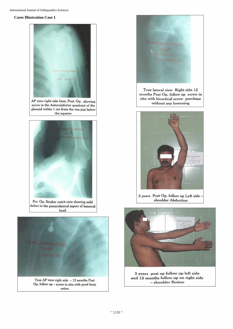
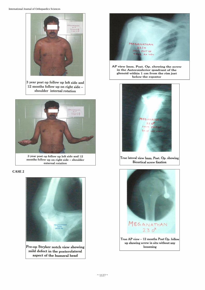
Pre-op Stryker Notch view showing mild degree of Hill Sachs
(Moderate to severe degrees are excluded from the study)
All patients without any fracture of the proximal humerus.
All patients without any ligamentous laxity
Based on the inclusion criteria, 2 patients who had generalized ligamentious laxity were
excluded from the study thus leaving 18 patients in our study.
All the patients were in the age group of 20 to 25 years with mean age of 22.5 years, average
age at the time of index dislocation being 19.8yrs. In two patients the index dislocation
occurred above the age of 20 (in one who had bilateral dislocation, on the right side at the age
of 23 and another person sustained the index dislocation at the age of 22). All the patients were
evaluated clinically and radiologically. Clinically all the patients had positive apprehension
sign, full ROM, no associated posterior instability and no previous sugery for shoulder
instability. Radiologically all the patients were evaluated by taking AP view of the shoulder,
True AP view of the shoulder, True lateral view of the shoulder, internal rotation (45 deg) AP
~ 1124 ~
International Journal of Orthopaedics Sciences view of the shoulder, Stryker Notch view of the shoulder. 17
shoulders had mild degree of Hill Sachs lesion and 2
shoulders had no Hill Sachs lesion. All the patients underwent
Bristow-Laterjet procedure
Operative Technique Under GA, with a sand bag under the scapula, through a
deltopectoral approach, cephalic vein is identified and
retracted medially with a cuff of muscle. Then going through
the interval between the deltoid and pectoralis major,
coracobrachialis and short head of biceps. In some cases
musculocutaneous nerve was seen and in other cases it was
palpated and in none of the cases the musculocutaneous nerve
was affected. The upper and lower limits of subscapularis
muscle were identified. The lower border of the muscle was
identified by the plexus of the anterior humeral circumflex
vessels. Split the subscapularis muscle in line with its fibre
from lateral to medial at approximately the junction of middle
and lower third of the muscle. Once the subscapularis muscle
was split, periosteal elevator was used to reflect it from the
outer surface of the shoulder capsule to expose the anterior
capsule. The anterior capsule was split in a manner to the split
made in the subscapularis muscle. The joint is explored for
intra-articular pathology.
Medial exposure of the anterior scapular neck is necessary for
proper placement of the transferred coracoids. The anterior
scapular neck was exposed by subperiosteal dissection. It is
important that this transfer site be inferior to the equator of
the glenoid and if possible less than 1 cm from its rim. At this
position on the anteroinferior portion of the scapular neck,
drill a 3.2 mm hole through the posterior cortex of the
scapular neck. The surface of scapular neck were the screw
has to be fixed was roughened with an osteotome. Position the
transferred carocoid tip with its muscle attachment through
the horizontal slit in the subscapularis onto the neck of
scapula. Then fix the coracoids tip with a 4.5 mm malleolar
screw with washer to avoid fragmentation of the coracoids.
Then close the subscapular, longitudinal split. Then after
attaining the hemostasis, wound closed in layers. All the
patients were given I. V. Antibiotics for 3 days and oral
antibiotics for 5 days.
Post-Operative Protocol
1st Week
Shoulder immobilization
1 to 3 week
Shoulder in arm sling
Circumduction exercises (Pendulum exercises) started.
It is aimed to achieve shoulder flexion, adduction and
external rotation of 60deg each.
Shoulder extension is not allowed
Elbow flexion is allwed but neither active nor passive
elbow extension is allowed.
3 to 6 weeks
Sling discarded
Increasing range of movements at the shoulder
Isometric strengthening of shoulder muscles (especially
of the rotator cuff).
It is aimed to achieve 90 degrees each of shoulder
flexion, external rotation and abduction
6 to 12 weeks
Gradual weight bearing is allowed in the shoulder.
Elbow extension is allowed.
Patient should be able to get full ROM in the shoulder
3 to 6 months
Normal day to day activities allowed.
Non-contact sports are allowed
Complications
The complications that are cited in the literature were
Neurovascular complications [27, 28] (Brachial plexus injury,
musculocutaneous nerve injury), screw penetration into the
joint, screw loosening, glenohumeral arthritis. In our series till
the latest follow up none of the patients had neurovascular
complications, screw loosening, screw penetration or
glenohumeral arthritis. None of the patients had recurrence of
dislocation or subluxation. We have not encountered any
complication such as nonunion of coracoid or problem with
the screw.
Results
All the patients were followed up periodically both clinically
and radiologically till their shoulder regained full range of
movements and radiological bony union of the coracoid graft.
All the patients were thoroughly examined and evaluated
subjectively and objectively and outcome was assessed by
Rowe’s scoring system.
Subjective Evaluation
Based on the ability return to work to their pre-dislocation
level, satisfaction, any shoulder instability or pain. All the
patients were satisfied with the surgery. They were able to
return to their pre-dislocation level activity. None of the
patients had pam or episodes of shoulder instability.
Objective evaluation (by clinical examination and Rowe’e
scoring system)
In all the patients apprehension sign was negative. All the
patients had regained full shoulder flexion, abduction, internal
rotation. Although patients were not aware the terminal 5 to
20 deg of external rotation (average) was uniformly restricted
in all the patients.
Radiological Evaluation
All the patients were evaluated by True AP view and True
lateral view of the shoulder to assess the bony union of the
transferred coracoid graft and to assess any screw loosening
or screw breakage. In all the patients there was bony union
and there is no evidence of any screw loosening.
Outcome assessment by Rowe’s scoring system
It includes a maximum potential score of 100 points which
were subdivided into stability (50 points), motion (20 points)
and function (30 points). The rating scale is heavily weighed
to the recurrence of instability (50 points). Results according
to this system were excellent in 14 shoulders and good in 5
shoulders.
Results
All the patients were followed up periodically both clinically
and radiologically till their shoulder regained full range of
movements and radiological bony union of the coracoid graft.
All the patients were thoroughly examined and evaluated
subjectively and objectively and outcome was assessed by
Rowe’s scoring system.

~ 1125 ~
International Journal of Orthopaedics Sciences Subjective evaluation
Based on the ability return to work to their pre-dislocation
level, satisfaction, any shoulder instability or pain. All the
patients were satisfied with the surgery. They were able to
return to their pre-dislocation level activity. None of the
patients had pam or episodes of shoulder instability.
Objective evaluation (by clinical examination and Rowe’e
scoring system)
In all the patients apprehension sign was negative. All the
patients had regained full shoulder flexion, abduction, internal
rotation. Although patients were not aware the terminal 5 to
20 deg of external rotation (average) was uniformly restricted
in all the patients.
Radiological evaluation
All the patients were evaluated by True AP view and True
lateral view of the shoulder to assess the bony union of the
transferred coracoid graft and to assess any screw loosening
or screw breakage. In all the patients there was bony union
and there is no evidence of any screw loosening.
Outcome assessment by Rowe’s scoring system
It includes a maximum potential score of 100 points which
were subdivided into stability (50 points), motion (20 points)
and function (30 points). The rating scale is heavily weighed
to the recurrence of instability (50 points). Results according
to this system were excellent in 14 shoulders and good in 5
shoulders.
~ 1126 ~
International Journal of Orthopaedics Sciences Cases Illustration Case 1
~ 1127 ~
International Journal of Orthopaedics Sciences
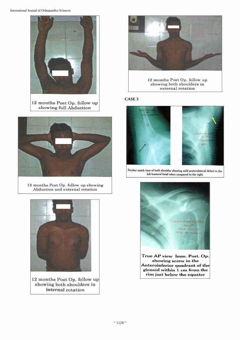
CASE 2
~ 1128 ~
International Journal of Orthopaedics Sciences
CASE 3

~ 1129 ~
International Journal of Orthopaedics Sciences
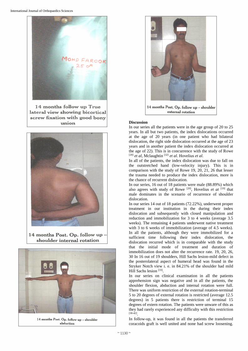
CASE 4
~ 1130 ~
International Journal of Orthopaedics Sciences
Discussion
In our series all the patients were in the age group of 20 to 25
years. In all but two patients, the index dislocations occurred
at the age of 20 years (in one patient who had bilateral
dislocation, the right side dislocation occurred at the age of 23
years and in another patient the index dislocation occurred at
the age of 22). This is in concurrence with the study of Rowe [20] et al, Mclaughtin [32] et al. Hovelius et al.
In all of the patients, the index dislocation was due to fall on
the outstretched hand (low-velocity injury). This is in
comparison with the study of Rowe 19, 20, 21, 26 that lesser
the trauma needed to produce the index dislocation, more is
the chance of recurrent dislocation.
In our series, 16 out of 18 patients were male (88.89%) which
also agrees with study of Rowe [20], Hovelius et at [19] that
male dominates in the scenario of recurrence of shoulder
dislocation.
In our series 14 out of 18 patients (72.22%), underwent proper
treatment in our institution in the during their index
dislocation and subsequently with closed manipulation and
reduction and immobilization for 3 to 4 weeks (average 3.5
weeks). The remaining 4 patients underwent native treatment
with 3 to 6 weeks of immobilization (average of 4.5 weeks).
In all the patients, although they were immobilized for a
sufficient time following their index dislocation, the
dislocation recurred which is in comparable with the study
that the initial mode of treatment and duration of
immobilization does not alter the recurrence rate. 19, 20, 26,
30 In 16 out of 19 shoulders, Hill Sachs lesion-mild defect in
the posterolateral aspect of humeral head was found in the
Stryker Notch view i. e. in 84.21% of the shoulder had mild
Hill Sachs lesion [33].
In our series on clinical examination in all the patients
apprehension sign was negative and in all the patients, the
shoulder flexion, abduction and internal rotation were full.
There was uniform restriction of the external rotation-terminal
5 to 20 degrees of external rotation is restricted (average 12.5
degrees) in 5 patients there is restriction of terminal 15
degrees of extern rotation. The patients were unware of this as
they had rarely experienced any difficulty with this restriction [30-42].
In follow-up, it was found in all the patients the transferred
coracoids graft is well united and none had screw loosening.
~ 1131 ~
International Journal of Orthopaedics Sciences Although bony union was not necessary (even fibrous union
is enough to hold the graft in situ) [34, 35], we achieved bony
union in all the cases. Complications cited in the literatures
were screw loosening, screw cut through, non-union of the
graft, neurovascular complication. We rarely encountered
these complications.
Result analysis as per function, all were excellent Rowe’s
Scoring 14 out of 19 shoulders (73.68%) showed excellent
results and the remaining 5 had good results (26.32%)
There are as many as 150 surgical procedures being described
for the treatment of recurrent dislocation of shoulder which
includes both soft tissue procedures and bony procedures. We
chose Bristow-Laterjet procedure for the following reasons.
The Principles of Bristow-Laterjet procedure is by Dynamic
Musculotendinous sling.
1. The transferred coracoids graft with the conjoined tendon
of short head of biceps and coracobranchialis muscles are
placed so as to produce a strong dynamic buttress across the
anterior and inferior aspects of the joint when the shoulder is
in the vulnerable position of abducted and externally rotated
position.
2. The transfer when passed through a split in the
subscapularis muscle, also functions to hold the lower half of
the subscapularis muscle in position and prevent it from
slipping superiorly over the humeral head when the shoulder
is abducted, and also it provides a new dynamic
musculotendinous sling to hold humeral head posteriorly.
3. The transferred coracoids fragment used for reconstruction
of the glenoid cavity is very effective at the end of throwing
movement as well as with low abduction of the arm.
Putti-Platt procedure is intended to shorten the subscapularis
muscle and according to Osmond Clarke [34], results in
permanent limitation of external rotation in most case and has
a high recurrence rate in younger patients, Glenohumeral
Osteoarthritis is also a late complication with this procedure.
The Bankart procedure repair of the detached capsule from
the glenoid-not only has technical difficulty, but also results
in restriction of lateral rotation by approximately 20 deg [37,
38]. Magnuson and Stack [39] are of the opinion that the
shoulder muscles are the only structures that maintain the
head of humerus in contact with the glenoid and in proper
position. In their operation, the insertion of the subscapularis
tendon into the lesser tuberosity of the humerus is transferred
laterally to the greater tuberosity. This overcomes the
weakness of the subscapularis due to it overstretching.
Magnuson and Stack’s operation diminishes the range of
outward rotation to a considerable extent.
Plain (50%), postoperative instability (22%) and loosening or
migration of the staple (12%) were reported after staple
capsulorraphy in a study conducted by Driscoil et al. [40]
In Boytchev’s [41] technique the coracoids tip was
osteotomised with conjoint tendons of coracobrachialis and
short head of biceps and pectorals minor and war re-routed
under the subscapularis muscle and was re-attached to its
original anatomical position with a screw. Even though no
restriction to its original anatomical position with a screw.
Even though no restriction of movement was reported with
this procedure, injury to the musculocutaneous nerve was an
important complication with this procedure [42].
Table 1: Comparison of Rate of Redislocation in various procedures
Series Year Procedure No of Cases Rat of % redislocation
Hel fet 1958 Bristow 30
Torg et al. 1987 Bristow 212 3.8
Miller et al. 1984 Magnusan stack 43 17
Hovelius et al. 1979 Putti piatt 68 19
Murrey and Jones 1976 Bankart 47 4.1
Our series 2012 Bristow-Later jet 18
In our small series none of the patients had recurrence of
subluxation or dislocation. Although there was restriction of
external rotation 5 to 20 degrees, functionally all the patients
had no/mild limitation which did not interfered with their
daily activities. Our results are comparable to the result of
various authorities in Bristow-Laterjet procedure.
Conclusion
An ideal surgical procedure for recurrent anterior shoulder
dislocation should, Obliterate the anterior glenohumeral rent
and Act as a glenoid block to force the humeral head into
glenoid cavity in the vulnerable position of abduction and
external rotation. These objectives are achieved by Bristow-
Laterjet procedure through its dynamic musculotendinous
sling mechanism.
The success of the procedure depends upon the correct
positioning of the transferred coracoids process (Hovelius et
al.) [29, 42]. The coracoid process should be less than one cm
medical to the glenoid rim. The coracoid is positioned inferior
to the transverse equator of the glenoid. There should be
bicortical screw purchase, screw should not penetrate the
articular surface, bony union develops between the coracoid
graft and scapula (anterior aspect of the neck). When the
above said technical points are clearly followed, excellent
results can be achieved by this procedure.
References
1. Chapman’s Orthopaedic Surgry. 3rd Edition, Chapter 76-
Biomechanics and functional anatomy of shoulder, II,
2063.
2. Moseley. Anterior capsular mechanism in recurrent
anterior dislocation of shoulder. JBJS Br.1962; 44:913.
3. Camp Bell, Operative Orthopaedics, 10th edition, 3, 2397-
2414.
4. O Brien S, Neves M, Amowsky S. The anatomy and
histology of the inferior glenohumeral ligament complex
of the shoulder, Am J Sports. Med. 1990; 18:449.
5. Chapman’s Orthopaedic Surgery, 3rd Edition Chapter 80-
Shoulder instability, II, 2145.
6. Itoi. Stabilizing function of biceps in stable and unstable
shoulder. JBJS Br. 1993; 75:546.
7. Last’s Anatomy, Regional and Applied 10th edition,
Chapter, 2, 47.
8. Gray’s Anatomy 3rd edition Chapter 6 skeletal system,
627-632.
9. Howell. Glenoid-Labral Socket; Clinic; Orthop. 1989,
243:122.
10. Flatow. Shoulder joint anatomy. Orthop. Trans. 1999;
15:803.
11. Symeonides. Humeral head torsion in recurrent anterior
dislocation of shoulder JBJS Br.1995; 77:687-690.

~ 1132 ~
International Journal of Orthopaedics Sciences 12. Kumar VP, Balasubramaniyam P. The role of atmosphere
pressure in stabilizing the shoulder. JBJS Br. 1985;
67:719-721
13. Rockwood and Green’s Fractures in Adults 5th edition,
Chapter 28, 2, 1154.
14. Turkel. Stabilizing mechanisms preventing anterior
dislocation and glenohumeral joint; JBJS. 1981;
634:1208-1217.
15. Bigilani LU, Pollock RG, Soslowsky LJ. Tensile
properties of the inferior glenohumeral ligament J Ortho.
Res. 1992; 10:187.
16. Clelant J. On the action of muscles passing over more
than one joint. J Anat. Physiol, 1866, 85-93.
17. Symenoides PP. Significance of subscapulars muscle in
the pathogenesis of recurrent anterior dislocation of
shoulder. JBJS Br. 1972; 54:476-483.
18. Matsen F, Harryman D, Slides J. Biomechanics of
glenohumeral Instability, Clinic sports Med. 10;
783:1991.
19. Hovelius. Primary anterior dislocation of the shoulder in
younger patients. JBJS Am. 1996; 61:1677-1684.
20. Rower CR. Factor related to the recurrences of anterior
dislocation of shoulder Clinical Orthop. 1961; 20:40.
21. Rower CR Prognosis in dislocation of shoulder JBJS Am.
1956; 38:957-977.
22. Hil HA, Sachs MD. The grooved defect in humetal head;
an unrecognized complication of dislocation of shoulder
point J Radiology. 1940; 38:23-29.
23. Bankart ASB. The pathology and treatment of recurrent
dislocation of the shoulder Joint Br. Jr. Sung. 1938;
26:23-29.
24. Mizuno K, Nahesshima Y, Hirohata K. Analysis of
Bankart lesion in the recurrent anterior dislocation or
subluxation of the shoulder. Clinic Orthop. 1993;
288:158-165.
25. Warren RF, Komblatt IB, Marchand R. Static factors
affecting posterior shoulder instability, Orthop Trans.
1984; 8:1-89.
26. Rockwood CA, Masten FA. The Shoulder II edition.
1998; 11:611-754.
27. Richards RR, Hudson AR, Bertoia JT. Injury to Brachial
Plexus during Putti-Piatt and Bristow Procedure; a report
of 8 cases. Am J Sports Med. 1987; 15:374.
28. Bach. Neurovascular complications JBJS Am, 1988, 70-
458.
29. Hovelius. The coracoid transfer for recurrent dislocation
of the shoulder. Technical aspects of Bristow-Laterjet
procedure. JBJS 65-A, 1983, 926-934.
30. Laterjet MA. propus du traitement des Luxations
recidivantos, de 1 epaule Lyon Chri. 1954; 49:994-997.
31. Allain AJ. Long-term results of the Laterjet procedure for
the treatment of anterior instability of the shoulder JBJS
Am. 1998; 80:842.
32. Shcauder KS. Tullos HS Role of the coracoid bone block
in the modified Bristow Procedure Am J Sports Med,
1992, 20-31
33. Mclaughlin. Recurrent Anterior dislocation of the
shoulder II. A Comparative study J Trauma. 1967; 7:191-
201.
34. Osmond Clarke H. Habitual dislocation of the shoulder.
ThePutti-Piatt Op JBJS. 1948; 303:19-25.
35. Hovelius L. Incidence of shoulder dislocation in Sweden,
Clinical Orthop. 1982; 166:127-131.
36. Hovelius L, Tholing J, French H. Recurrent anterior
dislocation of shoulder results after Bankart and Putti
Piatt Operation JBJS. 1979; 61:566-569.
37. Morrey BF, James JM. Recurrent anterior dislocation of
the shoulder long term follow up of the Patti-Piatt and
Bankart Procedure. JBJS A. 1976; 58:252-256.
38. Rowe CR, Patel D, Southmayd WN. The Bankart
procedure along term end result study. JBJS A, 1978, 60-
1
39. Magnuson PB, Stack JK. Recurrent dislocation of the
shoulder JAMA. 1943; 123:889-892.
40. Driscolland WO, Evans DC. Long term results of staple
capsuloraphy for anterior instability of the shoulder. JBJS
Am. 1993; 75:249-258.
41. Boytchev B. Treatment of recurrent shoulder instability
Minerva ortho. 1951; 2(2):377-379.
42. Conforty B. Boytchev’s procedure for recurrent
dislocation of shulder int. Orthopeadics. 1980; 4:127-132.
43. Conforty B. boytchev’s procedure for recurrent
dislocation of shoulder int. Orthopaedics. 1980; 4:127-
132.
44. Barry TP. The corcacoid transfer for recurrer ar; erior
instability of the shoulder in Adolescents JBJS Am. 1985;
67:382-387.
45. Banas MP, Dalldorf PG, Sebastianelli WJE. Long term
follow upof the modified Bristow-Procedure/Sports Med.
1993:21:666.
46. Ferlic DC, Digiovin NM. A long term retrospective
UiLk-O1 the modified Bristown Procedure Am J Sports
Med. 19S8; 16:469.
47. Ragon WD, Webster-Bagaert S, Hawkins RJ, Fowler PJ.
Comparative functional analysis of the Bristow.
Magnusor-stack and Putti-PIatt procedures for recurrent
dislocation of shoulder. Am J Sports Mec, 1989, 176-42.