Embed Size (px)
Citation preview
An Uncharacterized Apocarotenoid-Derived Signal Generatedin z-Carotene Desaturase Mutants Regulates LeafDevelopment and the Expression of Chloroplastand Nuclear Genes in ArabidopsisC W
Aida-Odette Avendaño-Vázquez,a Elizabeth Cordoba,a Ernesto Llamas,a Carolina San Román,a Nazia Nisar,b
Susana De la Torre,a Maricela Ramos-Vega,a María de la Luz Gutiérrez-Nava,a Christopher Ian Cazzonelli,c
Barry James Pogson,b and Patricia Leóna,1
a Departamento de Biología Molecular de Plantas, Instituto de Biotecnología, Universidad Nacional Autónoma de México,Cuernavaca, Morelos 62210, Mexicob Australian Research Council Centre of Excellence in Plant Energy Biology, Research School of Biology, Australian NationalUniversity, Canberra, Australian Capital Territory 0200, Australiac Hawkesbury Institute for the Environment, University of Western Sydney, Richmond, New South Wales 2753, Australia
In addition to acting as photoprotective compounds, carotenoids also serve as precursors in the biosynthesis of severalphytohormones and proposed regulatory signals. Here, we report a signaling process derived from carotenoids that regulates earlychloroplast and leaf development. Biosynthesis of the signal depends on z-carotene desaturase activity encoded by thez-CAROTENE DESATURASE (ZDS)/CHLOROPLAST BIOGENESIS5 (CLB5) gene in Arabidopsis thaliana. Unlike other carotenoid-deficient plants, zds/clb5 mutant alleles display profound alterations in leaf morphology and cellular differentiation as well asaltered expression of many plastid- and nucleus-encoded genes. The leaf developmental phenotypes and gene expressionalterations of zds/clb5/spc1/pde181 plants are rescued by inhibitors or mutations of phytoene desaturase, demonstrating thatphytofluene and/or z-carotene are substrates for an unidentified signaling molecule. Our work further demonstrates that this signalis an apocarotenoid whose synthesis requires the activity of the carotenoid cleavage dioxygenase CCD4.
INTRODUCTION
Chloroplasts are essential for plant survival, and they differentiatefrom a proplastid in response to environmental and developmentalcues. Chloroplast differentiation requires the participation of manyproteins with diverse structural, metabolic, and regulatory functions.Most of these proteins are nucleus encoded and are imported intothe developing organelle (Barkan and Goldschmidt-Clermont,2000). In this form, the nucleus regulates essential aspects ofchloroplast development (anterograde regulation).
Multiple lines of evidence have demonstrated that the de-veloping chloroplast also signals to the nucleus its metabolic anddevelopmental status (retrograde regulation). Retrograde feedbackmechanisms coordinate gene expression in both compartments toensure that appropriate levels of protein complexes are presentduring chloroplast differentiation and function. Multiple signalingpathways are recognized as participating in retrograde communi-cation. Some of these pathways participate during chloroplast
development, while others are involved in the operational control ofthe organelle (Pogson et al., 2008). All of these pathways modulatethe expression of a differential set of nuclear genes, many involvedin organelle functionality. The crosstalk of these pathways is es-sential for dynamic acclimation of the plant to fluctuating de-velopmental and environmental conditions (Pogson et al., 2008).Genetic screens using inhibitors such as norflurazon or linco-
mycin that repress carotenoid biosynthesis or plastid translation,respectively, have been invaluable toward identifying mutants insignaling pathways impaired in retrograde regulation (Susek et al.,1993; Sullivan and Gray, 1999; Gray et al., 2003; Nott et al., 2006).The characterization of these mutants, and more recently the use ofgenomic approaches, have led to the identification of the geneticcomponents of different retrograde signaling pathways (Kleineet al., 2007; Koussevitzky et al., 2007; Sun et al., 2011; Kindgrenet al., 2012).Although retrograde regulation has been a subject of study for
over 30 years, clarity is still needed with respect to the identity ofthe signals that initiate these signaling pathways. The charac-terization of retrograde mutants provided evidence that theprecursor of chlorophyll Mg-protoporphyrin IX acts as a signalfor the tetrapyrrole pathway (Mochizuki et al., 2001; Strand et al.,2003), but such function is still being debated (Mochizuki et al.,2008). Other molecules, such as heme, 1O2, and H2O2, have alsobeen suggested as possible signals for different retrogradepathways (Kim et al., 2009; Woodson et al., 2011), but conclusivedemonstration of these functions is still lacking. Experimental
1 Address correspondence to [email protected] author responsible for distribution of materials integral to the findingspresented in this article in accordance with the policy described in theInstructions for Authors (www.plantcell.org) is: Patricia León ([email protected]).C Some figures in this article are displayed in color online but in black andwhite in the print edition.W Online version contains Web-only data.www.plantcell.org/cgi/doi/10.1105/tpc.114.123349
This article is a Plant Cell Advance Online Publication. The date of its first appearance online is the official date of publication. The article has been
edited and the authors have corrected proofs, but minor changes could be made before the final version is published. Posting this version online
reduces the time to publication by several weeks.
The Plant Cell Preview, www.aspb.org ã 2014 American Society of Plant Biologists. All rights reserved. 1 of 14
evidence of true retrograde signals exists for 39-phosphoadeno-sine 59-phosphate, an abiotic stress–induced molecule that wasshown to move from the plastid to the nucleus in response toabiotic stresses and for which a nuclear target was identified(Estavillo et al., 2011). Two plastid-derived isoprenoid derivatives,methylerythritol cyclodiphosphate and b-cyclocitral, have alsobeen shown to induce retrograde signaling, but their receptor(s)or sites of action in the nucleus are unknown (Ramel et al., 2012;Xiao et al., 2012). Methylerythritol cyclodiphosphate is an in-termediate of the methylerythritol phosphate (MEP) pathway thataccumulates under abiotic stress (Xiao et al., 2012). b-Cyclocitralis a volatile apocarotenoid derived from b-carotene that accu-mulates in response to 1O2 and light stresses and regulates nu-clear gene expression (Ramel et al., 2012, 2013). These findingsindicate that the nature of retrograde signaling is largely un-defined territory.
Many apocarotenoids are generated by the cleavage action ofan ancient family of oxidative enzymes that belong to the ca-rotenoid cleavage deoxygenases (CCD) (Walter et al., 2010). Inmost plant species, the CCD family comprises multiple mem-bers, nine in the case of Arabidopsis thaliana. Although someof these enzymes have the capacity to cleave different ca-rotenoids, substrate specificity has been found for several ofthese enzymes (Auldridge et al., 2006b). The 9-cis-epoxy-carotenoid dioxygenase (NCED) subfamily has been shown tocleave 9-cis-isomers of epoxycarotenoids to yield the abscisicacid (ABA) precursor (Tan et al., 2003). By contrast, CCD7 andCCD8 are involved in the production of strigolactone (SL)(Ruyter-Spira et al., 2013). The substrate specificities of someother members, such as Arabidopsis CCD1 and CCD4, are lessclear.
In this work, we show that loss of function of the z-carotenedesaturase (ZDS; encoded by ZDS/CLB5/SPC1/PDE181) ar-rests chloroplast biogenesis at a very early stage of de-velopment. It is well known that various compounds derivedfrom carotenoids have signaling functions that modulate de-velopmental and stress responses in many organisms (Walteret al., 2010; Cazzonelli, 2011). Plants produce different ca-rotenoids, all derived from a common 40-carbon phytoeneprecursor synthesized by the action of phytoene synthase (PSY).A series of desaturation and isomerization reactions catalyzedby two evolutionarily related desaturases (phytoene desaturase[PDS] and ZDS) and two isomerases (15-cis-z-carotene isom-erase and carotenoid isomerase [CRTISO]) convert phytoene toall-trans-lycopene (Cazzonelli and Pogson, 2010).
ZDS catalyzes the second set of desaturation reactions sub-sequent to those catalyzed by PDS. While mutations in PSY,PDS, and ZDS result in bleached seedlings, here we show thatloss-of-function alleles of ZDS also exhibit dramatic alterations inleaf morphology and in the expression of a variety of chloroplastproteins and genes required for leaf development. These phe-notypes were not observed in other carotenoid-deficient mutants,including mutants deficient in the closely related desaturase,PDS. We present biochemical and genetic data to demonstratethat molecular and morphological changes are caused by theaccumulation of specific linear carotenes in zds mutants and thatthe process responsible for these phenotypes requires CCD4activity. We propose that a cis-carotene signal regulates the
expression of a variety of chloroplast- and nucleus-encodedgenes and dramatically affects early leaf development.
RESULTS
The CLB5 Mutation Disrupts ZDS Activity
To identify genes that mediate or regulate chloroplast de-velopment, several mutants that affect chloroplast biogenesiswere selected (Gutiérrez-Nava et al., 2004). chloroplast bio-genesis5 (clb5) was of particular interest, as it arrests chloroplastdevelopment at a very early stage of proplastid-to-chloroplastdifferentiation. Three independent clb5 alleles were isolated, andin all of them the albino phenotype segregated as a single re-cessive locus. clb5-1 was derived from an ethyl methanesulfonatecollection, and clb5-2 was produced by fast neutron mutagenesis.clb5-3 was selected from a T-DNA mutant collection, but themutation responsible for the albino phenotype did not segregatewith the T-DNA. Cosegregation analysis of PCR-based molecularmarkers showed that the clb5 mutation was linked to the upperarm of chromosome 3 (Gutiérrez-Nava et al., 2004).Fine mapping using a final population of 1610 chromosomes
located the CLB5 gene between the simple sequence lengthpolymorphism (SSLP) markers F7018(C) and T19J14(11), whichborder 13 annotated loci (Figure 1A). One of the loci represents theZDS gene (also referred as PIGMENT DEFECTIVE EMBRYO181[PDE181] and SPONTANEOUS CELL DEATH1 [SPC1]), involved incarotenoid biosynthesis (Figure 1B), and mutant alleles segregatefor pigment-defective plants (McElver et al., 2001; Dong et al.,2007). Progeny from crosses between clb5-1 and spc1-2(SALK_033039/spc1-2) heterozygous mutants produced ;25%albino seedlings, demonstrating that these mutants were allelic.Confirmation that all of the clb5 phenotypes are attributable to
a defect in ZDS gene expression was obtained from molecularcomplementation. A full-length cDNA of the wild-type ZDS generegulated by the cauliflower mosaic virus (CaMV) 35S promoterwas introduced into clb5-1 heterozygous plants. The clb5-1mutation results in the loss of a BamHI restriction site presentin the wild-type allele and was used as a polymorphic markerto genotype the CaMV35S:ZDS T2 transgenic green progeny(Supplemental Figure 1). Plants homozygous for the clb5-1 mu-tation (Figure 2B) displayed a phenotype indistinguishable fromwild-type plants (Figure 2A) in the presence of the transgene(Figure 2C), which demonstrates that a mutation in the ZDS locuswas responsible for the arrested chloroplast phenotype.Next, we sequenced the ZDS gene locus and identified the
precise mutation for each of the clb5 alleles. clb5-1 has a G-to-Atransition at nucleotide 693 that generates a stop codon andresults in a truncated protein of 230 amino acids. In the clb5-2allele, a 28-bp deletion at the beginning of the eighth exonremoves the acceptor sequence of the intron/exon splicingboundary. Finally, in the clb5-3 mutant, we found an A-to-Gtransition that results in a Lys-to-Glu amino acid substitution(Figure 1A). This Lys residue is highly conserved in the ZDSproteins of monocots, dicots, and other photosynthetic organ-isms (Supplemental Figure 2). This residue lies within the pro-posed NADP binding region (Linden et al., 1994).
2 of 14 The Plant Cell
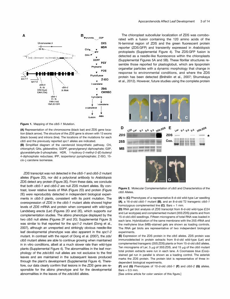
ZDS transcript was not detected in the clb5-1 and clb5-2mutantalleles (Figure 2D), nor did a polyclonal antibody to ArabidopsisZDS detect any protein (Figure 2E). From these data, we concludethat both clb5-1 and clb5-2 are null ZDS mutant alleles. By con-trast, lower relative levels of RNA (Figure 2D) and protein (Figure2E) were reproducibly detected in independent biological experi-ments in clb5-3 plants, consistent with its point mutation. Theoverexpression of ZDS in the clb5-1 mutant allele showed higherlevels of ZDS mRNA and protein when compared with wild-typeLandsberg erecta (Ler) (Figures 2D and 2E), which supports ourcomplementation studies. The albino phenotype displayed by thetwo clb5 null alleles (Figures 2F and 2G; Supplemental Figure 3)was similar to that reported for the spc1-2 mutant (Dong et al.,2007), although an unreported and strikingly obvious needle-likeleaf developmental phenotype was also apparent in the spc1-2mutant. In contrast with the report of Dong et al. (2007), the zds/clb5 mutant alleles are able to continue growing when maintainedin in vitro conditions, albeit at a much slower rate than wild-typeplants (Supplemental Figure 4). The abnormalities in the leaf mor-phology of the zds/clb5 null alleles are not exclusive to the firstleaves and are maintained in the subsequent leaves producedthrough the plant’s development (Supplemental Figure 4). There-fore, our data clearly confirm that lesions in the ZDS gene are re-sponsible for the albino phenotype and for the developmentalabnormalities in the leaves of the zds/clb5 alleles.
The chloroplast subcellular localization of ZDS was corrobo-rated with a fusion containing the 120 amino acids of theN-terminal region of ZDS and the green fluorescent proteinreporter (ZDS:GFP) and transiently expressed in Arabidopsisprotoplasts (Supplemental Figure 4). The ZDS:GFP fusion isdetected as a needle-like fluorescence within the chloroplasts(Supplemental Figures 5A and 5B). These fibrillar structures re-semble those reported for plastoglobuli, which are lipoproteinorganellar particles with a dynamic morphology that change inresponse to environmental conditions, and where the ZDSprotein has been detected (Bréhélin et al., 2007; Shumskayaet al., 2012). However, future studies using the complete protein
Figure 1. Mapping of the clb5-1 Mutation.
(A) Representation of the chromosome (black bar) and ZDS gene loca-tion (black arrow). The structure of the ZDS gene is shown with 13 exons(black boxes) and introns (line). The locations of the mutations for eachclb5 and the previously reported spc1 alleles are indicated.(B) Simplified diagram of the carotenoid biosynthetic pathway. Chl,chlorophyll; GAs, gibberellins; GGPP, geranylgeranyl diphosphate; G3P,glyceraldehyde-3-phosphate; HDR, 1-hydroxy-2-methyl-2-(E )-butenyl4-diphosphate reductase; IPP, isopentenyl pyrophosphate; Z-ISO, 15-cis-z-carotene isomerase.
Figure 2. Molecular Complementation of clb5 and Characteristics of theclb5 Alleles.
(A) to (C) Phenotypes of a representative 8-d-old wild-type Ler seedling(A), a 16-d-old clb5-1 mutant (B), and an 8-d-old T2 transgenic clb5-1homozygous complemented line (C). Bars = 1 mm.(D) RNA gel blot analysis of ZDS transcript from 8-d-old wild-type (C24and Ler ecotypes) and complemented mutant (35S:ZDS) plants and from15-d-old clb5 seedlings. Fifteen micrograms of total RNA was loaded ineach lane. Hybridization of the same membrane with the 25S rRNA andthe methylene blue (MB)–stained gels are shown as loading controls.The RNA gel blots are representative of two independent biologicalexperiments.(E) Expression of the ZDS protein in the clb5 alleles. ZDS protein wasimmunodetected in protein extracts from 8-d-old wild-type (Ler) andcomplemented transgenic (35S:ZDS) plants or from 15-d-old clb5 alleles.Ten micrograms of Ler, 5 mg of 35S:ZDS, and 15 mg of the clb5 mutanttotal protein extracts were run in each lane. A Coomassie blue (Coo)–stained gel run in parallel is shown as a loading control. The asteriskmarks the ZDS protein. The protein blot is representative of three in-dependent biological experiments.(F) and (G) Phenotypes of 10-d-old clb5-1 (F) and clb5-2 (G) alleles.Bars = 0.5 mm.[See online article for color version of this figure.]
Apocarotenoids Affect Leaf Development 3 of 14
are required to confirm this possibility. Accordingly, proteinquantification by antibody detection showed higher levels of theZDS protein in photosynthetic tissues, which is consistent withthe function of this protein (Supplemental Figure 6). Further-more, the protein expression pattern observed is also consistentwith the mRNA transcript expression profile reported previously(Dong et al., 2007).
Loss of ZDS Alters Leaf and Chloroplast Development
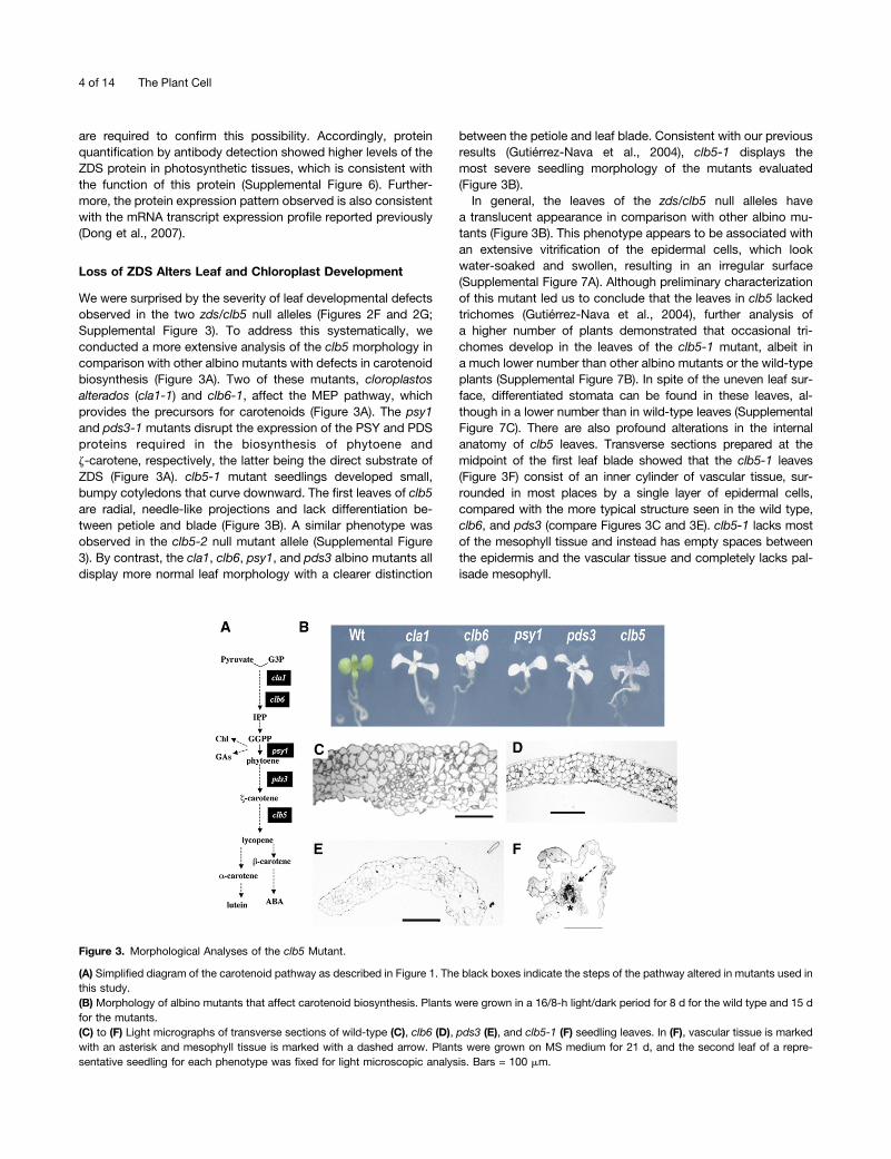
We were surprised by the severity of leaf developmental defectsobserved in the two zds/clb5 null alleles (Figures 2F and 2G;Supplemental Figure 3). To address this systematically, weconducted a more extensive analysis of the clb5 morphology incomparison with other albino mutants with defects in carotenoidbiosynthesis (Figure 3A). Two of these mutants, cloroplastosalterados (cla1-1) and clb6-1, affect the MEP pathway, whichprovides the precursors for carotenoids (Figure 3A). The psy1and pds3-1 mutants disrupt the expression of the PSY and PDSproteins required in the biosynthesis of phytoene andz-carotene, respectively, the latter being the direct substrate ofZDS (Figure 3A). clb5-1 mutant seedlings developed small,bumpy cotyledons that curve downward. The first leaves of clb5are radial, needle-like projections and lack differentiation be-tween petiole and blade (Figure 3B). A similar phenotype wasobserved in the clb5-2 null mutant allele (Supplemental Figure3). By contrast, the cla1, clb6, psy1, and pds3 albino mutants alldisplay more normal leaf morphology with a clearer distinction
between the petiole and leaf blade. Consistent with our previousresults (Gutiérrez-Nava et al., 2004), clb5-1 displays themost severe seedling morphology of the mutants evaluated(Figure 3B).In general, the leaves of the zds/clb5 null alleles have
a translucent appearance in comparison with other albino mu-tants (Figure 3B). This phenotype appears to be associated withan extensive vitrification of the epidermal cells, which lookwater-soaked and swollen, resulting in an irregular surface(Supplemental Figure 7A). Although preliminary characterizationof this mutant led us to conclude that the leaves in clb5 lackedtrichomes (Gutiérrez-Nava et al., 2004), further analysis ofa higher number of plants demonstrated that occasional tri-chomes develop in the leaves of the clb5-1 mutant, albeit ina much lower number than other albino mutants or the wild-typeplants (Supplemental Figure 7B). In spite of the uneven leaf sur-face, differentiated stomata can be found in these leaves, al-though in a lower number than in wild-type leaves (SupplementalFigure 7C). There are also profound alterations in the internalanatomy of clb5 leaves. Transverse sections prepared at themidpoint of the first leaf blade showed that the clb5-1 leaves(Figure 3F) consist of an inner cylinder of vascular tissue, sur-rounded in most places by a single layer of epidermal cells,compared with the more typical structure seen in the wild type,clb6, and pds3 (compare Figures 3C and 3E). clb5-1 lacks mostof the mesophyll tissue and instead has empty spaces betweenthe epidermis and the vascular tissue and completely lacks pal-isade mesophyll.
Figure 3. Morphological Analyses of the clb5 Mutant.
(A) Simplified diagram of the carotenoid pathway as described in Figure 1. The black boxes indicate the steps of the pathway altered in mutants used inthis study.(B) Morphology of albino mutants that affect carotenoid biosynthesis. Plants were grown in a 16/8-h light/dark period for 8 d for the wild type and 15 dfor the mutants.(C) to (F) Light micrographs of transverse sections of wild-type (C), clb6 (D), pds3 (E), and clb5-1 (F) seedling leaves. In (F), vascular tissue is markedwith an asterisk and mesophyll tissue is marked with a dashed arrow. Plants were grown on MS medium for 21 d, and the second leaf of a repre-sentative seedling for each phenotype was fixed for light microscopic analysis. Bars = 100 mm.
4 of 14 The Plant Cell
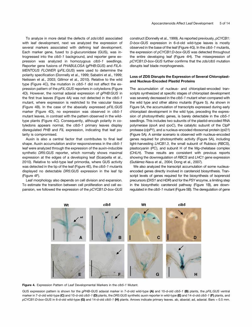
To analyze in more detail the defects of zds/clb5 associatedwith leaf development, next we analyzed the expression ofseveral markers associated with defining leaf development.Each marker gene, fused to b-glucuronidase (GUS), was in-trogressed into the clb5-1 background, and reporter gene ex-pression was analyzed in homozygous clb5-1 seedlings.Reporter gene fusions of PHABULOSA (gPHB:GUS) and FILA-MENTOUS FLOWER (pFIL:GUS) were used to determine thepolarity specification (Donnelly et al., 1999; Sabatini et al., 1999;Nelissen et al., 2003; Gillmor et al., 2010). Relative to the wildtype (Figure 4C), the mutation in clb5-1 did not affect the ex-pression pattern of the pFIL:GUS reporters in cotyledons (Figure4D). However, the normal adaxial expression of gPHB:GUS inthe first true leaves (Figure 4A) was not detected in the clb5-1mutant, where expression is restricted to the vascular tissue(Figure 4B). In the case of the abaxially expressed pFIL:GUSmarker (Figure 4D), no expression was observed in clb5-1mutant leaves, in contrast with the pattern observed in the wild-type plants (Figure 4C). Consequently, although polarity in co-tyledons appears normal, the clb5-1 primary leaves displaydisregulated PHB and FIL expression, indicating that leaf po-larity is compromised.
Auxin is also a central factor that contributes to final leafshape. Auxin accumulation and/or responsiveness in the clb5-1leaf were analyzed through the expression of the auxin-induciblesynthetic DR5:GUS reporter, which normally shows maximalexpression at the edges of a developing leaf (Scarpella et al.,2010). Relative to wild-type leaf primordia, where GUS activitywas detected in the tip of the leaf (Figure 4E), the clb5-1mutantsdisplayed no detectable DR5:GUS expression in the leaf tip(Figure 4F).
Leaf morphology also depends on cell division and expansion.To estimate the transition between cell proliferation and cell ex-pansion, we followed the expression of the pCYCB1:D-box-GUS
construct (Donnelly et al., 1999). As reported previously, pCYCB1:D-box-GUS expression in 6-d-old wild-type leaves is mostlyobserved in the base of the leaf (Figure 4G). In the clb5-1mutants,the expression of pCYCB1:D-box-GUS was detected throughoutthe entire developing leaf (Figure 4H). The misexpression ofpCYCB1:D-box-GUS further confirms that the zds/clb5 mutationdisrupts leaf blade morphogenesis.
Loss of ZDS Disrupts the Expression of Several Chloroplast-and Nucleus-Encoded Plastid Proteins
The accumulation of nucleus- and chloroplast-encoded tran-scripts synthesized at specific stages of chloroplast developmentwas severely decreased in the clb5-1mutant when compared withthe wild type and other albino mutants (Figure 5). As shown inFigure 5A, the accumulation of transcripts expressed during earlychloroplast development in the wild type, preceding the expres-sion of photosynthetic genes, is barely detectable in the clb5-1seedlings. This includes two subunits of the plastid-encoded RNApolymerase (rpoA and rpoC), the catalytic subunit of the ClpPprotease (clpP1), and a nucleus-encoded ribosomal protein (rpl21)(Figure 5A). A similar scenario is observed with nucleus-encodedgenes required for photosynthetic activity (Figure 5A), includinglight-harvesting LHCB1.3, the small subunit of Rubisco (RBCS),plastocyanin (PC), and subunit H of the Mg-chelatase complex(CHLH). These results are consistent with previous reportsshowing the downregulation of RBCS and LHC1 gene expression(Gutiérrez-Nava et al., 2004; Dong et al., 2007).We also analyzed the transcript accumulation of some nucleus-
encoded genes directly involved in carotenoid biosynthesis. Tran-script levels of genes required for the biosynthesis of isoprenoidprecursors (DXS1 andHDR) and for the PSY enzyme, a limiting stepin the biosynthetic carotenoid pathway (Figure 1B), are down-regulated in the clb5-1mutant (Figure 5B). The deregulation of gene
Figure 4. Expression Pattern of Leaf Developmental Markers in the clb5-1 Mutant.
GUS expression pattern is shown for the gPHB-GUS adaxial marker in 7-d-old wild-type (A) and 10-d-old clb5-1 (B) plants, the pFIL:GUS ventralmarker in 7-d-old wild-type (C) and 10-d-old clb5-1 (D) plants, the DR5:GUS synthetic auxin reporter in wild-type (E) and 14-d-old clb5-1 (F) plants, andpCYCB1:D-box-GUS in 6-d-old wild-type (G) and 14-d-old clb5-1 (H) plants. Arrows indicate primary leaves. ab, abaxial; ad, adaxial. Bars = 0.5 mm.
Apocarotenoids Affect Leaf Development 5 of 14

expression in clb5-1 is not global, as the accumulation of PDS orCRTISO transcripts remains unaltered in clb5-1 in comparison withother albino mutants and wild-type plants.
Because of the substantial posttranscriptional regulation ofproteins in chloroplasts, we also checked whether the RNAabundance defects observed in clb5-1 are also reflected at theprotein level. We confirmed that the abundance of the a-subunitof the chloroplast-encoded RNA polymerase (RpoA) and the1-deoxy-D-xylulose 5-phosphate synthase (DXS) enzymes isalmost undetectable in clb5-1 compared with the wild type and
other albino mutants (Figure 5C), which demonstrates that thechanges observed at the transcription level are also reflected atthe protein level.
The Accumulation of Specific Carotenes Is Responsible forthe clb5 Mutant Phenotype and Affects Gene Expression
The hormones ABA and SL are two signals derived from ca-rotenoids that affect plant development, including leaf morphol-ogy (Liu et al., 2012; Ramel et al., 2013). Thus, we next askedwhether or not treatment with these hormones could restore theleaf phenotype of clb5. We found that the exogenous addition ofthese two hormones does not alleviate the finger-like leaf mor-phology typically observed in the clb5 mutant (SupplementalFigure 8). However, the presence of ABA (0.5 and 1 mM) severelyretarded the development of wild-type and mutant seedlingscompared with its absence, supporting the action of this hormonein this assay. Also, the presence of SL resulted in the productionof higher numbers of leaves in the clb5-1 and pds3 albino mu-tants in a shorter time, supporting the effectiveness of the SLtreatment.Carotenoids also serve as essential photoprotectors, and
carotenoid-deficient mutants are subject to extensive photoox-idative stress (Triantaphylidès and Havaux, 2009). It has alsobeen documented that reactive oxygen species (ROS) might actas a signal to modulate plastid-to-nucleus communications anddevelopment (Pogson et al., 2008; Bräutigam et al., 2009), andthe clb5 phenotypes could be due to the accumulation of ROS.We tested this possibility by quantifying the levels of H2O2 andO2
2 in the clb5 mutant. We also analyzed the expression of theROS-responsive gene ZAT12 (Davletova et al., 2005) and thephotosystem II–associated protein EXECUTER1 (EX1 gene),which is an essential component of O2
2 signaling (Lee et al.,2007). We could not detect any major differences in the ROSlevels or in the expression levels of the ZAT12 and EX1 genes inthe clb5-1 mutant compared with other carotenoid-deficient mu-tants (Supplemental Figure 9). In summary, the clb5 finger-like leafmorphology does not appear to be a consequence of low ABA orSL levels, nor is it a result of the overaccumulation of ROS.Other possibilities to explain the phenotypic alterations asso-
ciated with clb5 are that ZDS directly regulates gene expressionand development through an activity on a substrate other thancarotenoids or that the absence of ZDS enzyme generates orinhibits the production of a specific carotenoid-derived signal. Forexample, in addition to hormones, an oxidative cleavage productof b-carotene acts as signaling molecule in response to oxidativestress, and cis-carotenes have been implicated as signals thatmediate gene expression in Arabidopsis (Ramel et al., 2012). Todistinguish between these possibilities, clb5-1 seedlings weregrown in the presence or absence of fluridone. Fluridone inhibitsPDS activity, which functions in the enzymatic step prior to ZDS(Figure 1B) (Bartels and Watson, 1978). We reasoned that ifa signal is derived from phytofluene and/or z-carotenoids thataccumulate in ZDS mutants, then the needle-like leaf morphologyobserved in clb5-1mutants may be partially complemented in thepresence of fluridone.Homozygous clb5-1 and pds3mutants and wild-type controls
were transferred to medium with fluridone 4 d after germination
Figure 5. Expression of Nucleus- and Chloroplast-Encoded Transcriptsand Proteins.
(A) Expression of chloroplast marker genes: clpP1, rpoA, rpoC, rpl21,LHCB1.3, RBCS, PC, and CHLH.(B) Nucleus-encoded genes involved in the carotenoid biosyntheticpathway: DXS1, HDR, PSY, PDS, ZDS, and CRTISO probes.Each lane contains 15 mg of total RNA from 8-d-old wild-type and 15-d-old mutant seedlings. The hybridization of the 25S rRNA and methyleneblue (MB)–stained gels are shown as loading controls. These data arerepresentative of two independent biological experiments.(C) Analysis of the protein levels of RpoA and nucleus-encoded DXS1.Total protein extracts were isolated from 8-d-old wild-type (Ler) and15-d-old cla1-1, clb6, pds3, and clb5-1 mutant seedlings. Immunoblot-ting was performed with specific antibodies against the RpoA and DXS1proteins. Each lane contains 15 mg of total protein extract. A Coomassieblue–stained gel run in parallel with the same samples is shown asa loading control (Coo).Asterisks mark the clb5 mutant lanes. The protein blots shown are rep-resentative from three independent biological replicates.
6 of 14 The Plant Cell
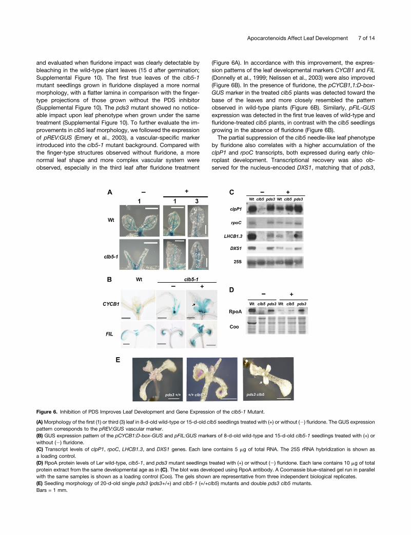
and evaluated when fluridone impact was clearly detectable bybleaching in the wild-type plant leaves (15 d after germination;Supplemental Figure 10). The first true leaves of the clb5-1mutant seedlings grown in fluridone displayed a more normalmorphology, with a flatter lamina in comparison with the finger-type projections of those grown without the PDS inhibitor(Supplemental Figure 10). The pds3 mutant showed no notice-able impact upon leaf phenotype when grown under the sametreatment (Supplemental Figure 10). To further evaluate the im-provements in clb5 leaf morphology, we followed the expressionof pREV:GUS (Emery et al., 2003), a vascular-specific markerintroduced into the clb5-1 mutant background. Compared withthe finger-type structures observed without fluridone, a morenormal leaf shape and more complex vascular system wereobserved, especially in the third leaf after fluridone treatment
(Figure 6A). In accordance with this improvement, the expres-sion patterns of the leaf developmental markers CYCB1 and FIL(Donnelly et al., 1999; Nelissen et al., 2003) were also improved(Figure 6B). In the presence of fluridone, the pCYCB1,1:D-box-GUS marker in the treated clb5 plants was detected toward thebase of the leaves and more closely resembled the patternobserved in wild-type plants (Figure 6B). Similarly, pFIL-GUSexpression was detected in the first true leaves of wild-type andfluridone-treated clb5 plants, in contrast with the clb5 seedlingsgrowing in the absence of fluridone (Figure 6B).The partial suppression of the clb5 needle-like leaf phenotype
by fluridone also correlates with a higher accumulation of theclpP1 and rpoC transcripts, both expressed during early chlo-roplast development. Transcriptional recovery was also ob-served for the nucleus-encoded DXS1, matching that of pds3,
Figure 6. Inhibition of PDS Improves Leaf Development and Gene Expression of the clb5-1 Mutant.
(A)Morphology of the first (1) or third (3) leaf in 8-d-old wild-type or 15-d-old clb5 seedlings treated with (+) or without (2) fluridone. The GUS expressionpattern corresponds to the pREV:GUS vascular marker.(B) GUS expression pattern of the pCYCB1:D-box-GUS and pFIL:GUS markers of 8-d-old wild-type and 15-d-old clb5-1 seedlings treated with (+) orwithout (2) fluridone.(C) Transcript levels of clpP1, rpoC, LHCB1.3, and DXS1 genes. Each lane contains 5 mg of total RNA. The 25S rRNA hybridization is shown asa loading control.(D) RpoA protein levels of Ler wild-type, clb5-1, and pds3 mutant seedlings treated with (+) or without (2) fluridone. Each lane contains 10 mg of totalprotein extract from the same developmental age as in (C). The blot was developed using RpoA antibody. A Coomassie blue–stained gel run in parallelwith the same samples is shown as a loading control (Coo). The gels shown are representative from three independent biological replicates.(E) Seedling morphology of 20-d-old single pds3 (pds3+/+) and clb5-1 (+/+clb5) mutants and double pds3 clb5 mutants.Bars = 1 mm.
Apocarotenoids Affect Leaf Development 7 of 14
and to lesser extent for the LHCB1.3 and RBCS genes (Figure6C), which are expressed during later stages of plastid de-velopment. The level of the RpoA protein was partially restoredby fluridone treatment to levels similar to those detected in thefluridone-treated wild-type seedlings (Figure 6D). By contrast,no difference in the RpoA protein levels was observed in thepds3 seedlings in the absence or presence of fluridone (Figure6D). Taken together, these data support the notion that it is notthe ZDS protein itself but rather the accumulation of phytoflueneand z-carotenoids or their derivatives that is responsible for theneedle-like leaf morphology as well as the gene expressiondefects observed in the clb5 mutant.
To further support that the phenotypes observed in clb5 arecaused by the accumulation of specific carotenoids and are nota secondary effect of the fluridone inhibitor used in the previousanalyses, a double pds3 clb5mutant was generated (SupplementalFigure 11). In agreement with our previous results, the leaf phe-notype displayed by pds3 clb5 double mutants appeared similar tothe pds3 allele, showing a flat lamina and the presence of tri-chomes, which was very different from the finger-type projectionsobserved in clb5 (Figure 6E). These data conclusively demonstratethat the leaf defects observed in the clb5 mutant result from theaccumulation of phytofluene and z-carotenoids as a consequenceof the absence of ZDS.
Deficiency in ZDS activity results in the accumulation of higherlevels of cis-carotenoids (Dong et al., 2007). We used HPLC toconfirm that z-carotene was the predominant carotenoid in clb5-1.In wild-type plants grown under standard light conditions, thepigment profile consists of b-carotene plus the three oxygenatedxanthophylls, lutein, violaxanthin, and neoxanthin (SupplementalFigure 12). In the pds3 mutant, no significant levels of chlorophyllor carotenoid pigments are detected, with the exception of phy-toene (assayed spectrophotometrically at 286 nm) (SupplementalFigure 12), as reported previously (Qin et al., 2007). In contrast, themost abundant carotenoid in clb5-1 was z-carotene, with traceamounts of phytofluene and cis-isomers of z-carotene alsodetected (Supplemental Figure 12). This is similar to previouslypublished spectral data for ZDS mutants (Matthews et al., 2003;Conti et al., 2004; Dong et al., 2007).
CCD4 Is Required to Promote clb5 Mutant Leaf andExpression Phenotypes
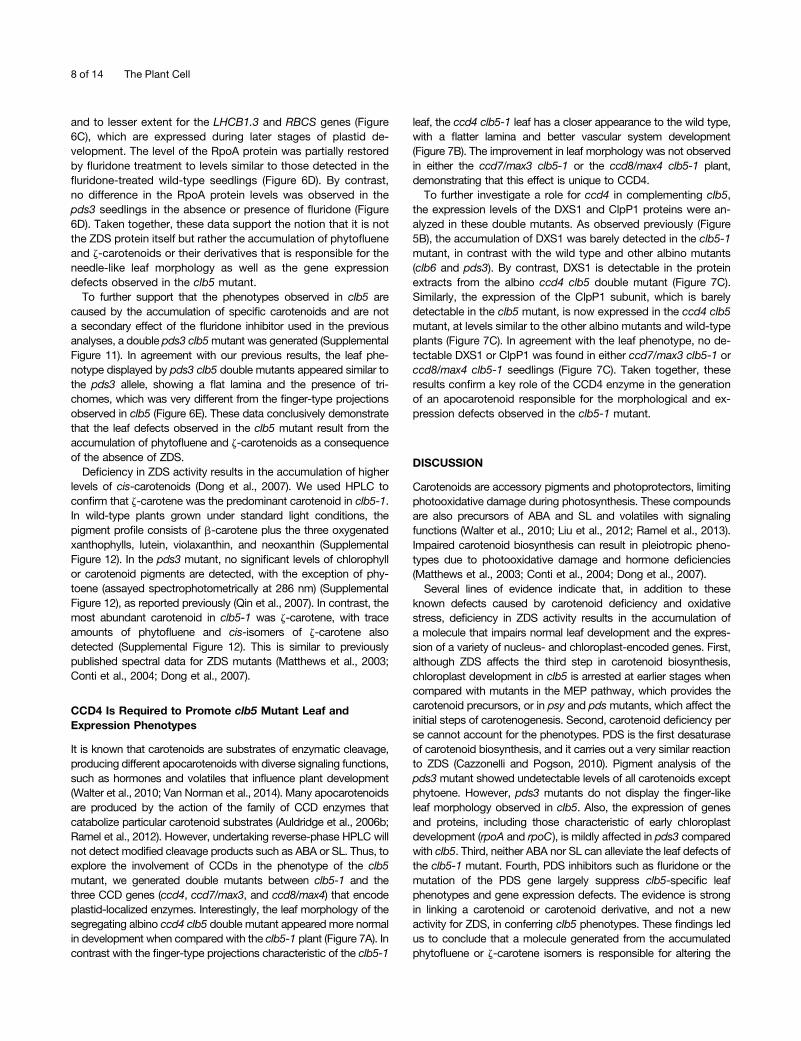
It is known that carotenoids are substrates of enzymatic cleavage,producing different apocarotenoids with diverse signaling functions,such as hormones and volatiles that influence plant development(Walter et al., 2010; Van Norman et al., 2014). Many apocarotenoidsare produced by the action of the family of CCD enzymes thatcatabolize particular carotenoid substrates (Auldridge et al., 2006b;Ramel et al., 2012). However, undertaking reverse-phase HPLC willnot detect modified cleavage products such as ABA or SL. Thus, toexplore the involvement of CCDs in the phenotype of the clb5mutant, we generated double mutants between clb5-1 and thethree CCD genes (ccd4, ccd7/max3, and ccd8/max4) that encodeplastid-localized enzymes. Interestingly, the leaf morphology of thesegregating albino ccd4 clb5 double mutant appeared more normalin development when compared with the clb5-1 plant (Figure 7A). Incontrast with the finger-type projections characteristic of the clb5-1
leaf, the ccd4 clb5-1 leaf has a closer appearance to the wild type,with a flatter lamina and better vascular system development(Figure 7B). The improvement in leaf morphology was not observedin either the ccd7/max3 clb5-1 or the ccd8/max4 clb5-1 plant,demonstrating that this effect is unique to CCD4.To further investigate a role for ccd4 in complementing clb5,
the expression levels of the DXS1 and ClpP1 proteins were an-alyzed in these double mutants. As observed previously (Figure5B), the accumulation of DXS1 was barely detected in the clb5-1mutant, in contrast with the wild type and other albino mutants(clb6 and pds3). By contrast, DXS1 is detectable in the proteinextracts from the albino ccd4 clb5 double mutant (Figure 7C).Similarly, the expression of the ClpP1 subunit, which is barelydetectable in the clb5 mutant, is now expressed in the ccd4 clb5mutant, at levels similar to the other albino mutants and wild-typeplants (Figure 7C). In agreement with the leaf phenotype, no de-tectable DXS1 or ClpP1 was found in either ccd7/max3 clb5-1 orccd8/max4 clb5-1 seedlings (Figure 7C). Taken together, theseresults confirm a key role of the CCD4 enzyme in the generationof an apocarotenoid responsible for the morphological and ex-pression defects observed in the clb5-1 mutant.
DISCUSSION
Carotenoids are accessory pigments and photoprotectors, limitingphotooxidative damage during photosynthesis. These compoundsare also precursors of ABA and SL and volatiles with signalingfunctions (Walter et al., 2010; Liu et al., 2012; Ramel et al., 2013).Impaired carotenoid biosynthesis can result in pleiotropic pheno-types due to photooxidative damage and hormone deficiencies(Matthews et al., 2003; Conti et al., 2004; Dong et al., 2007).Several lines of evidence indicate that, in addition to these
known defects caused by carotenoid deficiency and oxidativestress, deficiency in ZDS activity results in the accumulation ofa molecule that impairs normal leaf development and the expres-sion of a variety of nucleus- and chloroplast-encoded genes. First,although ZDS affects the third step in carotenoid biosynthesis,chloroplast development in clb5 is arrested at earlier stages whencompared with mutants in the MEP pathway, which provides thecarotenoid precursors, or in psy and pds mutants, which affect theinitial steps of carotenogenesis. Second, carotenoid deficiency perse cannot account for the phenotypes. PDS is the first desaturaseof carotenoid biosynthesis, and it carries out a very similar reactionto ZDS (Cazzonelli and Pogson, 2010). Pigment analysis of thepds3 mutant showed undetectable levels of all carotenoids exceptphytoene. However, pds3 mutants do not display the finger-likeleaf morphology observed in clb5. Also, the expression of genesand proteins, including those characteristic of early chloroplastdevelopment (rpoA and rpoC), is mildly affected in pds3 comparedwith clb5. Third, neither ABA nor SL can alleviate the leaf defects ofthe clb5-1 mutant. Fourth, PDS inhibitors such as fluridone or themutation of the PDS gene largely suppress clb5-specific leafphenotypes and gene expression defects. The evidence is strongin linking a carotenoid or carotenoid derivative, and not a newactivity for ZDS, in conferring clb5 phenotypes. These findings ledus to conclude that a molecule generated from the accumulatedphytofluene or z-carotene isomers is responsible for altering the
8 of 14 The Plant Cell
expression of a number of chloroplast-related genes and for dis-rupting leaf development.
Pigment analysis showed that clb5 seedlings accumulate phy-tofluene and z-carotene isomers. However, given the low levels ofthe phytofluene and z-carotenoid species that accumulate in the
clb5 mutant, the lack of appropriate standards, and the observa-tion that most hormones and signals are produced at very lowlevels, it would be a substantial challenge to identify the signalresponsible for the clb5 phenotypes by HPLC fractionation. In-deed, the structure of SL is very different from that of the apo-carotenoid it is derived from (Alder et al., 2012), so geneticevidence is fundamental to identifying the nature of the signal.It is well established that carotenoids are subject to extensive
enzymatic and nonenzymatic modifications, including oxidationand cleavage. Many apocarotenoids produced through theseremodeling processes have signaling activities, as they can moveto different subcellular compartments in which they perform vari-ous biological functions, including modulating developmentalprocesses such as lateral root pattering (Bouvier et al., 2005;Walter et al., 2010; Van Norman et al., 2014). We reasoned that itwas possible that an apocarotenoid derived from the linear ca-rotenoids that accumulate in clb5 could generate a signal re-sponsible for the described clb5 phenotypes. From the nine CCDgenes in Arabidopsis, five participate in the biosynthesis of ABA(NCED2, -3, -5, -6, and -9) and two in the synthesis of SL (CCD7and CCD8). The functions of the cytosolic CCD1 and plastidicCCD4 enzymes are less clear, although they have been implicatedin the production of some plant volatiles (Walter et al., 2010; Ramelet al., 2012). Apparently, both enzymes can cleave linear ca-rotenoids at C9-C10, C99, and C109 (Vogel et al., 2008; Huang et al.,2009).One of the most significant findings of this work is that the
absence of CCD4 rescues the phenotypic and gene expressiondefects of the clb5-1 mutant. This is a specific function of CCD4,as it was not observed with CCD7 or CCD8. We demonstrate thatCCD4 has the capacity to recognize and cleave one or more of thecarotenes that accumulate in clb5 and that this cleavage product,or a derivative thereof, is responsible for the morphological andgene expression defects observed in the clb5 mutant. This resultalso supports that the clb5 phenotypes result from the accumu-lation of phytofluene and z-carotene isomers and not from theaccumulation of a new toxic metabolite. However, this findingcontrasts with previous data using in vitro Escherichia coli assays,where it was found that CCD4 does not accept z-carotene as thepreferred substrate (Huang et al., 2009). One possible explanationis that z-carotene itself might not be the direct substrate of CCD4but instead phytofluene or even a not-yet identified compoundderived from these cis-linear carotenoids. An alternative explana-tion is that the substrate specificity of CCD4 differs between the invitro and in vivo studies. Interestingly, a recent analysis in tomato(Solanum lycopersicum) speculates that two distantly relatedCRTISO genes have the capacity to use cis-z-carotenes andtransform them to all-trans-z-carotene, initiating a new metabolicbranch (Fantini et al., 2013). Putative orthologs of these genes(CRTISO-L) also exist in Arabidopsis, opening the possibility thatall-trans-z-carotene could also be a substrate for the CCD4 en-zyme. Interestingly, recent work with tomato mutants providedevidence of a regulatory signal produced from linear cis-carotenoids,such as neurosporene and prolycopene, that regulate PSY expres-sion (Kachanovsky et al., 2012).CCD4-related genes are found in diverse plant species, sup-
porting an important role for this gene (Ahrazem et al., 2010). Re-cent evidence indicated that CCD4 is important in the turnover of
Figure 7. Role of CCDs in the Morphological and Expression Defects ofthe clb5-1 Mutant.
(A) Morphology of seedlings from 8-d-old wild-type and ccd4 mutantseedlings or 16-d-old clb5-1, ccd4 clb5-1, ccd7/max3 clb5-1, and ccd8/max4 clb5-1 mutant seedlings. Bars 5 1 mm.(B) Close-ups of the primary leaf of 16-d-old clb5 and two representativeccd4 clb5-1 leaves. Bars = 1 mm.(C) Analysis of the protein levels of the nucleus-encoded DXS1 and thechloroplast-encoded ClpP1. Total protein extracts were isolated from 8-d-old wild-type (Ler) and ccd4 mutant seedlings or from 15-d-old cla1-1,clb6, pds3, clb5-1, ccd4 clb5-1, ccd7/max3 clb5-1, and ccd8/max4 clb5-1mutant seedlings. Immunoblotting was performed with polyclonal anti-bodies against the DXS1 and ClpP proteins as described in Methods.Each lane contains 15 mg of total protein extract. The Ponceau-stainedmembrane is shown as a loading control (P). Asterisks mark the lanescorresponding to clb5-1 and ccd4 clb5-1 mutants. The protein gel shownis representative from three independent biological replicates.[See online article for color version of this figure.]
Apocarotenoids Affect Leaf Development 9 of 14
b-carotenoids during seed maturation and leaf senescence inArabidopsis (Gonzalez-Jorge et al., 2013). Accordingly, the ex-pression analysis through quantitative RT-PCR and from trans-genic plants showed that the CCD4 genes are preferentiallyexpressed in late seed development and flowers, although lowexpression levels were also detected in mature leaves (Huanget al., 2009; Ahrazem et al., 2010). Moreover CCD4-related genesin other plants have undergone gene duplications that appear to besubjected to stringent regulation. However, so far, no leaf pheno-type associated with the ccd4 mutant has been reported. Clearly,a more detailed analysis of the expression pattern of CCD4 duringleaf development and of the phenotype of the ccd4 mutant couldshed light on a possible signaling role of the apocarotenoid derivedfrom this enzymatic activity.
When and where does the signal act? It is tempting to speculatethat the molecule responsible for the defects described in this workinvolves a signal that regulates gene expression and leaf de-velopment, although we cannot exclude that (1) a clb5-generatedapocarotenoid does not become synthesized during normal Ara-bidopsis development, (2) the observed defects result from thefortuitous interaction of this molecule with one or more plant tar-gets, and (3) the apocarotenoid is produced during a particulardevelopmental stage or in response to an environmental change.In fact, based on the pleiotropic phenotypes that result from thisclb5 carotenoid-derived signal, one could speculate that the pro-duction of this signal is probably regulated in response to specificphysiological conditions. In this sense, it is interesting that pro-teomic studies demonstrated that CCD4 colocalizes with ZDS inplastoglobules, which are the site of high carotenoid accumulationunder stressful conditions (Ytterberg et al., 2006). Whether theCCD4 colocalization or cell-specific expression patterns are im-portant for the generation of this signal is a matter for future re-search. In addition, recent data showed that the rate-limitingenzymes of the carotenoid pathway, such as PSY, display a par-ticular expression pattern (Van Norman et al., 2014).
From a physiological point of view, plastid-derived regulation ofgene expression and development might provide an importantfeedback mechanism that reflects the capacity of the organelle toestablish a functional photosynthetic apparatus in response toenvironmental cues such as light. Indeed, the SAL1–39-phos-phoadenosine 59-phosphate retrograde signaling pathway thatoperates during abiotic stress also alters developmental pro-cesses such as flowering time and petiole length (Estavillo et al.,2011). It is interesting that during evolution, the CrtI-type desa-turases capable of catalyzing the entire desaturation steps toproduce lycopene present in archea, bacteria, and fungi weresubstituted in cyanobacteria and maintained in all plants by fourenzymes, including the PDS and ZDS desaturases (Sandmann,2009). Thus, this substitution provides the capacity of these en-zymes to link carotenogenesis with the photosynthetic electronchain via the plastoquinone pool (Foudree et al., 2010). Perhapsthe reactions of these two desaturases are strategic in their ca-pacity to perceive the metabolic status and functionality of theorganelle. One could speculate that a signal derived from theintermediate linear cis-carotenoid molecules might be an impor-tant link to the metabolic status of the organelle by adjusting thedevelopment of complex body plans present in vascular plants(Norris et al., 1995).
The severe abnormality of leaf morphology and anatomy inclb5 is striking compared with other albino mutants. However,this is not exclusive to clb5, since it is well recognized how im-portant the chloroplast status is in defining leaf architecture (Pykeet al., 2000). Several mutants affected in chloroplast developmentand functionality display defects in specific aspects of plant de-velopment. For example, altered differentiation and division of thepalisade cells has been observed in several mutants that affectchloroplast biogenesis, such as dag and scabra3 (Chatterjeeet al., 1996; Hricová et al., 2006), and in carotenoid biosynthesismutants, such as ghost (Scolnik et al., 1987). Such defects havebeen attributed to photooxidative damage induced by alteredcarotenoid profiles that, in turn, regulate terminal stages of me-sophyll cell differentiation (Woodson and Chory, 2008). The factthat several mutants affected in chloroplast biogenesis displaydefects in leaf anatomy and in chloroplast- and nucleus-encodedgenes further supports the existence of a plastid-derived signalthat regulates leaf development. Clearly, the plastid status canaffect the differentiation of a particular cell type and the leafshape. Early studies have demonstrated that the genotype of theplastids in evening primrose (Oenothera biennis) can underpin leafshape (Tiller and Bock, 2014).Leaf development is a process that involves the differentiation of
specialized cells along different axes to achieve the correct sizeand shape. It is well known that this process requires the partici-pation of a variety of regulators, including hormones, small RNAs,and transcription factors, that function in a multilayered regulatorynetwork to influence development (Tsukaya, 2013). The morpho-logical abnormalities observed in clb5 occur earlier in developmentand are more severe than the ones observed in mutants like cla1and pds3. This finding, together with the altered expression profileobserved in clb5, indicates that the disruption of ZDS generatesa molecule in the chloroplast that affects the earlier stages ofchloroplast and leaf development. Our analysis of the expressionpattern of the adaxial PHB and abaxial FIL markers demonstratedthat the finger-type leaves in the clb5mutant display defects in thedorsoventrality differentiation of the leaf, resembling an abaxializedleaf (Liu et al., 2012). Interestingly, HD-ZIPIII–type transcriptionfactors required for adaxial specification contain a START domain,related to the ABA binding domain from the PYR ABA receptor,that is predicted to bind a yet unidentified ligand (Liu et al., 2012). Itis tempting to speculate that the apocarotenoid derived from cis-z-carotenes could interact with some of these transcription factors,a possibility that remains to be addressed in the future. It is worthnoting that these defects are restricted to the primary leaves andare not observed in the cotyledons, exemplifying the differences inthe developmental regulation between these two organs with re-spect to plastid communication.A consideration of mutants that have leaf development pheno-
types similar to clb5 brought us to enlarged fil expression domain2(enf2), which is a pale seedling that harbors a mutation in a chloro-plast protein (ENF2) necessary for correct plastid gene expressionand exhibits narrow, serrated leaf defects (Tameshige et al., 2013).Similar to clb5, the enf2mutant displays defects in the expression ofthe abaxial FIL gene and in the leaf lamina morphology. The authorsdemonstrated that the enf2 defects correlate with an abaxializationof the leaf that, in turn, affects expansion of the lamina. Our analysesdemonstrated major defects in the expansion of clb5 lamina, which
10 of 14 The Plant Cell
contain fewer mesophyll cells with big air spaces and bumpy epi-dermal cells, all phenotypes that are consistent with abaxializedleaves (Liu et al., 2012; Tameshige et al., 2013). An additional sim-ilarity of profound defects in chloroplast gene expression and inorganelle protein synthesis was also observed between these mu-tants. According to this work, impairment in plastid transcription ortranslation results in a retrograde signal that modulates leaf de-velopment (Tameshige et al., 2013). In summary, the plastid playsa critical role in defining leaf morphology.
Another mutant with morphological similarities with clb5 isbypass1 (bps1). bps1 affects a protein of unknown function thatgenerates a mobile signal generated by the root that arrests leafdevelopment (Van Norman et al., 2011). Similar to clb5, thephenotype of bps1 is partially rescued by fluridone, suggestingthe participation of carotenoids (Van Norman and Sieburth, 2007).However, unlike bps1, we never observed major root defects inclb5. At this point, any connection between bps1 and clb5 isspeculative.
In conclusion, we show that, in addition to the known ABA andSL hormones, linear carotenoids can generate an apocarotenoidthat plays important roles in the regulation of leaf developmentand in the expression of a variety of chloroplast- and nucleus-encoded genes. This molecule originates from phytofluene andz-carotenoids and requires the action of the CCD4 tailoring en-zyme. This study exemplifies the importance of the carotenoidpathway in generating regulatory signals that, besides their well-established functions, can clearly affect major developmentalprocesses such as leaf development and function as feedbacksignals responding to the status of organelle development.
METHODS
Plant Material and Growth Conditions
Arabidopsis thaliana Columbia-0 and Ler ecotypes were used in thisstudy. Seed growth under sterile conditions was done in 13 Murashigeand Skoog (MS) medium with Gamborg vitamins (PhytotechnologyLaboratories) supplemented with 1% (w/v) Suc, solidified with 0.8% (w/v)phytoagar, and for adult plants in Metro-Mix 200 (SunGro). Seedlingswere grown under a 16/8-h light/dark cycle (120 mmol m22 s21) at 22°C.To break dormancy, seeds were vernalized at 4°C for 4 d. Seeds seg-regating for pds3 (ZHJ070204) were provided by Li-Jia Qu (Peking Uni-versity), and the spc1-2 (SALK_033039) mutant was obtained from theABRC. Seeds of pFIL:GUS and pREV:GUS lines were provided by JohnBowman (Monash University), pCYCB1,1:D-box-GUS by Dirk Inzé (GhentUniversity), and gPHB:GUS by Stewart Gillmor (Gillmor et al., 2010).Homozygous lines for each marker were crossed with clb5 mutant het-erozygous plants. The presence of the corresponding transgene and theclb5 mutation was corroborated through GUS histological staining andthe albino phenotype in the F2 generation. For ABA and SL treatments,clb5 heterozygous seedlings were germinated in MS medium andtransferred to MS normal medium or MS medium supplemented with0.5 mM ABA, 1 mM ABA, or 0.5 mM GR24 SL 2 d after germination, wherethey were grown in a 16/8-h light/dark period until morphology wasanalyzed (Supplemental Figure 8).
Mutant Complementation and Double Mutant Generation
The full ZDS cDNA was obtained by RT-PCR using the oligonucleotidesZDS5 and ZDS3 (Supplemental Table 1). Independent homozygous lines
for the transgene were selected, and the presence of the clb5 mutationwas corroborated by the absence of a BamHI site in a fragment amplifiedwith the ZDST5 and ZDST3 primers (Supplemental Figure 1). For gen-eration of the pds3 clb5 double mutant, single heterozygous pds3 andclb5-1mutants were crossed and the F2 progeny were genotyped by PCRusing genomic DNA. To genotype the clb5-1 allele, the CLB5 gene wasamplified using ZDS-3F and ZDS-4R oligonucleotides (SupplementalTable 1). The 586-bp product was digested with BamHI and analyzed ona 1% agarose gel (Supplemental Figures 1 and 11). PDS3 genotyping wasdone as reported (Qin et al., 2007). The ccd4 clb5-1 double mutant wasgenerated by crossing the homozygous ccd4 mutant (SALK_097984C),donated by Peter Lundquist (Heinrich-Heine-Düsseldorf University), withheterozygous clb5-1. ccd4 homozygous mutant plants were genotypedusing theCD4FWand LB T-DNA oligonucleotides (Supplemental Table 1).Double mutants between ccd7/max3 and ccd8/max4 were obtained bycrossing the homozygous ccd mutants with the clb5-1 heterozygousplants. Homozygous ccd7 and ccd8 plants were selected based on thepresence of the branching phenotype (Auldridge et al., 2006a). Thepresence of the clb5 mutation in all these lines was verified throughthe segregation of albino embryos.
Histochemical GUS Staining
The GUS histochemical assay was performed as reported previously(Jefferson, 1987). Seedlings were mounted in 50% glycerol and analyzedusing a light microscope (Nikon Eclipse E600).
Herbicide Treatment
Plants were germinated for 4 d in MS medium, and wild-type, clb5-1, andpds3 albino seedlings were transferred to medium containing 5 mMfluridone (Phytotechnology Laboratories). Plants were collected after 8 dfor the wild-type control and after 15 d for the wild-type, clb5-1, and pds3fluridone-treated seedlings.
Genetic Mapping and Sequence Analysis
A mapping population was generated by crossing CLB5+/2 plants withthe Columbia-0 ecotype. Fine-map positions were assigned by examiningrecombination frequencies between the gene of interest and linkedflanking SSLP markers in a population of 805 F2 clb5 albino and het-erozygous plants (CLB5+/2) (Lukowitz et al., 2000). SSLP markers weredesigned based on the insertion/deletion database produced by theCereon Arabidopsis polymorphism database (https://www.arabidopsis.org/browse/Cereon/index.html). Alignment of At-ZDS with the ZDS fromphotosynthetic organisms (Supplemental Figure 2) was performed usingInterPro functional analysis (http://www.ebi.ac.uk/interpro/).
Gene Expression Analyses
Total RNAwas isolated using TRIZOL (Invitrogen). For RNA gel blot analysis,the RNA was fractionated by electrophoresis and transferred onto HybondN+ nylon membranes (GE Biosciences). Hybridizations and washes wereperformed in high-stringency conditions according to standard proceduresusing 32P-radiolabeled probes produced with the Megaprime DNA labelingsystem (Amersham). The fragments used as probes were obtained by PCRusing specific oligonucleotides (Supplemental Table 1). Specific fragmentswere used for the Arabidopsis PC, RCBS, and LHCB1.3 genes. The RPL21probe was obtained from the 146E8 clone from the ABRC. As a loadingcontrol, final hybridization was performed using the 25S rRNA with a shorttime exposure. For quantitative real-time RT-PCR (Supplemental Figure 9),total RNAwas treatedwithRNase-freeDNase I (Promega) and repurifiedwiththe RNA cleanup protocol in the RNeasy kit (Qiagen). First-strand cDNA wassynthesized with MMLV reverse transcriptase (Invitrogen) using 3 mg of total
Apocarotenoids Affect Leaf Development 11 of 14
RNA and oligo(dT) primer, according to the manufacturer. Two microlitersfrom a 1:10 dilution of the first-strand solution was used for the subsequentquantitative PCR with FastStart DNA MasterPLUS SYBR Green I in theLightCycler carousel-based system (Roche). The primers used for EX1 andZAT12 genes are described in Supplemental Table 1. All relative transcriptlevels are referred to EF1. Normalized values come from three technicalreplicates from two independent experiments. Tukey statistical analysis wasused to evaluate the confidence of the data (http://www.r-project.org/).
Antibody Preparation
Polyclonal antibodies were generated against the protein product of theZDS cDNA cloned in the pDEST17 vector (Invitrogen). The recombinantproteins were purified under denaturing conditions from Escherichia colicrude extracts according to the QIAexpressionist protocol (Qiagen). Forantibody generation, 50 mg of the gel-purified ZDS protein was used toimmunize New Zealand rabbits. The ZDS antibodies were purified byimmune adsorption to nitrocellulose according to a published protocol(Harlow and Lane, 1988). The specificity of the antibodies against the ZDSprotein was confirmed by analysis of the clb5 mutants.
Protein Gel Blot Analysis
Total protein samples were obtained from frozen tissues in SDS samplebuffer (0.125 M Tris-HCl, pH 6.8, 20% [v/v] glycerol, 4% [w/v] SDS, and 2%[v/v] 2-mercaptoethanol). The protein concentration was determined withBradford reagent (Bio-Rad). Proteins were separated by SDS-PAGE. Toverify equal protein loading, a parallel gel was run and stained with Coo-massie Brilliant Blue R 250. The proteins were transferred onto nitrocellulose(Hybond C; GE Biosciences). Immunodetection was performed using thefollowing antibody dilutions: 1:500 of ZDS, 1:1000 of DXS, 1:5000 of ClpP(Uniplastomic), and 1:2000 of RpoA (donated by Yuzuru Tozawa, EhimeUniversity). An anti-rabbit immunoglobulin horseradish peroxidase conjugateor an anti-mouse alkaline phosphatase was used as the secondary antibody(Invitrogen). The detected proteins were visualized using an enhancedchemiluminescence detection kit (Thermo Scientific) or a BCIP/NBT sub-strate kit (Invitrogen). A minimum of two independent biological experimentswere done for each of the protein analyses included in this article.
Light Microscopy
Tissues were fixed with 6% (w/v) glutaraldehyde and 2% paraformaldehydein PBS (pH 7.2) for 18 h. After dehydration in a graded series of ethanol,sampleswere embedded in epoxy resin, and0.5- to 0.7-mmsemithin sectionswere obtained. The sections were stained with 1% (w/v) toluidine blue andobserved in a bright field with a light microscope (Nikon Eclipse E600).
Pigment Analysis
Carotenoid pigments were quantified in 15-d-old mutants and 8-d-oldwild-type plants grown in MS medium under standard conditions (150mmol m22 s21) or in the presence of 10 mM norflurazon (SupplementalFigure 12). Pigments were extracted from frozen tissues (50 to 100 mg);sampleswere prepared in 500mL of acetone:ethyl acetate (3:2, v/v). HPLCscans of carotenoids were extracted from pds3 and clb5-1 mutants aswell as the wild type. Pigments were monitored at lmax of 400 nm (majorcarotenoids and chlorophyll) and 286 nm (phytoene). There was a mini-mum of three replicates per mutant and treatment.
Accession Numbers
Sequence data from this article can be found in the GenBank/EMBL li-braries under the following accession numbers: ZDS (At3g04870), PDS
(At4g14210), DXS1 (At4g15560), HDR (At4g34350), PSY (At5g17230),CCD4 (At4g19170), CCD7 (At2g44990), CCD8 (At4g32810), PC(At1g76100), RCBS (At1g67090), LHCB1.3 (At1g29930), and RPL21(At1g33680).
Supplemental Data
The following materials are available in the online version of this article.
Supplemental Figure 1. Genotyping of the clb5-1 ComplementedPlants.
Supplemental Figure 2. Amino Acid Alignment of the ConservedRegion of ZDS.
Supplemental Figure 3. Phenotypes of the Different clb5 Alleles.
Supplemental Figure 4. Phenotype of an 8-Week-Old clb5-1 MutantPlant.
Supplemental Figure 5. Subcellular Localization of ZDS.
Supplemental Figure 6. ZDS Protein Abundance in ArabidopsisTissues.
Supplemental Figure 7. Morphological Analysis of the clb5-1 Leaves.
Supplemental Figure 8. Effect of the ABA and Strigolactone Hor-mones over the clb5 Leaf Morphology.
Supplemental Figure 9. Analysis of the ROS in the clb5 Mutant.
Supplemental Figure 10. Fluridone Treatment.
Supplemental Figure 11. Genotyping of the Double pds3 clb5Mutant.
Supplemental Figure 12. HPLC Chromatograms of Carotenoids.
Supplemental Table 1. Oligonucleotides Used in This Work.
ACKNOWLEDGMENTS
We dedicate this work to the memory of Carolina San Román. We thankVirginia Walbot and Kenneth Luehrsen for helpful comments andGuadalupe Zavala and Andrés Saralegui for assistance with light andconfocal microscopy. We thank the ABRC for providing seeds and TAIRfor organizing Arabidopsis resources. This work was supported by theConsejo Nacional de Ciencia y Tecnología (Grant CONACYT127546 andfellowships to A.-O.A.-V., E.L., and S.D.l.T.), the Dirección General deAsuntos para el Personal Académico-UNAM (Grant IN208211-3 andIN207214), the Howard Hughes Medical Institute, and the AustralianResearch Council Centre of Excellence in Plant Energy Biology (GrantsCE14100008 and DP130102593).
AUTHOR CONTRIBUTIONS
A.-O.A.-V. mapped and identified the mutant and performed expressionanalysis, leaf marker analysis, mutant phenotypic characterization,electron microscopy analysis, and pigment analysis. E.C. performedexpression analysis, double mutant generation, and analysis. E.L.performed double mutant generation and analysis, leaf marker genera-tion, and analysis. N.N. performed pigment analysis and double mutantgeneration. C.S.R. performed antibody generation and protein analysis.S.D.l.T performed transcriptional analysis and inhibitor analysis. M.R.-V.performed protein analysis. M.d.l.L.G.-N. generated the mappingpopulation. C.I.C. discussed and edited the article. B.J.P. supervised,discussed, and edited the article. P.L. designed the project, supervised,discussed the results, and wrote the article.
12 of 14 The Plant Cell
Received January 20, 2014; revised May 6, 2014; accepted May 16,2014; published June 6, 2014.
REFERENCES
Ahrazem, O., Trapero, A., Gómez, M.D., Rubio-Moraga, A., andGómez-Gómez, L. (2010). Genomic analysis and gene structure ofthe plant carotenoid dioxygenase 4 family: A deeper study in Crocussativus and its allies. Genomics 96: 239–250.
Alder, A., Jamil, M., Marzorati, M., Bruno, M., Vermathen, M.,Bigler, P., Ghisla, S., Bouwmeester, H., Beyer, P., and Al-Babili,S. (2012). The path from b-carotene to carlactone, a strigolactone-like plant hormone. Science 335: 1348–1351.
Auldridge, M.E., Block, A., Vogel, J.T., Dabney-Smith, C., Mila, I.,Bouzayen, M., Magallanes-Lundback, M., DellaPenna, D.,McCarty, D.R., and Klee, H.J. (2006a). Characterization of threemembers of the Arabidopsis carotenoid cleavage dioxygenase familydemonstrates the divergent roles of this multifunctional enzyme family.Plant J. 45: 982–993.
Auldridge, M.E., McCarty, D.R., and Klee, H.J. (2006b). Plantcarotenoid cleavage oxygenases and their apocarotenoid products.Curr. Opin. Plant Biol. 9: 315–321.
Barkan, A., and Goldschmidt-Clermont, M. (2000). Participation ofnuclear genes in chloroplast gene expression. Biochimie 82: 559–572.
Bartels, P.G., and Watson, C.W. (1978). Inhibition of carotenoidsynthesis by fluridone and norflurazon. Weed Sci. 26: 198–203.
Bouvier, F., Isner, J.C., Dogbo, O., and Camara, B. (2005). Oxidativetailoring of carotenoids: A prospect towards novel functions in plants.Trends Plant Sci. 10: 187–194.
Bräutigam, K., et al. (2009). Dynamic plastid redox signals integrate geneexpression and metabolism to induce distinct metabolic states inphotosynthetic acclimation in Arabidopsis. Plant Cell 21: 2715–2732.
Bréhélin, C., Kessler, F., and van Wijk, K.J. (2007). Plastoglobules:Versatile lipoprotein particles in plastids. Trends Plant Sci. 12: 260–266.
Cazzonelli, C. (2011). Carotenoids in nature: Insights from plants andbeyond. Funct. Plant Biol. 38: 833–847.
Cazzonelli, C.I., and Pogson, B.J. (2010). Source to sink: Regulationof carotenoid biosynthesis in plants. Trends Plant Sci. 15: 266–274.
Chatterjee, M., Sparvoli, S., Edmunds, C., Garosi, P., Findlay, K., andMartin, C. (1996). DAG, a gene required for chloroplast differentiation andpalisade development in Antirrhinum majus. EMBO J. 15: 4194–4207.
Conti, A., Pancaldi, S., Fambrini, M., Michelotti, V., Bonora, A.,Salvini, M., and Pugliesi, C. (2004). A deficiency at the gene codingfor zeta-carotene desaturase characterizes the sunflower nondormant-1 mutant. Plant Cell Physiol. 45: 445–455.
Davletova, S., Schlauch, K., Coutu, J., and Mittler, R. (2005). Thezinc-finger protein Zat12 plays a central role in reactive oxygen andabiotic stress signaling in Arabidopsis. Plant Physiol. 139: 847–856.
Dong, H., Deng, Y., Mu, J., Lu, Q., Wang, Y., Xu, Y., Chu, C., Chong,K., Lu, C., and Zuo, J. (2007). The Arabidopsis Spontaneous CellDeath1 gene, encoding a zeta-carotene desaturase essential for carotenoidbiosynthesis, is involved in chloroplast development, photoprotection andretrograde signalling. Cell Res. 17: 458–470.
Donnelly, P.M., Bonetta, D., Tsukaya, H., Dengler, R.E., andDengler, N.G. (1999). Cell cycling and cell enlargement in developingleaves of Arabidopsis. Dev. Biol. 215: 407–419.
Emery, J.F., Floyd, S.K., Alvarez, J., Eshed, Y., Hawker, N.P., Izhaki, A.,Baum, S.F., and Bowman, J.L. (2003). Radial patterning of Arabidopsisshoots by class III HD-ZIP and KANADI genes. Curr. Biol. 13: 1768–1774.
Estavillo, G.M., et al. (2011). Evidence for a SAL1-PAP chloroplastretrograde pathway that functions in drought and high light signaling inArabidopsis. Plant Cell 23: 3992–4012.
Fantini, E., Falcone, G., Frusciante, S., Giliberto, L., and Giuliano,G. (2013). Dissection of tomato lycopene biosynthesis throughvirus-induced gene silencing. Plant Physiol. 163: 986–998.
Foudree, A., Aluru, M., and Rodermel, S. (2010). PDS activity actsas a rheostat of retrograde signaling during early chloroplast biogenesis.Plant Signal. Behav. 5: 1629–1632.
Gillmor, C.S., Park, M.Y., Smith, M.R., Pepitone, R., Kerstetter,R.A., and Poethig, R.S. (2010). The MED12-MED13 module ofMediator regulates the timing of embryo patterning in Arabidopsis.Development 137: 113–122.
Gonzalez-Jorge, S., et al. (2013). Carotenoid cleavage dioxygenase4is a negative regulator of b-carotene content in Arabidopsis seeds.Plant Cell 25: 4812–4826.
Gray, J.C., Sullivan, J.A., Wang, J.H., Jerome, C.A., and MacLean,D. (2003). Coordination of plastid and nuclear gene expression.Philos. Trans. R. Soc. Lond. B Biol. Sci. 358: 135–145.
Gutiérrez-Nava, M.d.l.L., Gillmor, C.S., Jiménez, L.F., Guevara-García, A., and León, P. (2004). CHLOROPLAST BIOGENESISgenes act cell and noncell autonomously in early chloroplast development.Plant Physiol. 135: 471–482.
Harlow, E., and Lane, D. (1988). Immunizations. (Plainview, NY: ColdSpring Harbor Laboratory Press).
Hricová, A., Quesada, V., and Micol, J.L. (2006). The SCABRA3nuclear gene encodes the plastid RpoTp RNA polymerase, which isrequired for chloroplast biogenesis and mesophyll cell proliferationin Arabidopsis. Plant Physiol. 141: 942–956.
Huang, F.C., Molnár, P., and Schwab, W. (2009). Cloning andfunctional characterization of carotenoid cleavage dioxygenase 4genes. J. Exp. Bot. 60: 3011–3022.
Jefferson, R.A. (1987). Assaying chimeric genes in plants: The GUSgene fusion system. Plant Mol. Biol. Rep. 5: 387–405.
Kachanovsky, D.E., Filler, S., Isaacson, T., and Hirschberg, J.(2012). Epistasis in tomato color mutations involves regulation ofphytoene synthase 1 expression by cis-carotenoids. Proc. Natl. Acad.Sci. USA 109: 19021–19026.
Kim, C., Lee, K.P., Baruah, A., Nater, M., Göbel, C., Feussner, I.,and Apel, K. (2009). 1O2-mediated retrograde signaling during lateembryogenesis predetermines plastid differentiation in seedlings byrecruiting abscisic acid. Proc. Natl. Acad. Sci. USA 106: 9920–9924.
Kindgren, P., Kremnev, D., Blanco, N.E., de Dios Barajas López, J.,Fernández, A.P., Tellgren-Roth, C., Kleine, T., Small, I., andStrand, A. (2012). The plastid redox insensitive 2 mutant of Arabidopsis isimpaired in PEP activity and high light-dependent plastid redox signallingto the nucleus. Plant J. 70: 279–291.
Kleine, T., Kindgren, P., Benedict, C., Hendrickson, L., and Strand,A. (2007). Genome-wide gene expression analysis reveals a criticalrole for CRYPTOCHROME1 in the response of Arabidopsis to highirradiance. Plant Physiol. 144: 1391–1406.
Koussevitzky, S., Nott, A., Mockler, T.C., Hong, F., Sachetto-Martins, G., Surpin, M., Lim, J., Mittler, R., and Chory, J. (2007).Signals from chloroplasts converge to regulate nuclear geneexpression. Science 316: 715–719.
Lee, K.P., Kim, C., Landgraf, F., and Apel, K. (2007). EXECUTER1-and EXECUTER2-dependent transfer of stress-related signals fromthe plastid to the nucleus of Arabidopsis thaliana. Proc. Natl. Acad.Sci. USA 104: 10270–10275.
Linden, H., Misawa, N., Saito, T., and Sandmann, G. (1994). A novelcarotenoid biosynthesis gene coding for zeta-carotene desaturase:Functional expression, sequence and phylogenetic origin. Plant Mol.Biol. 24: 369–379.
Liu, T., Reinhart, B.J., Magnani, E., Huang, T., Kerstetter, R., andBarton, M.K. (2012). Of blades and branches: Understanding andexpanding the Arabidopsis ad/abaxial regulatory network through
Apocarotenoids Affect Leaf Development 13 of 14
target gene identification. Cold Spring Harb. Symp. Quant. Biol. 77:31–45.
Lukowitz, W., Gillmor, C.S., and Scheible, W.R. (2000). Positionalcloning in Arabidopsis. Why it feels good to have a genome initiativeworking for you. Plant Physiol. 123: 795–805.
Matthews, P.D., Luo, R., and Wurtzel, E.T. (2003). Maize phytoenedesaturase and zeta-carotene desaturase catalyse a poly-Z desaturationpathway: Implications for genetic engineering of carotenoid contentamong cereal crops. J. Exp. Bot. 54: 2215–2230.
McElver, J., et al. (2001). Insertional mutagenesis of genes required forseed development in Arabidopsis thaliana. Genetics 159: 1751–1763.
Mochizuki, N., Brusslan, J.A., Larkin, R., Nagatani, A., and Chory,J. (2001). Arabidopsis genomes uncoupled 5 (GUN5) mutant revealsthe involvement of Mg-chelatase H subunit in plastid-to-nucleussignal transduction. Proc. Natl. Acad. Sci. USA 98: 2053–2058.
Mochizuki, N., Tanaka, R., Tanaka, A., Masuda, T., and Nagatani,A. (2008). The steady-state level of Mg-protoporphyrin IX is nota determinant of plastid-to-nucleus signaling in Arabidopsis. Proc.Natl. Acad. Sci. USA 105: 15184–15189.
Nelissen, H., Clarke, J.H., De Block, M., De Block, S.,Vanderhaeghen, R., Zielinski, R.E., Dyer, T., Lust, S., Inzé, D.,and Van Lijsebettens, M. (2003). DRL1, a homolog of the yeastTOT4/KTI12 protein, has a function in meristem activity and organgrowth in plants. Plant Cell 15: 639–654.
Norris, S.R., Barrette, T.R., and DellaPenna, D. (1995). Geneticdissection of carotenoid synthesis in Arabidopsis defines plastoquinoneas an essential component of phytoene desaturation. Plant Cell 7:2139–2149.
Nott, A., Jung, H.S., Koussevitzky, S., and Chory, J. (2006). Plastid-to-nucleus retrograde signaling. Annu. Rev. Plant Biol. 57: 739–759.
Pogson, B.J., Woo, N.S., Förster, B., and Small, I.D. (2008). Plastidsignalling to the nucleus and beyond. Trends Plant Sci. 13: 602–609.
Pyke, K., Zubko, M.K., and Day, A. (2000). Marking cell layers withspectinomycin provides a new tool for monitoring cell fate duringleaf development. J. Exp. Bot. 51: 1713–1720.
Qin, G., Gu, H., Ma, L., Peng, Y., Deng, X.W., Chen, Z., and Qu, L.J.(2007). Disruption of phytoene desaturase gene results in albino anddwarf phenotypes in Arabidopsis by impairing chlorophyll, carotenoid,and gibberellin biosynthesis. Cell Res. 17: 471–482.
Ramel, F., Birtic, S., Cuiné, S., Triantaphylidès, C., Ravanat, J.L.,and Havaux, M. (2012). Chemical quenching of singlet oxygen bycarotenoids in plants. Plant Physiol. 158: 1267–1278.
Ramel, F., Mialoundama, A.S., and Havaux, M. (2013). Nonenzymiccarotenoid oxidation and photooxidative stress signalling in plants.J. Exp. Bot. 64: 799–805.
Ruyter-Spira, C., Al-Babili, S., van der Krol, S., and Bouwmeester, H.(2013). The biology of strigolactones. Trends Plant Sci. 18: 72–83.
Sabatini, S., Beis, D., Wolkenfelt, H., Murfett, J., Guilfoyle, T.,Malamy, J., Benfey, P., Leyser, O., Bechtold, N., Weisbeek, P.,and Scheres, B. (1999). An auxin-dependent distal organizer ofpattern and polarity in the Arabidopsis root. Cell 99: 463–472.
Sandmann, G. (2009). Evolution of carotene desaturation: The complicationof a simple pathway. Arch. Biochem. Biophys. 483: 169–174.
Scarpella, E., Barkoulas, M., and Tsiantis, M. (2010). Control of leaf andvein development by auxin. Cold Spring Harb. Perspect. Biol. 2: a001511.
Scolnik, P.A., Hinton, P., Greenblatt, I.M., Giuliano, G., Delanoy,M.R., Spector, D.L., and Pollock, D. (1987). Somatic instability ofcarotenoid biosynthesis in the tomato ghost mutant and its effecton plastid development. Planta 171: 11–18.
Shumskaya, M., Bradbury, L.M., Monaco, R.R., and Wurtzel, E.T.(2012). Plastid localization of the key carotenoid enzyme phytoene
synthase is altered by isozyme, allelic variation, and activity. PlantCell 24: 3725–3741.
Strand, A., Asami, T., Alonso, J., Ecker, J.R., and Chory, J. (2003).Chloroplast to nucleus communication triggered by accumulation ofMg-protoporphyrinIX. Nature 421: 79–83.
Sullivan, J.A., and Gray, J.C. (1999). Plastid translation is required forthe expression of nuclear photosynthesis genes in the dark and inroots of the pea lip1 mutant. Plant Cell 11: 901–910.
Sun, X., Feng, P., Xu, X., Guo, H., Ma, J., Chi, W., Lin, R., Lu, C., andZhang, L. (2011). A chloroplast envelope-bound PHD transcription factormediates chloroplast signals to the nucleus. Nat. Commun. 2: 477.
Susek, R.E., Ausubel, F.M., and Chory, J. (1993). Signal transductionmutants of Arabidopsis uncouple nuclear CAB and RBCS gene expressionfrom chloroplast development. Cell 74: 787–799.
Tameshige, T., Fujita, H., Watanabe, K., Toyokura, K., Kondo, M.,Tatematsu, K., Matsumoto, N., Tsugeki, R., Kawaguchi, M.,Nishimura, M., and Okada, K. (2013). Pattern dynamics in adaxial-abaxial specific gene expression are modulated by a plastid retrogradesignal during Arabidopsis thaliana leaf development. PLoS Genet. 9:e1003655.
Tan, B.C., Joseph, L.M., Deng, W.T., Liu, L., Li, Q.B., Cline, K., andMcCarty, D.R. (2003). Molecular characterization of the Arabidopsis9-cis epoxycarotenoid dioxygenase gene family. Plant J. 35: 44–56.
Tiller, N., and Bock, R. (2014). The translational apparatus of plastidsand its role in plant development. Mol. Plant in press.
Triantaphylidès, C., and Havaux, M. (2009). Singlet oxygen in plants:Production, detoxification and signaling. Trends Plant Sci. 14: 219–228.
Tsukaya, H. (2013). Leaf development. The Arabidopsis Book 11:e0163, doi/10.1199/tab.0163.
Van Norman, J.M., and Sieburth, L.E. (2007). Dissecting thebiosynthetic pathway for the bypass1 root-derived signal. Plant J.49: 619–628.
Van Norman, J.M., Murphy, C., and Sieburth, L.E. (2011). BYPASS1:Synthesis of the mobile root-derived signal requires active rootgrowth and arrests early leaf development. BMC Plant Biol. 11: 28.
Van Norman, J.M., Zhang, J., Cazzonelli, C.I., Pogson, B.J.,Harrison, P.J., Bugg, T.D., Chan, K.X., Thompson, A.J., andBenfey, P.N. (2014). Periodic root branching in Arabidopsis requiressynthesis of an uncharacterized carotenoid derivative. Proc. Natl.Acad. Sci. USA 111: E1300–E1309.
Vogel, J.T., Tan, B.C., McCarty, D.R., and Klee, H.J. (2008). Thecarotenoid cleavage dioxygenase 1 enzyme has broad substratespecificity, cleaving multiple carotenoids at two different bondpositions. J. Biol. Chem. 283: 11364–11373.
Walter, M.H., Floss, D.S., and Strack, D. (2010). Apocarotenoids:Hormones, mycorrhizal metabolites and aroma volatiles. Planta 232: 1–17.
Woodson, J.D., and Chory, J. (2008). Coordination of gene expressionbetween organellar and nuclear genomes. Nat. Rev. Genet. 9:383–395.
Woodson, J.D., Perez-Ruiz, J.M., and Chory, J. (2011). Hemesynthesis by plastid ferrochelatase I regulates nuclear gene expressionin plants. Curr. Biol. 21: 897–903.
Xiao, Y., Savchenko, T., Baidoo, E.E., Chehab, W.E., Hayden, D.M.,Tolstikov, V., Corwin, J.A., Kliebenstein, D.J., Keasling, J.D., andDehesh, K. (2012). Retrograde signaling by the plastidial metaboliteMEcPP regulates expression of nuclear stress-response genes. Cell149: 1525–1535.
Ytterberg, A.J., Peltier, J.B., and van Wijk, K.J. (2006). Proteinprofiling of plastoglobules in chloroplasts and chromoplasts. Asurprising site for differential accumulation of metabolic enzymes.Plant Physiol. 140: 984–997.
14 of 14 The Plant Cell
DOI 10.1105/tpc.114.123349; originally published online June 6, 2014;Plant Cell
Cazzonelli, Barry James Pogson and Patricia LeónNisar, Susana De la Torre, Maricela Ramos-Vega, María de la Luz Gutiérrez-Nava, Christopher Ian Aida-Odette Avendaño-Vázquez, Elizabeth Cordoba, Ernesto Llamas, Carolina San Román, Nazia
ArabidopsisRegulates Leaf Development and the Expression of Chloroplast and Nuclear Genes in -Carotene Desaturase MutantsζAn Uncharacterized Apocarotenoid-Derived Signal Generated in
This information is current as of May 16, 2018
Supplemental Data /content/suppl/2014/06/02/tpc.114.123349.DC1.html
Permissions https://www.copyright.com/ccc/openurl.do?sid=pd_hw1532298X&issn=1532298X&WT.mc_id=pd_hw1532298X
eTOCs http://www.plantcell.org/cgi/alerts/ctmain
Sign up for eTOCs at:
CiteTrack Alerts http://www.plantcell.org/cgi/alerts/ctmain
Sign up for CiteTrack Alerts at:
Subscription Information http://www.aspb.org/publications/subscriptions.cfm
is available at:Plant Physiology and The Plant CellSubscription Information for
ADVANCING THE SCIENCE OF PLANT BIOLOGY © American Society of Plant Biologists