Embed Size (px)
Citation preview
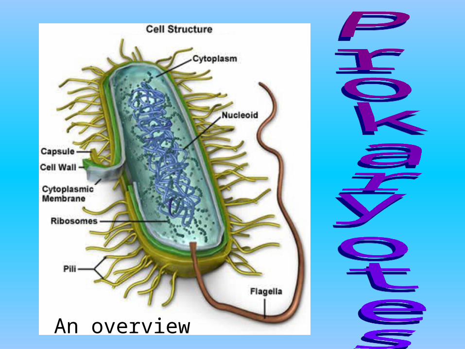
An overview

What to Expect:
• These notes focus on –Cell theory
–Prokaryotes
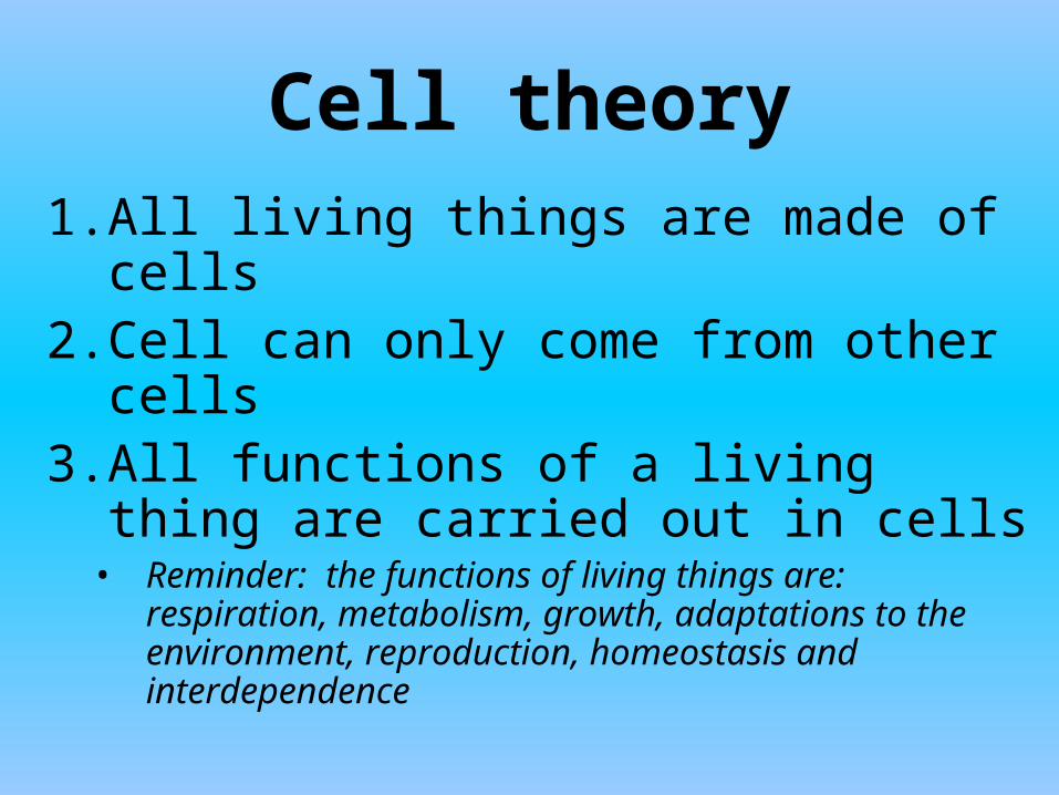
Cell theory
1. All living things are made of cells2. Cell can only come from other
cells3. All functions of a living thing are
carried out in cells • Reminder: the functions of living things are:
respiration, metabolism, growth, adaptations to the environment, reproduction, homeostasis and interdependence
On your worksheet1.State the 3 points of
cell theory.
1.List the 7 characteristics of life
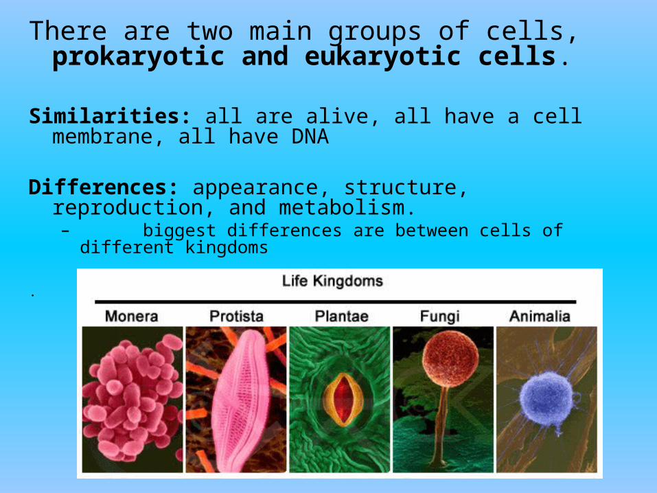
There are two main groups of cells, prokaryotic and eukaryotic cells.
Similarities: all are alive, all have a cell membrane, all have DNA
Differences: appearance, structure, reproduction, and metabolism. – biggest differences are between cells of different
kingdoms
.
On your worksheet
• List the 2 types of cells
Where do we find Prokaryotes?
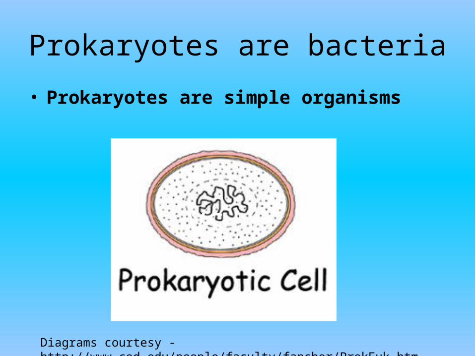
Prokaryotes are bacteria
• Prokaryotes are simple organisms
VS
Diagrams courtesy - http://www.cod.edu/people/faculty/fancher/ProkEuk.htm

According to current scientific thought; Prokaryotes were formed 2 billion years before eukaryotes (or about
3.5 billion years ago)
On your worksheet
• According to current scientific thought, about how old are prokaryotes?
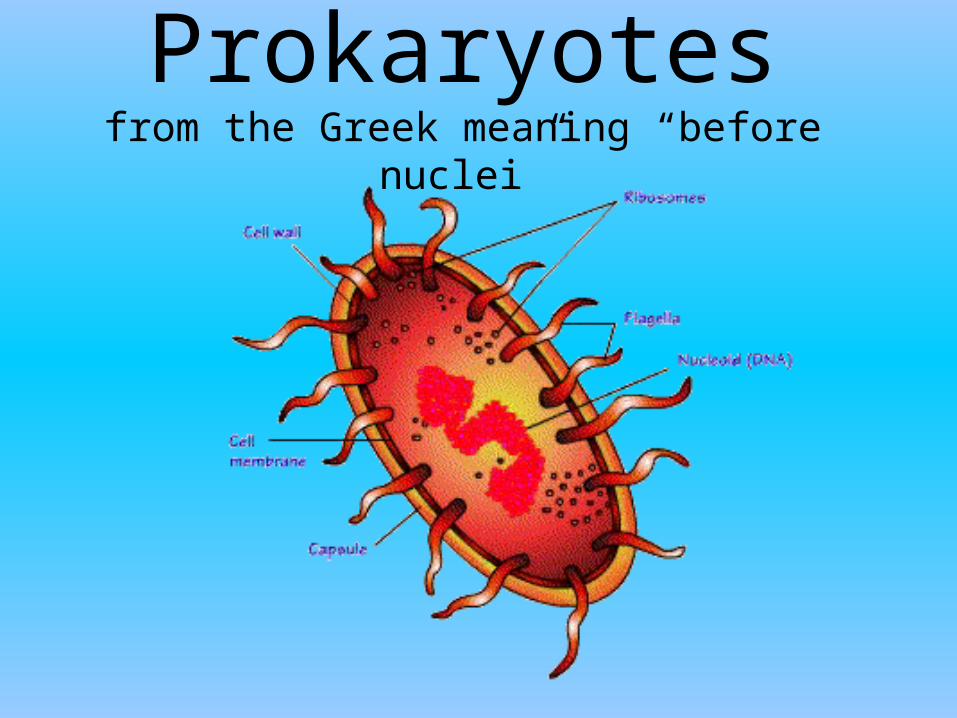
Prokaryotesfrom the Greek meaning “before nuclei”
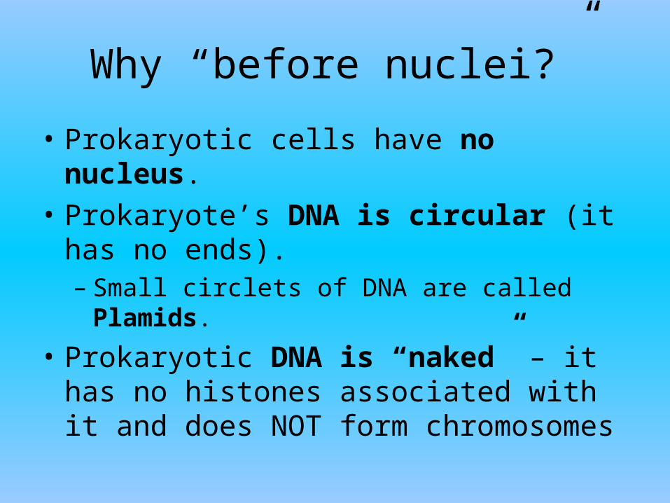
Why “before nuclei?”
• Prokaryotic cells have no nucleus.
• Prokaryote’s DNA is circular (it has no ends).– Small circlets of DNA are called Plamids.
• Prokaryotic DNA is “naked” – it has no histones associated with it and does NOT form chromosomes
On your worksheet
• What does prokaryote mean?
• Why is the term prokaryote used to describe the cells we are talking about?
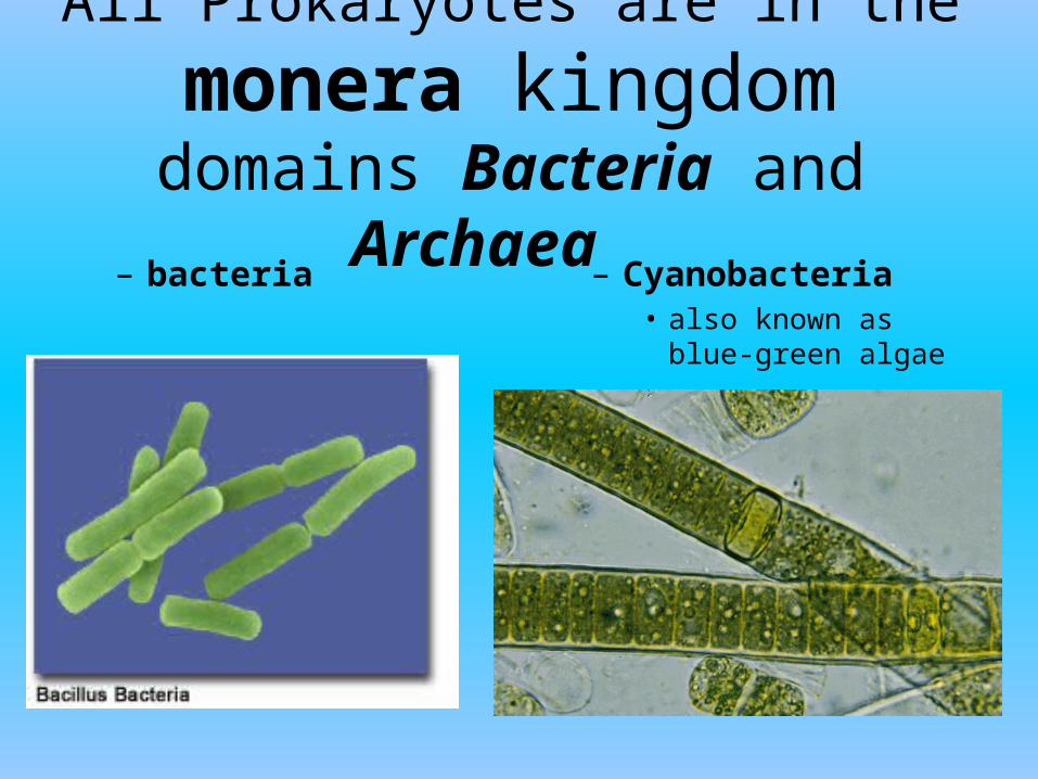
All Prokaryotes are in the
monera kingdomdomains Bacteria and Archaea
– bacteria – Cyanobacteria• also known as blue-
green algae
On your worksheet
• What kingdom to do all prokaryotes belong to?
Prokaryote Characteristics1. Very small size.
2. Lack membrane-bound organelles inside the cell
3. have few internal structures that are distinguishable under a microscope.
4. genetic information is in a circular loop called a plasmid
5. Strong cell walls: resistant to environmental changes
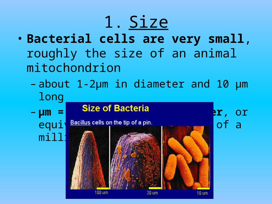
1. Size• Bacterial cells are very small, roughly
the size of an animal mitochondrion – about 1-2µm in diameter and 10 µm long – µm = one millionth of a meter, or
equivalently one thousandth of a millimeter.

Video:
On your worksheet
•What is the size of an average Prokaryote?
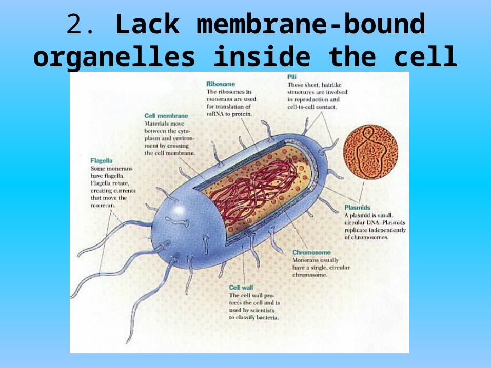
2. Lack membrane-bound organelles inside the cell
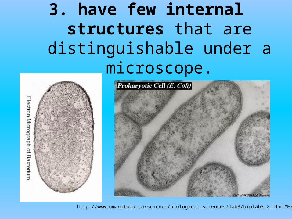
3. have few internal structures that are distinguishable under a
microscope.
http://www.umanitoba.ca/science/biological_sciences/lab3/biolab3_2.html#Examine

4. genetic information is in a circular loop called a plasmid
• E. coli cell dividing.
• E. Coli Grows in human intestine; – Has a single, circular
chromosome– contains DNA as
plasmids • Plasmids are extra-
chromosomal DNA
http://www.bio.mtu.edu/campbell/prokaryo.htm
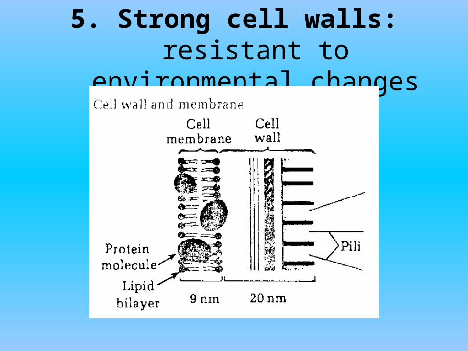
5. Strong cell walls: resistant to environmental changes

On your worksheet
Describe the 5 items used to classify a prokaryote
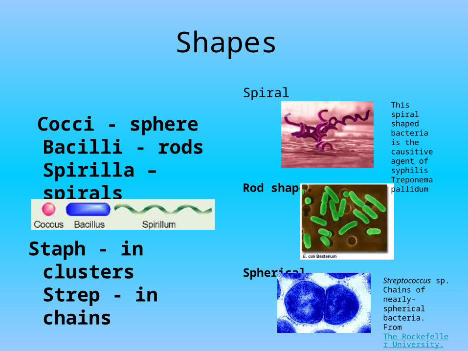
Shapes
Cocci - sphereBacilli - rodsSpirilla – spirals
Staph - in clustersStrep - in chains
Spiral
Rod shaped
SphericalStreptococcus sp. Chains of nearly-spherical bacteria.From The Rockefeller University.
This spiral shaped bacteria is the causitive agent of syphilis Treponema pallidum
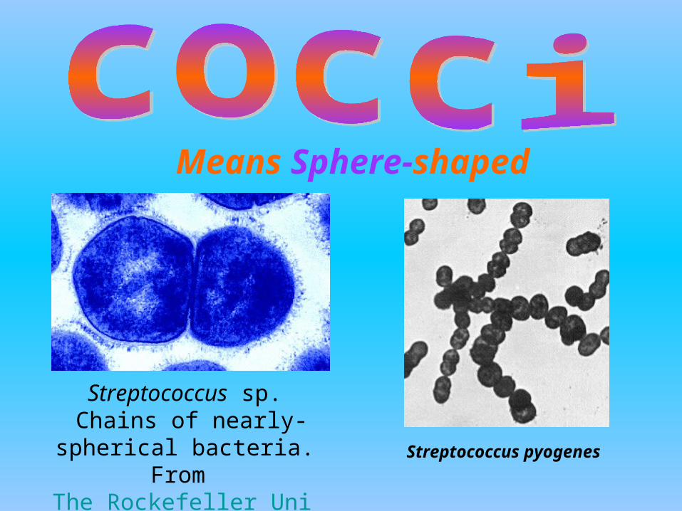
Streptococcus sp. Chains of nearly-spherical bacteria.
From The Rockefeller University.
Means Sphere-shaped
Streptococcus pyogenes
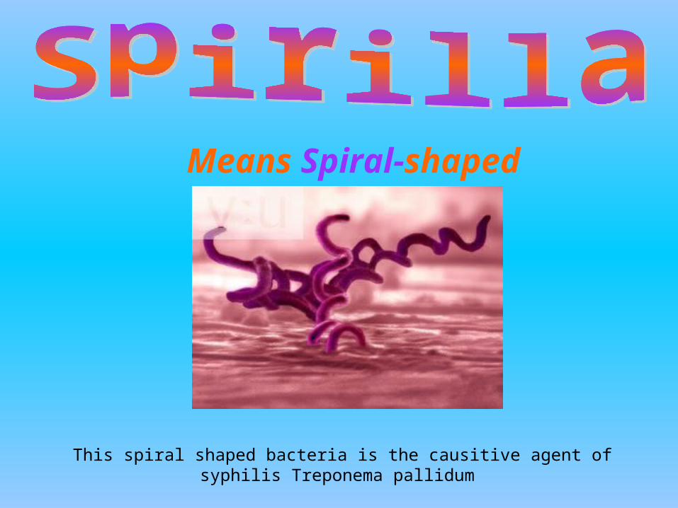
This spiral shaped bacteria is the causitive agent of syphilis Treponema pallidum
Means Spiral-shaped
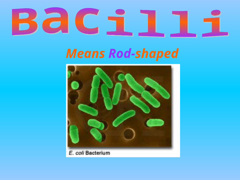
Means Rod-shaped
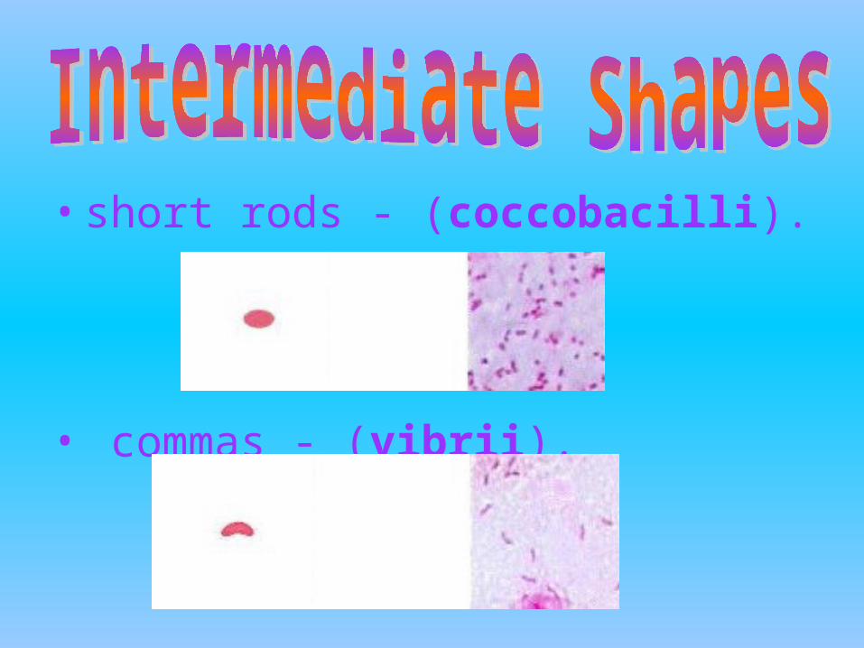
• short rods - (coccobacilli).
• commas - (vibrii).

squares stars irregular
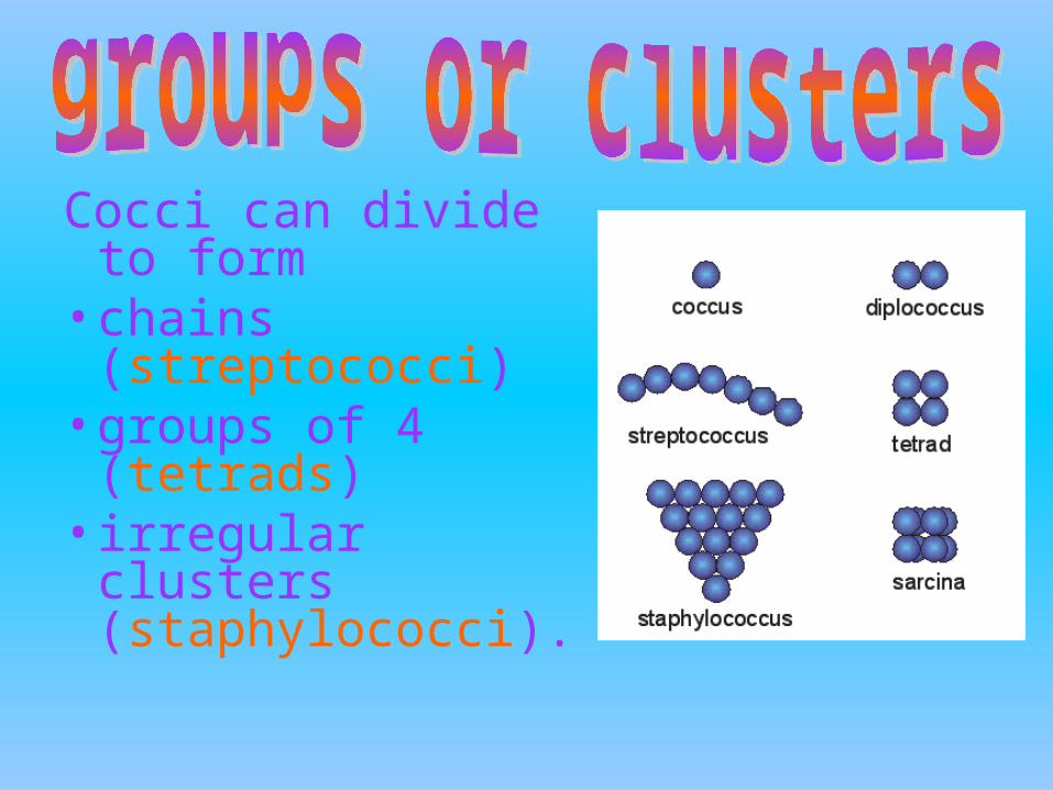
Cocci can divide to form
• chains (streptococci)
• groups of 4 (tetrads)
• irregular clusters (staphylococci).
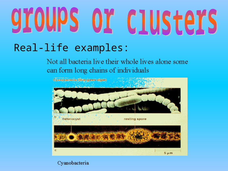
Real-life examples:
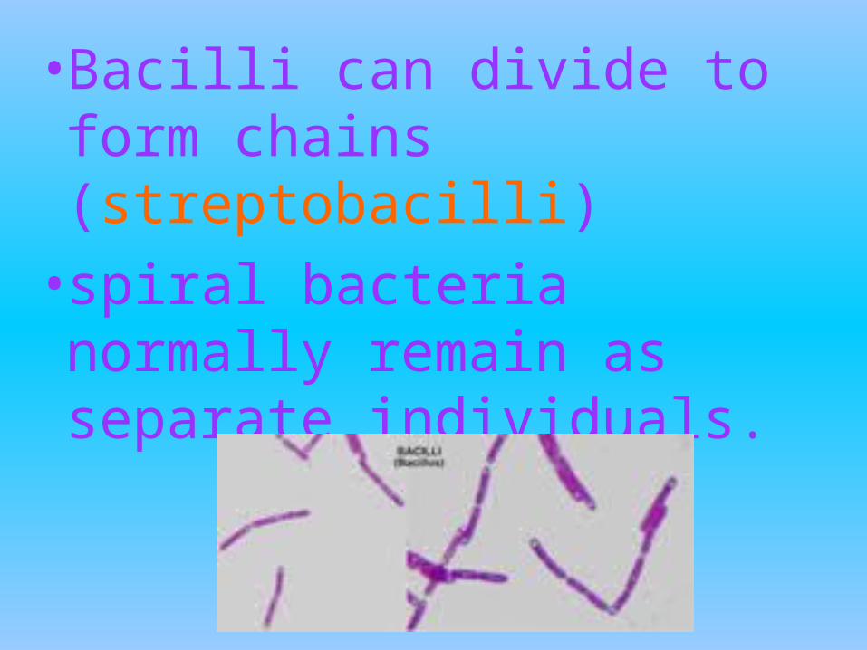
• Bacilli can divide to form chains (streptobacilli)
• spiral bacteria normally remain as separate individuals.
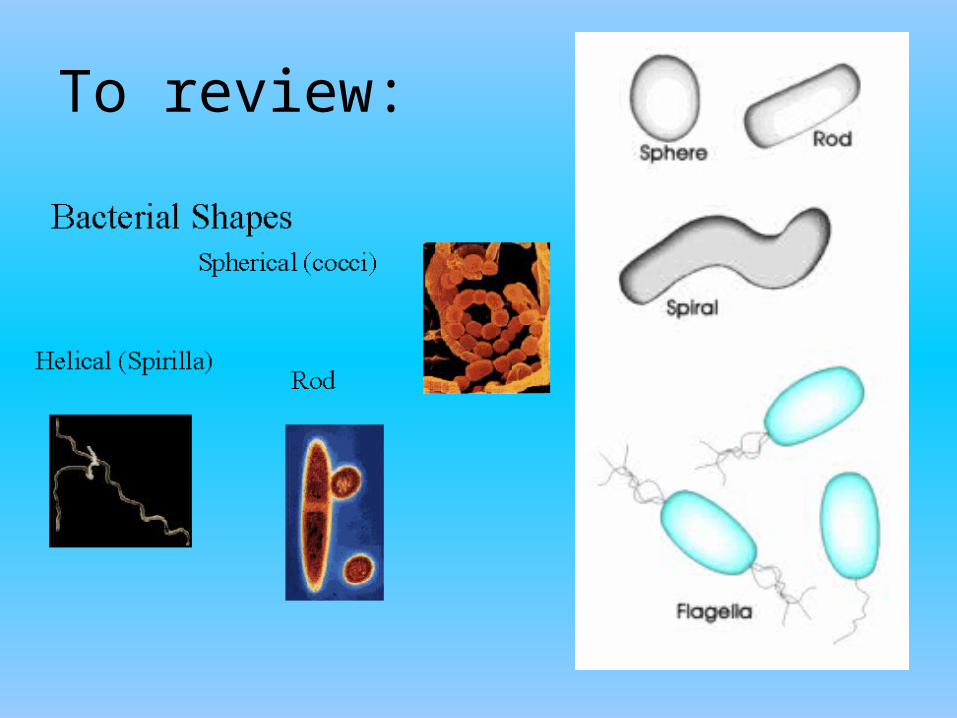
To review:
Or, how bacteria move
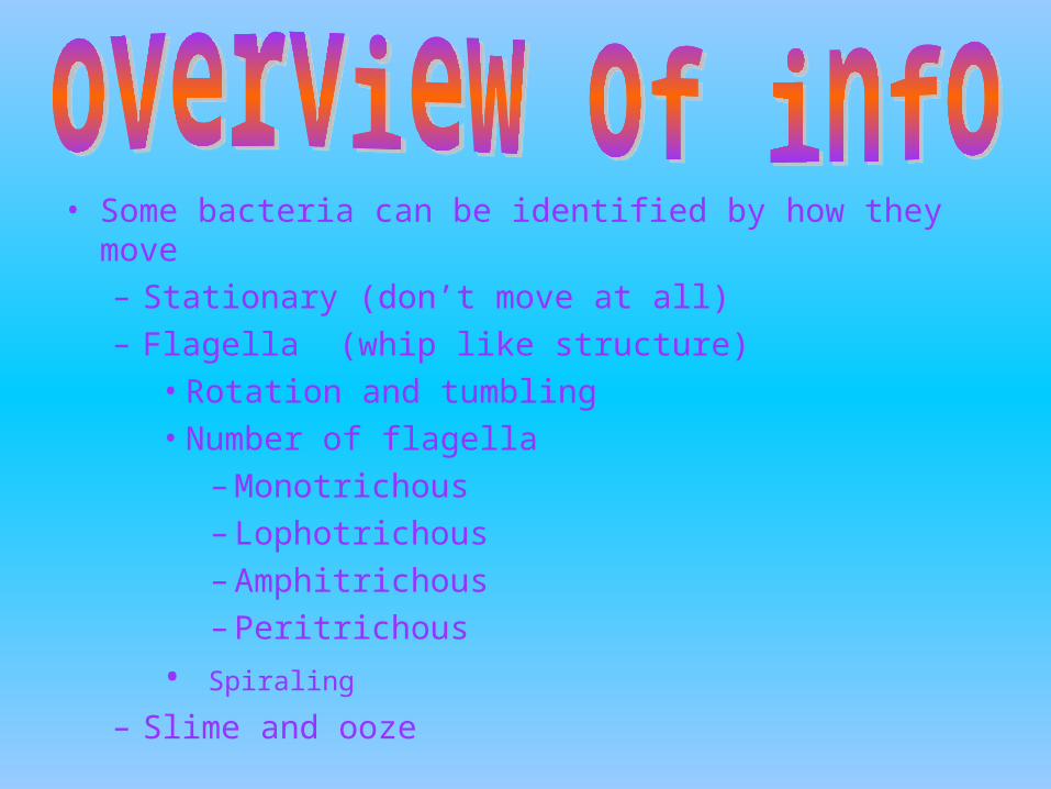
• Some bacteria can be identified by how they move– Stationary (don’t move at all)– Flagella (whip like structure)
• Rotation and tumbling• Number of flagella
– Monotrichous – Lophotrichous– Amphitrichous– Peritrichous
• Spiraling
– Slime and ooze
• Which means, some bacteria simply do not move - - at all, ever.
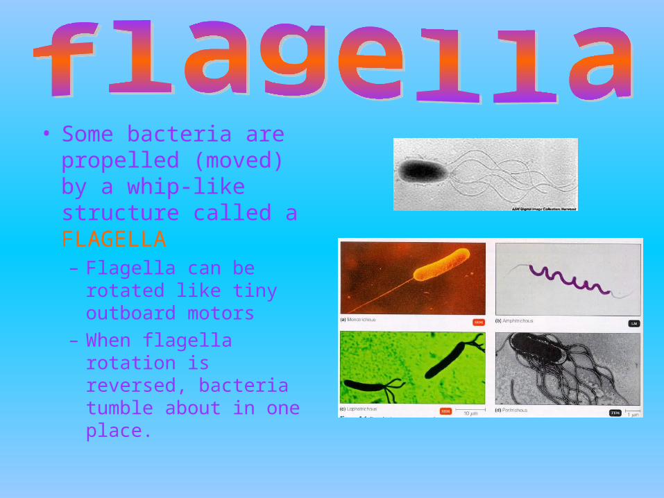
• Some bacteria are propelled (moved) by a whip-like structure called a FLAGELLA– Flagella can be rotated
like tiny outboard motors
– When flagella rotation is reversed, bacteria tumble about in one place.
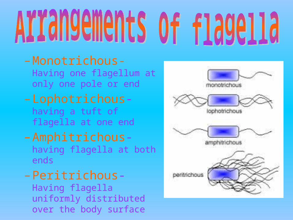
– Monotrichous- Having one flagellum at only one pole or end
– Lophotrichous- having a tuft of flagella at one end
– Amphitrichous- having flagella at both ends
– Peritrichous- Having flagella uniformly distributed over the body surface
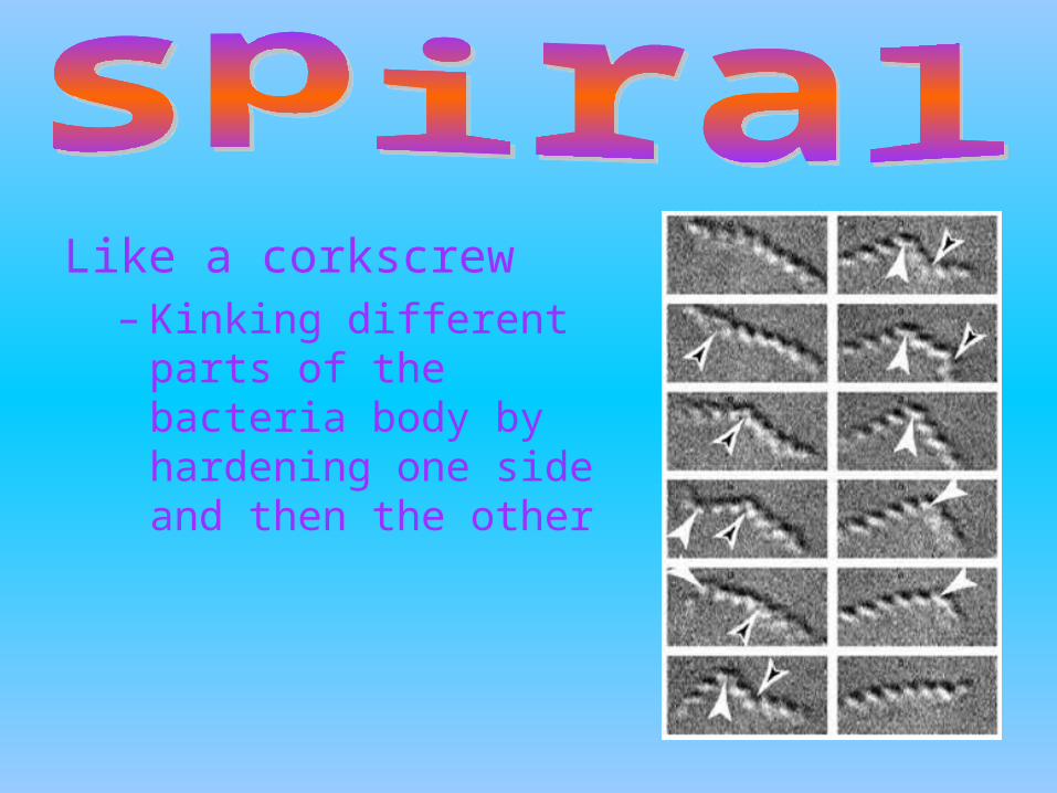
Like a corkscrew– Kinking different parts of
the bacteria body by hardening one side and then the other
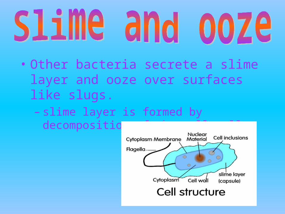
• Other bacteria secrete a slime layer and ooze over surfaces like slugs. – slime layer is formed by decomposition of the
cell wall.
• Or, how one bacteria can become many
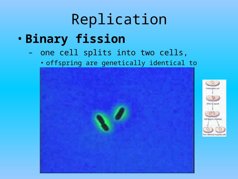
Replication• Binary fission
– one cell splits into two cells,• offspring are genetically identical to parent
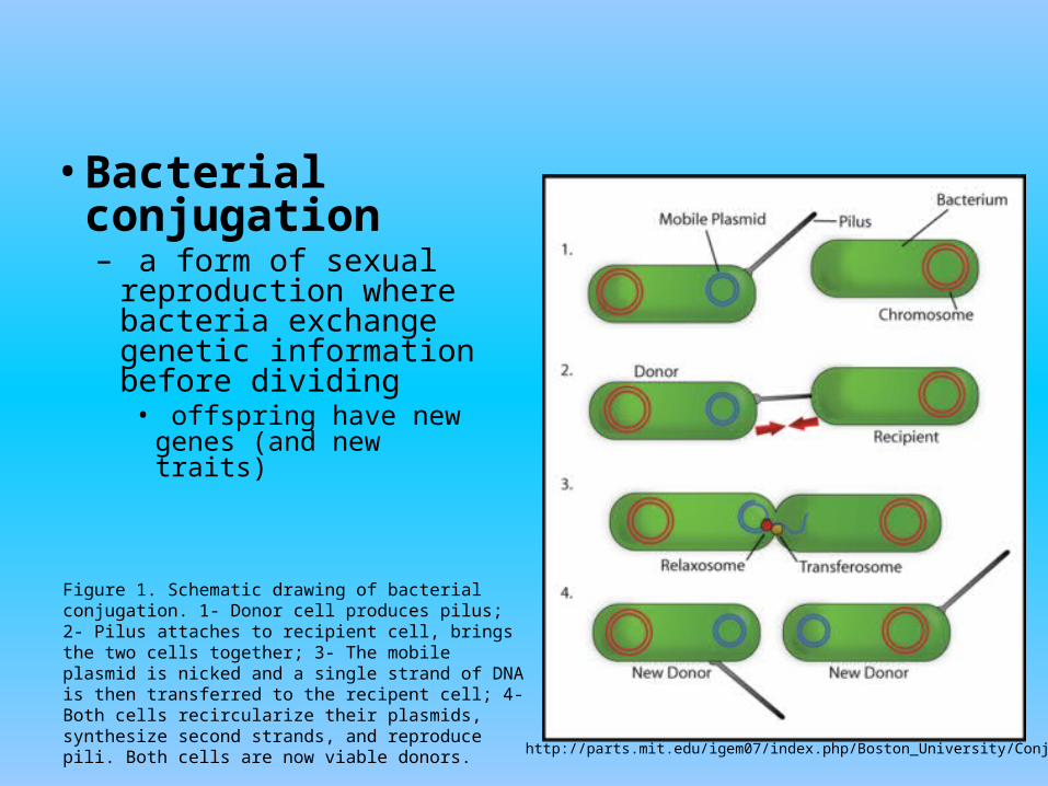
• Bacterial conjugation– a form of sexual
reproduction where bacteria exchange genetic information before dividing
• offspring have new genes (and new traits)
Figure 1. Schematic drawing of bacterial conjugation. 1- Donor cell produces pilus; 2- Pilus attaches to recipient cell, brings the two cells together; 3- The mobile plasmid is nicked and a single strand of DNA is then transferred to the recipent cell; 4- Both cells recircularize their plasmids, synthesize second strands, and reproduce pili. Both cells are now viable donors.
http://parts.mit.edu/igem07/index.php/Boston_University/Conjugation
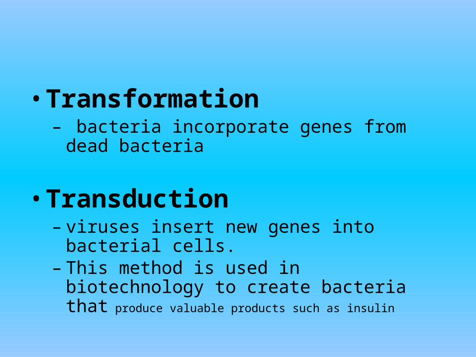
• Transformation – bacteria incorporate genes from dead
bacteria
• Transduction – viruses insert new genes into bacterial cells. – This method is used in biotechnology to
create bacteria that produce valuable products such as insulin
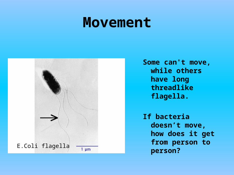
Movement
Some can't move, while others have long threadlike flagella.
If bacteria doesn’t move, how does it get from person to person?
E.Coli flagella
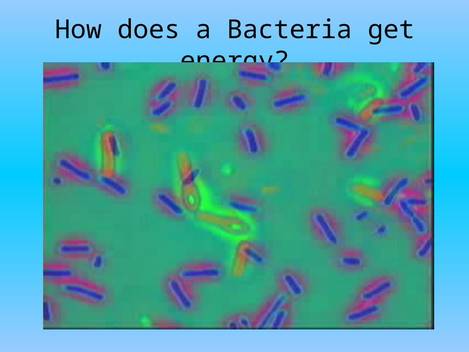
How does a Bacteria get energy?
Or, how bacteria get energy
– 4 main ways bacteria get energy• Chemoheterotrophs• Photoheterotrophs• Photoautotroph• Chemoautotroph
– Energy is released through either cellular respiration or fermentation
– Oxygen demands vary• Obligate aerobe• Obligate anaerobe• Facultative anaerobe
• Heterotrophs get energy by eating other organisms– Chemoheterotrophs
• Eat other organisms for Energy• Eat other organisms for carbon supply
– Photoheterotrophs• Use sunlight for energy• Eat other organisms for carbon supply
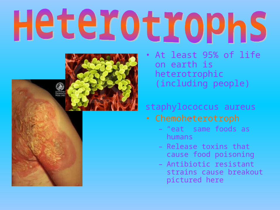
• At least 95% of life on earth is heterotrophic (including people)
staphylococcus aureus• Chemoheterotroph
– “eat” same foods as humans
– Release toxins that cause food poisoning
– Antibiotic resistant strains cause breakout pictured here
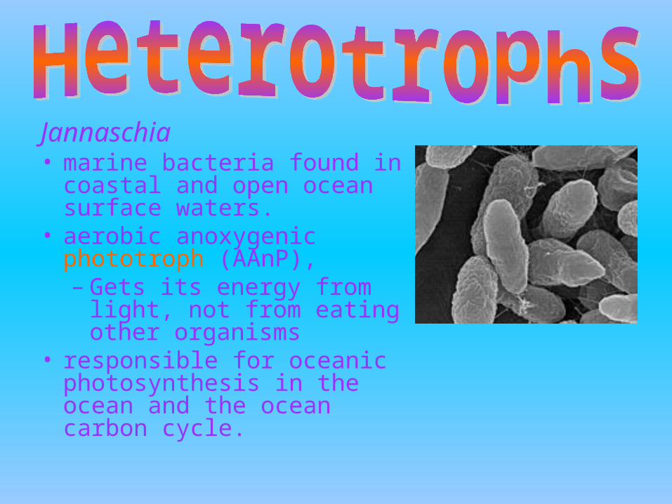
Jannaschia • marine bacteria found in
coastal and open ocean surface waters.
• aerobic anoxygenic phototroph (AAnP), – Gets its energy from light,
not from eating other organisms
• responsible for oceanic photosynthesis in the ocean and the ocean carbon cycle.
• Autotrophs make their own energy from inorganic (not-living) molecules– Photoautotroph
• Uses sunlight (light energy) to convert CO2 and H2O into Carbon compound and oxygen
– Chemoautotroph• Make organic carbon molecules from CO2 using
energy from chemical reactions involving ammonia, hydrogen sulfide, nitrites or iron
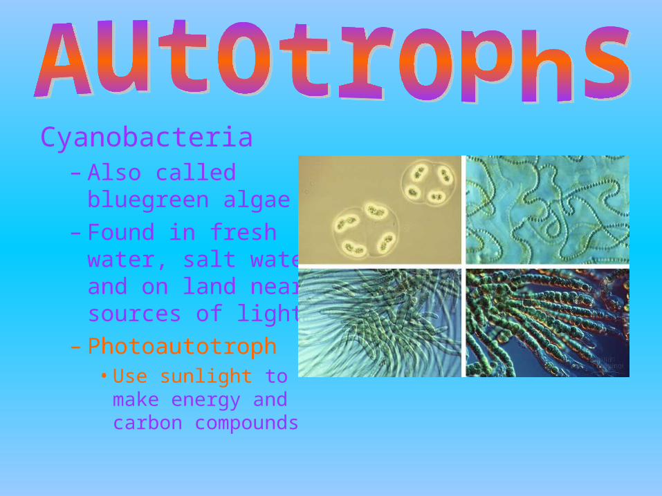
Cyanobacteria– Also called bluegreen
algae– Found in fresh water,
salt water and on land near sources of light
– Photoautotroph • Use sunlight to make
energy and carbon compounds
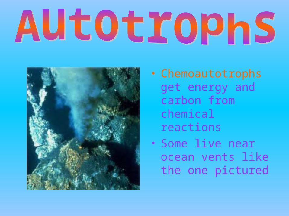
• Chemoautotrophs get energy and carbon from chemical reactions
• Some live near ocean vents like the one pictured
• Once bacteria have “eaten” they need to break down their “food” to make energy
• The process of breaking down organic compounds into ATP (energy cells can use) is called cellular respiration
• Same 1st step to begin with, but the lack or presence of oxygen determines the 2nd step– Step one – Glycolisis– Step two – Fermentation or Kreb Cycle
• To oversimplify the first step
–called glycolysis• Doesn’t require Oxygen (anaerobic)
• Takes place in the cytosol (fluid surrounding organelles) of a cell
• Breaks glucose into pyruvate creating ATP and H in the process
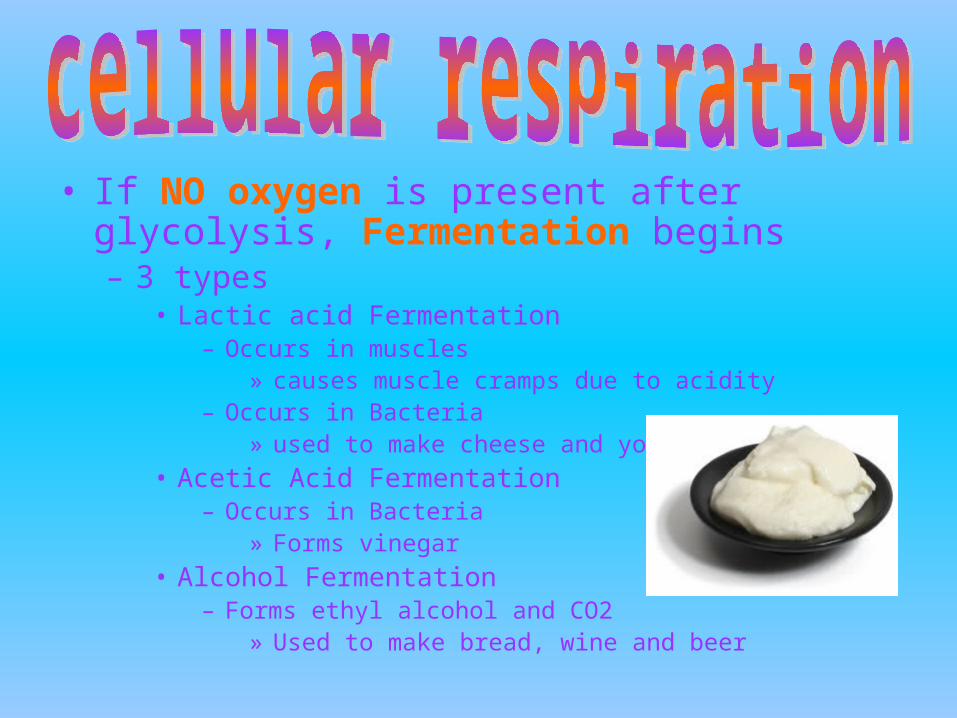
• If NO oxygen is present after glycolysis, Fermentation begins– 3 types
• Lactic acid Fermentation– Occurs in muscles
» causes muscle cramps due to acidity– Occurs in Bacteria
» used to make cheese and yogurt
• Acetic Acid Fermentation – Occurs in Bacteria
» Forms vinegar
• Alcohol Fermentation– Forms ethyl alcohol and CO2
» Used to make bread, wine and beer
• If Oxygen IS present after glycolysis, Acetyl CoA is made and the Kreb cycle begins– we’ll save the chemical details of this process
for another class, but, basically it produces a whole lot of ATP for the cell to use
• SOME BACTERIA REQUIRE OXYGEN, SOME DON’T– OBLIGATE AEROBES– OBLIGATE ANAEROBES– FACULTATIVE ANAEROBES
• Obligate aerobes NEED oxygen to live– OBLIGATE means required to – AEROBE means oxygen
• Release energy through cellular respiration or fermentation
• Example: myobacterium tuberculosis
• Obligate anaerobes DO NOT need oxygen to live– OBLIGATE means required to – ANAEROBE means without oxygen
• Release energy through cellular respiration or fermentation
• Example: clostridium botulinum
• Facultative anaerobes can survive with or without oxygen– Facultative – means able to function in
different ways
• These bacteria can live just about anywhere
• Example: E. coli

FunctionsWhat does Bacteria do?
• decomposers, agents of fermentation, and they play an important role in our own digestive system.
• involved in many nutrient cycles such as the nitrogen cycle, which restores nitrate into the soil for plants.
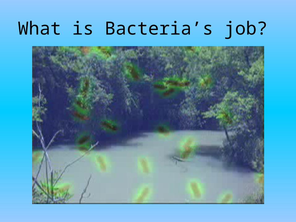
What is Bacteria’s job?
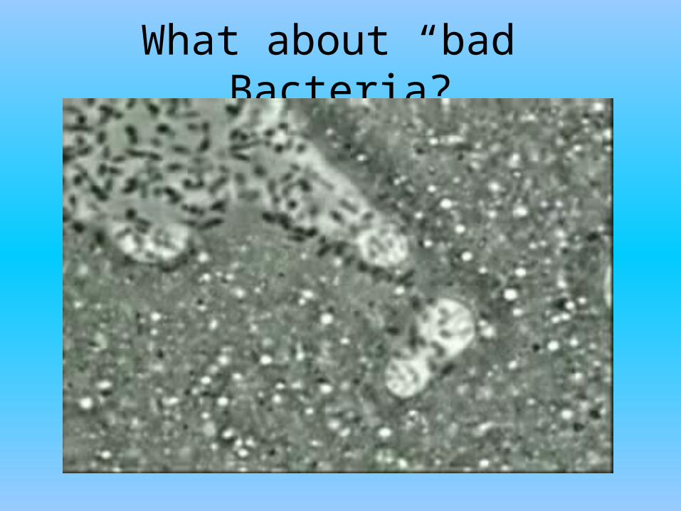
What about “bad” Bacteria?
• Describe a benefit of having bacteria on Earth
• Describe a “bad” bacteria and how it affects people.
Images of Bacteria
• http://www.ulb.ac.be/sciences/biodic/ImBacterie2.html
• http://www.buckman.com/eng/micro101/bacteria.htm