Embed Size (px)
Citation preview
Haskins Laboratories Status Report on Speech Research1993, SR-113, 107-130
An MRI-based Study of Pharyngeal VolumeContrasts in Akan
Mark K. Tiede
Characteristic differences in pharyngeal volume between Akan +I-Advanced Tongue Root(ATR) vowel pairs have been investigated using Magnetic Resonance Imaging (MRI)techniques, and compared to the similar TenselLax vowel distinction in English. Twosubjects were scanned during steady-state phonation of three pairs of contrasting vowels.Analysis of the resulting images shows that it is the overall difference in pharyngealvolume that is relevant to the Akan vowel contrast, not just the tongue root advancementand laryngeal lowering previously reported from xray studies. The data also show that theATR contrast is articulatorily distinct from the English TenseILax contrast.
INTRODUCTION
I am grateful to the many people who assisted me in thisproject, particularly Cathe Browman, Alice Faber, LouisGoldstein, Carol Gracco, Robin Greene, Kathy Harris, Pat Nye,Alfred Opoku, Elliot Saltzman, Doug Whalen, and all thosewho agreed to be magnetized for Science. Goofs and garblesremain of course my responsibility. This work was supportedby NIH grant DC-00121.
In general vowels in an Akan word harmonize;that is all vowels are drawn exclusively from oneset or the other. (The low mid vowel Ia! may occurin a word with vowels from either set.2 ) Affixeshave two forms for compatibility with stemshaving type 1 or type 2 vowels. For example (fromDolphyne 1988:15):
Several West African languages have aphonological process of Vowel Harmony that splitsthe distribution of their vowels into two congruentand mutually exclusive sets. The Akan language,a member of the Niger-Congo Kwa group (Stewart1971) spoken in Ghana, is typical of this pattern.The vowels of Akan may be grouped as follows: 1
(1) Set 1 Set 2
di --> midi "I eat"[eat]
dI --> mIdI "I am called"[be called]
dI --> :JdI "he is called"[be called]
di --> odi "he eats"[eat]
mI +[1st Sg]
o +[3rd Sg]
(2) mI +[1st Sg]
o +[3rd Sg]
Drawing on a cineradiographic study of the related language Igbo by Ladefoged (1964), Stewart(1967) proposed an analysis of Akan in whichcorresponding vowels from the two harmonygroups are distinguished articulatorily bydifferences in tongue root position.Cineradiographic studies of Akan undertaken byLindau (1975, 1979) confirmed the primacy oftongue root position in the harmony mechanism,but also showed correlated variation in larynxheight: advanced tongue root was combined withlowered larynx, and retracted root with raisedlarynx. In reporting this work Lindau suggestedthat the relevant contrast was not just the relativepositions of these articulators but rather theoverall difference in pharyngeal volume producedby their cooperative positioning, and proposed thefeature "Expanded" to describe it. A contrastbased on differences in pharyngeal volume
£.
a
uoe
107
108 Tiede
vocal tract area functions of four point vowels, andin similar work by Lakshminarayanan, Lee, andMcCutcheon (1991).
The English TenselLax distinction eludesprecise articulatory description, but is similar inmany ways to the Akan contrast: tense vowels aregenerally articulated with an advanced tongueroot, and sometimes a lowered larynx. Englishvowels comparable to those used in Akan may begrouped as in (1) above:
Previous attempts to identify the English distinction with the same mechanism used in theAkan ATR contrast include a proposal by Halleand Stevens (1969), and a cineradiographic studyby Perkell (1971). However a more extensivecineradiographic study conducted by Ladefoged etai. (1972) showed that tongue root advancement isjust one of several complementary strategies usedto implement the TenselLax contrast, and is notused consistently by all speakers. Similarlyelectromyographic data reported by Raphael andBell-Berti (1975) showed differences betweensubjects in patterns of muscle tension used todistinguish between tense and lax vowels. Factoranalysis of xray-derived tongue shapes byHarshman, Ladefoged, and Goldstein (1977)showed that tongue position for English tense/laxpairs can be predicted very completely byreference to just two parameters along which eachof the tongue root and tongue dorsum positionscovary, whereas a similar analysis of Akan byJackson (1988) found three parameters necessaryfor tongue shape specification. While it is probablythe case that different mechanisms are involved ineach language, they are similar enough to makedirect comparison feasible and interesting, and sothey have been treated in parallel in the currentstudy.
Method
In this experiment the contrast in crosssectional area was examined at adjacent levelsthrough the pharynx for corresponding expandedand constricted vowels (i.e. +I-ATR; TenselLax).5The experiment involved two subjects, both male,in their early thirties. Subject AO is a nativespeaker of the Asante dialect of Akan; subject MTis a native speaker of Midwestern AmericanEnglish. The vowels selected for comparison wereIi : II, Ie : e/, and lu : u/, chosen because of theirreasonable similarity across the two languages.
suggests that vowels from the two groups mayalso differ in the left-to-right or lateral dimensionof the pharynx, assuming that this is subject tovoluntary control, but studies based oncineradiography are inherently unable to explorethis possibility, since the technique collapses alllateral information into a flat (sagittal) image.The purpose of the study discussed here was toinvestigate the predicted difference in pharyngealvolume using Magnetic Resonance Imaging, whichis not subject to the same limitation.
The magnetic resonance technique has onlyrecently become a viable imaging alternative.Developed primarily for medical diagnosticpurposes, MRI exploits the behavior of hydrogennuclei in a magnetic field to construct an imagecorrelated with the concentration of hydrogen inthe scanned tissue.3 Because different tissue typeshave differing hydrogen densities and bondings,MR images provide soft tissue definition over arange inaccessible to xray techniques. Twoadditional advantages make MRI an especiallyattractive imaging modality: there are currentlyno known health risks for the subject associatedwith the technique, and because the imagingplane may be reoriented without moving thesubject, three dimensional data collection ispossible.
Despite these advantages the MR technique hassubstantial drawbacks in its potential for phoneticresearch, chief of which is the tradeoff betweenimaging time and resulting image quality.Although image acquisition rates continue to dropas the technology evolves, they are currently stilltoo slow by an order of magnitude for capturingdynamic speech. In addition, three dimensionalscanning requires multiple passes through thesame volume, further increasing acquisition time.This limits the current usefulness of MRI inphonetics to studies involving static vocal tractshapes and sustainable patterns of phonation.
The current study was able to proceed underthese constraints. Although vowels sustained formany times their normal speaking duration represent an admittedly artificial source of data, allof the vowels examined (except English lax vowels) can occur in open syllables, and are thereforeartificial only in duration, and not in syllablestructure. Furthermore, sung vowels of constantpitch and quality and extended duration occur inthe musical traditions of Ghanaian and Americancultures.4 There is also precedent for use of theMRI technique applied to measurement of staticvowel shapes in the work of Baer, Gore, Gracco, &Nye (in press), who successfully used it to obtain
(3)
e
Tenseuo
Laxu;)
An MRI-based Study ofPharyngeal Volume Contrasts in Allan 109
These words were also recorded for acousticanalysis:
Prior to the experiment a target stimulus tapewas prepared for each subject by twice recordingeach of the target vowels in a characteristic word,extracting the vowel portion using digital editingtechniques, then recording it as a continuousutterance by concatenation. The following targetwords were used:
(4) Vowel Akan English
[i] pi "many" hid "heed"
[I] fl "to vomit" hId "hid"
[e] Jqe "empty" held "hayed"
[E] ;:,JqE "he looked at" hEd "head"
[u] bu "to break" hud "who'd"
[u] bu "to be drunk" hud "hood"
The experiment was performed on a GeneralElectric Signa machine installed at the Yale NewHaven Hospital. The Signa system consists of atoroidal superconducting electromagnetdeveloping a 1.5 Tesla flux density, placed in ascanning room designed to minimized interferencefrom external electromagnetic noise. The magnetis controlled from an operator's console outside thescanning room, and an attached computer is usedfor image reconstruction, collation, and storage.
After divesting themselves of all ferrous material subjects were fitted with an earphone, microphone, and neck RF transceiver imaging coil,and positioned on their backs inside the bore ofthe magnet. The earphone was the terminus of alength of plastic tubing connected to a smallspeaker placed as far away from the magnet aspossible (because the strength of the magneticfield tended to overwhelm the speaker coil). Thespeaker was driven by an amplifier outside thescanning room, and was used to communicatewith the subject and to play the prerecordedtarget stimuli during scanning. The microphonewas used to record subject phonation immediatelyprior to and following scanning; while phonationduring scanning was also recorded it wasunusable for analysis because of the intensity ofthe noise produced by the machine.
2:322:322:322:322:322:32
2:252:362:442:442:362:36
Time
TimeVowel TR TE
32 13I 37 11e 39 13I:: 39 12u 37 IIu 37 11
Vowel TR TE
36 1036 10
e 37 11I:: 37 11u 36 10u 37 11
Subject MT (English)
Subject AO (Akan)
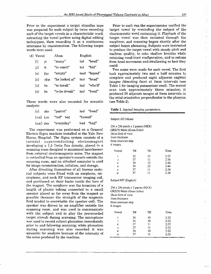
Table 1. Sagittal imaging parameters.
256 x 256 pixels x 2 passes (NEX)GRPJ30 Multi (Grass Echo)28cm field of view3mm thicknessOmm interscan skip8 images
Prior to each run the experimenter verified thetarget vowel by reminding the subject of thecharacteristic word containing it. Playback of thetarget vowel was then initiated through theearphone, and scanning begun shortly after thesubject began phonating. Subjects were instructedto produce the target vowel with steady pitch anduniform quality, to take shallow breaths whileretaining vocal tract configuration, and to refrainfrom head movement and swallowing as best theycould.
Two scans were made for each vowel. The firsttook approximately two and a half minutes tocomplete and produced eight adjacent sagittalimages (bisecting face) at 3mm intervals (seeTable 1 for imaging parameters used). The secondscan took approximately three minutes; itproduced 28 adjacent images at 5mm intervals inthe axial orientation perpendicular to the pharynx(see Table 2).
256 x 256 pixels x 2 passes (NEX)GRPJ30 Multi (Grass Echo)28cm field of view3mm thicknessOmm interscan skip8 images
hod "hoed"
hred "had"
"hawed"
[0] ako "parrot"
[;:':0] b;:, "red" hod
[a:re] daa "everyday"
110
Table 2. Axial imaging parameters
Subject AO (Akan)
256 x 128 pixels x I pass (NEX)
SPGRl45 Volume (Spoiled grass)
28cm field of view
5mm thickness
Omm interscan skip
28 images
TR45.
TE5
Time 3:05
Subject MT (English)
256 x 128 pixels x I pass (NEX)
SPGRl45 Volume (Spoiled grass)
30cm field of view 0, I, u, U)
28cm field of view (e, E)
5mm thickness
Omm interscan skip
28 images
TR45
TE5
Time 3:05
JawHeight
Tiede
AnalysisThe images obtained were converted from 16 bit
Signa format to an 8 bit (255 gray level) formatcompatible with display and analysis software.The axial images were normalized for a standardimage density so that a given pixel magnituderepresented the same value across all series; thiswas done so that air-tissue boundary measurements could be made consistently. Image analysiswas performed on a Macintosh II computer usingthe NIH IMAGE program.6
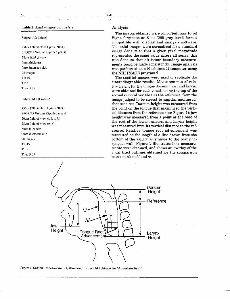
The sagittal images were used to replicate thecineradiographic results. Measurements of relative height for the tongue dorsum, jaw, and larynxwere obtained for each vowel, using the top of thesecond cervical vertebra as the reference, from theimage judged to be closest to sagittal midline forthat scan set. Dorsum height was measured fromthe point on the tongue that maximized the vertical distance from the reference (see Figure 1); jawheight was measured from a point at the base ofthe root of the lower incisors; and larynx heightwas measured from its vertical distance to the reference. Relative tongue root advancement wasmeasured as the length of a line drawn from thebottom of the vallecular sinuses to the rear pharyngeal wall. Figure 1 illustrates how measurements were obtained, and shows an overlay of thevocal tract outlines obtained for the comparisonbetween Akan Iii and hi.
___:;;:::=~_~_---':~----..,..-- Dorsumt Height
Reference
Tongue Root LarynxAdvancement.--:::_.t--b_"---":"'- Height
\\0Figure 1. Sagittal measurements, showing Subject AO (Akan) for lil overlain by lil.
An MRI·based Study ofPharyngeal Volume Contrasts in Akan 111
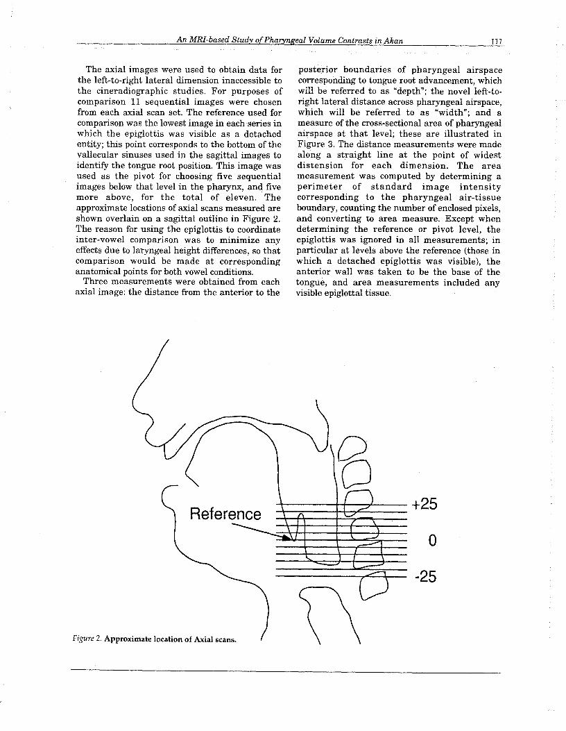
The axial images were used to obtain data forthe left-to-right lateral dimension inaccessible tothe cineradiographic studies. For purposes ofcomparison 11 sequential images were chosenfrom each axial scan set. The reference used forcomparison was the lowest image in each series inwhich the epiglottis was visible as a detachedentity; this point corresponds to the bottom of thevallecular sinuses used in the sagittal images toidentify the tongue root position. This image wasused as the pivot for choosing five sequentialimages below that level in the pharynx, and fivemore above, for the total of eleven. Theapproximate locations of axial scans measured areshown overlain on a sagittal outline in Figure 2.The reason for using the epiglottis to coordinateinter-vowel comparison was to minimize anyeffects due to laryngeal height differences, so thatcomparison would be made at correspondinganatomical points for both vowel conditions.
Three measurements were obtained from eachaxial image: the distance from the anterior to the
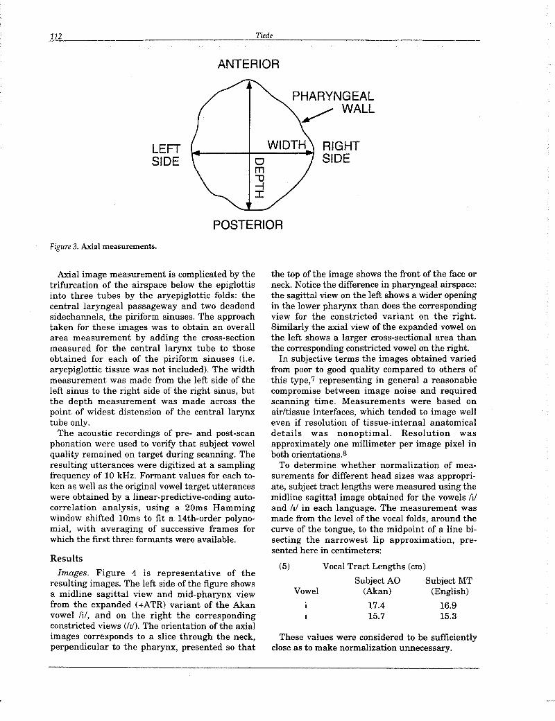
posterior boundaries of pharyngeal airspacecorresponding to tongue root advancement, whichwill be referred to as "depth"; the novel left-toright lateral distance across pharyngeal airspace,which will be referred to as "width"; and ameasure of the cross-sectional area of pharyngealairspace at that level; these are illustrated inFigure 3. The distance measurements were madealong a straight line at the point of widestdistension for each dimension. The areameasurement was computed by determining aperimeter of standard image intensitycorresponding to the pharyngeal air-tissueboundary, counting the number of enclosed pixels,and converting to area measure. Except whendetermining the reference or pivot level, theepiglottis was ignored in all measurements; inparticular at levels above the reference (those inwhich a detached epiglottis was visible), theanterior wall was taken to be the base of thetongue, and area measurements included anyvisible epiglottal tissue.
6+25
Reference
0
0 -25
Figure 2. Approximate location of Axial scans.
112
LEFTSIDE
Figure 3. Axial measurements.
Tiede
ANTERIOR
POSTERIOR
PHARYNGEAL~ WALL
RIGHTSIDE
Axial image measurement is complicated by thetrifurcation of the airspace below the epiglottisinto three tubes by the aryepiglottic folds: thecentral laryngeal passageway and two deadendsidechannels, the piriform sinuses. The approachtaken for these images was to obtain an overallarea measurement by adding the cross-sectionmeasured for the central larynx tube to thoseobtained for each of the piriform sinuses (i.e.aryepiglottic tissue was not included). The widthmeasurement was made from the left side of theleft sinus to the right side of the right sinus, butthe depth measurement was made across thepoint of widest distension of the central larynxtube only.
The acoustic recordings of pre- and post-scanphonation were used to verify that subject vowelquality remained on target during scanning. Theresulting utterances were digitized at a samplingfrequency of 10 kHz. Formant values for each token as well as the original vowel target utteranceswere obtained by a linear-predictive-coding autocorrelation analysis, using a 20ms Hammingwindow shifted 10ms to fit a 14th-order polynomial, with averaging of successive frames forwhich the first three formants were available.
Results
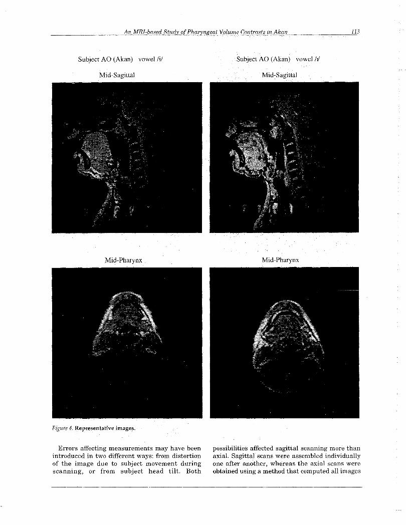
Images. Figure 4 is representative of theresulting images. The left side of the figure showsa midline sagittal view and mid-pharynx viewfrom the expanded (+ATR) variant of the Akanvowel Iii, and on the right the correspondingconstricted views (hi). The orientation of the axialimages corresponds to a slice through the neck,perpendicular to the pharynx, presented so that
the top of the image shows the front of the face orneck. Notice the difference in pharyngeal airspace:the sagittal view on the left shows a wider openingin the lower pharynx than does the correspondingview for the constricted variant on the right.Similarly the axial view of the expanded vowel onthe left shows a larger cross-sectional area thanthe corresponding constricted vowel on the right.
In subjective terms the images obtained variedfrom poor to good quality compared to others ofthis type,7 representing in general a reasonablecompromise between image noise and requiredscanning time. Measurements were based onairltissue interfaces, which tended to image welleven if resolution of tissue-internal anatomicaldetails was nonoptimal. Resolution wasapproximately one millimeter per image pixel inboth orientations.S
To determine whether normalization of measurements for different head sizes was appropriate, subject tract lengths were measured using themidline sagittal image obtained for the vowels Iiiand hi in each language. The measurement wasmade from the level of the vocal folds, around thecurve of the tongue, to the midpoint of a line bisecting the narrowest lip approximation, presented here in centimeters:
(5) Vocal Tract Lengths (em)
Subject AO Subject MTVowel (Akan) (English)
17.4 16.915.7 15.3
These values were considered to be sufficientlyclose as to make normalization unnecessary.
Subject AO (Akan) vowel iii
Mid-Sagittal
Mid-Pharynx
Figure 4. Representative images.
Errors affecting measurements may have beenintroduced in two different ways: from distortionof the image due to subject movement duringscanning, or from subject head tilt. Both
Subject AO (Akan) vowel!J/
Mid-Sagittal
Mid-Pharynx
possibilities affected sagittal scanning more thanaxial. Sagittal scans were assembled individuallyone after another, whereas the axial scans wereobtained using a method that computed all images
• Akan +~ Akan-• English +/, English.DOISum Height
35
30
25
20
E 15E
10
5
0E U
Jaw Height
0
10
20
EE30
40
50
concurrently. Subject movement in a sagittal scanset resulted in blurring of the single image beingacquired at that moment, making it unusable formeasurement, while movement in an axial setcaused only a loss of definition across all images inthe set, leaving them all still viable formeasurement.
The effect of any head tilt or rotation was tocause the scanning plane to intersect with thesubject at an oblique angle, rather than producinga true bisection of the head. This was not aproblem for axial images, since any tilting wouldhave been too slight to cause measurabledistortion in that orientation. For the sagittalimages however, even slight tilting was sufficientto make determination of the head midlinedifficult; for example, one image might showmidline at the level of the larynx, yet be off centerat the level of the palate, thus affecting tonguedorsum height measurements. Therefore allsagittal tongue measurements were confirmed onimages adjacent to the one chosen as midline.
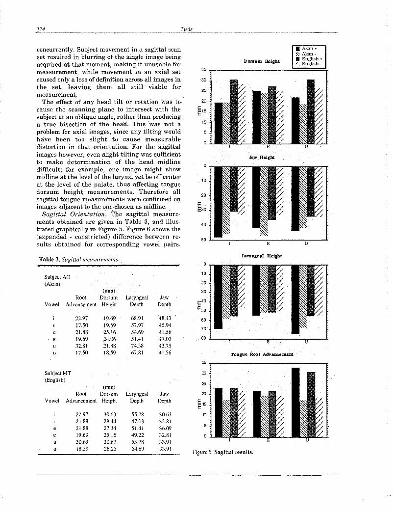
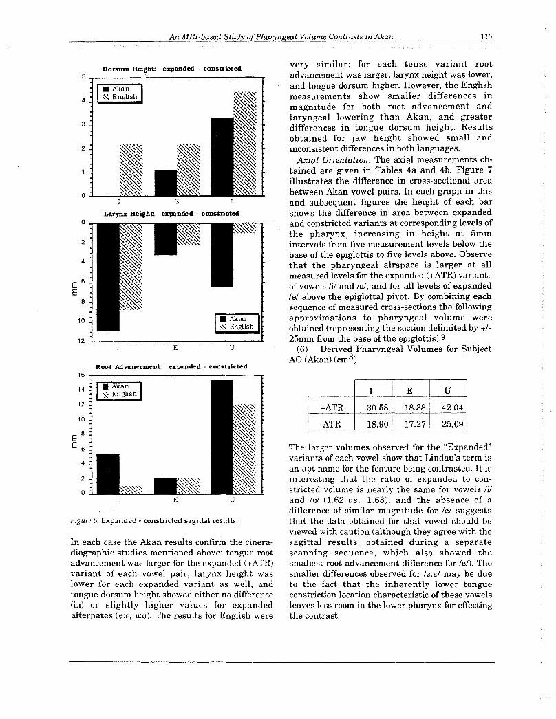
Sagittal Orientation. The sagittal measurements obtained are given in Table 3, and illustrated graphically in Figure 5. Figure 6 shows the(expanded - constricted) difference between results obtained for corresponding vowel pairs.
Table 3. Sagittal measurements.
Subject AO(Akan)
(mm)Root Dorsum Laryngeal Jaw
Vowel Advancement Height Depth Depth
22.97 19.69 68.91 48.13I 17.50 19.69 57.97 45.94e 21.88 25.16 54.69 41.56£ 19.69 24.06 51.41 47.03u 32.81 21.88 74.38 43.75u 17.50 18.59 67.81 41.56
laryngeal Height
o
60
70
80 ~---:-------""E-------"""'U""""-.......t
Tongue RoQt Advancement
~ .,....---------------....Subject MT(English)
(mm)Root Dorsum Laryngeal Jaw
Vowel Advancement Height Depth Depth
22.97 30.63 55.78 30.6321.88 28.44 47.03 32.81
e 21.88 27.34 51.41 36.09£ 19.69 25.16 49.22 32.81u 30.63 30.63 55.78 33.91u 18.59 26.25 54.69 33.91
10
5
o
Figure 5. Sagittal results.
An MRI-based Study ofPharyngeal Volume Contrasts in Akan 115
12 ..1- ---'-
very similar: for each tense variant rootadvancement was larger, larynx height was lower,and tongue dorsum higher. However, the Englishmeasurements show smaller differences inmagnitude for both root advancement andlaryngeal lowering than Akan, and greaterdifferences in tongue dorsum height. Resultsobtained for jaw height showed small andinconsistent differences in both languages.
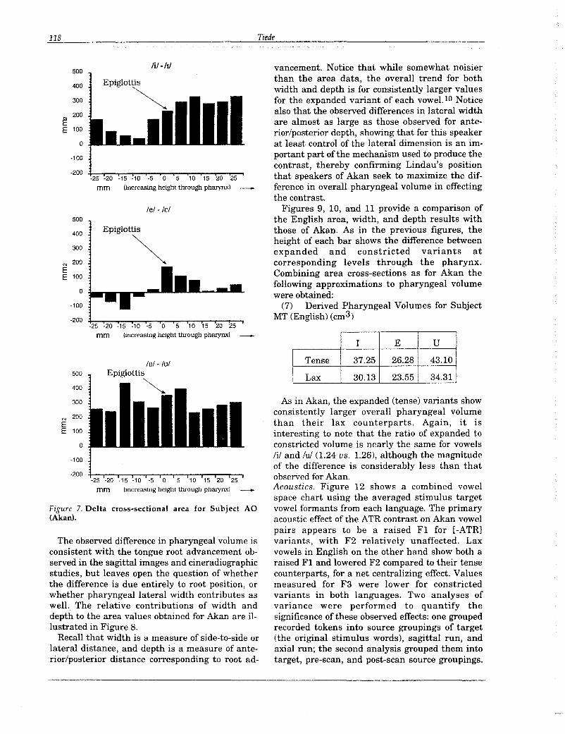
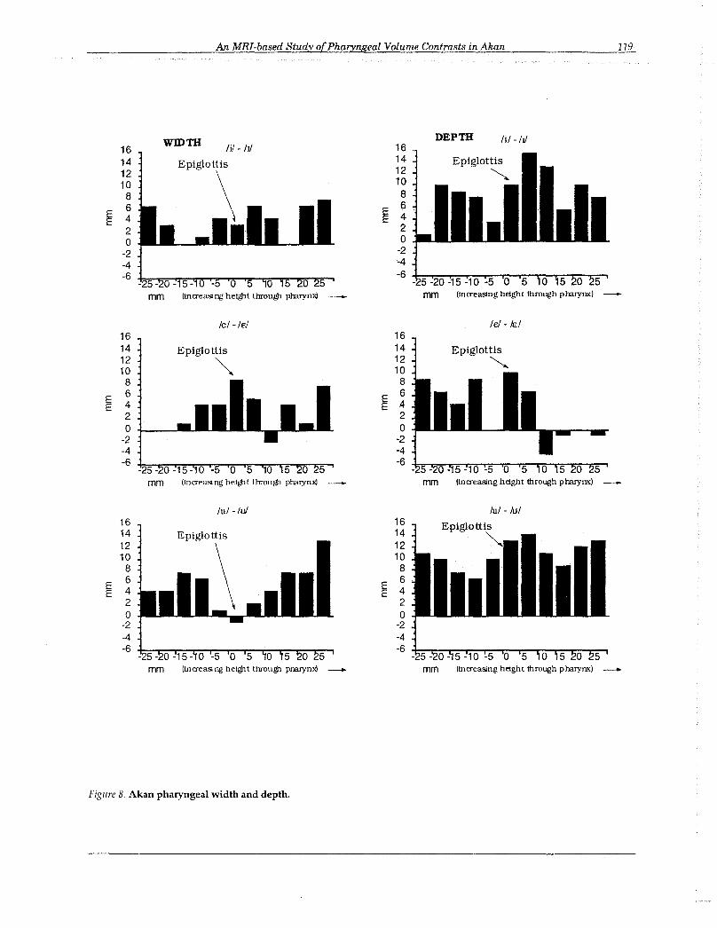
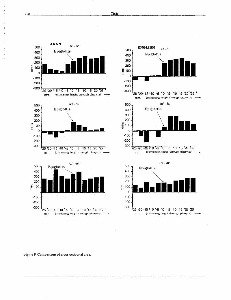
Axial Orientation. The axial measurements obtained are given in Tables 4a and 4b. Figure 7illustrates the difference in cross-sectional areabetween Akan vowel pairs. In each graph in thisand subsequent figures the height of each barshows the difference in area between expandedand constricted variants at corresponding levels ofthe pharynx, increasing in height at 5mmintervals from five measurement levels below thebase of the epiglottis to five levels above. Observethat the pharyngeal airspace is larger at allmeasured levels for the expanded (+ATR) variantsof vowels iii and lui, and for all levels of expandedlei above the epiglottal pivot. By combining eachsequence of measured cross-sections the followingapproximations to pharyngeal volume wereobtained (representing the section delimited by +125mm from the base of the epiglottis):9
(6) Derived Pharyngeal Volumes for SubjectAO (Akan) (cm3)
uE
2
Root Advancement: expanded· constricted
10
4
o
E
Larynx Height: expanded - constricted
2
3
4
5.,.... ..,.Dorsum Height: expanded - constricted
E 6E
8
16 ..,------------------r14
12
10
E 8
E 6
4
2
o
Figure 6. Expanded - constricted sagittal results.
In each case the Akan results confirm the cineradiographic studies mentioned above: tongue rootadvancement was larger for the expanded (+ATR)variant of each vowel pair, larynx height waslower for each expanded variant as well, andtongue dorsum height showed either no difference0:1) or slightly higher values for expandedalternates (e:e, u:u). The results for English were
I E U
+ATR 30.58 18.38 42.04
-ATR 18.90 17.27 25.09
The larger volumes observed for the "Expanded"variants of each vowel show that Lindau's term isan apt name for the feature being contrasted. It isinteresting that the ratio of expanded to constricted volume is nearly the same for vowels Iiiand lui (1.62 us. 1.68), and the absence of adifference of similar magnitude for lei suggeststhat the data obtained for that vowel should beviewed with caution (although they agree with thesagittal results, obtained during a separatescanning sequence, which also showed thesmallest root advancement difference for lei). Thesmaller differences observed for Ie:£! may be dueto the fact that the inherently lower tongueconstriction location characteristic of these vowelsleaves less room in the lower pharynx for effectingthe contrast.
116 Tiede
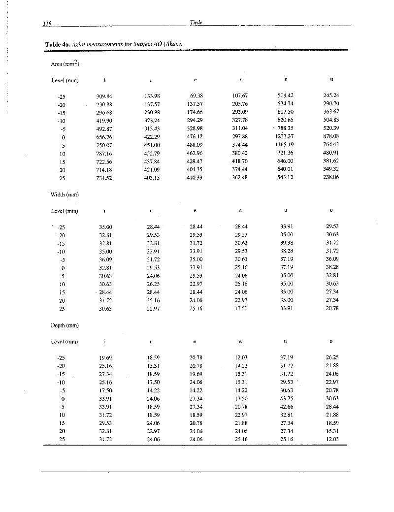
Table 4a. Axial measurements for Subject AO (Akan).
Area (mm2)
Level (mm) e e u u
-25 309.84 133.98 69.38 107.67 508.42 245.24
-20 230.88 137.57 137.57 205.76 534.74 290.70
-15 296.68 230.88 174.66 293.09 807.50 363.67
-10 419.90 373.24 294.29 327.78 820.65 504.83
-5 492.87 313.43 328.98 311.04 788.35 520.39
0 656.76 422.29 476.12 297.88 1233.37 878.08
5 750.07 451.00 488.09 374.44 1165.19 764.43
10 787.16 455.79 462.96 380.42 721.36 480.91
15 722.56 437.84 429.47 418.70 646.00 381.62
20 714.18 421.09 404.35 374.44 640.01 349.32
25 734.52 403.15 410.33 362.48 543.12 238.06
Width (mm)
Level (mm) e e u u
-25 35.00 28.44 28.44 28.44 33.91 29.53
-20 32.81 29.53 29.53 29.53 35.00 30.63
-15 32.81 32.81 31.72 30.63 39.38 31.72
-10 35.00 33.91 33.91 29.53 38.28 31.72
-5 36.09 31.72 35.00 30.63 37.19 36.09
0 32.81 29.53 33.91 25.16 37.19 38.28
5 30.63 24.06 29.53 24.06 35.00 32.81
10 30.63 26.25 22.97 25.16 35.00 30.63
15 28.44 . 28.44 28.44 24.06 35.00 27.34
20 31.72 25.16 24.06 22.97 35.00 27.34
25 30.63 22.97 25.16 17.50 33.91 20.78
Depth (mm)
Level (mm) e e u u
-25 19.69 18.59 20.78 12.03 37.19 26.25
-20 25.16 15.31 20.78 14.22 31.72 21.88
-15 27.34 18.59 19.69 15.31 31.72 24.06
-10 25.16 17.50 24.06 15.31 29.53 22.97
-5 17.50 14.22 14.22 14.22 30.63 20.78
0 33.91 24.06 27.34 17.50 43.75 30.63
5 33.91 18.59 27.34 20.78 42.66 28.44
10 31.72 18.59 18.59 22.97 32.81 21.88
15 29.53 24.06 20.78 21.88 27.34 18.59
20 32.81 22.97 24.06 24.06 27.34 15.31
25 31.72 24.06 24.06 25.16 25.16 12.03
An MRI-based Study ofPharyngeal Volume Contrasts in Akan 117
Table 4b. Axial measurements for Subject MT (English).
Area (mm2)
Level (mm) e E u u
-25 438.08 508.12 470.14 486.89 753.94 618.48
-20 517.73 519.10 411.52 514.40 699.01 617.29
-15 527.34 616.61 391.19 614.89 806.12 608.91
-10 661.93 649.57 576.61 590.97 862.43 752.47
-5 606.99 585.02 523.97 644.80 896.76 718.97
0 914.61 659.18 608.91 532.35 1031.34 848.17
5 951.69 630.34 653.17 368.46 958.56 802.71
10 942.08 611.11 575.42 295.48 944.82 724.95
15 948.94 597.38 517.99 324.19 855.56 628.05
20 940.70 649.57 526.37 337.35 811.61 541.92
25 961.30 708.62 525.17 394.78 752.56 485.69
Width (mm)
Level (mm) e E u u
-25 32.81 33.98 33.91 33.91 33.98 32.81
-20 37.50 36.33 36.09 36.09 36.33 33.91
-15 39.84 39.84 38.28 38.28 39.84 36.09
-10 41.02 39.84 39.38 41.56 41.02 39.38
-5 41.02 41.02 39.38 40.47 41.02 40.47
0 38.67 38.67 40.47 38.28 39.84 39.38
5 37.50 38.67 39.38 37.19 38.67 39.38
10 42.19 36.33 36.09 36.09 39.84 39.38
15 43.36 41.02 35.00 33.91 41.02 31.72
20 44.53 38.67 36.09 35.00 41.02 36.09
25 45.70 42.19 33.91 33.91 39.84 38.28
Depth (mm)
Level e E u u
-25 26.95 29.30 29.53 26.25 30.47 30.63
-20 21.09 22.27 21.88 22.97 28.13 28.44
-15 19.92 19.92 21.88 24.06 29.30 26.25
-10 21.09 19.92 17.50 19.69 24.61 29.53
-5 17.58 17.58 17.50 17.50 24.61 24.06
0 31.64 23.44 24.06 17.50 31.64 26.25
5 30.47 22.27 22.97 15.31 30.47 27.34
10 30.47 21.09 20.78 14.22 29.30 19.69
15 30.47 18.75 20.78 12.03 25.78 18.59
20 31.64 23.44 18.59 12.03 24.61 16.41
25 29.30 23.44 20.78 17.50 21.09 15.31
118 Tiede
-100
-200
-200 +.2-5~.2-:-0""""15~'1~0""".5~~0--"~5"""1-:-0.."..15~2:-:::0...,..2:':'5 ....
mm (increasing height through pharynx)
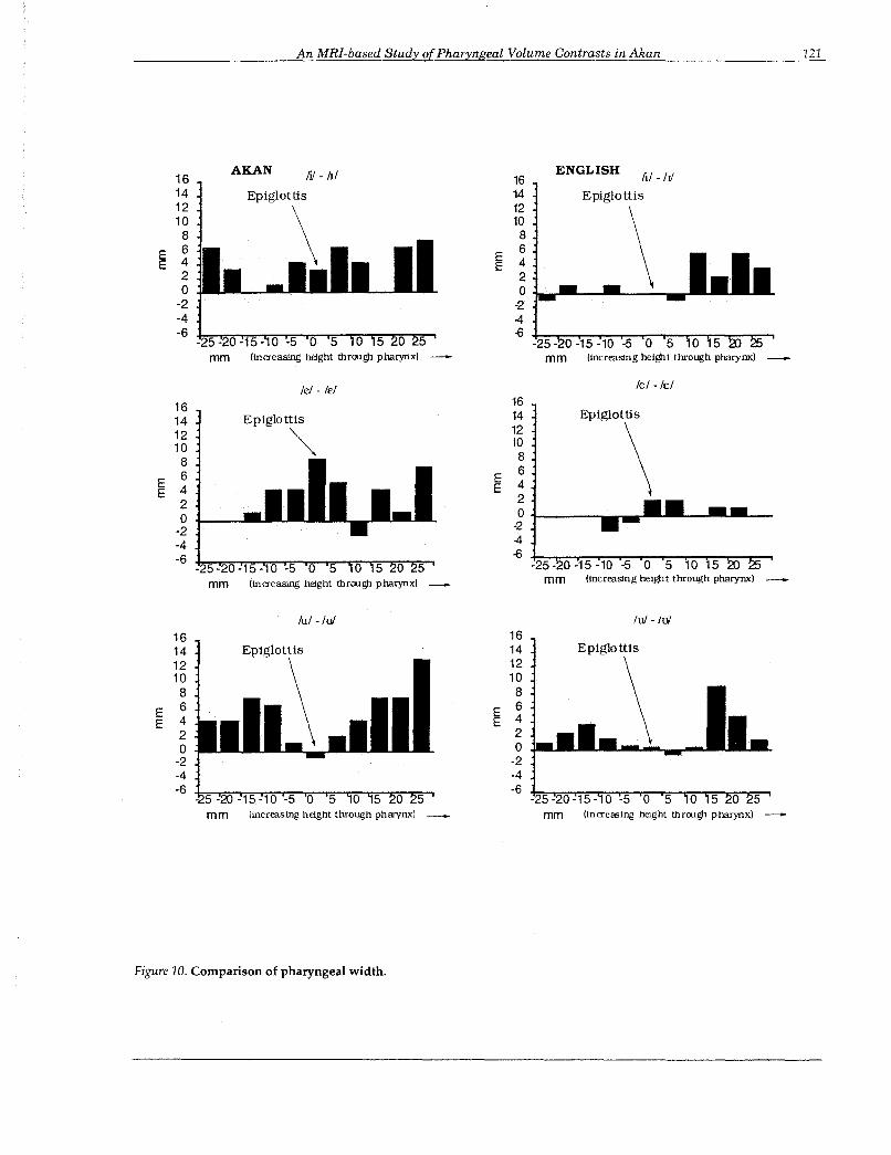
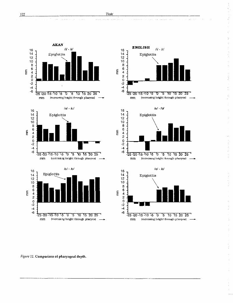
vancement. Notice that while somewhat noisierthan the area data, the overall trend for bothwidth and depth is for consistently larger valuesfor the expanded variant of each vowe1. 10 Noticealso that the observed differences in lateral widthare almost as large as those observed for anterior/posterior depth, showing that for this speakerat least control of the lateral dimension is an important part of the mechanism used to produce thecontrast, thereby confirming Lindau's positionthat speakers of Akan seek to maximize the difference in overall pharyngeal volume in effectingthe contrast.
Figures 9, 10, and 11 provide a comparison ofthe English area, width, and depth results withthose of Akan. As in the previous figures, theheight of each bar shows the difference betweenexpanded and constricted variants atcorresponding levels through the pharynx.Combining area cross-sections as for Akan thefollowing approximations to pharyngeal volumewere obtained:
(7) Derived Pharyngeal Volumes for SubjectMT (English) (cm3)
IiI-IIi500
400 Epiglottis
300 ~200
~E 100
0
leI - leI500
400Epiglottis
300 ~'"
200EE 100
0
-100
-25 -20 -15 -10-5 0 5 10 15 20 25mm (increaSing height through pharynx) _
500
400
300
200EE 100
o
-100
-200-25 -20 -15 -10 -5 0 5 10 15 20 25
mm llncreasing height through pharynx) .........
Figure 7. Delta cross-sectional area for Subject AD(Akan).
The observed difference in pharyngeal volume isconsistent with the tongue root advancement observed in the sagittal images and cineradiographicstudies, but leaves open the question of whetherthe <iifference is due entirely to root position, orwhether pharyngeal lateral width contributes aswell. The relative contributions of width anddepth to the area values obtained for Akan are illustrated in Figure 8.
Recall that width is a measure of side-to-side orlateral distance, and depth is a measure of anterior/posterior distance corresponding to root ad-
I E U
Tense 37.25 26.28 43.10
Lax 30.13 23.55 34.31
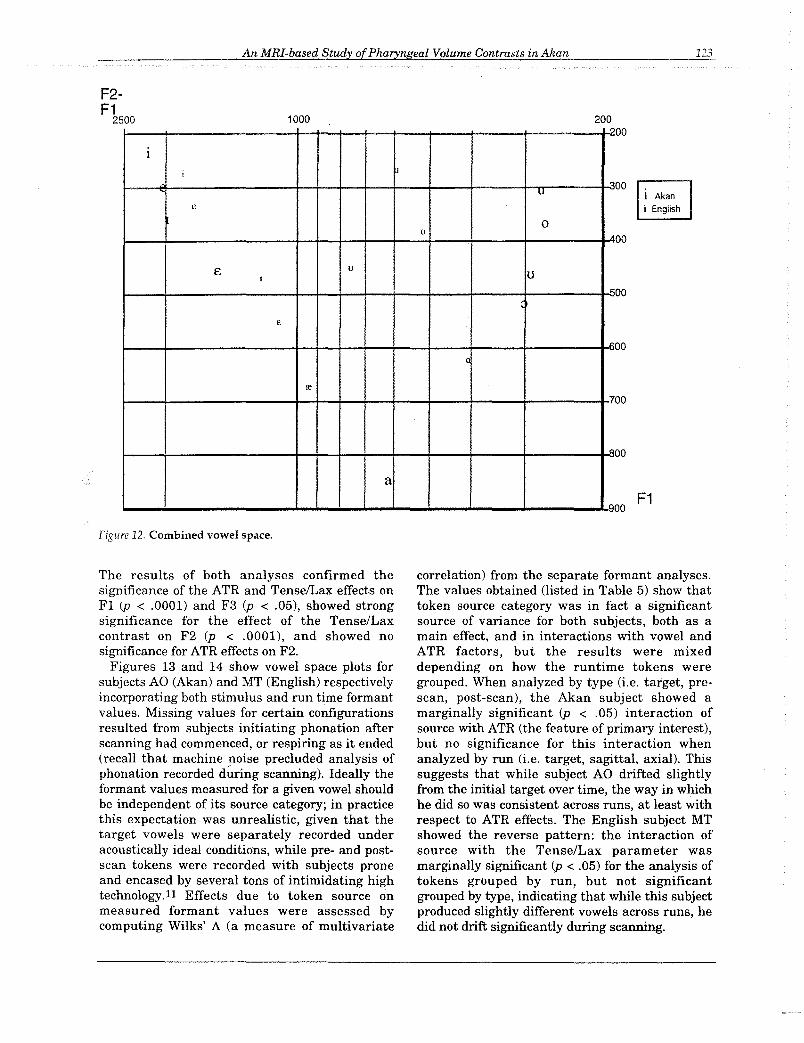
As in Akan, the expanded (tense) variants showconsistently larger overall pharyngeal volumethan their lax counterparts. Again, it isinteresting to note that the ratio of expanded toconstricted volume is nearly the same for vowels/i/ and /ul (1.24 us. 1.26), although the magnitudeof the difference is considerably less than thatobserved for Akan.Acoustics. Figure 12 shows a combined vowelspace chart using the averaged stimulus targetvowel formants from each language. The primaryacoustic effect of the ATR contrast on Akan vowelpairs appears to be a raised Fl for [-ATRJvariants, with F2 relatively unaffected. Laxvowels in English on the other hand show both araised Fl and lowered F2 compared to their tensecounterparts, for a net centralizing effect. Valuesmeasured for F3 were lower for constrictedvariants in both languages. Two analyses ofvariance were performed to quantify thesignificance of these observed effects: one groupedrecorded tokens into source groupings of target(the original stimulus words), sagittal run, andaxial run; the second analysis grouped them intotarget, pre-scan, and post-scan source groupings.
An MRI-based Study ofPharyngeal Volume Contrasts inAkan 119
WIDTH DEPTH iii - hi16 1614 1412 1210 10
8 86 E 6
~ 4 E 42 20 0
-2 -2-4 -4-6 -6
- 5 -20 -15 -10 ·5 0 5 0 5 0 5mm mm (lncreaslIlg height through pharynx)
leI - IFf lei - leI16 1614 Epiglottis 14 Epiglottis12
~12 ~
10 108 8
E 6 E 6E 4 E 4
2 20 0
-2 -2-4 -4-6 -6
mm
lui -lui luI - luI16 16 Epiglottis14 14
""12 1210 10
8 8
E 6~
6E 4 4
2 20 0
-2 -2-4 -4-6 -6
5Iincreaslng hel~t throu~ pharynJd --.... (lncreaslng height through pharynx) --....
Figure 8. Akan pharyngeal width and depth.
120 Tiede
AKANENGLISH ftI-!J1
Epiglottis
~
500
400
300
200
E100E
o i.....--
-100
-200
-300 -6~......,.........,,.,,,..,..,,.....,~.,..,,,.......,.,.,,-.............,,.,,.......,........-25 -20-15 -10 -5 0 5 0 15 0 25
mm (increasing height thnugh pharynx)
Iii -!J1
Epiglottis
~
500
400
300
200
E100E
o-100
-200
-300 f=~:-T"':"=-r:'::'"T-:--1r-::-r::-'"T':"::~:-r':"""'r.:::-1-25-20 -15 -10 -5 0 5 10 5 0 25
mm (Increasing height through pharynx)
lei -IE!
Epiglottis
\Ie! - lei
Epiglottis
\500
400
300
200
~ 100E 0
-100
-200
-300 -t.::-::~::-T::":"T~r.::-'T'::'"-r::~'::'"""'I;-:-r::::::-"'t:":.,-25 -20 -15 -10 -5 0 5 0 5 5
mm (lncreastng height through pharynx)
500
400
300
200'"E 100E 0 ___
-100
-200
-300 -6,..."..,,,...,.,,.,,....,..,..,....,........,...,.....,...,.-,,,..,,.....,.,,,.......,...-.=..,-25 -20 -15 -10 -5 0 5 10 5 0 25
mm Uncreaslng height through pharynx)
lui -lui
Epiglottis
~
500
400
300
200
~ 100E
o-100
-200
-300 +.::-::,.,.,.,,,...,.,,,.,,..,..,..,,...,..........,...,.....,...,.,..-.,.,..,,.-,,...,...~ ...........,-25 -20 -15 -10 -5 0 5 0 5 2) 5
mm (lncreastng height through pharynx) --
500
400
300
200
~ 100o
-100
-200
-300 -1-::-:=-=~~-.-::-...-:-...-:--r::~"'::'"""T:-=-",.."..-r=...,-25 -20 -15 -10 -5 0 5 10 15 0 25
mm Una-easing height through pharynx) __
Figure 9. Comparison of cross-sectional area.
An MRI-based Study ofPharyngeal Volume Contrasts in Akan 121
16AKAN ENGLISH
1614 1412 1210 10
8 8
E 6E
6E 4 E 4
2 20 0
-2 -2-4 -4-6 -6
-25 -20 -15 - 0 -5 0 5 0 5 0 5 -25 - 0 -15 -10 -5 0 5 10 15mm (increasing height thrcugh pha.rynx) mm (increasing heIgh t through pharynx)
lei - lEi leI -lEI
16 16
14 Epiglottis 14 Epiglottis
12~
12
\10 10
8 8
6 E6
E 4E 4 E
2 2
0 0
-2 -2
-4 -4
-6 -6-25 -20 -15 - 0 -5 0 5 -25 - 0 -15 -10 -5 0 5 10 15
mm (increasing height thrcugh pha.rynx) mm (increaSIng heIght through pharynx)
luI -lui lui -lui
16 1614 14 Epiglottis12 12
\10 10
8 8
E 6 E 6
E 4 E 42 20 0
-2 -2-4 -4-6 -6
- 5 -m -15 -10 -5 0 5 -25 -20 -15 - 0 -5 0 5mm (increasing height through pharynx) - mm (IncreasIng height thrcugh pha.rynx) -
Figure 10. Comparison of pharyngeal width.
122 Tiede
AKANIii - !JI ENGLISH Iii - !JI16 16
14 Epiglottis 14 Epiglottis12
~12
~10 108 8
~6 E
64 E 42 20 0
-2 -2-4 -4-6 -6
.......... mm
16lei - fel 16
luI - lui
14 Epiglottis 14 Epiglottis12
~12
\10 108 8
E6 E
6E 4 E 4
2 20 0
-2 -2-4 -4-6 -6
(lna-easlng heIght through pharymq -- --16
luI - luI16
lei - fe/
14 Epiglottis 1412 -------. 1210 108 8
E6
E6
E 4 E 42 20 0
-2 -2-4 -4-6 -6
- 5 -20 -15 -10 -5 0 5 0 5 0 5mm mm (lna-easlng height through pharynx) -
Figure 11. Comparison of pharyngeal depth.
An MRI-based Study ofPharyngeal Volume Contrasts in Akan 123
F1
00
00
001 Akani English
00
00
00
00
20000
1000 ,.,
1
i"u
e
00
£ uUI
c:
f.';
c
0
re7
"
a
"
F2F1
2500
Figure 12. Combined vowel space.
The results of both analyses confirmed thesignificance of the ATR and TenseILax effects onFl (p < .0001) and F3 (p < .05), showed strongsignificance for the effect of the Tense/Laxcontrast on F2 (p < .0001), and showed nosignificance for ATR effects on F2.
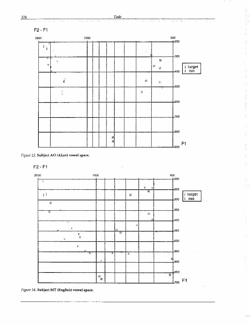
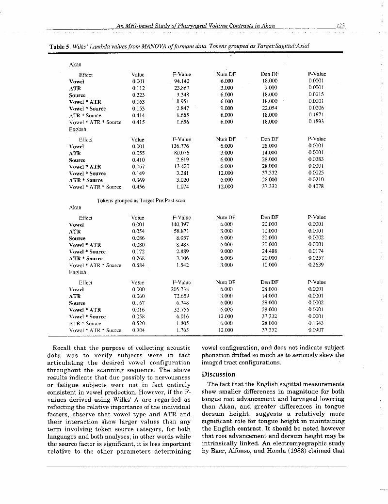
Figures 13 and 14 show vowel space plots forsubjects AO (Akan) and MT (English) respectivelyincorporating both stimulus and run time formantvalues. Missing values for certain configurationsresulted from subjects initiating phonation afterscanning had commenced, or respiring as it ended(recall that machine noise precluded analysis ofphonation recorded during scanning). Ideally theformant values measured for a given vowel shouldbe independent of its source category; in practicethis expectation was unrealistic, given that thetarget vowels were separately recorded underacoustically ideal conditions, while pre- and postscan tokens were recorded with subjects proneand encased by several tons of intimidating hightechnology.ll Effects due to token source onmeasured formant values were assessed bycomputing Wilks' A (a measure of multivariate
correlation) from the separate formant analyses.The values obtained (listed in Table 5) show thattoken source category was in fact a significantsource of variance for both subjects, both as amain effect, and in interactions with vowel andATR factors, but the results were mixeddepending on how the runtime tokens weregrouped. When analyzed by type (i.e. target, prescan, post-scan), the Akan subject showed amarginally significant (p < .05) interaction ofsource with ATR (the feature of primary interest),but no significance for this interaction whenanalyzed by run (i.e. target, sagittal, axial). Thissuggests that while subject AO drifted slightlyfrom the initial target over time, the way in whichhe did so was consistent across runs, at least withrespect to ATR effects. The English subject MTshowed the reverse pattern: the interaction ofsource with the Tense/Lax parameter wasmarginally significant (p < .05) for the analysis oftokens grouped by run, but not significantgrouped by type, indicating that while this subjectproduced slightly different vowels across runs, hedid not drift significantly during scanning.
124 Tiede
F2 - F1
1000 2001--;-t--i-------+~-_r__;_-_r_-_r_-_r_--_;_---.....,l_2noo
2500
u1-_e-r~-------+_+_+-+-+-+--+_---l:r_----i_3n·oo
~
1---f--e-------+-+-+-+--t--l--+--...;-----t-'l'00
(J o o
u uI---+--------t-+-+-+--t---+--+----+--~--t-O~·OO
I---+--------t-+-+-+--t---+--+----+-----t-O"IOO
1---+--------t--+--+-+--t--+--+----+-----t-7OO
1---+--------+_+-+-+-+--+--+_---+-----t-Bn ,OOaa
n. F1L..-_-'- -'-.......L_.J..-.......L_..J..._-'-__.J..-__-'- ..L.900
Figure 13. Subject AD (Akan) vowel space.
F2 - F1
50
00
00 F1
50
00
50
00
00
50
50
40000
10002
i
u II n.
Ui i u
,>,
e,>,
c0
r AI
I
(l
I 111 AI
IU
Ie ",
e0 ".
<;.. £ /\
£(
6
6a: Q
c.e7
2000
Figure 14. Subject MT (English) vowel space.
An MRI-based Study ofPharyngeal Volume Contrasts in Akan
Table 5. Wilks' Lambda values from MANOVA offonnant data. Tokens grouped as Target:Sagittal:Axial
Akan
Effect Value F-Value NUIllDF DenDF P-Value
Vowel 0.001 94.142 6.000 18.000 0.0001
ATR 0.112 23.867 3.000 9.000 0.0001
Source 0.223 3.348 6.000 18.000 0.0215
Vowel * ATR 0.063 8.951 6.000 18.000 0.0001
Vowel * Source 0.153 2.847 9.000 22.054 0.0206
ATR * Source 0.414 1.665 6.000 18.000 0.1871
Vowel * ATR * Source 0.415 1.656 6.000 18.000 0.1893
English
Effect Value F-Value NumDF DenDF P-Value
Vowel 0.001 136.776 6.000 28.000 0.0001
ATR 0.055 80.075 3.000 14.000 0.0001
Source 0.410 2.619 6.000 28.000 0.0383
Vowel * ATR 0.067 13.420 6.000 28.000 0.0001
Vowel * Source 0.149 3.281 12.000 37.332 0.0025
ATR * Source 0.369 3.020 6.000 28.000 0.0210
Vowel * ATR * Source 0.456 1.074 12.000 37.332 0.4078
Tokens grouped as Target:Pre:Post scanAkan
Effect Value F-Value NumDF DenDF P-Value
Vowel 0.001 140.397 6.000 20.000 0.0001
ATR 0.054 58.871 3.000 10.000 0.0001
Source 0.086 8.057 6.000 20.000 0.0002
Vowel * ATR 0.080 8.483 6.000 20.000 0.0001Vowel * Source 0.172 2.889 9.000 24.488 0.0174
ATR * Source 0.268 3.106 6.000 20.000 0.0257
Vowel * ATR * Source 0.684 1.542 3.000 10.000 0.2639English
Effect Value F-Value NumDF DenDF P-Value
Vowel 0.000 205.738 6.000 28.000 0.0001ATR 0.060 72.659 3.000 14.000 0.0001Source 0.167 6.748 6.000 28.000 0.0002Vowel * ATR 0.016 32.756 6.000 28.000 0.0001Vowel * Source 0.058 6.016 12.000 37.332 0.0001ATR * Source 0.520 1.805 6.000 28.000 0.1343Vowel * ATR * Source 0.304 1.765 12.000 37.332 0.0907
125
Recall that the purpose of collecting acousticdata was to verify subjects were in factarticulating the desired vowel configurationthroughout the scanning sequence. The aboveresults indicate that due possibly to nervousnessor fatigue subjects were not in fact entirelyconsistent in vowel production. However, if the Fvalues derived using Wilks' A are regarded asreflecting the relative importance of the individualfactors, observe that vowel type and ATR andtheir interaction show larger values than anyterm involving token Source category, for bothlanguages and both analyses; in other words whilethe source factor is significant, it is less importantrelative to the other parameters determining
vowel configuration, and does not indicate subjectphonation drifted so much as to seriously skew theimaged tract configurations.
Discussion
The fact that the English sagittal measurementsshow smaller differences in magnitude for bothtongue root advancement and laryngeal loweringthan Akan, and greater differences in tonguedorsum height, suggests a relatively moresignificant role for tongue height in maintainingthe English contrast. It should be noted howeverthat root advancement and dorsum height may beintrinsically linked. An electromyographic studyby Baer, Alfonso, and Honda (1988) claimed that
126 Tiede
Subject MT(English)
580.0900.008-0.687n.s.
above epiglottis
280.6800.4624.91
<0.01
above epiglottis
280.7440.5535.89
<0.01
Subject AO(Akan)
580.4430.1963.76
<0.01
All Vowels
dJ.Pearson's rr 2
I-ratio
English Vowels (Subject MT)below epiglottis
d.f. 28Pearson's r -0.671r2 0.450I-ratio -4.79p <0.01
Akan Vowels (SUbject AO)below epiglottis
d.f. 28Pearson's r 0.545r 2 0.297t-ratio 3.44p <0.01
p
Table 6. Regression ofAxial Width and Depth.
are encompassed in the Language x Group x ATRinteraction. Results of the analysis for the widthparameter showed no significance for this interaction (F=1.12), but moderate significance for depth(F=9.48, p<.05), and strong significance for area(F=15.84, p<.005), reflecting the difference insubepiglottal behavior between the two subjects.
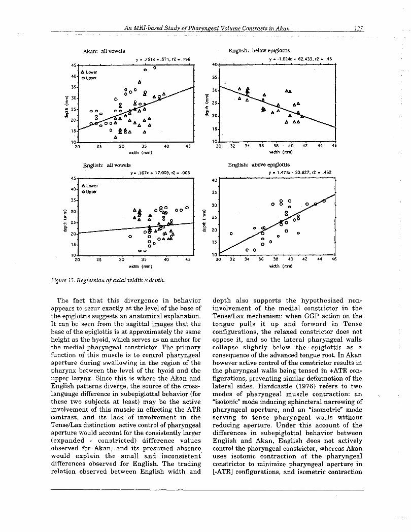
Further evidence of divergent behavior isprovided by a regression analysis of width againstdepth for corresponding measurement levels,summarized in Table 6, and illustrated in Figure15. The results show a significant (p < .01)positive correlation between Akan width anddepth across all measurement levels, but nocorrelation for the same analysis on English.When the English values are analyzed separatelyby upper/lower grouping however, the resultsshow a significant (p < .01) positive correlationbetween width and depth for measurement levelsabove the epiglottal pivot, and a significant (p <.01) negative correlation at levels below it; in otherwords below the level of the epiglottis in Englishan increase in anterior/posterior depth isaccompanied by a decrease in lateral width.Separate analysis of the Akan values by groupshows significant (p < .01) positive correlationsbetween width and depth for measurement levelsabove and below the epiglottal pivot.
SubjectMT(English)
40.7820.6112.51
<0.05n.s.
d.f.Pearson's rr2
t-ratiop
the same contraction of the posterior genioglossus(GGP) muscle effecting tongue root advancementalso forces the tongue dorsum upwards, whilecontraction of the separately controlled anteriorgenioglossus (GGA) pulls the dorsum forward anddown. The results obtained here suggest that GGAactivity is greater in Akan than in English: activecontrol of the GGA in Akan works against thedorsum-raising effect of the GGP, resulting insmaller net dorsum height differences than thoseobserved for English. This predicts that thecorrelation of dorsum height with rootadvancement should be stronger in English thanin Akan, which is confirmed by a regressionanalysis outlined in the table below:
(8) Dorsum Height regressed against RootAdvancement (all vowels)
SubjectAO(Akan)40.2260.0510.465
Assuming the pattern of muscle activity mentioned above, one apparent difference thereforebetween the ATR and TenselLax mechanisms liesin how each language treats the inherent dorsumraising effect of root advancement: Akan seeks toneutralize its effect, whereas English (at least inthis subject's dialect) appears to exploit it.
Another difference is apparent from thepatterning of axial data at measured levels belowthe epiglottis. With one exception (area measuredat the three lowest levels of lei), the area, width,and depth measurements obtained for Akan showconsistently larger values for expanded (+ATR)variants at all measured levels, above and belowthe epiglottis. But while the English data alsoshow consistently larger values above theepiglottal pivot, at levels below that pointdifferences between tense and lax variants areinconsistent in sign and considerably smaller inmagnitude. The change occurs abruptly, and isevident to some degree in all three parametersmeasured.
An attempt was made to quantify the significance of this divergence by performing an analysisof variance on each measurement parameter,treating the individual levels as repeated measures nested within an upper or lower groupingfactor.l2 Under this design, inter-language differences in behavior above and below the epiglottisbetween expanded and constricted configurations
An MRI-based Study ofPharyngeal Volume Contrasts in Akan 127
Akan: all vowels
34 36 38' 40 42 44 46
Y<idth (nrol
English: below epIglottis
y. -1.02« + 62.433. r2 • .4540
35
30
E.s. 25iiit" 20
15
1030 32
A
goo e80
AAA
A A AAooOA AA A
o itA AA
y • .751x + .571, r2 •.19645+-~-"""'--..r..---","--;t'"-"----'""r
o 0A LONer
40 ouwer
35
15
10J-__...-_-~--...---_--~20 25 30 35 40 45
width (nrol
E 30.s.or: 25
!20
English: all vowelsy • .167x + 17.009, r2 •.008
English: above epIglottisy. 1.475x - 33.627, r2 • .462
45ot-.......- .....--.....--------o-y 40
15
A LONer40 0 Uprer
35
E 30 A j A 0 8~ 0 °°-5. AA A .8"or: 25L_-----jrA1A~~~ti 20 0' A&l~ A,..
° ° OA.H:go00
10J-_-_~-_~-_~-__-.-l20 25 30 35 40 45
width (nrol
35
30
~ 25
ii.,.-8 20
15
32 34 36 38 40 42 44
Y<idth (nrol
Figure 15. Regressioll of axial width x depth.
The fact that this divergence in behaviorappears to occur exactly at the level of the base ofthe epiglottis suggests an anatomical explanation.It can be seen from the sagittal images that thebase of the epiglottis is at approximately the sameheight as the hyoid, which serves as an anchor forthe medial pharyngeal constrictor. The primaryfunction of this muscle is to control pharyngealaperture during swallowing in the region of thepharynx between the level of the hyoid and theupper larynx. Since this is where the Akan andEnglish patterns diverge, the source of the crosslanguage difference in subepiglottal behavior (forthese two subjects at least) may be the activeinvolvement of this muscle in effecting the ATRcontrast, and its lack of involvement in theTenselLax distinction: active control of pharyngealaperture would account for the consistently larger(expanded - constricted) difference valuesobserved for Akan, and its presumed absencewould explain the small and inconsistentdifferences observed for English. The tradingrelation observed between English width and
depth also supports the hypothesized noninvolvement of the medial constrictor in theTenselLax mechanism: when GGP action on thetongue pulls it up and forward in Tenseconfigurations, the relaxed constrictor does notoppose it, and so the lateral pharyngeal wallscollapse slightly below the epiglottis as aconsequence of the advanced tongue root. In Akanhowever active control of the constrictor results inthe pharyngeal walls being tensed in +ATR configurations, preventing similar deformation of thelateral sides. Hardcastle (1976) refers to twomodes of pharyngeal muscle contraction: an"isotonic" mode inducing sphincteral narrowing ofpharyngeal aperture, and an "isometric" modeserving to tense pharyngeal walls withoutreducing aperture. Under this account of thedifferences in subepiglottal behavior betweenEnglish and Akan, English does not activelycontrol the pharyngeal constrictor, whereas Akanuses isotonic contraction of the pharyngealconstrictor to minimize pharyngeal aperture in[-ATR] configurations, and isometric contraction
128 Tiede
in [+ATRJ configurations to prevent deformationof pharyngeal lateral walls. Pharyngeal isometrictension may be significant in a different context:Hardcastle (1973) has suggested that it may beimportant in the production of tensed initial stopsin Korean.
Assuming that the patterns observed for thesetwo subjects are in fact representative of Akanand English, it is evident that the ATR andTense/Lax distinctions are only superficiallysimilar. In producing +ATR vowels Akan speakersenlarge the pharyngeal cavity by advancing theroot of the tongue, lowering the larynx, andmaintaining tension in the pharyngeal walls. In-ATR configurations the root is retracted,pharyngeal aperture is constricted, and the larynxis raised, minimizing pharyngeal volume. Becausespeakers appear to make adjustments to maintainrelatively constant dorsum height across the twoconfigurations, the ATR distinction is essentially acontrast in pharyngeal volume. Tense vowels inEnglish are also articulated with an advancedtongue root, enlarging the pharyngeal cavity as aconsequence; but below the level of the epiglottisthis enlargement is counteracted by thedeformation of the lateral walls from lack ofconstrictor tension. In lax configurationspharyngeal volume is smaller, but it is unclearwhether this is due to actual constriction by themedial constrictor, or simply the relaxed positionof the tongue root. The dorsum-raising effect ofroot advancement is not adjusted for in English,and instead constitutes an integral part of thecontrast.
Generalizations of this sort are somewhat presumptuous given the limited scope of this study,and those made for English in particular shouldbe viewed in the context of the Ladefoged et al.(1972) and Raphael & Bell-Berti (1975) findingsmentioned above, showing that different Englishspeakers produce the Tense/Lax contrast differently. Separate studies by Lindau in 1975 and1979 involving four subjects each found consistentarticulatory implementation of the ATR mechanism, so the generalizations made here for Akanare perhaps on firmer ground, especially as thesagittal results of this study replicated her findings. In any case, while different speakers ofEnglish may approximate more or less closely theAkan ATR articulatory mechanism, the point isthat the TenselLax contrast is not identical to it,and must be represented by a different feature.Furthermore, because the Akan distinction appears to involve two muscles that the Englishcontrast does not exploit (the medial pharyngeal
constrictor and possibly the anterior genioglossus), from an articulatory standpoint it appears tobe more complex with respect to the active controland coordination needed to produce it.
ConclusionsAlthough exploratory in scope, this study did
succeed in achieving certain objectives. Itdemonstrated that despite its inherent drawbacksMagnetic Resonance Imaging can be a usefultechnique for obtaining information about staticvocal tract configurations, one that willundoubtedly increase in importance as thetechnology is further refined. Measurements fromthe sagittal images collected here successfullyreplicated previous results from cineradiographicstudies showing the significance of tongue rootadvancement and larynx height in effecting theAkan ATR contrast, and those obtained from axialimages extended these results for the first timeinto the dimension of lateral width. The axialmeasurements confirm Lindau's position that it isthe overall difference in pharyngeal volume that isrelevant to the Akan vowel contrast, not justrelative larynx or tongue root positions. Finallythe parallel analysis of the English Tense/Laxcontrast gave results showing that despitesuperficial similarities with the Akan ATRdistinction, the two contrasts are not the same.
REFERENCESBaer, T., Alfonso, P., & K. Honda (1988) Electromyography of the
tongue muscles during vowels in /@pVp/ environment.Annual Bulletin of the Research Institute for Logopedics andPlwniatrics (University ofTokyo), 22, 7-19.
Baer. T., Gore, J. C, Gracco, 1. C, & Nye, P. W. (1991). Analysis ofvocal tract shape and dimensions using magnetic resonanceimaging: Vowels. Journal of the Acoustical Society of America, 90(2), 799-828.
Bradley, W. G., Newton, T. H., & Crooks, 1. E. (1983). Physicalprinciples of nuclear magnetic resonance. In T. H. Newton & D.G. Potts (Eds.), Modern neuroradiology: Advanced imagingtechniques (pp. 15-62). San Francisco: Clavadel Press.
Clements, G. N. (1980). Vowel harmony in a nonlinear generativephonology: An autosegmental model. Indiana UniversityLinguistics Club.
Dolphyne, F. A. (1988) The Akan (Twi-Fante) Language. Accra:Ghana Universities Press.
Halle, M., & Stevens, K. N. (1969). On the feature AdvancedTongue Root. Quorterly Progress Report, 94, Research Laboratoryof Electronics, Massachusetts Institute of Technology, 209-215.
Hardcastle, W. J. (1973). Some observations on the tense-laxdistinction in initial stops in Korean. Journal of Phonetics, 1, 263272.
Hardcastle, W. J. (1976). Physiology of speech production. New York:Academic Press.
Harshman, R., Ladefoged, P., & Goldstein, L. (1977). Factoranalysis of tongue shapes. Journal of the Acoustical Society ofAmerica, 62, 693-707.
An MRI-based Study ofPharyngeal Volume Contrasts in Man 129
Jackson, M. T. (1988). Phonetic theory and cross-linguisticvariation in vowel articulation. Working Papers in Phonetics, 71,Los Angeles: University of California.
Ladefoged, P. (1964). A phonetic study of West African languages.Cambridge: University Press.
Ladefoged, P., DeClerk, J., Lindau, M., & Papcun, G. (1972). Anauditory-motor theory of speech production. Working Papers inPhonetics, 22, 48-75. Los Angeles: University of California.
Lakshminaravanan, A. V., Lee, S., & McCutcheon, M. J. (1991).MR Imagrr;'g of the vocal tract during vowel production. JournalofMagnetic Resonance Imaging, 1, 71-76.
Lindau, M. (1975). [Features] For Vowels. Working Papers inPhonetics, 30. Los Angeles: University of California.
Lindau, M. (1979). The feature expanded. Journal of Phonetics, 7,163-176.
Perkell, J. (1971). Physiology of speech production: a preliminarystudy of two suggested revisions of the features specifyingvowels. Quarterly Progress Report, 102, Research Laboratory ofElectronics, Massachusetts Institute of Technology, 123-139.
Raphael, L. J., & Bell-Berti, F. (1975). Tongue musculature and thefeature of tension in English vowels. Phonetica, 32, 61-73.
Stewart, J. M. (1967). Tongue root position in Akan vowelharmony. Phonetica, 16, 185-204.
Stewart, J. M. (1971). Niger-Congo, Kwa. In T. Sebeok (Eds.),Current trends in linguistics (pp. 179-212). The Hague: Mouton.
FOOTNOTESIHigh vowels from both harmony groups (and low la/) also
have nasalized counterparts which were not investigated inthis study.2In Clements' (1980) analysis of Akan vowel harmony lal istreated as an opaque vowel that induces the [-ATR] feature onany subsequent vowels. Some dialects of Akan have anadditional ([+ATRJ) front vowel 131 that harmonizes with I a/;however the exact distribution of this vowel is unclear, and it isignored here.3The magnetic resonance technique relies on strong magneticfields to align the magnetic moments of hydrogen nuclei, andpulsed radio-frequency energy to set them into resonance. Thesignal intensity in a MR image depends on the density of thehydrogen nuclei in the scanned tissue and its resonance decaycharacteristics, determined by the atomic environment of theprotons within each molecule. See Bradley et al. (1983) forfurther information.
4Personal communication from my Akan informant.
5To facilitate cross-language comparison in the followingdiscussion I freely apply the terms "expanded" and"constricted" to both Akan and English, but do not mean toimply by this that the same feature mechanism is involved inboth languages; nor am I referring to Lindau's term forthe Akan feature. The terms are simply meant as shorthandphysical descriptions of the respective contrasts,so that "expanded" for example should be understood as[+ATR] in the context of Akan, and [+Tense] in the context ofEnglish.
6Image version 1.29q by W. Rasband, NIH.7lmage quality was inferior to that obtained by Baer et al. (1991);
however those researchers used custom-made cephalostats andlonger scanning times in obtaining their best results.
8The scanning process resolved a given volume into a flat 256 x256 pixel image. Sagittal scanning used a 280mm width x280mm depth x 3mm thickness giving a resolution ofl.09mm/pix. Half the axial scans were done using a 280mm x280mm x 5mm volume resulting in the same l.09mm/pixresolution, and half were done using a 300mm x 300mm x 5mmvolume giving a resolution of 1.17mm/pix. See Tables 1 and 2for full scanning specifications used.
9Each volume element represents L (scan area' scan thickness),where scanned image thickness was a constant 5mm (seepreceding footnote).
laThe uppermost axial levels measured for Akan le:EI show(slightly) larger depth values for the contracted variant,reversing the pattern found everywhere else. One possibleexplanation is that at these levels scanning has moved out ofthe region manipulated for the ATR contrast, and into therelatively invariant region of the tongue constrictioncharacterizing the vowel.
II Both subjects reported feelings of nervousness when first placedinside the magnet; as the diameter of the central bore isapproximately two feet only, claustrophobia is definitely apotential problem in studies of this type.
12For purposes of symmetry the uppermost level was droppedfrom the analysis, so that the five subepiglottal levelsconstituted the 'lower' group, and the epiglottal pivot and itsfour succeeding levels the 'upper.' ANOVA design (levels asrepeated measures nested in group): level (measurement level,1-5); group (below labove epiglottis); vowel (I/E/U); ATR(expanded I constricted); language (Akan/English). Thelevel'vowel>ATR>language interaction was used as the errorterm (l6df).