Embed Size (px)
Citation preview
1
AN INVESTIGATION INTO THE PREVALENCE OF POINT MUTATIONS
ASSOCIATED WITH ANTIMALARIAL DRUG RESISTANCE IN PLASMODIUM
FALCIPARUM FOUND IN THE ZAMBEZI REGION OF NAMIBIA
A RESEARCH THESIS SUBMITTED IN FULFILMENT OF THE REQUIREMENTS FOR
THE DEGREE OF
MASTER OF SCIENCE
OF
THE UNIVERSITY OF NAMIBIA
BY
LUCILLE L. DAUSAB
201174901
March 2018
MAIN SUPERVISOR: Prof. Davis Mumbengegwi
CO-SUPERVISOR: Dr Jaishree Raman, National Institute of Communicable Diseases, South
Africa
i
Abstract
Surveillance and monitoring of emerging drug resistance is important as Namibia
moves towards malaria elimination. In 2005 artemisinin combination therapies
(ACT’s) were introduced as first-line treatment for uncomplicated falciparum malaria
in Namibia. However, reduced ACT efficacy has been reported in Asia and in some
parts of Western Africa, raising concerns around the efficacy of artemisinin. This
study aimed to identify the different plasmodium species found in Zambezi region,
Namibia as well as to investigate the prevalence of antimalarial drug resistance
polymorphisms in the pfcrt, pfmdr1 and Kelch 13 genes in the region. A QIAamp
DNA mini-kit (Qiagen, Germany) was used to extract DNA from malaria positive
Dried Blood Spots (DBS). Prior to investigating the prevalence of point mutations,
multiplex PCR was performed to investigate the different plasmodium species found
in the Zambezi region of Namibia. Seventy-two P. falciparum positive samples out of
a total of 143 malaria positive samples were analysed by PCR-RFLP at codon N86Y
in the pfmdr1 whereas the prevalence of haplotypes at codons 72-76 in the pfcrt gene
were analysed using Quantitative-PCR. Additionally, mutations at 25 codons in the
Kelch 13 gene were analysed by n-PCR followed by sequencing. The study found that
76.2% of the cases were caused by P. falciparum which was less than expected.
Additionally, 7.1% of the cases were found to be caused by P. vivax followed by 6.0%
of P. malariae infections. In the pfcrt gene 92.5% samples contained the wild type
haplotype, 3.8% contained the mutant haplotype while 3.8% samples had mixed
haplotype. Furthermore, 18.5% mutant and 59.3% wild and type alleles were observed
in pfmdr1 gene at N86Y. No mutant alleles were observed in the Kelch 13 gene.
This study provides the first data on point mutations in the pfcrt, pfmdr1 and Kelch
13 genes in Namibia. A low prevalence of mutations was observed in this study which
shows that ACTs are still effective in Namibia. However, continued surveillance is
recommended with similar studies in other endemic regions of Namibia. Additionally,
it is recommended that further studies such as in vivo and in vitro responses to drug
treatment be done to determine the role of these polymorphisms in drug resistance to
support these findings.
Key words: Plasmodium falciparum; Artemisinin Combination Therapies;
Antimalarial drug resistance; Pfmdr1; Pfcrt; Kelch 13; Namibia.
ii
Table of Contents
Abstract ........................................................................................................................... i
List of Figures ............................................................................................................... iv
List of Tables ................................................................................................................ vi
List of Conferences and Posters ................................................................................... vii
Acknowledgements .................................................................................................... viii
Declaration ..................................................................................................................... x
Dedication ..................................................................................................................... xi
List of Abbreviations ................................................................................................... xii
1. INTRODUCTION ..................................................................................................... 1
1.1. Background of the study ..................................................................................... 1
1.1.1. Global Malaria Burden ................................................................................. 1
1.1.2. The Plasmodium parasite, vector and its Life Cycle .................................... 2
1.1.3. Malaria Symptoms ........................................................................................ 7
1.1.4. Malaria Control ........................................................................................ 8
1.1.5. The Impact of Antimalarial Drug Resistance ........................................ 13
1.2. Problem Statement ............................................................................................ 14
1.3. General Objectives of the study ........................................................................ 15
1.3.1. Specific Objectives of this study were to: .................................................. 15
1.4. Significance of the study ................................................................................... 15
1.5. Limitations of the study..................................................................................... 16
1.6. Delimitations of the study ................................................................................. 16
2. Literature Review..................................................................................................... 17
2.1. Antimalarial drugs ............................................................................................. 18
2.1.1. Quinolines ................................................................................................... 19
2.1.2. Antifolates .................................................................................................. 24
2.1.3. Artemisinin ................................................................................................. 26
2.2. Antimalarial drug resistance.............................................................................. 27
2.3. Development and Spread of Drug Resistance ................................................... 28
2.4. Mechanisms of Antimalarial drug resistance .................................................... 29
2.4.1. Resistance mediated by transporter mutations ........................................... 29
iii
2.4.2. Resistance to Antifolates ............................................................................ 33
2.4.3. Artemisinin and Derivatives ....................................................................... 34
3. Research Methods .................................................................................................... 37
3.1. Research Design ................................................................................................ 37
3.2. Methods ............................................................................................................. 37
3.2.1. Study Population and Study Site ................................................................ 37
3.2.2. Sample Collection....................................................................................... 38
3.3. Molecular Methods ............................................................................................... 39
3.3.1. Extraction of plasmodium parasite DNA using the QIAamp DNA mini-kit
(Qiagen Germany) ................................................................................................ 39
3.3.2. Plasmodium Species Identification ............................................................ 39
3.3.3. SNP Analysis .............................................................................................. 40
3.3.4. Restriction Digest ....................................................................................... 42
3.3.6. Quantitative Real Time PCR for the Detection Of crt72-76 Haplotypes ... 43
3.3.7. Amplification and Sequencing of the K13 Propeller Gene ........................ 45
3.4. Data Analysis .................................................................................................... 47
4. Research Ethics ..................................................................................................... 47
5. Results .................................................................................................................. 48
5.1. Identification of Plasmodium species by Multiplex PCR ................................. 48
5.2. Prevalence of the pfcrt 72-76 haplotypes .......................................................... 50
5.3. Prevalence of the pfmdr1 N86Y mutation......................................................... 51
5.4. Prevalence of SNPs in the Kelch 13 propeller gene.......................................... 52
6. Discussion ............................................................................................................. 54
7. Conclusion ............................................................................................................ 64
8. Recommendations .................................................................................................... 65
9. References ................................................................................................................ 67
10. Appendices ........................................................................................................ 92
Appendix 1: Carestart RDT Manufactures guideline ............................................... 92
Appendix 2: Eiken Japan PAN/Pf LAMP guidelines .............................................. 94
Appendix 3: New England Biolabs Restriction Digest Guidelines .......................... 97
Appendix 4: Qiagen DNA Extraction Protocol...................................................... 101
iv
List of Figures
FIGURE 1; MORPHOLOGICAL DIFFERENCES BETWEEN THE FOUR-PLASMODIUM SPECIES IN
HUMAN BLOOD SMEARS SOURCE: HTTP://WWW.DPD.CDC.GOV/DPDX ....................... 5
FIGURE 2: PLASMODIUM PARASITE LIFE CYCLE, SOURCE:
HTTP://WWW.DPD.CDC.GOV/DPDX............................................................................ 7
FIGURE 3. THE MALARIA DISTRIBUTION MAP SHOWING THE GLOBAL EFFORT TO
ERADICATE MALARIA (FEACHEM ET AL., 2009). .................................................... 17
FIGURE 4. CHEMICAL STRUCTURE OF CHLOROQUINE (WHO, 2015B) ........................... 20
FIGURE 5. CHEMICAL STRUCTURE OF AMODIAQUINE (DESHPANDE AND KUPPAST, 2016)
.............................................................................................................................. 21
FIGURE 6. CHEMICAL STRUCTURE OF MEFLOQUINE (WHO, 2015B). ............................ 22
FIGURE 7. CHEMICAL STRUCTURE OF LUMEFANTRINE (AMADI, OTUOKERE AND
CHINEDUM, 2017). ................................................................................................ 23
FIGURE 8. CHEMICAL STRUCTURE OF PRIMAQUINE (DELVES ET AL., 2012) .................. 24
FIGURE 9. CHEMICAL STRUCTURES OF BOTH TYPE 1 AND TYPE 2 ANTIFOLATES (DELVES
ET AL., 2012) ......................................................................................................... 25
FIGURE 10. CHEMICAL STRUCTURE OF ARTEMISININ AND ITS DERIVATIVES (STAINES AND
KRISHNA, 2012) .................................................................................................... 27
FIGURE 11. STRUCTURE OF THE THE P-GLYCOPROTEIN AND AMINO ACID POSITIONS
(IBRAHEEM ET AL., 2014) ...................................................................................... 31
FIGURE 12. STRUCTURE OF THE TRANSPORTER PROTEIN AND AMINO ACID POSITIONS
(PULCINI ET AL., 2015) .......................................................................................... 33
FIGURE 13. SHOWING THE ZAMBEZI REGION OF NAMIBIA WHERE THIS STUDY WAS
CONDUCTED (IN DARK GREEN) ............................................................................... 38
FIGURE 14. REPRESENTATIVE PICTURES OF RDTS (LEFT) SHOWING EXAMPLES OF A
POSITIVE (A) AND A NEGATIVE (B) RESULT. AN EXAMPLE OF A DBS (RIGHT)
SHOWING THE COLLECTION OF BLOOD FROM A FINGER PRICK ON FILTER PAPER. ... 39
FIGURE 15; GEL REPRESENTATIVE SHOWING THE IDENTIFICATION OF THE FOUR
DIFFERENT PLASMODIUM SPECIES. FOUR POSITIVE CONTROLS (P. FALCIPARUM, P.
MALARIAE, P. OVALE AND P. VIVAX) WERE INCLUDED DURING EACH RUN AS WELL
AS A NEGATIVE CONTROL. SAMPLE 2 SHOWS AN EXAMPLE OF A P. MALARIAE
INFECTION, SAMPLE 20 SHOWS AN EXAMPLE OF A P. VIVAX INFECTION WHEREAS
v
SAMPLES 30-34 SHOWS EXAMPLES OF P. FALCIPARUM INFECTIONS. ADDITIONALLY,
LANES 9 AND 26 CONTAIN NO TEMPLATE SAMPLES. ............................................... 49
FIGURE 15.1. PREVALENCE OF THE PLASMODIUM SPECIES IN WESTERN ZAMBEZI REGION
.............................................................................................................................. 50
FIGURE 16; GEL REPRESENTATIVE SHOWING RESTRICTION DIGEST PCR PRODUCT FROM
THE PFMDR1 GENE. LANE M=MOLECULAR WEIGHT; NEC= NEGATIVE CONTROL,
LANE 3=NO TEMPLATE CONTROL, LANE 4,5,7,8,9,10,11,12 AND 13=P. FALCIPARUM
MALARIA POSITIVE ISOLATES CARRYING THE WILD TYPE ALLELE, LANE 6 AND 14= P.
FALCIPARUM MALARIA POSITIVE ISOLATES CARRYING THE MUTANT ALLELE, PEC=
POSITIVE CONTROL ................................................................................................ 51
FIGURE 17. PREVALENCE OF DRUG RESISTANCE POLYMORPHISMS, WILD TYPE AND
MUTANT ALLELES AT CODON N86Y IN THE PFMDR1 GENE ..................................... 52
FIGURE 18. GEL REPRESENTATIVE SHOWING NESTED PCR PRODUCT FROM THE KELCH 13
PROPELLER GENE. LANE M=MOLECULAR WEIGHT LANE PEC= POSITIVE CONTROL,
LANE 1- 23= P. FALCIPARUM MALARIA POSITIVE ISOLATES ................................... 53
vi
List of Tables
TABLE 1 CURRENT MALARIA VACCINES UNDER DEVELOPMENT (COELHO ET AL., 2017).
.............................................................................................................................. 12
TABLE 2; SUMMARY OF MOLECULAR MARKERS ASSOCIATED WITH ANTIMALARIAL DRUG
RESISTANCE FOR THE DIFFERENT DRUGS ................................................................ 35
TABLE 3. SUMMARY OF PRIMER NAMES AND SEQUENCES USED TO AMPLIFY THE
DIFFERENT PLASMODIUM SPECIES .......................................................................... 40
TABLE 4. SUMMARY OF THE PRIMER NAMES AND SEQUENCES USED FOR THE
AMPLIFICATION OF THE PFMDR1 GENE AT CODON 86 ............................................. 42
TABLE 5. PRIMERS USED IN THE MULTIPLEX REAL-TIME PCR FOR THE DETECTION OF TWO
P, FALCIPARUM CRT ALLELES ................................................................................ 44
TABLE 5.1. PROBES USED IN THE MULTIPLEX REAL-TIME PCR FOR THE DETECTION OF
TWO P, FALCIPARUM CRT ALLELES ........................................................................ 45
TABLE 6. PRIMER NAMES AND SEQUENCES USED DURING THE AMPLIFICATION OF THE
KELCH 13 GENE ..................................................................................................... 46
TABLE 7; PREVALENCE OF WILD TYPE MUTANT AND MIXED HAPLOTYPES FOUND IN THE
PFCRT GENE 72-76 IN GENE FROM THE ZAMBEZI REGION OF NAMIBIA. ................. 51
vii
List of Conferences and Posters
1. Tambo M, Dausab L, Mukosha C, McCreesh P, Roberts K, Böck R, Cueto C,
Hsiang MS, Gosling R and Mumbengegwi D (2017). Evaluation of Loop-
mediated isothermal amplification (LAMP) as a surveillance tool for the
detection of asymptomatic low-density malaria in the Zambezi region of
Namibia. Paper presented at the 2nd Multi-disciplinary Research Centre
Conference 2017, Windhoek, Namibia. Oral Presentation

viii
Acknowledgements
Firstly, I would like to express my sincere gratitude to my supervisor, Prof. Davis
Mumbengegwi for the continuous support through my study and related research, for
his patience, motivation, and immense knowledge. His guidance helped me through all
the research and writing of this thesis. I could not have imagined having a better advisor
and mentor for my master’s study.
My sincere thanks also go to Dr. Jaishree Raman who provided an opportunity for me
to join her team, who gave access to the laboratory and research facilities. Without their
precious support it would not be possible to conduct this research. I would also like to
thank Ms. Noxolo for her technical support in the lab and for working those long hours
with no rest with me every day, thank you!
I thank my fellow lab mates and my friends Ms. Mukosha Chisenga, Mr. Erastus
Haindongo, Ms. Hatago Stuurmann, Ms. Florence Dushimemaria and Mr. Munyaradzi
Tambo for their stimulating discussions, assistance in the laboratory, support and
encouragement throughout the study period and for all the fun we have had in the last
two years.
I thank The Global Fund through the University of California at San Francisco for the
scholarship for funding this research. The Malaria research laboratory management,
Department of Biological Sciences and Multidisciplinary Research Center (MRC) for
hosting this research in their facilities.
Elisha Sinyinza thank you for sitting up with me late at night while I was writing and
for proofreading my thesis.
Last but not the least, I would like to thank my family: my parents Josophine Dausab
and Stephanus Dausab, to my brothers Brian Witbeen and Heino Witbeen and my sister

ix
Lubiza Dausab for supporting me spiritually throughout my degree and my life in
general.
May God bless each one of you for being part of my journey because without Him, I
wouldn’t have reached this far.
x
Declaration
I, Lucille Lorendana Dausab, hereby declare that this study is a true reflection of my
own research, and that this work, or part thereof has not been submitted for a degree in
any other institution of higher education.
No part of this report may be reproduced, stored in any retrievable system, or
transmitted in any form, or by means (e.g. electronic, mechanical, photocopying,
recording or otherwise) without the prior permission of the author, or the University of
Namibia in that behalf.
I, Lucille Lorendana Dausab, grant the University of Namibia the right to reproduce
this thesis in whole or in part, in any manner of format, which the University of Namibia
may deem fit for any person or institution requiring it for study and research: providing
that the University of Namibia shall waive this right in the whole thesis has been or is
being published in a manner satisfactory to the University.
.................................................................... Date...........................................
Lucille Lorendana Dausab

xi
Dedication
I dedicate this thesis to my parents. Baie dankie vir alles wat jul vir my gedoen mammie
en pa.

xii
List of Abbreviations
ACT: Artemisinin Combination Therapy
AL: Artemether-Lumefantrine
AQ: Amodiaquine
AS-AQ: Artesunate-Amodiaquine
BLAST: Basic Local Alignment Search Tool
bp: base pairs
CDC: Centre for Disease Control and Prevention
Ct: Cycle Threshold
CQ: Chloroquine
D (Asp): Aspartic Acid
DBS: Dried Blood Spots
DNA: Deoxyribonucleic Acid
DNTP’s: Deoxynucleotide Triphosphates
DV: Digestive vacuole
E (Glu): Glutamic Acid
G6PD: Glucose-6-Phosphate Dehydrogenase
K (Lys): Lysine
LAMP: Loop-mediated isothermal Amplification
xiii
IPT: Intermittent Preventive Treatment
IRS: Indoor Residual Spraying
ITN: Insecticide Treated Net
LLINs: Long Lasting Insecticide Treated Nets
MQ: Mefloquine
N (Asn): Asparagine
nPCR: nested Polymerase Chain Reaction
PCR: Polymerase Chain Reaction
PBS: Phosphate Buffered Saline
pfATPase6: Sarco-/endoplasmic reticulum Ca2+ - ATPase orthologue of P.
falciparum gene
Pfcrt: P. falciparum chloroquine resistance transporter gene
Pfdhfr: P. falciparum dihydrofolate reductase gene
Pfdhps: P. falciparum dihydropteroate synthase gene
pfmdr1: P. falciparum multidrug resistance protein 1 gene
Pgh1: P-glycoprotein Homologue 1
QN: Quinine
qPCR: Quantitative PCR
RDTs: Rapid Diagnostic Tests

xiv
RFLP: Restriction Fragment Length Polymorphism
SNP: Single Nucleotide Polymorphism
SP: Sulfadoxine-pyrimethamine
T (Thr): Threonine
Y (Tyr): Tyrosine
1
1. INTRODUCTION
1.1. Background of the study
1.1.1. Global Malaria Burden
Malaria is the most important parasitic protozoan infection that poses a threat to
human health globally (Feng et al., 2015). This disease which is caused by protozoan
parasites of the genus Plasmodium, is reported to affect 300-500 million people who
reside in malaria endemic areas (Hasab et al., 2012). Although malaria is preventable
and treatable, the World Health Organization (WHO) reported approximately 216
million malaria cases and an estimated 445 000 deaths in the world in 2016 (WHO,
2017c). Nearly 3.3 billion people are at risk of malaria transmission with Africa being
the most affected continent (Bayih et al., 2016). Sub-Saharan Africa carries a
disproportionally high share of the global malaria burden (World Health Organization,
2017). In 2016, Africa reported 90% of global malaria cases and 91% of global
malaria deaths (WHO, 2017c). South East Asia and the Eastern Mediterranean Region
represented 7% and 2% of global malaria cases respectively while 6% of all the
malaria deaths in the world were reported in South East Asia (WHO, 2017c). Out of
91 malaria endemic countries, fifteen countries in sub-Saharan Africa are said to carry
80% of the global malaria burdern as well as 80% of the global malaria deaths
excluding India (WHO, 2017c). In Namibia, 25 198 cases and 65 deaths were
reported by WHO in 2016. (WHO, 2017c). Malaria in Namibia is confined to the
northern parts of the country (Kavango (East and West), Kunene, Ohangwena,
Omusati, Oshana, Oshikoto, Otjozunjupa, and the Zambezi region) where a high risk
of infection is reported from November to June (Kamwi et al., 2015).
2
Chloroquine was the first line treatment for malaria in several malaria endemic
countries before resistant P. falciparum parasites were discovered along the Thailand
and Cambodia border in 1957, resistance further spread to the rest of the world
(Lucchi et al., 2015). Following drug resistance to chloroquine, WHO recommended
the use of Sulfadoxine-pyrimethamine (SP) as first-line treatment for falciparum
malaria (Ashley et al., 2014). However shortly after the introduction of SP, mutant
strands were discovered which then led to the change in drug policy, subsequently
leading to the introduction of artemisinin combination therapies (ACTs) (Winzeler
and Manary, 2014). Thus, WHO recommends the routine monitoring of drug
resistance to detect early emergence of resistance and using molecular makers as these
tools is effective, especially in a low transmission setting such a Namibia where in
vivo tests may be found difficult to conduct because of the lack of symptomatic
participants.
1.1.2. The Plasmodium parasite, vector and its Life Cycle
Malaria parasites are micro-organisms that belong to the genus Plasmodium (Gatc et
al., 2013). More than 100 Plasmodium species have been identified that can infect
animal species such as reptiles, birds and there are a number of other mammals that
are also infected (WHO, 2016). Four species of Plasmodium namely: Plasmodium
vivax (P.vivax), Plasmodium ovale (P. ovale), Plasmodium malariae (P. malariae),
and Plasmodium falciparum (P. falciparum) have been recognized to infect humans
(Eyasu, 2015). Recently, a fifth new species, Plasmodium knowlesi (P. Knowlesi)
which was known to only infect long-tailed and pig-tail macaque monkeys was
discovered to cause human malaria (Zhang et al., 2016) (Nkumama, O’Meara and
Osier, 2017). Malaria parasites are differentiated by examination of thin blood smears
3
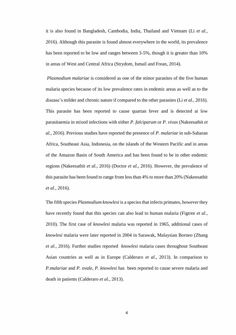
under a light microscope. These differences have been tabulated in figure 1 showing
how the parasites differ at the various stages during the life cycle.
The most severe illness and deaths from malaria and the most drug resistant infections
are due to infection with Plasmodium falciparum (Wongsrichanalai and Sibley, 2013).
This parasite is known as the most virulent human malaria parasite and is responsible
for the majority of malaria deaths globally (Kwenti et al., 2017). P. falciparum is the
most prevalent species in sub-Saharan Africa (WHO, 2016). In Namibia, P falciparum
accounts for more than 90% of the malaria infections (Smith-Gueye et al., 2014). The
P. falciparum parasite is principally transmitted by the vector Anopheles gambiae.
However, other vectors such as Anopheles albimanus, Anopheles freeborni,
Anopheles maculatus and Anopheles stephensi among others have been found to
transmit P. falciparum (Molina-Cruz and Barillas-Mury, 2014).
Plasmodium vivax is the second most significant species that infects humans causing
25-40% of malaria cases worldwide, and is most prevalent in Southeast Asia and Latin
America (Golassa et al., 2015). Previous studies observed that P. vivax remains
dormant during the liver stage, but can be reactivated at a later stage even in the
absence of a mosquito bite, thus leading to clinical symptoms (Le Bras and Durand,
2003) (WHO, 2015c).
Plasmodium ovale is also a species of parasitic protozoa of which two distinct sub
species have been described; P. ovale curtisi and P. ovale wallikeri that cause tertian
malaria in humans (Plasmodium parasites | Scientists Against Malaria, no date).
Unlike P. falciparum and P. vivax, P. ovale has been reported to be less dangerous
and the distribution of this parasite is said to be limited to West and Central Africa,
Philippines, eastern Indonesia, and Papua New Guinea (Li et al., 2016). Additionally,
4
it is also found in Bangladesh, Cambodia, India, Thailand and Vietnam (Li et al.,
2016). Although this parasite is found almost everywhere in the world, its prevalence
has been reported to be low and ranges between 3-5%, though it is greater than 10%
in areas of West and Central Africa (Strydom, Ismail and Frean, 2014).
Plasmodium malariae is considered as one of the minor parasites of the five human
malaria species because of its low prevalence rates in endemic areas as well as to the
disease’s milder and chronic nature if compared to the other parasites (Li et al., 2016).
This parasite has been reported to cause quartan fever and is detected at low
parasitaemia in mixed infections with either P. falciparum or P. vivax (Nakeesathit et
al., 2016). Previous studies have reported the presence of P. malariae in sub-Saharan
Africa, Southeast Asia, Indonesia, on the islands of the Western Pacific and in areas
of the Amazon Basin of South America and has been found to be in other endemic
regions (Nakeesathit et al., 2016) (Doctor et al., 2016). However, the prevalence of
this parasite has been found to range from less than 4% to more than 20% (Nakeesathit
et al., 2016).
The fifth species Plasmodium knowlesi is a species that infects primates, however they
have recently found that this species can also lead to human malaria (Figtree et al.,
2010). The first case of knowlesi malaria was reported in 1965, additional cases of
knowlesi malaria were later reported in 2004 in Sarawak, Malaysian Borneo (Zhang
et al., 2016). Further studies reported knowlesi malaria cases throughout Southeast
Asian countries as well as in Europe (Calderaro et al., 2013). In comparison to
P.malariae and P. ovale, P. knowlesi has been reported to cause severe malaria and
death in patients (Calderaro et al., 2013).
5
Figure 1; Morphological differences between the four-plasmodium species in human
blood smears Source: http://www.dpd.cdc.gov/dpdx
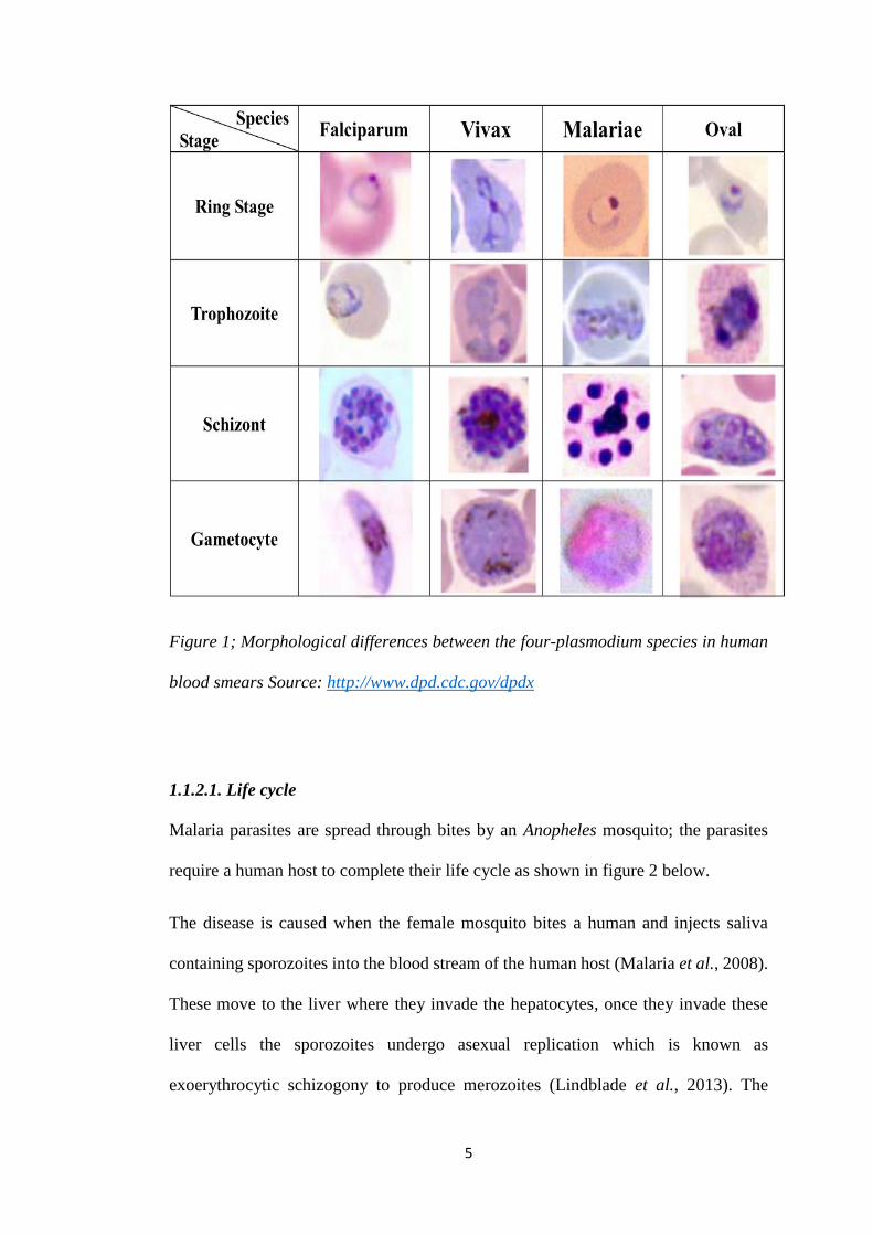
1.1.2.1. Life cycle
Malaria parasites are spread through bites by an Anopheles mosquito; the parasites
require a human host to complete their life cycle as shown in figure 2 below.
The disease is caused when the female mosquito bites a human and injects saliva
containing sporozoites into the blood stream of the human host (Malaria et al., 2008).
These move to the liver where they invade the hepatocytes, once they invade these
liver cells the sporozoites undergo asexual replication which is known as
exoerythrocytic schizogony to produce merozoites (Lindblade et al., 2013). The
6
merozoites are then released into the bloodstream when the cell ruptures. However,
with P. ovale and P, vivax, the merozoites remain dormant as hypnozoites for a few
weeks to several years instead of immediately undergoing asexual reproduction but
can reactivate even in the absence of a mosquito bite and cause the disease (Deshpande
and Kuppast, 2016). The merozoites move through the bloodstream and invade the
red blood cells (erythrocytes), they then mature into trophozoites and undergo a
second phase of asexual replication which is known as erythrocytic schizongony
(Soulard et al., 2015). During this stage about 36 merozoites are released to invade
new erythrocytes (Hawkins, Burton and Labarre, 2014). This process is repeated and
is responsible for the disease. Some of the merozoites transform into male and female
gametocytes that enter the peripheral blood where they are taken up into anopheles
mosquito once it feeds on an infected individual (Lindblade et al., 2013).
In the mosquito the gametocytes form male and female gametes that undergo sexual
replication and produce a zygote (ookinete). The ookinete moves to the gut of the
mosquito and develops into an oocyte, the oocyte in turn undergoes asexual
replication and produce sporozoites (Lindblade et al., 2013). These rounds of
multiplication results in the rupture of the oocyte which releases the sporozoites that
later migrates to the salivary gland of the mosquito, ready to infect another host
(Ouattara and Laurens, 2015).
7
Figure 2: Plasmodium parasite life cycle, Source: http://www.dpd.cdc.gov/dpdx
1.1.3. Malaria Symptoms
The onset of malaria symptoms from the initial bite of a mosquito, to the appearance
of symptoms is variable and it depends on the species of plasmodium (Lindblade et
al., 2013). For P. falciparum it takes 9 to 14 days whereas for P. ovale and P. vivax it
takes about 12 to18 days however, some P. vivax strains may have an incubation
period of 8 to 10 months or longer where they remain dormant. Additionally, it takes
8
P. malariae 18 to 40 days and P. knowlesi 9 to 12 days for symptoms to appear after
the initial bite (Malaria - including symptoms, treatment and prevention, 2012).
The first symptoms of malaria are non-specific and are similar to that of a flu (CDC,
2013). These symptoms include: headaches, fatigue, abdominal discomfort and
muscle and joint aches, followed by fever, chills, perspiration, anorexia and vomiting
(WHO, 2015a). With treatment the symptoms and parasite should be cleared.
However, if left untreated, especially with P. falciparum malaria, the disease could
progress to severe malaria and cause cerebral malaria, metabolic acidosis, anaemia,
hypoglycaemia, acute renal failure, acute pulmonary oedema and eventually death if
not treated. (WHO, 2015a).
1.1.4. Malaria Control
The current goal of many malaria control programs and activities is to reduce the
number of malaria cases and deaths to a level where malaria is no longer a public
health problem in malaria endemic countries (CDC, 2014). In Africa the strategies
that have been used to reduce malaria morbidity and mortality include, prevention
through integrated vector control (LLINs and IRS), early diagnosis of all malaria
cases, and the administration of effective and appropriate treatment once an individual
has been diagnosed with malaria (Eyasu, 2015) (Korenromp et al., 2016). Additional
interventions that have reduced malaria over the years is the confirmation of malaria
diagnostics through the use of rapid diagnostics tests (RDTs).
1.1.4.1. Vector Control
Karunamoorthi describes vector control as any kind of measures which are directed
against a vector (mosquito) of a disease (malaria) and intending to limit its ability to
transmit the disease (Karunamoorthi, 2011). Over the years, the use of Indoor residual
9
spraying (IRS) and long-lasting insecticidal nets (LLINs) have been reported to
contribute greatly to reductions in malaria cases globally (Ngufor et al., 2017). Both
these interventions involve the use of insecticides by either spraying them on walls
and ceilings of houses (IRS) or by treating mosquito nets with insecticides (LLINs) to
prevent human-mosquito contact and thus preventing the onward transmission of the
parasite (Karunamoorthi, 2011). The WHO recently reported an increase in household
ownership of LLINs from 50% in 2010 to 80% in 2016 across sub-Saharan Africa
(WHO, 2017c). In addition to the above mentioned vector control strategies,
larviciding is another form of vector control which according to WHO, involves the
killing of mosquito larvae or creating unfavourable conditions which prevents
mosquitoes from breeding (WHO, 2012). Namibia has been using IRS, LLNs and
larvaciding vector control interventions to reduce mosquito to human transmission of
the parasite (WHO, 2016b).
1.1.4.2. Parasite Control
In order to treat all malaria effectively, it is important to detect all cases of malaria.
Microscopy detects the presence of malaria parasites by the visualization of a patient’s
blood under light microscopy, this is the WHO recommended gold standard for
detecting malaria (WHO, 2018). However, this method has some drawbacks,
especially in a low transmission such as Namibia where there are asymptomatic
patients (WHO, 2017c). When these cases are left undetected and not treated they
contribute to the onward transmission of malaria (Hawkins, Burton and Labarre,
2014). Additionally, microscopy is not reliable in a field setting where highly skilled
microscopists, high quality microscopes and electricity is needed (Ilesanmi et al.,
2017). Thus, it is important to introduce more cost-effective tools to effectively
diagnose and treat all cases of malaria (M. L. McMorrow, M. Aidoo, 2015).
10
1.1.4.3. Case Detection and Rapid Diagnostic Tests (RDTs)
According to WHO after the introduction of Rapid Diagnostic Tests (RDTs) an
increase in malaria diagnostics in Africa was reported from 36% of suspected malaria
cases in 2005 to 65% of suspected cases (WHO, 2016a). As compared to microscopy,
RDTs are more cost effective, results can be viewed within 15 minutes and they can
be used in field settings (Jimenez et al., 2017). RDTs are point of care lateral flow
immunochromatographic assays and work by detecting species specific antigens in
human blood (Thompson, 2012). In addition to RDTs being easy to use and cost
effective, different types of RDTs can detect different antigens in malaria parasites
(Jimenez et al., 2017). Three main antigens are detected by RDTs, these are histidine-
rich protein 2 (HRP2) which is specific to P. falciparum, the parasite specific
plasmodium lactate dehydrogenase (pLDH) which is P. falciparum specific, pan-
specific (can detect all species of plasmodium), and P. vivax-specific and the
plasmodium aldolase which is pan-specific (Kakkilaya, 2015). Although standard
RDTs have a detection limit of 200ul/parasite, which may result in missing some cases
in low transmission settings, they are more affordable and easy to use as compared to
microscopy (Kakkilaya, 2015). As early diagnosis is important for disease
management and surveillance.
1.1.4.4. Antimalarial Treatment
In order to ensure that all the previously mentioned malaria control interventions are
effective, equal efforts should be directed towards antimalarial treatment (WHO,
2015a). WHO recommends that all patients with uncomplicated malaria be treated
with ACTs which is the recommended first line treatment, except for pregnant women
in their 1st trimester, which are treated with quinine + clindamycin (WHO, 2015a). In
patients with severe malaria, including pregnant women WHO strongly recommends
11
treatment with intravenous or intramuscular artesunate for at least 24 hours and until
they are able to tolerate oral medication (WHO, 2015a). Once a patient has received
at least 24 hours of parenteral therapy and can tolerate oral therapy, they should
complete the treatment with 3 days of ACT with a single dose primaquine in areas of
low transmission (WHO, 2015a). Although there have been previous reports on drug
resistance in Sulfadoxin-Pyrimethamine (SP) as a first line treatment (Shah et al.,
2015), WHO recommends that SP should be provided with Intermittent Preventative
Treatment (IPTp-SP) to pregnant women in Africa, who are in their 2nd trimester
(WHO, 2014b).In addition to IPTp-SP WHO also recommends that Intermittent
Preventive Treatment with SP be given to infants (< 12 months of age) (SP-IPTi) in
moderate to high transmission areas where SP is still effective (WHO, 2015b). The
use of chemoprophylactic regimens such as mefloquine, atovaquone/proguanil
(Malarone) or doxycycline is recommended to people travelling from non-endemic to
endemic to prevent malaria (Cui et al., 2015)
1.1.4.5. Malaria Vaccines
According to WHO a vaccine is a “biological preparation that improves immunity to
a disease which contains an agent that resembles a disease-causing microorganism, in
its weakened or killed form. This agent activates the body's immune system to
recognize the agent as foreign, destroy it, and "remember" it, so that the immune
system can easily recognize and destroy any of these microorganisms that it later
encounters” (WHO, 2017b). There are currently 10 vaccines, listed in table 1 below
which are either in their preclinical development or their clinical trial development
against malaria (Coelho et al., 2017). Only one which is commonly known as
Mosquirix (RTS,S/AS01) is in phase IV of its clinical trials and has been approved by
the European Medicines Agency’s Committee for Medicinal Products for Human Use
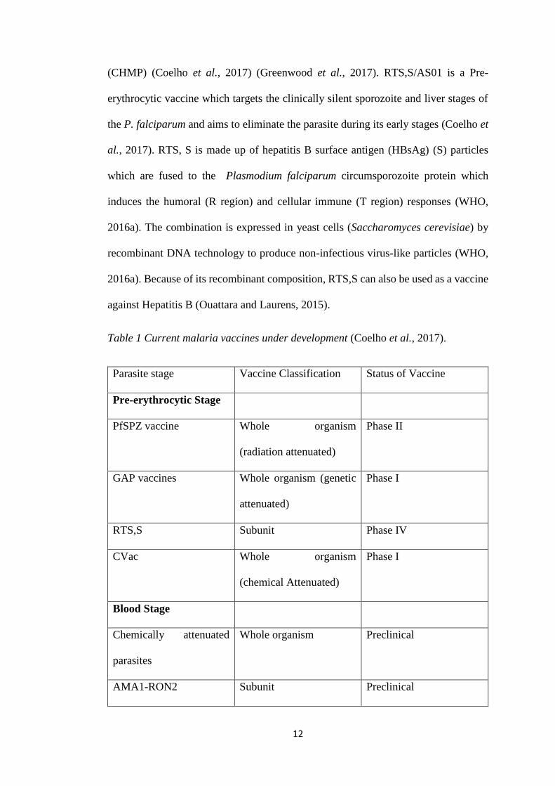
12
(CHMP) (Coelho et al., 2017) (Greenwood et al., 2017). RTS,S/AS01 is a Pre-
erythrocytic vaccine which targets the clinically silent sporozoite and liver stages of
the P. falciparum and aims to eliminate the parasite during its early stages (Coelho et
al., 2017). RTS, S is made up of hepatitis B surface antigen (HBsAg) (S) particles
which are fused to the Plasmodium falciparum circumsporozoite protein which
induces the humoral (R region) and cellular immune (T region) responses (WHO,
2016a). The combination is expressed in yeast cells (Saccharomyces cerevisiae) by
recombinant DNA technology to produce non-infectious virus-like particles (WHO,
2016a). Because of its recombinant composition, RTS,S can also be used as a vaccine
against Hepatitis B (Ouattara and Laurens, 2015).
Table 1 Current malaria vaccines under development (Coelho et al., 2017).
Parasite stage Vaccine Classification Status of Vaccine
Pre-erythrocytic Stage
PfSPZ vaccine Whole organism
(radiation attenuated)
Phase II
GAP vaccines Whole organism (genetic
attenuated)
Phase I
RTS,S Subunit Phase IV
CVac Whole organism
(chemical Attenuated)
Phase I
Blood Stage
Chemically attenuated
parasites
Whole organism Preclinical
AMA1-RON2 Subunit Preclinical

13
PfRH5 Subunit Phase I
Mosquito Stage
Pfs25 Subunit Phase I
Pfs230 Subunit Phase I
Pfs47 Subunit Preclinical
1.1.5. The Impact of Antimalarial Drug Resistance
Widespread P. falciparum resistance to the formerly used antimalarial medicines such
as Chloroquine (CQ), Sulfadoxine-pyrimethamine (SP) and Amodiaquine (AQ) has
led to changes in the malaria treatment policy in endemic countries, leading to the
introduction of artemisinin-based combination therapies (ACTs) (Chilongola et al.,
2014). WHO recommends that all uncomplicated falciparum malaria in both adults
and children, except in pregnant women who are their 1st trimester be treated with
either one of the following combination therapies: artemether + lumefantrine,
artesunate + amodiaquine, artesunate + mefloquine, dihydroartemisinin + piperaquine
and artesunate + sulfadoxine–pyrimethamine (SP) (WHO, 2015a). Recently there
have been reports on declining efficacy of one of the ACTs which is the artesunate-
mefloquine combination in Senegal, French Guiana, and on the Cambodia and
Thailand border region of Southeast Asia, the same countries where emergence
Chloroquine resistance originated from (Chilongola et al., 2015). These findings are
raising concerns around the efficacy of the other ACTs that are being used as first-
line treatment.
The use of artemisinin in Namibia was introduced in 2005 and it is currently the first-
line of treatment in Namibia, it is used in the combination therapy known as
artemether + lumefantrine (Smith-Gueye et al., 2014). This change in treatment was
14
made due to the high resistance levels of P. falciparum parasites to chloroquine in
Namibia (Smith-Gueye et al., 2014).
The development of antimalarial drug resistance is said to be associated with single
nucleotide polymorphisms (SNPs) in genes such as the transporter genes which is the
Plasmodium falciparum chloroquine resistance transporter (pfcrt) gene, or the
increase in copy number of the Plasmodium falciparum multidrug resistance1
(pfmdr1) gene (Petersen, Eastman and Lanzer, 2011). Additionally, antimalarial drug
resistance may also be caused by the change in the parasite target of the antimalarial
drug (Ouji et al., 2018). These changes may be due to mutations at the cytosol level
of genes encoding dihydropteroate synthase (Pfdhps) and dihydrofolate reductase
(Pfdhfr) in sulfadoxine-pyrimethamine resistance, or at the mitochondrion level,
cytochrome b which leads to atovaquone resistance (Ouji et al., 2018). These markers
can be an effective surveillance tools for monitoring the emergence of antimalarial
drug resistance in malaria endemic countries such as Namibia.
1.2. Problem Statement
As countries move towards malaria elimination it becomes increasingly important to
strengthen all malaria elimination strategies, including the effective use of
antimalarial drugs as there are currently no new drugs available on the market. To
date malaria control interventions have brought down malaria to a state of controlled
low endemic malaria in Namibia. However, these control interventions effectiveness
will be limited without equal efforts directed against antimalarial drug resistance.
Hence, as Namibia is moving towards malaria elimination, the effectiveness of
malaria case management without surveillance for resistance to antimalarial
medicines will be limited. Therefore, monitoring and surveillance using molecular
markers for drug resistance should be conducted to strengthen malaria control and
15
elimination efforts, as currently there is no data available on antimalarial drug
resistance to ACTs and on Chloroquine among the P. falciparum population in
Namibia.
1.3. General Objectives of the study
The main objective of this study was to determine the prevalence of P. falciparum
point mutations in the Zambezi region of Namibia, that have been reported to be
associated with resistance to the WHO recommended Artemisinin Combination
Therarapy (ACT) and to Cholroquine, Amodiaquine and Lumefantrine. Artemether
and lumefantrine is currently the first-line treatment in Namibia.
1.3.1. Specific Objectives of this study were to:
Identify the different species of plasmodium parasites using multiplex PCR.
Determine the prevalence of drug resistance polymorphisms at codon
N86Y in the pfmdr1 gene.
Determine the prevalence of the three Plasmodium falciparum haplotypes
formed by codons crt72-76 associated with antimalarial drug resistance in
the pfcrt gene.
Determine the prevalence of the 25 validated drug resistance markers in the
Kelch 13 propeller protein encoded by the PF3D7_1343700 gene.
1.4. Significance of the study
As many countries in Sub-Saharan Africa continue to scale-up malaria control
measures, countries in Southern Africa are progressing towards malaria elimination
(WHO, 2013). Thus, WHO recommends the routine monitoring of these antimalarial
drugs using molecular markers as surveillance tools amongst other surveillance
strategies to detect early emergence of drug resistance. This study will provide the
16
data on antimalarial drug resistance polymorphisms among the P. falciparum
population in Namibia, in addition will attempt to provide baseline data on the level
mutations associated with antimalarial drug resistance in the country. Such data will
ensure the appropriate and effective use of antimalarial drugs in the country.
1.5. Limitations of the study
This study will be limited by the fact that only samples from Zambezi region, of
Namibia will be analysed and will not be representative of the whole country. In
addition, it will only focus on known mutations associated with artemisinin,
chloroquine and lumefantrine resistance and not on the new and emerging ones.
1.6. Delimitations of the study
This study will only focus on drug resistance in P. falciparum and not on the other
plasmodium species in the Zambezi region of Namibia. Quantitative PCR will not be
conducted on all markers although copy number of markers may be a key factor in
determining resistance.
17
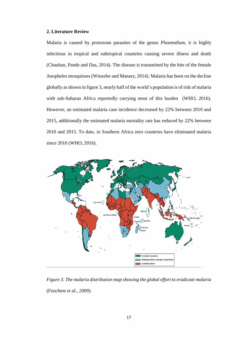
2. Literature Review
Malaria is caused by protozoan parasites of the genus Plasmodium, it is highly
infectious in tropical and subtropical countries causing severe illness and death
(Chauhan, Pande and Das, 2014). The disease is transmitted by the bite of the female
Anopheles mosquitoes (Winzeler and Manary, 2014). Malaria has been on the decline
globally as shown in figure 3, nearly half of the world’s population is of risk of malaria
with sub-Saharan Africa reportedly carrying most of this burden (WHO, 2016).
However, an estimated malaria case incidence decreased by 22% between 2010 and
2015, additionally the estimated malaria mortality rate has reduced by 22% between
2010 and 2015. To date, in Southern Africa zero countries have eliminated malaria
since 2010 (WHO, 2016).
Figure 3. The malaria distribution map showing the global effort to eradicate malaria
(Feachem et al., 2009).
18
Among the five species (P.vivax, P.ovale, P.malaria, P.knowlesi and P.falciparum)
that infect humans, P. falciparum is the most severe form of the disease (Winzeler and
Manary, 2014). Therefore, the development and spread of P. falciparum resistance to
antimalarial drugs represents a major threat to global malaria control (Ghanchi et al.,
2011).
Following results of drug resistance to (CQ) and (SP), ACTs are now recommended
by the (WHO) as first-line treatment of uncomplicated falciparum malaria in all areas
in which malaria is endemic (Dondorp et al., 2009). ACTs are the current treatment
for P. falciparum malaria globally and they have reduced the morbidity and mortality
associated with malaria. However, studies have shown signs that the efficacy of
artemisinin-based combination therapy and artesunate monotherapy recently have
declined in Southeast Asia (Dhorda et al., 2015).
2.1. Antimalarial drugs
The currently available antimalarial drugs fall into three broad categories according
to their chemical structure and mode of action (Fig 4):
1. Aryl amino alcohol compounds (Quinolines): quinine, quinidine,
chloroquine, amodiaquine, mefloquine, halofantrine, lumefantrine,
piperaquine, tafenoquine
2. Antifolate compounds (“antifols”): pyrimethamine, proguanil,
chlorproguanil, trimethoprim
3. Artemisinin compounds (artemisinin, dihydroartemisinin, artemether,
artesunate) (Deshpande and Kuppast, 2016).
19
2.1.1. Quinolines
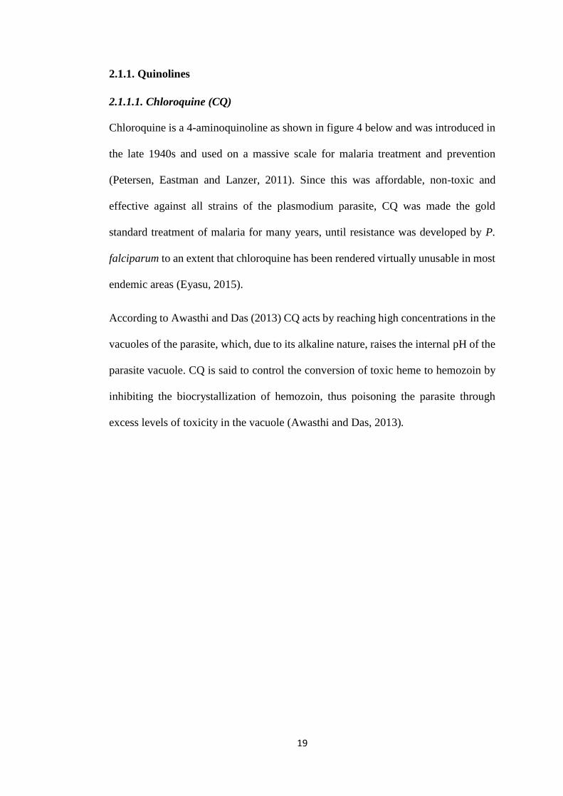
2.1.1.1. Chloroquine (CQ)
Chloroquine is a 4-aminoquinoline as shown in figure 4 below and was introduced in
the late 1940s and used on a massive scale for malaria treatment and prevention
(Petersen, Eastman and Lanzer, 2011). Since this was affordable, non-toxic and
effective against all strains of the plasmodium parasite, CQ was made the gold
standard treatment of malaria for many years, until resistance was developed by P.
falciparum to an extent that chloroquine has been rendered virtually unusable in most
endemic areas (Eyasu, 2015).
According to Awasthi and Das (2013) CQ acts by reaching high concentrations in the
vacuoles of the parasite, which, due to its alkaline nature, raises the internal pH of the
parasite vacuole. CQ is said to control the conversion of toxic heme to hemozoin by
inhibiting the biocrystallization of hemozoin, thus poisoning the parasite through
excess levels of toxicity in the vacuole (Awasthi and Das, 2013).
20
Figure 4. Chemical structure of Chloroquine (WHO, 2015b)
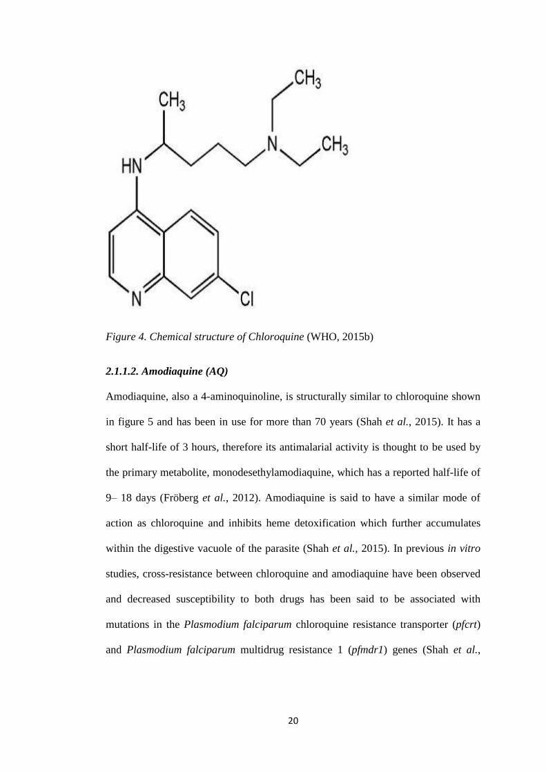
2.1.1.2. Amodiaquine (AQ)
Amodiaquine, also a 4-aminoquinoline, is structurally similar to chloroquine shown
in figure 5 and has been in use for more than 70 years (Shah et al., 2015). It has a
short half-life of 3 hours, therefore its antimalarial activity is thought to be used by
the primary metabolite, monodesethylamodiaquine, which has a reported half-life of
9– 18 days (Fröberg et al., 2012). Amodiaquine is said to have a similar mode of
action as chloroquine and inhibits heme detoxification which further accumulates
within the digestive vacuole of the parasite (Shah et al., 2015). In previous in vitro
studies, cross-resistance between chloroquine and amodiaquine have been observed
and decreased susceptibility to both drugs has been said to be associated with
mutations in the Plasmodium falciparum chloroquine resistance transporter (pfcrt)
and Plasmodium falciparum multidrug resistance 1 (pfmdr1) genes (Shah et al.,
21
2015). However, when cross-resistance is incomplete some chloroquine resistant
parasites remain susceptible to amodiaquine (Rosenthal, 2013).
Amodiaquine is a potent blood schizonticide that has been used for the treatment of
uncomplicated malaria particularly in Africa. As AQ can cause neutropenia in patients
it was not used for many years. However, it has recently been revived as part of an
ACT (Petersen, Eastman and Lanzer, 2011).
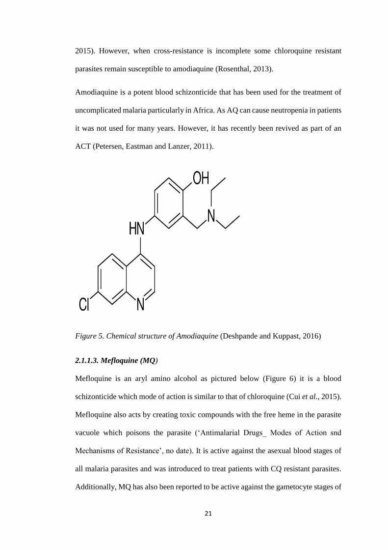
Figure 5. Chemical structure of Amodiaquine (Deshpande and Kuppast, 2016)
2.1.1.3. Mefloquine (MQ)
Mefloquine is an aryl amino alcohol as pictured below (Figure 6) it is a blood
schizonticide which mode of action is similar to that of chloroquine (Cui et al., 2015).
Mefloquine also acts by creating toxic compounds with the free heme in the parasite
vacuole which poisons the parasite (‘Antimalarial Drugs_ Modes of Action snd
Mechanisms of Resistance’, no date). It is active against the asexual blood stages of
all malaria parasites and was introduced to treat patients with CQ resistant parasites.
Additionally, MQ has also been reported to be active against the gametocyte stages of
22
P. vivax, P. ovale and P. malariae (Cui et al., 2015). MQ was initially used as
monotherapy in areas of low malaria transmission however due to resistance it is now
principally used in combination with artesunate to treat P. falciparum in Southeast
Asia (Phompradit et al., 2014).
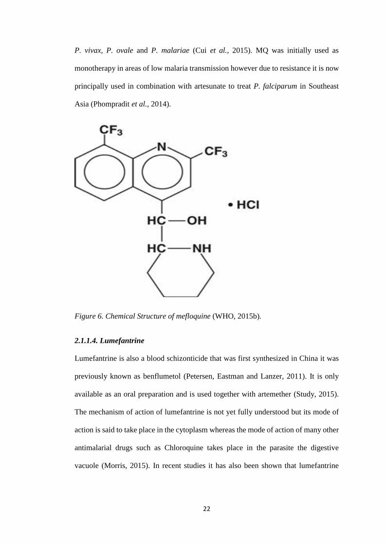
Figure 6. Chemical Structure of mefloquine (WHO, 2015b).
2.1.1.4. Lumefantrine
Lumefantrine is also a blood schizonticide that was first synthesized in China it was
previously known as benflumetol (Petersen, Eastman and Lanzer, 2011). It is only
available as an oral preparation and is used together with artemether (Study, 2015).
The mechanism of action of lumefantrine is not yet fully understood but its mode of
action is said to take place in the cytoplasm whereas the mode of action of many other
antimalarial drugs such as Chloroquine takes place in the parasite the digestive
vacuole (Morris, 2015). In recent studies it has also been shown that lumefantrine
23
inhibits haemozoin formation in the parasite cell, which shows that lumefantrine may
have a similar mechanism to chloroquine (Petersen, Eastman and Lanzer, 2011).
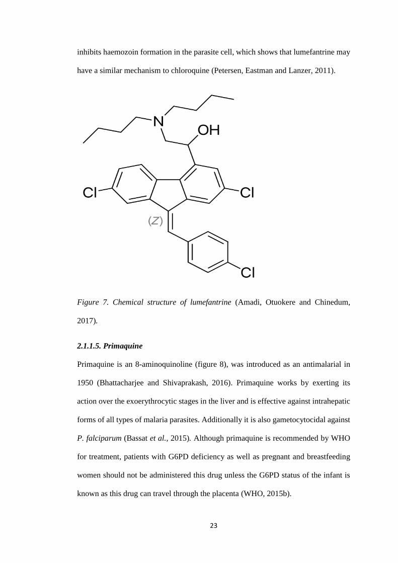
Figure 7. Chemical structure of lumefantrine (Amadi, Otuokere and Chinedum,
2017).
2.1.1.5. Primaquine
Primaquine is an 8-aminoquinoline (figure 8), was introduced as an antimalarial in
1950 (Bhattacharjee and Shivaprakash, 2016). Primaquine works by exerting its
action over the exoerythrocytic stages in the liver and is effective against intrahepatic
forms of all types of malaria parasites. Additionally it is also gametocytocidal against
P. falciparum (Bassat et al., 2015). Although primaquine is recommended by WHO
for treatment, patients with G6PD deficiency as well as pregnant and breastfeeding
women should not be administered this drug unless the G6PD status of the infant is
known as this drug can travel through the placenta (WHO, 2015b).
24
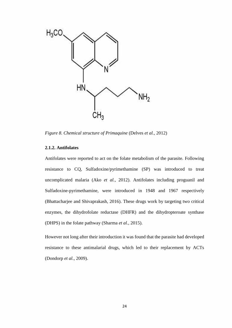
Figure 8. Chemical structure of Primaquine (Delves et al., 2012)
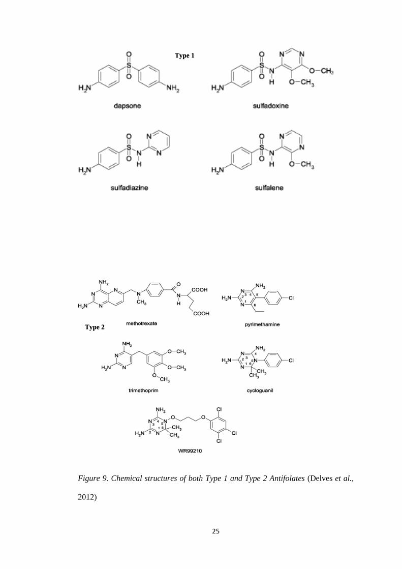
2.1.2. Antifolates
Antifolates were reported to act on the folate metabolism of the parasite. Following
resistance to CQ, Sulfadoxine/pyrimethamine (SP) was introduced to treat
uncomplicated malaria (Ako et al., 2012). Antifolates including proguanil and
Sulfadoxine-pyrimethamine, were introduced in 1948 and 1967 respectively
(Bhattacharjee and Shivaprakash, 2016). These drugs work by targeting two critical
enzymes, the dihydrofolate reductase (DHFR) and the dihydropteroate synthase
(DHPS) in the folate pathway (Sharma et al., 2015).
However not long after their introduction it was found that the parasite had developed
resistance to these antimalarial drugs, which led to their replacement by ACTs
(Dondorp et al., 2009).
25
Figure 9. Chemical structures of both Type 1 and Type 2 Antifolates (Delves et al.,
2012)
Type 1
Type 2

26
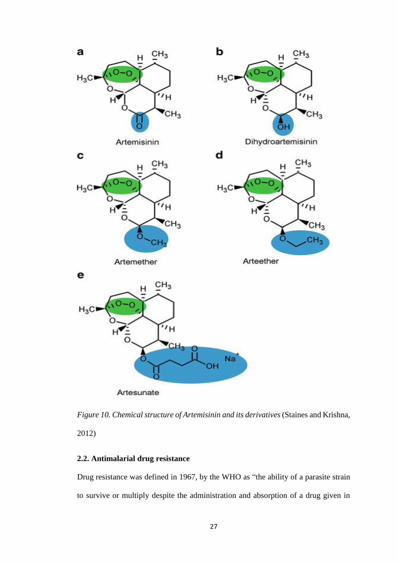
2.1.3. Artemisinin
Artemisinin is a potent antimalarial, and their short half-life of around 1-2 hours helps
prevent the selection of resistant parasites, by acting very fast and reducing the
parasite load quickly (Dhorda et al., 2015). They are active against the asexual stages
of the parasites and are said to act on the young gametocyte stage in the life cycle of
the parasite thus prevents the transmission of mature gametocytes (Aminake and
Pradel, 2013). Artemisinin-based combination therapy (ACT) is the recommended
standard first-line treatment for individuals with uncomplicated malaria (Bassat et al.,
2015). However, it is recommended that these drugs are used in combinations of
artemether-lumefantrine, artesunate-amodiaquine, artesunate-mefloquine,
dihydroartemisinin-piperaquine, artesunate-pyronaridine, artesunate-sulfadoxine-
pyrimethamine to protect drugs from resistance. The chemical structures of
artemisinin and its derivatives (artesunate, artemether, arteether and
dihydroartemisinin) are shown in figure 10 (Olasehinde et al., 2014).
27
Figure 10. Chemical structure of Artemisinin and its derivatives (Staines and Krishna,
2012)
2.2. Antimalarial drug resistance
Drug resistance was defined in 1967, by the WHO as “the ability of a parasite strain
to survive or multiply despite the administration and absorption of a drug given in
28
doses equal to or higher than those usually recommended but within the tolerance of
the subject” (WHO,1967).
However, once treatment fails it does not necessarily mean that there is drug
resistance, as treatment failure may be due to incorrect dosing, not finishing the
recommended regimen within the prescribed time, poor quality drugs, drug
interactions, poor or erratic absorption of the drug, and misdiagnosis (Sisowath,
2009). These factors nevertheless could lead to the development of resistant parasites
to the drug they were inadequately exposed to (WHO, 2010).
The emergence of resistance in Plasmodium parasites depends on several factors,
namely, the rate of mutation of the parasite, the fitness costs associated with the
resistance mutations, the parasite load and the strength of the drug used (Petersen,
Eastman and Lanzer, 2011).
2.3. Development and Spread of Drug Resistance
In order to successfully control and eliminate malaria, the use of effective drug therapy
is important and the increasing levels of resistant P. falciparum parasites to commonly
used drugs is hampering this goal (Nsanzabana et al., 2018). The development of
resistant parasites has been said to be caused by several different factors which
include; the development of de novo resistant mutations of the plasmodium parasite
which can be single or multiple, these mutations allows the parasite to survive the
administered treatment, and in turn multiplies and is transmitted by the vector (WHO,
2010). These mutations give the parasite a survival advantage over the susceptible
parasites, allowing them to be transmitted to the next host leading to the spread of
resistant parasites (Goswami et al., 2014).
29
2.4. Mechanisms of Antimalarial drug resistance
2.4.1. Resistance mediated by transporter mutations
From previous studies it has been established that there are several genes which
encode the parasite transporter proteins, P. falciparum chloroquine resistance
transporter ( pfcrt ) and P. falciparum multidrug resistance1 (pfmdr1), they are located
in membranes of digestive vacuoles, and are proposed to play a role as key
contributors of resistance of P. falciparum to antimalarial drugs (Muhamad et al.,
2011). The parasite digestive vacuole is where many antimalarial drugs act, and amino
acid substitutions at these sites may lead to antimalarial drug resistance by causing
efflux of the drugs from the cells in the digestive vacuoles out of the parasites
(Rosenthal, 2013).
2.4.1.1. The pfmrd1 gene
The pfmrd1 gene encodes a 162 kDa protein, P. falciparum homologue of the P-
glycoprotein (Pgh1), and it is located on chromosome 5, Pgh1 is in turn located in the
parasite food vacuole (Figure 11) (Wurtz et al., 2012). Pgh1 is believed to play a role
in resistance to several antimalarial drugs (Inoue et al., 2014). In many studies pfmdr1
point mutations have been observed in both CQ resistant and sensitive strains of P.
falciparum and are said to be responsible for the movement of antimalarial drugs from
the cytosol into the digestive vacuole of the parasite (Ibraheem et al., 2014). Studies
showed that the substitution of amino acid, asparagine (N) by tyrosine (Y) at codon
N86Y was linked to chloroquine and amodiaquine resistance, and it is also associated
with increased sensitivity to MQ and artemisinin derivatives (Inoue et al., 2014).
Furthermore, four other single nucleotide polymorphisms (SNP’s) have been
identified from field isolates in the pfmdr1 gene (Y184F, S1034C, N1042D and
30
D1246Y). The mutant alleles at codons 1034C, 1042D and 1246Y have been linked
to resistance in quinine and increased susceptibilities to artemisinin, mefloquine and
halofantrine (Kavishe et al., 2014). The mutant alleles at codons N86Y and D1246Y
has been associated with the decrease in drug sensitivity in artesunate-amodiaquine
(AS-AQ), whereas the wild type forms at these codons were shown to cause resistance
in artemether-lumefantrine (AL) which is responsible for the movement of Al from
the its site of action the cytoplasm into the food vacuole of the parasite (Kavishe et
al., 2014). According to Wurtz et al (2012), increased copy number of pfmdr1 is the
main cause of resistance to MQ in P. falciparum. Additionally, pfmdr1 amplification
has been linked to reduced sensitivity in artemisinin derivatives (Inoue et al.2014).
31
Figure 11. Structure of the the P-glycoprotein and amino acid positions (Ibraheem et
al., 2014)
2.4.1.2. The pfcrt gene
The pfcrt gene encodes a transporter protein of 424 amino acids and 48.6 kDa which
is located in the digestive vacuole of the parasite, and is located on chromosome 7
(Figure 12) (Saleh, Handayani and Anwar, 2014). Previous research indicate that
change in K76T on the pfcrt gene results in the resistance phenotype, and is said to be
the most reliable molecular marker of resistance (Olasehinde et al., 2014).
Mutations in the pfcrt gene which cause amino acid substitutions (Threonine (T),
Asparagine (N) or Isoleucine (I)) changes the electric charge of the membrane, leading
to the efflux of CQ and AQ from the digestive vacuole (Inoue et al., 2014). Mutations
32
in the pfcrt gene has been associated with artemisinin, quinine and AQ susceptibility
(Heuchert et al., 2015). AQ and quinine were found to show cross-resistance with CQ,
mediated by 76T, whereas lumefantrine displays an inverse cross-resistance, with the
wild type K76 which in turn leads to reduced susceptibility to lumefantrine (Petersen,
Eastman and Lanzer, 2011).
Previous studies show that 76T is usually found together with additional SNPs which
create specific pfcrt 72–76 and the 271 and 371 haplotypes that indicate the origin of
CQ resistance (Pathak et al., 2014).
33
Figure 12. Structure of the transporter protein and amino acid positions (Pulcini et
al., 2015)
2.4.2. Resistance to Antifolates
Antifolates, target parasite dihydrofolate reductase (DHFR) and dihydropteroate
synthase (DHPS), they bind to enzymes necessary for parasite folate biosynthesis and
are subdivided into two classes: Type 1 (Pyrimethamine, chlorproguanil,
trimethoprim) and Type 2 (sulphonamides: Sulfadoxine and dapsone) , they inhibit
the enzymes DHFR, and DHPS respectively (Agomo et al., 2016). According to
Sharma et al, (2015) the aforementioned drugs inhibit P. falciparum dihydrofolate
reductase (pfdhfr) thus indirectly blocks the synthesis of nucleic acids in the malaria
parasite.
34
Molecular studies show that resistance to pyrimethamine has been associated with
parasites that carry mutations at codons 51, 59, 108, and 164 in the pfdhfr gene, and
resistance to Sulfadoxine with mutations at codons 436, 437, 540, 581, and 613 in the
P. falciparum dihydropteroate synthase (pfdhps) gene (Ako et al., 2012).
2.4.3. Artemisinin and Derivatives
Partial artemisinin resistance is observed when the parasite clearance rate is slow after
treatment with ACT (Ashley et al., 2014). However this definition is said to be
affected by multiple factors such as patient immunity, blood drug concentration or
partner drug activity (WHO, 2014). From treatment failures which were observed in
South East Asia, studies showed that resistance was correlated either with lower
efficacy of the partner drug and with slower parasite clearance rate due to artemisinin
partial resistance (Escobar et al., 2015).
Artemisinin resistance was previously linked with the sarco-/endoplasmic reticulum
Ca 2+ -ATPase ortholog of P. falciparum (PfATP6), suggesting that mutations in
pfATP6 was involved in the mechanism that cause resistance in artemisinin
(Muhamad et al., 2011). This assumption was made because it was found that
artemisinin decreases the ATPase activity in Xenopus oocytes that expresses the
PfATP6, which has a similar potency to thapsigargin that is also another SERCA
inhibitor (Winzeler and Manary, 2014). However recent studies state that resistance
in artemisinin is caused by mutations on the Kelch 13 propeller protein K13 which is
encoded in the PF3D7_1343700 gene (Escobar et al., 2015). Artemisinin resistance
was first described in Cambodia, and the presence of K13 mutants (mainly Y493H,
R539T, I543T and C580Y) are associated with in vitro parasite survival rates and in
vivo parasite clearance rates (Taylor et al., 2015).
35
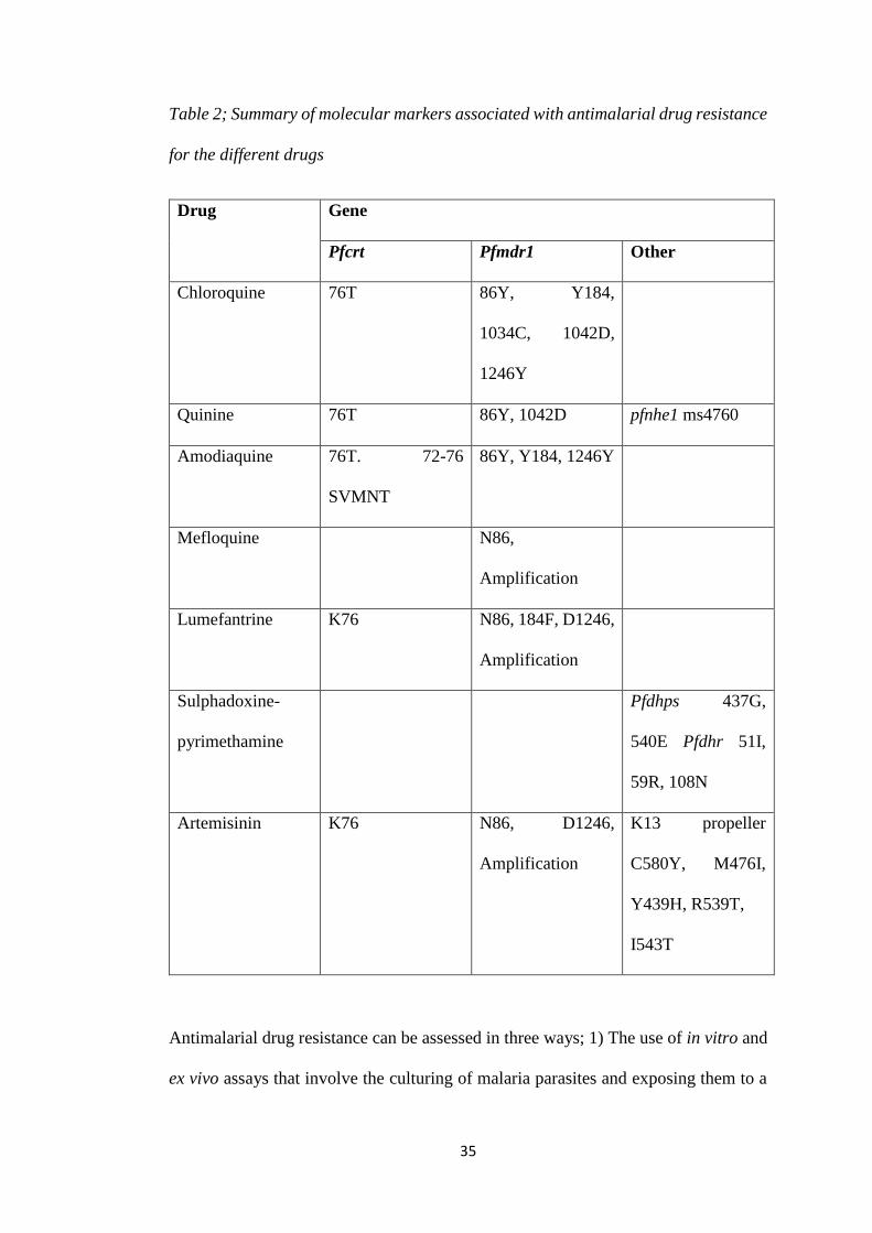
Table 2; Summary of molecular markers associated with antimalarial drug resistance
for the different drugs
Drug Gene
Pfcrt Pfmdr1 Other
Chloroquine 76T 86Y, Y184,
1034C, 1042D,
1246Y
Quinine 76T 86Y, 1042D pfnhe1 ms4760
Amodiaquine 76T. 72-76
SVMNT
86Y, Y184, 1246Y
Mefloquine N86,
Amplification
Lumefantrine K76 N86, 184F, D1246,
Amplification
Sulphadoxine-
pyrimethamine
Pfdhps 437G,
540E Pfdhr 51I,
59R, 108N
Artemisinin K76 N86, D1246,
Amplification
K13 propeller
C580Y, M476I,
Y439H, R539T,
I543T
Antimalarial drug resistance can be assessed in three ways; 1) The use of in vitro and
ex vivo assays that involve the culturing of malaria parasites and exposing them to a
36
particular drug at different levels for the assessment of sensitivities of cultured
parasites. 2) The use of in vivo test which involve the testing of malaria positive
individuals with a known dose of a particular drug, during this test the participants are
followed up and monitored for parasitological and clinical response for a specified
period of time depending on the drug in use. 3) The evaluation of genetic
polymorphisms associated with antimalarial drug resistance in specific genes (Eyasu,
2015).
Although the use of in vitro and in vivo tests provide data on clinical treatment failures,
these test can be expensive to conduct in some endemic countries as they require
highly trained personnel and well equipped laboratories (Nsanzabana et al., 2018).
Additionally using in vivo tests in a low transmission setting such as Namibia can
become challenging because it gets difficult to get the required sample size to conduct
efficacy studies (Nsanzabana et al., 2018).
This study will focus on the evaluation of genetic polymorphisms associated with drug
resistance, where the prevalence of these antimalarial drug resistance polymorphisms
will be investigated. Thus, antimalarial drug resistance polymorphisms used as
molecular markers may provide information on the emergence of drug resistance
patterns in the field and hence can be used to design malaria control strategies
particularly during case management studies (Olasehinde et al., 2014).
37
3. Research Methods
3.1. Research Design
This study was a qualitative study that employed the interpretation of results from
Quantitative PCR, nested PCR and allele specific restriction enzyme digestion
strategy. Malaria positive samples from the Zambezi region were investigated for the
prevalence of mutations in the pfmdr1, pfcrt and Kelch 13 genes.
3.2. Methods
3.2.1. Study Population and Study Site
The target population was individuals from all households that were reported at the
different randomly selected health facility catchment areas as well as the population
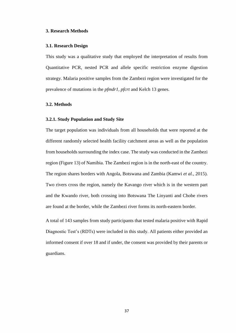
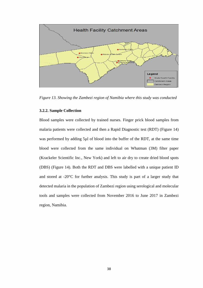
from households surrounding the index case. The study was conducted in the Zambezi
region (Figure 13) of Namibia. The Zambezi region is in the north-east of the country.
The region shares borders with Angola, Botswana and Zambia (Kamwi et al., 2015).
Two rivers cross the region, namely the Kavango river which is in the western part
and the Kwando river, both crossing into Botswana The Linyanti and Chobe rivers
are found at the border, while the Zambezi river forms its north-eastern border.
A total of 143 samples from study participants that tested malaria positive with Rapid
Diagnostic Test’s (RDTs) were included in this study. All patients either provided an
informed consent if over 18 and if under, the consent was provided by their parents or
guardians.
38
Figure 13. Showing the Zambezi region of Namibia where this study was conducted
3.2.2. Sample Collection
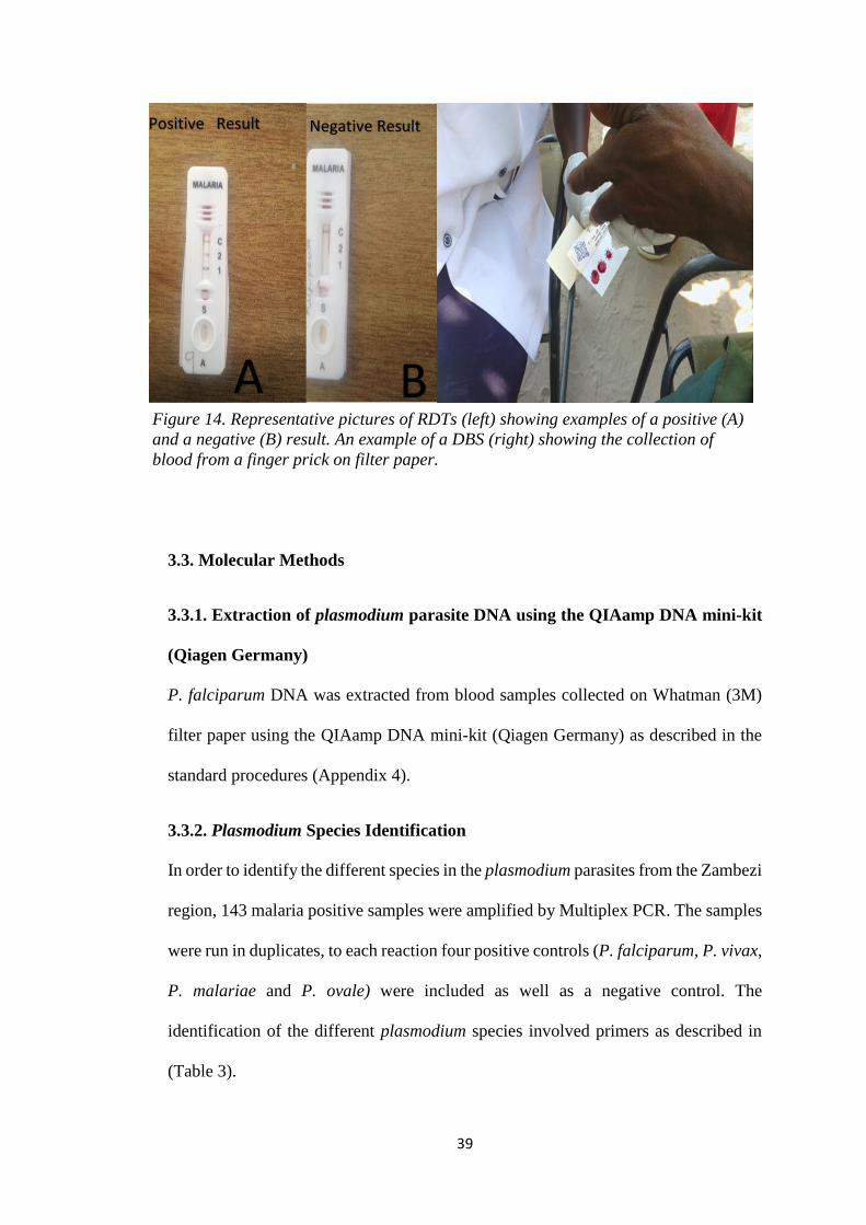
Blood samples were collected by trained nurses. Finger prick blood samples from
malaria patients were collected and then a Rapid Diagnostic test (RDT) (Figure 14)
was performed by adding 5µl of blood into the buffer of the RDT, at the same time
blood were collected from the same individual on Whatman (3M) filter paper
(Krackeler Scientific Inc., New York) and left to air dry to create dried blood spots
(DBS) (Figure 14). Both the RDT and DBS were labelled with a unique patient ID
and stored at -20°C for further analysis. This study is part of a larger study that
detected malaria in the population of Zambezi region using serological and molecular
tools and samples were collected from November 2016 to June 2017 in Zambezi
region, Namibia.
39
3.3. Molecular Methods
3.3.1. Extraction of plasmodium parasite DNA using the QIAamp DNA mini-kit
(Qiagen Germany)
P. falciparum DNA was extracted from blood samples collected on Whatman (3M)
filter paper using the QIAamp DNA mini-kit (Qiagen Germany) as described in the
standard procedures (Appendix 4).
3.3.2. Plasmodium Species Identification
In order to identify the different species in the plasmodium parasites from the Zambezi
region, 143 malaria positive samples were amplified by Multiplex PCR. The samples
were run in duplicates, to each reaction four positive controls (P. falciparum, P. vivax,
P. malariae and P. ovale) were included as well as a negative control. The
identification of the different plasmodium species involved primers as described in
(Table 3).
A B Figure 14. Representative pictures of RDTs (left) showing examples of a positive (A)
and a negative (B) result. An example of a DBS (right) showing the collection of
blood from a finger prick on filter paper.
Positive Result Negative Result
40
This procedure targets the conserved 18S rRNA genes of the four Plasmodium
species. During the amplification of each sample, a single reverse primer was included
in the master mix for all four species with four species-specific forward primers to
produce products of different sized amplicons. To each 0.2ml PCR tube 5µl of sample
DNA was added with 12.5µl of 2G fast multiple mix (2x) Master Mix and 5.5µl of
Nuclease free water with 1.0µl of forward primer mix and 1.0µl multi reverse primer.
The final amplification was carried out in a total of 25ul. The cycling conditions of
the PCR are as follows: the primary denaturing at 94°C for 5 minutes followed by 35
cycles of final denaturing at 94°C for 15 seconds, annealing at 60°C for 30 seconds,
an extension step at 68°C for 15 seconds and a final extension step at 68°C for 2
minutes with the holding step at 4°C.
Table 3. Summary of Primer names and sequences used to amplify the different
plasmodium species
Primer Name Primer Sequence
P. falciparum 5'- -AACAGACGGGTAGTCATGATTGAG-3’
P. vivax 5'-AACAGACGGGTAGTCATGATTGAG-3’
P. ovale 5' -CTGTTCTTTGCATTCCTTATGC-3’
P. malariae 5'-CGTTAAGAATAAACGCCAAGCG-3’
Reverse Primer 5' -GTATCTGATCGTCTTCACTCCC-3’
3.3.3. SNP Analysis
To analyse the SNPs, the extracted DNA sample was amplified in a nested PCR
process. The PCR process was carried out using mutant specific primers to the SNPs
41
3.3.3.1. Plasmodium falciparum multidrug resistance gene (pfmdr1) codon N86Y
amplification
Amplification of pfmdr1 gene by outer PCR for pfmdr1 codon N86Y, involved
primers as described in (Table 4). For pfmdr1 codon N86Y amplification outer
primers MDR-A and MDR-B were used. To each of the 0.2ml PCR tubes 5µl of
sample DNA was added to 12.5µl OneTaq® 2X Master Mix with Standard Buffer,
6.5µl Nuclease free water and 0.5 µl of each of the two primers. The cycling
conditions for the reaction was programmed on PCR machine as one cycle of primary
denaturing at 94°C for 30seconds followed by 30 cycles of final denaturing at 94°C
for 30 seconds, annealing at 65°C 30 seconds, an extension step at 68°C for 1 minute
and a final extension step at 68°C for 5 minutes with the holding step at 4°C.
Nested PCR of the pfmdr1 gene at codon N86Y was performed in order to increase
the sensitivity of low parasitaemia samples (Atroosh et al., 2012). The product of the
outer PCR was used as a template for the nested PCR (Table 4).
Inner primers MDR-D1 and MDR-D2 were used for codon 86, 5µl of the outer PCR
products 12.5µl of Master Mix 6.5µl of Nucleus free water with 0.5 µl of each primer
was added to the 0.2ml PCR tubes. The cycling conditions of the nested PCR was
allowed to proceed for one cycle of primary denaturing at 94°C for 30 seconds
followed by 30 cycles of final denaturing 92°C for 30 seconds, annealing at 53°C for
1 minute, an extension step at 68°C for 1 minute and a final extension step at 68°C
for 5 minutes with the holding step at 4°C.
42
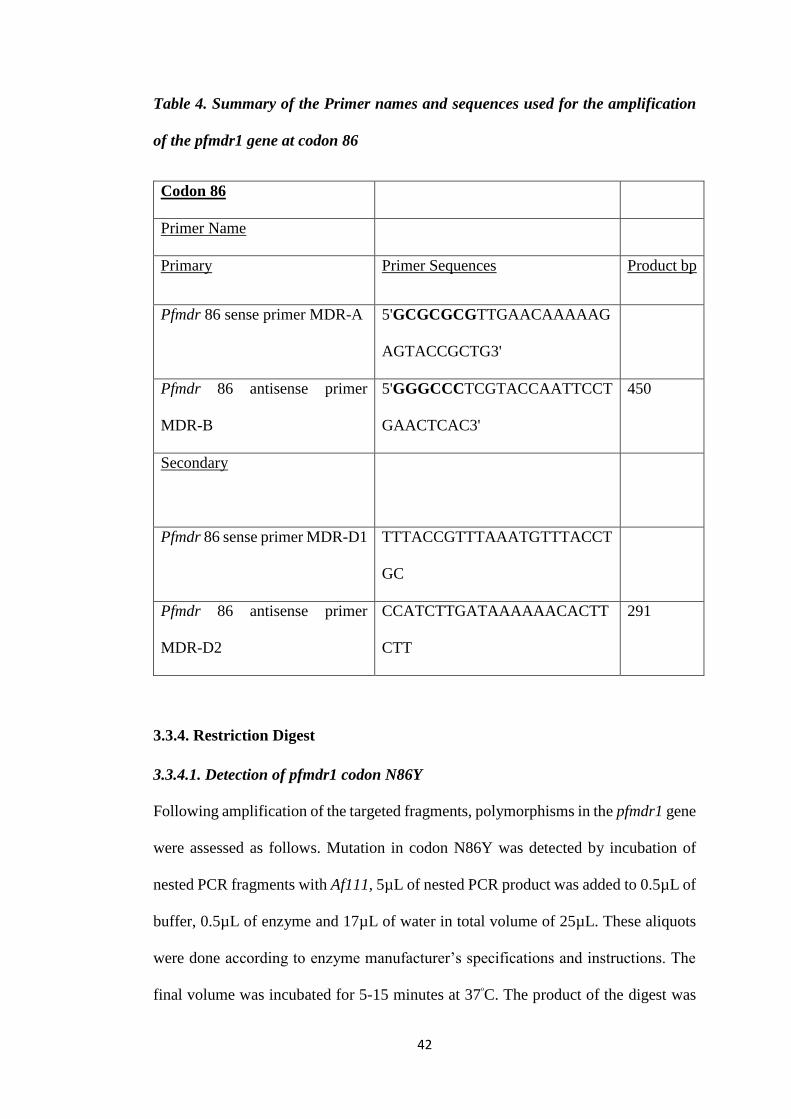
Table 4. Summary of the Primer names and sequences used for the amplification
of the pfmdr1 gene at codon 86
Codon 86
Primer Name
Primary Primer Sequences Product bp
Pfmdr 86 sense primer MDR-A 5'GCGCGCGTTGAACAAAAAG
AGTACCGCTG3'
Pfmdr 86 antisense primer
MDR-B
5'GGGCCCTCGTACCAATTCCT
GAACTCAC3'
450
Secondary
Pfmdr 86 sense primer MDR-D1 TTTACCGTTTAAATGTTTACCT
GC
Pfmdr 86 antisense primer
MDR-D2
CCATCTTGATAAAAAACACTT
CTT
291
3.3.4. Restriction Digest
3.3.4.1. Detection of pfmdr1 codon N86Y
Following amplification of the targeted fragments, polymorphisms in the pfmdr1 gene
were assessed as follows. Mutation in codon N86Y was detected by incubation of
nested PCR fragments with Af111, 5µL of nested PCR product was added to 0.5µL of
buffer, 0.5µL of enzyme and 17µL of water in total volume of 25µL. These aliquots
were done according to enzyme manufacturer’s specifications and instructions. The
final volume was incubated for 5-15 minutes at 37ᵒC. The product of the digest was
43
run on a 2.5% gel stained with ethidium bromide and bands were visualized using Gel
documentation system.
The endonuclease AfIII was purchased from Inqaba Biotechnical Industries (Pty) Ltd
South Africa and incubations were setup following the manufacturer’s instructions.
Appropriate control DNA of samples with known pfmdr1sequences was used in
parallel with field-collected parasite isolates in every PCR-RFLP protocol; these were
Dd2 (genotype pfmdr1 86Y).
A 2% agarose gel was prepared by boiling 4g of LE agarose powder in 200ml of 1X
TAE buffer (40mM Tris, 20mM Borate, and 1mM EDTA, pH=8). A total of 6µl of
Ethidium Bromide (EtBr) was added to stain the agarose gel. After casting the gel
using a gel casting apparatus, 3µl of each nested PCR product of either mdr1 or crt
genes were mixed with 2µl of 6X gel loading dye that contained bromophenol blue
on a plastic sheet (para film). Then, each mixed nested product was loaded into the
wells of the solidified gel, 3µl molecular weight marker with 100bp-ladder were
loaded on to the gel as well.
3.3.6. Quantitative Real Time PCR for the Detection Of crt72-76 Haplotypes
To detect the two Plasmodium falciparum crt alleles, multiplex Real Time
quantitative PCR (RT-PCR) (CFX Real Time Machine) was performed. Parasite DNA
was extracted from dried bloodspots using the Qiagen extraction kit extracted DNA
was stored at -20°C. For this experiment, labelled probes, CVMNK and CVIET were
used to detect three common haplotypes at codons 72 to 76 of the pfcrt gene. Each
probe was labelled with a reporter dye at the 5’ end and a quencher moiety at the 3’
end. Primers which are specific to the conserved region of pfcrt and the two probes
were combined in one reaction, the oligonucleotide and probe sequences are described
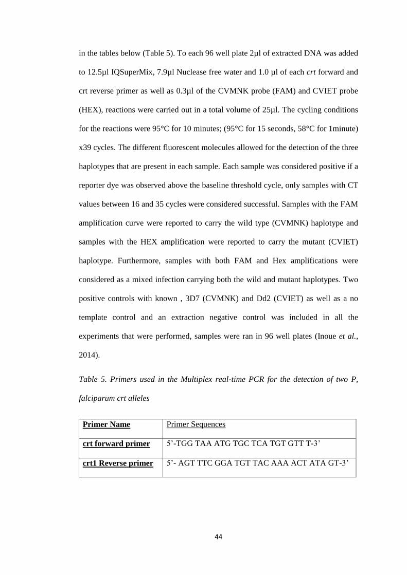
44
in the tables below (Table 5). To each 96 well plate 2µl of extracted DNA was added
to 12.5µl IQSuperMix, 7.9µl Nuclease free water and 1.0 µl of each crt forward and
crt reverse primer as well as 0.3µl of the CVMNK probe (FAM) and CVIET probe
(HEX), reactions were carried out in a total volume of 25µl. The cycling conditions
for the reactions were 95°C for 10 minutes; (95°C for 15 seconds, 58°C for 1minute)
x39 cycles. The different fluorescent molecules allowed for the detection of the three
haplotypes that are present in each sample. Each sample was considered positive if a
reporter dye was observed above the baseline threshold cycle, only samples with CT
values between 16 and 35 cycles were considered successful. Samples with the FAM
amplification curve were reported to carry the wild type (CVMNK) haplotype and
samples with the HEX amplification were reported to carry the mutant (CVIET)
haplotype. Furthermore, samples with both FAM and Hex amplifications were
considered as a mixed infection carrying both the wild and mutant haplotypes. Two
positive controls with known , 3D7 (CVMNK) and Dd2 (CVIET) as well as a no
template control and an extraction negative control was included in all the
experiments that were performed, samples were ran in 96 well plates (Inoue et al.,
2014).
Table 5. Primers used in the Multiplex real-time PCR for the detection of two P,
falciparum crt alleles
Primer Name Primer Sequences
crt forward primer 5’-TGG TAA ATG TGC TCA TGT GTT T-3’
crt1 Reverse primer 5’- AGT TTC GGA TGT TAC AAA ACT ATA GT-3’
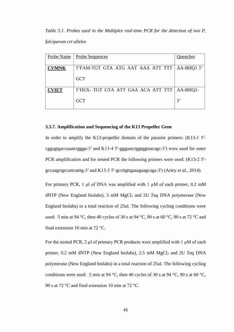
45
Table 5.1. Probes used in the Multiplex real-time PCR for the detection of two P,
falciparum crt alleles
Probe Name Probe Sequences Quencher
CVMNK 5’FAM-TGT GTA ATG AAT AAA ATT TTT
GCT
AA-BHQ1 3’
CVIET 5’HEX- TGT GTA ATT GAA ACA ATT TTT
GCT
AA-BHQ1-
3’
3.3.7. Amplification and Sequencing of the K13 Propeller Gene
In order to amplify the K13-propeller domain of the parasite primers: (K13-1 5′-
cggagtgaccaaatctggga-3′ and K13-4 5′-gggaatctggtggtaacagc-3′) were used for outer
PCR amplification and for nested PCR the following primers were used: (K13-2 5′-
gccaagctgccattcatttg-3′ and K13-3 5′-gccttgttgaaagaagcaga-3′) (Ariey et al., 2014).
For primary PCR, 1 μl of DNA was amplified with 1 μM of each primer, 0.2 mM
dNTP (New England biolabs), 3 mM MgCl2 and 2U Taq DNA polymerase (New
England biolabs) in a total reaction of 25ul. The following cycling conditions were
used: 5 min at 94 °C, then 40 cycles of 30 s at 94 °C, 90 s at 60 °C, 90 s at 72 °C and
final extension 10 min at 72 °C.
For the nested PCR, 2 μl of primary PCR products were amplified with 1 μM of each
primer, 0.2 mM dNTP (New England biolabs), 2.5 mM MgCl2 and 2U Taq DNA
polymerase (New England biolabs) in a total reaction of 25ul. The following cycling
conditions were used: 5 min at 94 °C, then 40 cycles of 30 s at 94 °C, 90 s at 60 °C,
90 s at 72 °C and final extension 10 min at 72 °C.
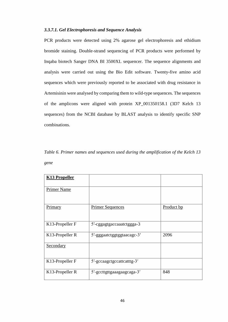
46
3.3.7.1. Gel Electrophoresis and Sequence Analysis
PCR products were detected using 2% agarose gel electrophoresis and ethidium
bromide staining. Double-strand sequencing of PCR products were performed by
Inqaba biotech Sanger DNA BI 3500XL sequencer. The sequence alignments and
analysis were carried out using the Bio Edit software. Twenty-five amino acid
sequences which were previously reported to be associated with drug resistance in
Artemisinin were analysed by comparing them to wild-type sequences. The sequences
of the amplicons were aligned with protein XP_001350158.1 (3D7 Kelch 13
sequences) from the NCBI database by BLAST analysis to identify specific SNP
combinations.
Table 6. Primer names and sequences used during the amplification of the Kelch 13
gene
K13 Propeller
Primer Name
Primary Primer Sequences Product bp
K13-Propeller F 5′-cggagtgaccaaatctggga-3
K13-Propeller R 5′-gggaatctggtggtaacagc-3′ 2096
Secondary
K13-Propeller F 5′-gccaagctgccattcatttg-3′
K13-Propeller R 5′-gccttgttgaaagaagcaga-3′ 848
47
3.4. Data Analysis
Base-line frequencies of P. falciparum point mutations associated with artemisinin
resistance in Northern Namibia will be calculated, using data that has been entered
and validated with Microsoft Office Excel® 2013 (Microsoft Corporation
4. Research Ethics
Approval for this study was obtained from the medical directors and administrators of
health facilities before samples were collected. The study was explained to the
participants in their own language. A written informed consent of the participants or
their parents /guardians /representatives, has been received before samples were
collected. The names of the participants were kept anonymous. Samples were
collected and handled by trained nurses. There was no risk to participants except for
mild pain during blood collection. Sterile techniques were enforced at all times during
the study.
This study was conducted within larger parent studies conducted in the Zambezi
region. Ethical approval for the parent study has been obtained from the University of
Namibia and the Ministry of Health and Social Services Biomedical Research Ethics
Committee. Research Ethical Clearance has been obtained from the University of
Namibia Research Ethics Committee.
48
5. Results
A total of 143 RDT positive malaria samples were speciated using multiplex PCR.
They were also analysed for polymorphisms at codon N86Y of the pfmdr1 gene and
crt 72-76 haplotypes of the pfcrt gene and sequenced of the Kelch 13 codons.
Additionally, mutations at codons crt 72-76 were analysed using quantitative real-
time PCR.
5.1. Identification of Plasmodium species by Multiplex PCR
To identify the different plasmodium species in the Zambezi region 143 samples were
amplified by multiplex PCR with species specific primers. Only 84/143 (58.7%)
samples were successfully amplified. The species distribution was as follows 64/84
(76.2%) P. falciparum, 6/84 (7.1%) P. vivax and 5/84 (6.0%) P. malariae parasites.
Additionally, 9 mixed infections were found of which 8/84 (9.5%) were P. falciparum
mixed with P. vivax mixed and 1/84 (1.2%) was P. falciparum mixed with P. malariae
and. No P. ovale parasites were found. Figure 15 below shows a representative of the
results obtained during the multiplex PCR. Additionally, frequency of the different
parasites found are summarized below in (figure. 15.1)
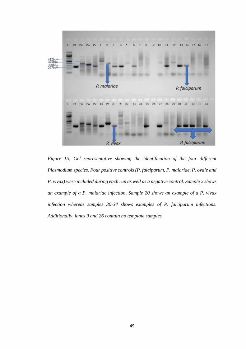
49
Figure 15; Gel representative showing the identification of the four different
Plasmodium species. Four positive controls (P. falciparum, P. malariae, P. ovale and
P. vivax) were included during each run as well as a negative control. Sample 2 shows
an example of a P. malariae infection, Sample 20 shows an example of a P. vivax
infection whereas samples 30-34 shows examples of P. falciparum infections.
Additionally, lanes 9 and 26 contain no template samples.
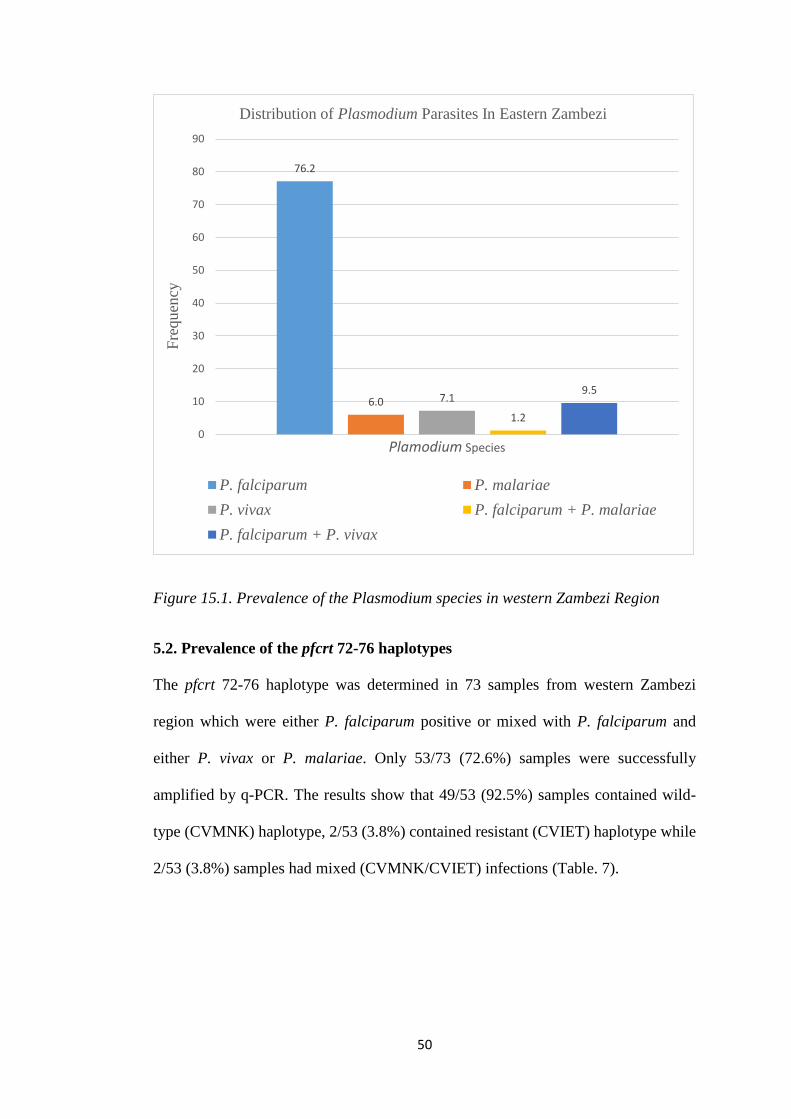
50
Figure 15.1. Prevalence of the Plasmodium species in western Zambezi Region
5.2. Prevalence of the pfcrt 72-76 haplotypes
The pfcrt 72-76 haplotype was determined in 73 samples from western Zambezi
region which were either P. falciparum positive or mixed with P. falciparum and
either P. vivax or P. malariae. Only 53/73 (72.6%) samples were successfully
amplified by q-PCR. The results show that 49/53 (92.5%) samples contained wild-
type (CVMNK) haplotype, 2/53 (3.8%) contained resistant (CVIET) haplotype while
2/53 (3.8%) samples had mixed (CVMNK/CVIET) infections (Table. 7).
76.2
6.0 7.1
1.2
9.5
0
10
20
30
40
50
60
70
80
90
Fre
quen
cy
Plamodium Species
Distribution of Plasmodium Parasites In Eastern Zambezi
P. falciparum P. malariae
P. vivax P. falciparum + P. malariae
P. falciparum + P. vivax
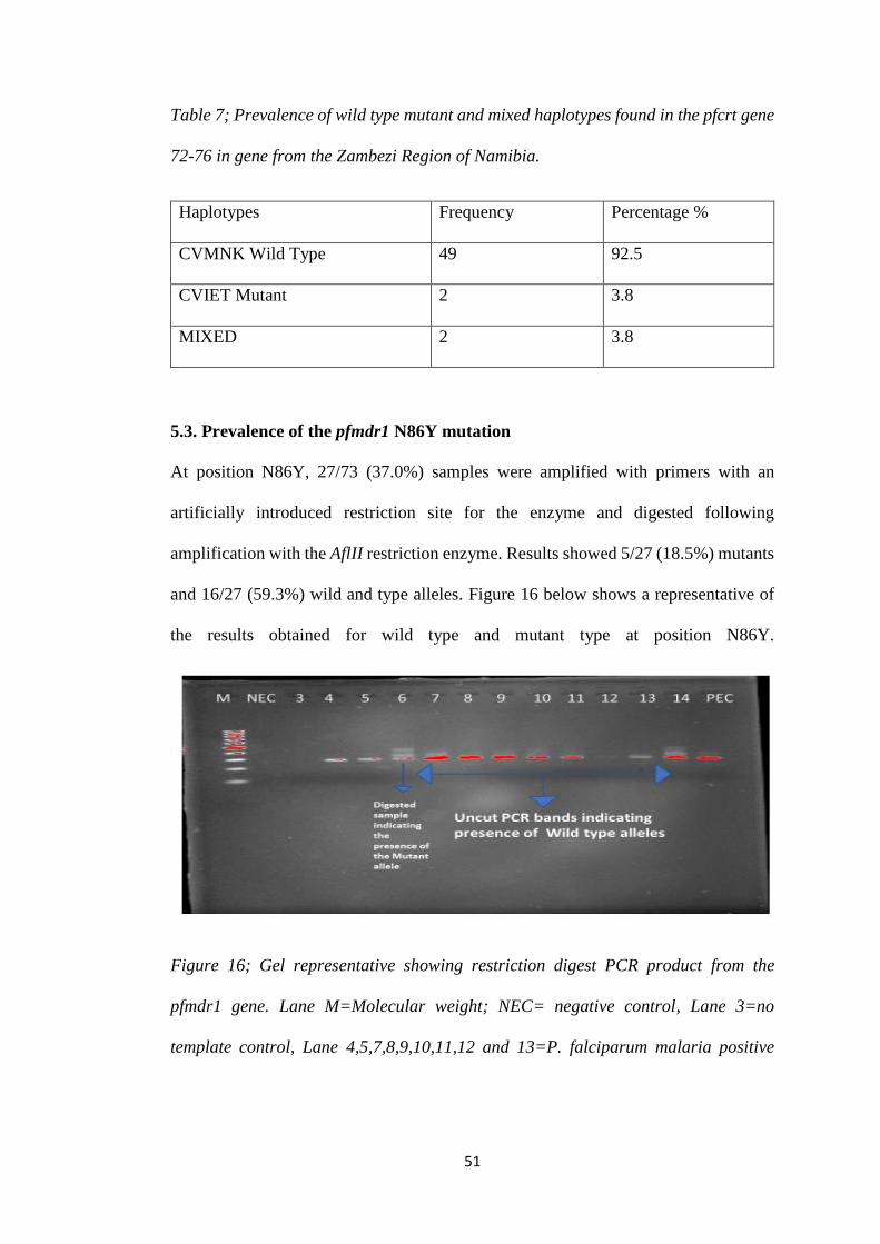
51
Table 7; Prevalence of wild type mutant and mixed haplotypes found in the pfcrt gene
72-76 in gene from the Zambezi Region of Namibia.
Haplotypes Frequency Percentage %
CVMNK Wild Type 49 92.5
CVIET Mutant 2 3.8
MIXED 2 3.8
5.3. Prevalence of the pfmdr1 N86Y mutation
At position N86Y, 27/73 (37.0%) samples were amplified with primers with an
artificially introduced restriction site for the enzyme and digested following
amplification with the AflII restriction enzyme. Results showed 5/27 (18.5%) mutants
and 16/27 (59.3%) wild and type alleles. Figure 16 below shows a representative of
the results obtained for wild type and mutant type at position N86Y.
Figure 16; Gel representative showing restriction digest PCR product from the
pfmdr1 gene. Lane M=Molecular weight; NEC= negative control, Lane 3=no
template control, Lane 4,5,7,8,9,10,11,12 and 13=P. falciparum malaria positive
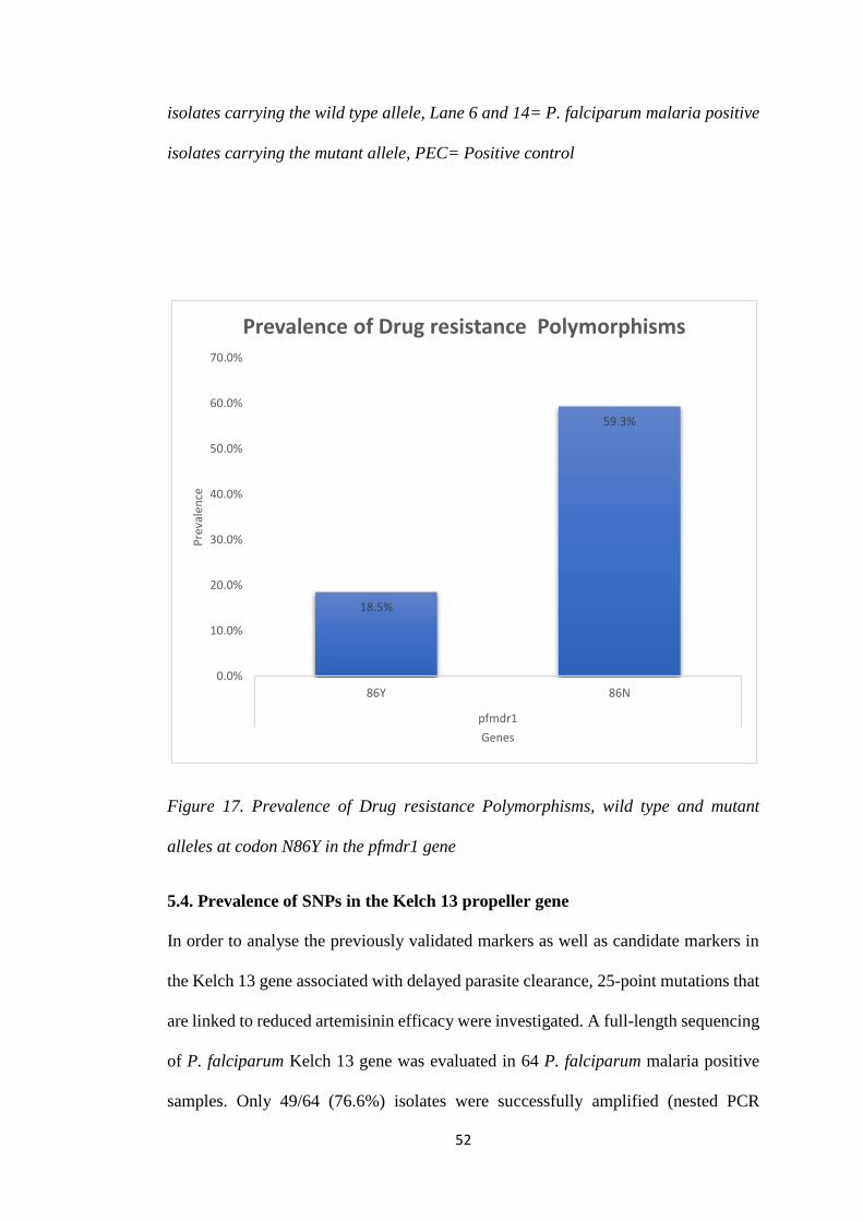
52
isolates carrying the wild type allele, Lane 6 and 14= P. falciparum malaria positive
isolates carrying the mutant allele, PEC= Positive control
Figure 17. Prevalence of Drug resistance Polymorphisms, wild type and mutant
alleles at codon N86Y in the pfmdr1 gene
5.4. Prevalence of SNPs in the Kelch 13 propeller gene
In order to analyse the previously validated markers as well as candidate markers in
the Kelch 13 gene associated with delayed parasite clearance, 25-point mutations that
are linked to reduced artemisinin efficacy were investigated. A full-length sequencing
of P. falciparum Kelch 13 gene was evaluated in 64 P. falciparum malaria positive
samples. Only 49/64 (76.6%) isolates were successfully amplified (nested PCR
18.5%
59.3%
0.0%
10.0%
20.0%
30.0%
40.0%
50.0%
60.0%
70.0%
86Y 86N
pfmdr1
Pre
vale
nce
Genes
Prevalence of Drug resistance Polymorphisms
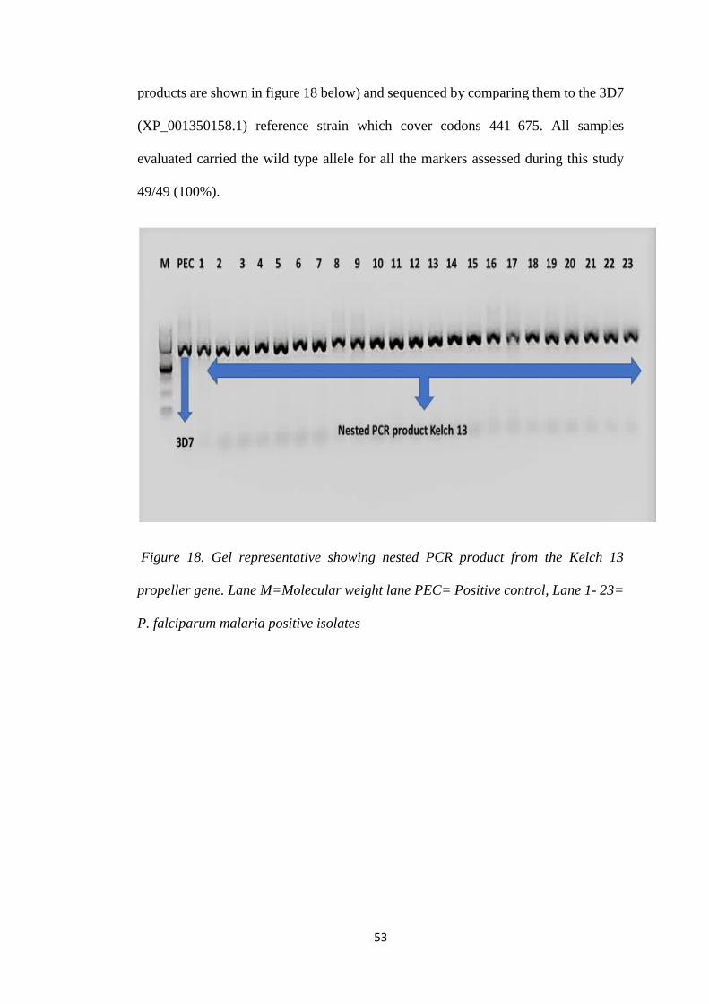
53
products are shown in figure 18 below) and sequenced by comparing them to the 3D7
(XP_001350158.1) reference strain which cover codons 441–675. All samples
evaluated carried the wild type allele for all the markers assessed during this study
49/49 (100%).
Figure 18. Gel representative showing nested PCR product from the Kelch 13
propeller gene. Lane M=Molecular weight lane PEC= Positive control, Lane 1- 23=
P. falciparum malaria positive isolates
54
6. Discussion
Using Single Nucleotide Polymorphisms (SNPs) as surveillance tools is an effective
way to monitor early emergence of antimalarial drug resistance before in vivo efficacy
studies are conducted as these studies can be time consuming and in some instances
can take long to get ethical approval (WHO 2009). To date there are no reports on the
prevalence of drug resistance markers in any part of Namibia although there are other
reports in the Southern African region such as in Angola, Mozambique and Zambia
(Foumane-Ngane et al., 2015) (Escobar et al., 2015) (Mwanza et al., 2016). This is
the first such study conducted in Namibia reporting on antimalarial drug resistance
markers in P. falciparum isolates. The study also investigated the distribution of
plasmodium species in malaria positive participants in the Zambezi region from the
different catchment areas. One hundred and forty-three samples collected from the
Zambezi region in the North eastern part of Namibia which were confirmed malaria
positive by RDT and LAMP were used to extract DNA to assess the presence of
molecular markers associated with antimalarial drug resistance. In this study three
genes were investigated namely pfcrt, pfmdr1 and the Kelch13 propeller gene.
The study revealed that the majority of the infections were due to P. falciparum
(76.2%), these findings were consistent with previous reports stating that P.
falciparum accounts for majority of the malaria infections in Namibia (Smith-Gueye
et al., 2014)(Noor et al., 2013). These results also support findings that show that the
majority of malaria cases are caused by P. falciparum in Africa (Noor et al., 2013)
(Balogun et al., 2016). This number however was found to be much lower than what
was reported previously by Noor et al 2013, of which showed that 90.97% of cases
were caused by P. falciparum in Namibia. The decrease could be due to the decrease
in the number of malaria cases since 2013 (Smith-Gueye et al., 2014). Studies have
55
shown that as malaria cases decrease, the parasite density also decreases, which makes
the identification of parasites using PCR more difficult (Vallejo et al., 2015). This
may have been the case during this study which resulted in the detection of the lower
prevalence of P. falciparum cases as previously compared and could also explain why
only 58.7% of the samples were successfully amplified during species identification
essay. Additionally, previous studies have shown that PCR is not as sensitive in
detecting the presence of parasites in a low transmission setting such as Namibia with
a detection limit of 6 parasites/μl of blood from dried spots (Laufer et al., 2010) thus
could explain the lower prevalence of 76.2% P. falciparum parasites as compared to
the 90% previously report by Noor et al, since Multiplex PCR was used for species
identification during this study.
Although mixed infections and other plasmodium species were not expected as
previously reported, during this study other species were found with P. vivax
accounting for 7.1% of the total malaria cases followed by 6.0% of P. malariae
infections, which could be as a result of imported malaria cases from other endemic
countries. The findings were in contrast with what was reported by WHO that stated
that 100% of all malaria infections were due to P. falciparum (WHO, 2016) and thus
calls for molecular species identification studies to detect and treat all malaria cases
effectively. It is however suspected that these plasmodium species may have been
missed during the previous studies, for the reason that firstly P. vivax is known to
remain dormant for weeks to years after infection (Lindblade et al., 2013).
Additionally P. vivax cannot be easily detected during asymptomatic patients and thus
can have been missed during the time these studies were conducted and may have
remerged during the present study (Lindblade et al., 2013). Secondly because
Namibia is a low transmission setting the tools used such as light microscopy which
56
is the gold standard for species detection may have missed these parasites at the time
of previous studies (Hopkins et al., 2013). Thirdly studies have found that P. malariae
is found at a lower prevalence and density in endemic areas (Kwenti et al., 2017).
Since Namibia is a low transmission setting, the conventional methods used
previously could have missed the presence of this particular parasite in Namibia
(Perera et al., 2017). Finally, because most Sub-Saharan African countries report that
P. falciparum is the predominant species, diagnostics tools such as RDTs and Loop-
mediated isothermal amplification (LAMP) kits are developed with the focus on P.
falciparum sensitivity only and with limited effectiveness directed towards the
sensitivity of other species thus could be missing these species during diagnosis
(Hayashida et al., 2017).
Nevertheless, the results observed during this study were similar to the findings
reported in Luanda, Angola. It was reported that P. falciparum (92%) accounted for
the majority of infections followed by low prevalence of infections caused by P. vivax
(7.0%) and P. malariae (3.0%) (Pembele, Rivero and Fraga, 2015). Furthermore, a
study done in Zambia also observed that 88% of their cases were P. falciparum
making up majority of the cases and 10.6% were mixed infections (Sitali et al., 2015).
These similarities in parasite distributions may be a result of the number of travellers
from these countries to Namibia and as previously reported, the majority of malaria
cases that occur in Northern Namibia are from Angola (Njovu et al., 2013) (Smith
Gueye et al., 2014). Thus, to eliminate malaria, countries sharing borders are proposed
to work together to detect and treat all cases of malaria as malaria parasites are known
to move freely between countries. Additionally, mono and mixed infections with P.
ovale species (P. ovale curtisi and P. ovale wallikeri) were observed in Angola and
Zambia, whereas during this study, 0% P. ovale infections were found (Pembele,
57
Rivero and Fraga, 2015)(Hayashida et al., 2017). Similarly, no P. ovale infections
have been reported in previous studies that were conducted in Namibia ( Noor et al.,
2013).
Although a low prevalence of P. malariae cases and no P. ovale cases were found in
the present study, a study conducted in Jiangsu Province, China has found that most
of their imported P. malariae and P. ovale cases came from Africa and stated that the
majority of the P. malariae cases were from Sub-Saharan Africa (Cao et al., 2016).
These findings by Cao et al have been confirmed by a study done in the Democratic
Republic of the Congo where they found that 26.3 % of the parasites tested positive
for P. malariae and 17.2 % were positive for P. ovale (Doctor et al., 2016).
Furthermore, a low frequency of mixed infections with P. falciparum was observed
in this study of which 1.2% were P. malariae mixed with P. falciparum and 9.5%
were P. vivax mixed with P. falciparum. These findings are however in contrast with
what was reported in Zambia, where they found that majority of their mixed infections
were a combination of P. falciparum and P. malariae (Sitali et al., 2015).
This study has shown that P. falciparum is not the only plasmodium species present
in Namibia that cause malaria infections as previously reported by WHO (WHO,
2016). Thus, in order to detect and treat all malaria cases with the appropriate
antimalarial drugs and to avoid the misuse of antimalarial medicine. Species
identification should be done after mixed infections are detected by RDTs using both
molecular methods and the recommended gold standard, as the gold standard has been
previously reported to not be as accurate in the successful differentiation between
different species (Stanis et al., 2016)(Dafalla et al., 2017). This will allow for the
accurate administration of correct antimalarial medicines once an individual is tested
positive for malaria.
58
Following the identification of the distinct species, samples that tested positive for P.
falciparum as well as mixed samples with P. falciparum were included to determine
the prevalence of antimalarial drug resistance makers in the pfcrt gene. The
occurrence of the CVIET (mutant), CVMNK (wild type) and the Mixed
(CVIET/CVMNK) haplotypes were studied at codons 72-76. Many studies have
linked point mutations in this gene with drug resistance in previously used
antimalarial drugs such as Chloroquine and Amodiaquine (Rosenthal, 2013)(Golassa
et al., 2014). Whereas the wild type form at codon K76T has been linked to the
reduced sensitivity in Lumefantrine which is currently being used as a partner drug
with artemisinin in Namibia (Rosenthal, 2013). Additionally, the presence of the
CVMNK haplotype has been linked with reduced sensitivity in Artemisinin’s which
are currently in use with partner drugs as a first line treatment in many African
countries.
The CVMNK and SVMNT are known as the “mother” haplotypes in the pfcrt gene.
However derivatives for SVMNT such as CVIET which has been previously reported
to be present in Africa is being used as the mutant haplotype in different parts of the
world (Zhou et al., 2016). During this study the CVIET derivative was used as the
mutant haplotype, because the SVMNK haplotype is said to be dominant in regions
outside Africa (South America, Indonesia, Papua New Guinea, and the Philippines in
Southeast Asia) (Reteng et al., 2017).
In order to test for the prevalence of resistant, sensitive and mixed haplotypes in the
pfcrt gene 73 samples found to be either P. falciparum positive or mixed with P.
falciparum and either P. vivax or P. malariae were genotyped. The 3D7 strain that
harbours the CVMNK haplotype and the Dd2 harbouring the CVIET haplotype were
59
included as controls during the amplification along with a no template and a PCR
negative control.
During this study a high prevalence of isolates carrying the wild type haplotype
(92.5%) and low prevalence of (3.8%) isolates carrying the mutant haplotype was
observed. Additionally, only 3.8% of the samples carried the mixed haplotype in the
isolates from Zambezi region. Similar results were found in studies done in Nigeria
(Salissou et al., 2014), Zambia (Mwanza et al., 2016) and Malawi (Laufer et al.,
2010). Studies conducted in Ethiopia (Golassa et al., 2014), North Cameroon (Ali et
al., 2013) , Gabon (Salissou et al., 2014) and Angola (Fancony, Brito and Gil, 2016)
presented contrasting findings to the present study where a high prevalence of the
CVIET haplotype was observed and low prevalence of the CVMNK haplotype. The
results from this study has shown that the withdrawal of Chloroquine as treatment for
P. falciparum malaria after CQR strains have been encountered has led to the increase
in wildtype haplotypes in the pfcrt gene, thus showing the revival of chloroquine
sensitive (CQS) populations of P. falciparum in Namibia. These findings support the
possible reintroduction of chloroquine as a partner drug with ACTs in the future with
continual observation of antimalarial drug resistance. Additionally, studies have
shown that the increase in the wild type allele at codon K76T can lead to the reduced
sensitivity in Lumefantrine as parasites have selected for the wild type allele during
ex vivo studies (Taylor et al., 2016). However, this selection alone does not lead to a
resistance phenotype in Lumefantrine therefore calling for studies on pfmdr1 copy
number which has been said to cause resistance in the presence of the K76 allele to
support these findings (Shah et al., 2015).
Furthermore, this study also aimed to observe the presence of point mutations in the
pfmdr1 gene at codon N86Y. Mutations in this gene have also been linked to
60
antimalarial drug resistance in Chloroquine, Amodiaquine and Quinoline (QN)
(Ibraheem et al., 2014). Whereas the wild type alleles have been linked to reduced
susceptibility in Halofantrine and Lumefantrine. Additionally the amplification of this
gene has been linked to reduced susceptibility in Mefloquine (MQ) and Artemisinin
(Heuchert et al., 2015).
At position N86Y, 37.0% samples were amplified and digested with the AflII
restriction enzyme. There was a low prevalence of (18.5%) mutant alleles and a high
prevalence of 59.3% wild type alleles. The low prevalence observed at this position
is consistent with the low prevalence found in the pfcrt CVIET haplotype also
observed in this study. These results do show that the withdrawal of chloroquine did
indeed lead to the selection of the wild type allele over the parasites with the mutant
allele which could no longer survive due to the better fitness of parasites which selects
for the wild type allele. Furthermore, these findings are in agreement with reports that
stated that the CQR phenotype is a result of both the presence of the N86Y mutation
and the K76T mutation (Norahmad et al., 2016). Additionally, Amodiaquine
resistance is also linked to the mutant haplotype in the pfcrt gene as well as to the
mutant allele in the pfmdr1 gene at codon N86Y (Fröberg et al., 2012) (Gadalla et al.,
2015). Although some studies found a low prevalence of mutations in both the pfcrt
gene and pfmdr1 gene at codon N86Y, like the study done in Nigeria (Agomo et al.,
2016), studies such as the one done in Jimma, Ethiopia observed a low prevalence of
mutations at position N86Y and a high prevalence of the pfcrt mutant haplotype
(Heuchert et al., 2015). Moreover, a high prevalence of wild type alleles were
observed at codon N86Y and this may be due to the introduction of artemether
lumefantrine as the first line treatment in Namibia, studies reported that this
61
antimalarial drug selects for the wild type allele which can explain this observation
(Nguetse et al., 2017).
Mutations observed at codon D1246Y together with S1034C and N1042D has been
linked to resistance in QN and correspondingly to increased susceptibility in MQ,
halofantrine (HF) and artemisinin (Mbaye et al., 2016). Additionally the presence of
the wild type alleles at codons N86Y, F184Y and D1246Y have been linked to
reduced artemether-lumefantrine (AL) susceptibility, whereas the presence of mutant
alleles at these codons have been linked to amodiaquine and chloroquine reduced
susceptibility (Kavishe et al., 2014). Thus, the low prevalence of the wild type allele
found at codon N86Y in this study could be an indication that the P. falciparum
parasites in Namibia are still susceptible to AL which is currently used as first line
treatment for malaria. However, although it has been shown that N86Y is an important
marker for antimalarial drug resistance in Al it has been suggested that studies look at
the DFN and YYY haplotypes in the pfmdr1 gene at codons (86,187 and 1256) to give
a more conclusive report (Dhorda et al., 2015)(Sondo et al., 2016). Furthermore, it is
suggested that in order to make a definite claim on the status of AL in a country, in
vitro and in vivo studies should be done to confirm the susceptibility of AL (Apinjoh
et al., 2016).
ACTs are currently used as first line treatment for falciparum malaria in majority of
malaria endemic countries in Africa including Namibia, with artemether-lumefantrine
and artesunate-Amodiaquine being the most commonly used antimalarial drugs (Li et
al., 2016). Namibia adopted artemether-lumefantrine as first treatment in 2006 and
since then the number of malaria cases have reduced (WHO, 2015c). However,
recently, delayed parasite clearance in ACTs were reported in the Greater Mekong
Sub region after in vivo and in vitro studies presented delayed parasite clearance in
62
the presence of non-synonymous single nucleotide polymorphisms (SNPs) found in
the Kelch 13 propeller gene (Lin and Zaw, 2015)(Huang et al., 2015)(Sondo et al.,
2016).
In order to determine the prevalence of Kelch 13 SNPs in the Zambezi region of
Namibia, 49 P. falciparum malaria positive samples were successfully amplified and
sequenced which covered codons 441–675 including the three mutations (C580Y,
R539T and Y493H) that were said to be strongly associated with delayed parasite
clearance in South Asia as well as codon M476I found in Tanzania (Huang et al.,
2015). During this study we found zero mutations at any of the sequenced codons,
with a high prevalence of wild type alleles. These findings were consistent with the
results observed in The Gambia and in Bioko Island, Equatorial Guinea which
observed no mutations at any of the codons that has been associated with delayed
parasite clearance (Amambua-Ngwa et al., 2017) (Li et al., 2016). Even though some
studies found a low prevalence of mutations in the Kelch 13 gene, studies done in
Angola and Mozambique observed synonymous mutations at low frequencies in
codons R471R, V494I, and R575R which are believed to play a role in artemisinin
resistance. This calls for further in vivo and in vitro studies to determine whether their
occurrence leads to delayed parasite clearance (Escobar et al., 2015) (Fancony, Brito
and Gil, 2016).
Nevertheless, another study done in Angola did however find lower than the expected
drug efficacy in the Zaire Province in northern Angola. This report showed that the
AL efficacy was less than 90% which is the lower limit recommended for ACTs by
WHO. However, they found that this reduced drug efficacy was not linked to any K13
mutations (Plucinski et al., 2017). This raises concerns as it may show that
artermisinin reduced drug efficacy may not be linked to K13 mutations in some
63
endemic regions, suggesting that antimalarial drug resistance should be routinely
monitored using both molecular markers which were previously linked to antimalarial
drug resistance, as well as clinical and parasitological response to antimalarial drugs
in patients as recommended by WHO (WHO, 2017a).
Additionally, majority of the studies done in Africa presented that none of the isolates
carried the mutant allele in any of the four candidate markers described in Asia that
are strongly linked to Artemisinin failure (Huang et al., 2015). These findings suggest
that ACTs in Africa including Namibia are still highly effective. Nonetheless,
continued surveillance is needed to detect early emergence of drug resistance as some
countries in East Africa and Angola have observed frequencies of synonymous
mutations (Lin and Zaw, 2015).
During this study the pfmdr1 copy number was also not evaluated which has been
linked to artemisinin resistance. Therefore, more studies need to be done as presence
of point mutations alone does not lead to artermisinin resistance. Additionally, due to
the smaller sample size used and the limited geographic region that was covered by
the samples, this study may have not have detected all the cases of artermisinin-
resistant malaria and the findings may be limited to the north eastern region of
Namibia where the study was conducted.
64
7. Conclusion
During this study a low prevalence of point mutations in both the pfmrd1 gene at
codon N86Y and at the codons 72-76 in the pfcrt gene was observed. Additionally, no
mutations were found in all 25 markers in the Kelch 13 gene associated with drug
resistance in artemisinin. These observations show that the use of artemether-
lumefantrine is still effective in western Namibia. Furthermore, the study showed that
the prevalence of P. falciparum malaria cases is not as high as previously reported
and that a higher prevalence of P. vivax malaria cases was found which was more than
expected. Additionally, the presence of P. malariae cases were also detected,
nonetheless no P. ovale cases were observed.
This study also showed that using multiplex PCR for plasmodium species
identification is an effective and faster way to identify different species as many
studies have found that the gold standard of using conventional light microscopy for
the examination and identification of plasmodium have some draw backs. Thus, it is
recommended to use multiplex PCR along with the gold standard for the accurate
identification of plasmodium species.
Moreover, this study showed that using molecular markers as surveillance tools for
antimalarial drug resistance may be an easier and faster way to detect early emergence
of drug resistance. However, studies have found that not all mutations lead to a
resistance phenotype as some antimalarial drugs may select for the wild type allele
such as with AL, thus it is recommended that in in vivo and in vitro drug efficacy
studies are done to see if the presence of these polymorphisms lead to treatment
failure.
65
8. Recommendations
During this study a low prevalence of point mutations was found, and this may suggest
that AL efficacy is still high in Namibia. However, a small sample size was used from
only one geographical area therefore, it is recommended that further studies be done
including other malaria endemic areas in Namibia with a larger sample size to give a
better representation of the country.
Findings from this study also suggest a need for continued monitoring of non-
falciparum infection prevalence to decide when species-specific RDTs should be
introduced for diagnostic purposes, because species-specific diagnosis will be
important when the prevalence of malaria in this population reduces, as the country
moves towards malaria elimination. Additionally while it should not necessarily be the
primary diagnostic tool, diagnostic approaches that have the capacity to investigate and
detect non-falciparum malaria may become important in future. In health facilities
where microscopy services are present, there is need to conduct refresher courses for
laboratory staff and microscopists with emphasis on Plasmodium species-wide
microscopy to strengthen non-falciparum malaria diagnosis
Although multiplex PCR is a much faster way compared to light microscopy to
identify different plasmodium parasites, this assay has some drawbacks, which
include that it is qualitative assay and it is not as sensitive as qPCR assays. Thus, it is
recommended that a further study be done using quantitative assays to supplement
these findings.

66
Finally, it is recommended that other studies should be done in different malaria
endemic areas to give a clear representation of the current antimalarial drug resistance
status in Namibia.
67
9. References
Agomo, C. O., Oyibo, W. A., Sutherland, C., Hallet, R. and Oguike, M. (2016)
‘Assessment of Markers of Antimalarial Drug Resistance in Plasmodium falciparum
Isolates from Pregnant Women in Lagos, Nigeria’, PLoS One. Edited by T. A.
Smith. Elsevier Science Limited, 11(1), p. e0146908. doi:
10.1371/journal.pone.0146908.
Ako, B. A., Offianan, A. T., Johansson, M., Penali, L. K., Assanvo, S. P., Nguetta
and Sibley, C. H. (2012) ‘Molecular analysis of markers associated with chloroquine
and sulfadoxine/pyrimethamine resistance in plasmodium falciparum malaria
parasites from southeastern c??te-d’ivoire by the time of artemisinin-based
combination therapy adoption in 2005’, Infection and Drug Resistance, 5(1), pp.
113–120. doi: 10.2147/IDR.S31409.
Ali, I. M., Netongo, P. M., Atogho-Tiedeu, B., Ngongang, E.-O., Ajua, A., Achidi,
E. a and Mbacham, W. F. (2013) ‘Amodiaquine-Artesunate versus Artemether-
Lumefantrine against Uncomplicated Malaria in Children Less Than 14 Years in
Ngaoundere, North Cameroon: Efficacy, Safety, and Baseline Drug Resistant
Mutations in pfcrt, pfmdr1, and pfdhfr Genes.’, Malaria research and treatment,
2013, p. 234683. doi: 10.1155/2013/234683.
Amadi, O. K., Otuokere, I. E. and Chinedum, N. C. (2017) ‘Synthesis and
Antibacterial Studies of Fe ( II ), Co ( II ), Ni ( II ) and Cu ( II ) Complexes of
Lumefantrine’, International journal of Chemical, Gas and Material Science
(IJCGM), 1(1).
Amambua-Ngwa, A., Okebe, J., Mbye, H., Ceesay, S., El-Fatouri, F., Joof, F.,
68
Nyang, H., Janha, R., Affara, M., Ahmad, A., Kolly, O., Nwakanma, D. and
D’Alessandro, U. (2017) ‘Sustained ex vivo susceptibility of Plasmodium
falciparum to artemisinin derivatives but increasing tolerance to artemisinin
combination therapy partner quinolines in The Gambia’, Antimicrobial Agents and
Chemotherapy, 61(12). doi: 10.1128/AAC.00759-17.
Aminake, M. and Pradel, G. (2013) ‘Antimalarial drugs resistance in Plasmodium
falciparum and the current strategies to overcome them’, Microbial pathogens and
strategies for combating them: science, technology and education, 1, pp. 269–282.
doi: 10.1089/ars.2011.3999.
‘Antimalarial Drugs_ Modes of Action snd Mechanisms of Resistance’ (no date).
Available at: https://www.medscape.com/viewarticle/734498_5 (Accessed: 16
February 2018).
Apinjoh, T., Anchang-Kimbi, J., Ajonina, M., Njonguo, E., Njua-Yafi, C., Ngwai,
A., Mugri, R. and Achidi, E. (2016) ‘In Vivo Efficacy of Artesunate/Sulphadoxine-
Pyrimethamine versus Artesunate/Amodiaquine in the Treatment of Uncomplicated
P. falciparium Malaria in Children around the Slope of Mount Cameroon: A
Randomized Controlled Trial’, Biomedicines, 4(1), p. 5. doi:
10.3390/biomedicines4010005.
Ariey, F., Witkowski, B., Amaratunga, C., Beghain, J., Langlois, A.-C., Khim, N.,
Kim, S., Duru, V., Bouchier, C., Ma, L., Lim, P., Leang, R., Duong, S., Sreng, S.,
Suon, S., Chuor, C. M., Bout, D. M., Ménard, S., Rogers, W. O., Genton, B.,
Fandeur, T., Miotto, O., Ringwald, P., Le Bras, J., Berry, A., Barale, J.-C., Fairhurst,
R. M., Benoit-Vical, F., Mercereau-Puijalon, O. and Ménard, D. (2014) ‘A
molecular marker of artemisinin-resistant Plasmodium falciparum malaria’, Nature.
69
NIH Public Access, 505(7481), pp. 50–5. doi: 10.1038/nature12876.
Ashley, E. a., Dhorda, M., Fairhurst, R. M., Amaratunga, C., Lim, P., Suon, S.,
Sreng, S., Anderson, J. M., Mao, S., Sam, B., Sopha, C., Chuor, C. M., Nguon, C.,
Sovannaroth, S., Pukrittayakamee, S., Jittamala, P., Chotivanich, K., Chutasmit, K.,
Suchatsoonthorn, C., Runcharoen, R., Hien, T. T., Thuy-Nhien, N. T., Thanh, N. V.,
Phu, N. H., Htut, Y., Han, K.-T., Aye, K. H., Mokuolu, O. A., Olaosebikan, R. R.,
Folaranmi, O. O., Mayxay, M., Khanthavong, M., Hongvanthong, B., Newton, P.
N., Onyamboko, M. a., Fanello, C. I., Tshefu, A. K., Mishra, N., Valecha, N., Phyo,
A. P., Nosten, F., Yi, P., Tripura, R., Borrmann, S., Bashraheil, M., Peshu, J., Faiz,
M. A., Ghose, A., Hossain, M. A., Samad, R., Rahman, M. R., Hasan, M. M., Islam,
A., Miotto, O., Amato, R., MacInnis, B., Stalker, J., Kwiatkowski, D. P., Bozdech,
Z., Jeeyapant, A., Cheah, P. Y., Sakulthaew, T., Chalk, J., Intharabut, B., Silamut,
K., Lee, S. J., Vihokhern, B., Kunasol, C., Imwong, M., Tarning, J., Taylor, W. J.,
Yeung, S., Woodrow, C. J., Flegg, J. a., Das, D., Smith, J., Venkatesan, M., Plowe,
C. V., Stepniewska, K., Guerin, P. J., Dondorp, A. M., Day, N. P. and White, N. J.
(2014) ‘Spread of Artemisinin Resistance in Plasmodium falciparum Malaria’, New
England Journal of Medicine, 371(5), pp. 411–423. doi: 10.1056/NEJMoa1314981.
Atroosh, W. M., Al-Mekhlafi, H. M., Mahdy, M. A. K. and Surin, J. (2012) ‘The
detection of pfcrt and pfmdr1 point mutations as molecular markers of chloroquine
drug resistance, Pahang, Malaysia’, Malaria Journal. Malaria Journal, 11(1), p. 1.
doi: 10.1186/1475-2875-11-251.
Awasthi, G. and Das, A. (2013) ‘Genetics of chloroquine-resistant malaria: A
haplotypic view’, Memorias do Instituto Oswaldo Cruz, 108(8), pp. 947–961. doi:
10.1590/0074-0276130274.
70
Balogun, S. T., Sandabe, U. K., Bdliya, D. N., Adedeji, W. A., Okon, K. O. and
Fehintola, F. A. (2016) ‘Asymptomatic falciparum malaria and genetic
polymorphisms of Pfcrt K76T and Pfmdr1 N86Y among almajirai in northeast
Nigeria’, Journal of Infection in Developing Countries, 10(3), pp. 290–297. doi:
10.3855/jidc.6853.
Bassat, Q., Ogutu, B., Djimde, A., Stricker, K. and Hamed, K. (2015) ‘Tailoring a
Pediatric Formulation of Artemether-Lumefantrine for Treatment of Plasmodium
falciparum Malaria’, Antimicrob Agents Chemother, 59(8), pp. 4366–4374. doi:
10.1128/aac.00014-15.
Bayih, A. G., Getnet, G., Alemu, A., Getie, S., Mohon, A. N. and Pillai, D. R.
(2016) ‘A unique plasmodium falciparum K13 gene mutation in Northwest
Ethiopia’, American Journal of Tropical Medicine and Hygiene, 94(1), pp. 132–135.
doi: 10.4269/ajtmh.15-0477.
Bhattacharjee, D. and Shivaprakash, G. (2016) ‘Drug resistance in Malaria-in a
nutshell’, Journal of Applied Pharmaceutical Science, 6(3), pp. 137–143. doi:
10.7324/JAPS.2016.60324.
Le Bras, J. and Durand, R. (2003) ‘The mechanisms of resistance to antimalarial
drugs in Plasmodium falciparum’, Fundamental and Clinical Pharmacology, 17(2),
pp. 147–153. doi: 10.1046/j.1472-8206.2003.00164.x.
Calderaro, A., Piccolo, G., Gorrini, C., Rossi, S., Montecchini, S., Dell’Anna, M.,
De Conto, F., Medici, M., Chezzi, C. and Arcangeletti, M. (2013) ‘Accurate
identification of the six human Plasmodium spp. causing imported malaria,
including Plasmodium ovale wallikeri and Plasmodium knowlesi’, Malaria Journal,
12(1), p. 321. doi: 10.1186/1475-2875-12-321.
71
Cao, Y., Wang, W., Liu, Y., Cotter, C., Zhou, H., Zhu, G., Tang, J., Tang, F., Lu, F.,
Xu, S., Gu, Y., Zhang, C., Li, J. and Cao, J. (2016) ‘The increasing importance of
Plasmodium ovale and Plasmodium malariae in a malaria elimination setting: An
observational study of imported cases in Jiangsu Province, China, 2011-2014’,
Malaria Journal. BioMed Central, 15(1), pp. 1–9. doi: 10.1186/s12936-016-1504-2.
CDC (2014) ‘CDC - Malaria - Malaria Worldwide - How Can Malaria Cases and
Deaths Be Reduced? - Insecticide-Treated Bed Nets’, Global Health - Division of
Parasitic Diseases and Malaria. Available at:
https://www.cdc.gov/malaria/malaria_worldwide/reduction/index.html (Accessed:
10 July 2018).
CDC, C. F. D. C. and P. (2013) Treatment of Malaria ( Guidelines For Clinicians ),
Treatment of Malaria (Guidelines for clinicians). doi: 10.1016/S0140-
6736(05)66420-3.
Chauhan, K., Pande, V. and Das, A. (2014) ‘DNA sequence polymorphisms of the
pfmdr1 gene and association of mutations with the pfcrt gene in Indian Plasmodium
falciparum isolates’, Infection, Genetics and Evolution. Elsevier B.V., 26, pp. 213–
222. doi: 10.1016/j.meegid.2014.05.033.
Chilongola, J., Ndaro, A., Tarimo, H., Shedrack, T., Barthazary, S., Kaaya, R.,
Masokoto, A., Kajeguka, D., Kavishe, R. A. and Lusingu, J. (2015) ‘Occurrence of
pfatpase6 Single Nucleotide Polymorphisms Associated with Artemisinin
Resistance among Field Isolates of Plasmodium falciparum in North-Eastern
Tanzania.’, Malaria research and treatment, 2015, p. 279028. doi:
10.1155/2015/279028.
Coelho, C. H., Doritchamou, J. Y. A., Zaidi, I. and Duffy, P. E. (2017) ‘Advances in
72
malaria vaccine development: Report from the 2017 malaria vaccine symposium’,
npj Vaccines. Springer US, 2(1). doi: 10.1038/s41541-017-0035-3.
Cui, L., Mharakurwa, S., Ndiaye, D., Rathod, P. K. and Rosenthal, P. J. (2015)
‘Antimalarial Drug Resistance: Literature Review and Activities and Findings of the
ICEMR Network.’, The American journal of tropical medicine and hygiene, 93(3
Suppl), pp. 57–68. doi: 10.4269/ajtmh.15-0007.
Dafalla, O. M., Alsheikh, A. A., D, A. A., Mohammed, W. S., MNour, B. Y., J, S.
K. and Noureldin, E. M. (2017) ‘Identifi cation of Plasmodium species from
outdated blood samples by nested-PCR compared with microscopy diagnosis in
Jazan region, Saudi Arabia’, Biosci. Biotech. Res. Comm. BBRCBA Thomson
Reuters ISI ESC and Crossref Indexed Journal NAAS Journal Score, 10(2), pp. 68–
75.
Delves, M., Plouffe, D., Scheurer, C., Meister, S., Wittlin, S., Winzeler, E. A.,
Sinden, R. E. and Leroy, D. (2012) ‘The Activities of Current Antimalarial Drugs on
the Life Cycle Stages of Plasmodium: A Comparative Study with Human and
Rodent Parasites’, PLoS Medicine. Edited by J. G. Beeson. Academic Press, 9(2), p.
e1001169. doi: 10.1371/journal.pmed.1001169.
Deshpande, S. and Kuppast, B. (2016) ‘4-aminoquinolines: An Overview of
Antimalarial Chemotherapy’, Medicinal chemistry, 06(01), pp. 1–11. doi:
10.4172/2161-0444.1000315.
Dhorda, M., Cheah, P. Y., Pukrittayakamee, S., Ashley, E. A., Anderson, T. J. C.,
Nair, S., Mcdew-white, M., White, N. J. and Woodrow, C. J. (2015) ‘Spread of
artemisinin-resistant Plasmodium falciparum in Myanmar: A cross-sectional survey
of the K13 molecular marker’, The Lancet Infectious Diseases, 15(4), pp. 415–421.
73
doi: 10.1016/S1473-3099(15)70032-0.
Doctor, S. M., Liu, Y., Anderson, O. G., Whitesell, A. N., Mwandagalirwa, M. K.,
Muwonga, J., Keeler, C., Emch, M., Likwela, J. L., Tshefu, A. and Meshnick, S. R.
(2016) ‘Low prevalence of Plasmodium malariae and Plasmodium ovale mono-
infections among children in the Democratic Republic of the Congo: a population-
based, cross-sectional study’, Malaria Journal. BioMed Central, 15(1), pp. 1–6. doi:
10.1186/s12936-016-1409-0.
Dondorp, A. M., Nosten, F., Yi, P., Das, D., Phyo, A. P., Tarning, J., Ph, D., Lwin,
K. M., Ariey, F., Hanpithakpong, W., Lee, S. J., Ringwald, P., Silamut, K.,
Herdman, T., An, S. S., Yeung, S., Socheat, D. and White, N. J. (2009) ‘Artemisinin
Resistance in Plasmodium falciparum Malaria’, Drug Therapy, 361(5), pp. 455–467.
doi: 10.1086/657120.
Escobar, C., Pateira, S., Lobo, E., Lobo, L., Teodosio, R., Dias, F., Fernandes, N.,
Arez, A. P., Varandas, L. and Nogueira, F. (2015) ‘Polymorphisms in Plasmodium
falciparum K13-propeller in angola and mozambique after the introduction of the
ACTs’, PLoS ONE, 10(3), pp. 2–7. doi: 10.1371/journal.pone.0119215.
Eyasu, M. (2015) ‘Antimalarial Drug Resistance : In the Past , Current Status and
Future Perspectives’, British Journal of Pharmacology and Toxicology, 6(1), pp. 1–
15.
Fancony, C., Brito, M. and Gil, J. P. (2016) ‘Plasmodium falciparum drug resistance
in Angola.’, Malaria journal. BioMed Central, 15(1), p. 74. doi: 10.1186/s12936-
016-1122-z.
Feng, J., Li, J., Yan, H., Feng, X. and Xia, Z. (2015) ‘Evaluation of antimalarial
resistance marker polymorphism in returned migrant workers in China’,
74
Antimicrobial Agents and Chemotherapy, 59(1), pp. 326–330. doi:
10.1128/AAC.04144-14.
Figtree, M., Lee, R., Bain, L., Kennedy, T., Mackertich, S., Urban, M., Cheng, Q.
and Hudson, B. J. (2010) ‘Plasmodium knowlesi in human, Indonesian Borneo’,
Emerging Infectious Diseases, 16(4), pp. 672–674. doi: 10.3201/eid1604.091624.
Foumane Ngane, V., Allico Djaman, J., Culeux, C., Piette, N., Carnevale, P.,
Besnard, P., Fortes, F., Basco, L. K. and Tahar, R. (2015) ‘Molecular epidemiology
of drug-resistant Plasmodium falciparum in Benguela province, Angola’, Malaria
Journal, 14(1), pp. 1–6. doi: 10.1186/s12936-015-0634-2.
Fröberg, G., Jörnhagen, L., Morris, U., Shakely, D., Msellem, M. M. I. M., Gil, J. P.
J. J. P., Björkman, A., Mårtensson, A., Bhattarai, A., Ali, A., Kachur, S.,
Mårtensson, A., Abbas, A., Khatib, R., Al-Mafazy, A., Ramsan, M., Rotllant, G.,
Gerstenmaier, J., Molteni, F., Abdulla, S., Montgomery, S., Kaneko, A., Björkman,
A., Childs, G., Boudreau, E., Milhous, W., Wimonwattratee, T., Pooyindee, N.,
Pang, L., Davidson, D., Olliaro, P., Mussano, P., Djimde, A., Doumbo, O., Cortese,
J., Kayentao, K., Doumbo, S., Diourte, Y., Dicko, A., Su, X., Nomura, T., Fidock,
D., Wellems, T., Plowe, C., Coulibaly, D., Nsobya, S., Kiggundu, M., Nanyunja, S.,
Joloba, M., Greenhouse, B., Rosenthal, P., Duraisingh, M., Drakeley, C., Muller, O.,
Bailey, R., Snounou, G., Targett, G., Greenwood, B., Warhurst, D., Holmgren, G.,
Gil, J. P. J. J. P., Ferreira, P. P. P., Veiga, M., Obonyo, C., Björkman, A., Picot, S.,
Olliaro, P., Monbrison, F. de, Bienvenu, A., Price, R., Ringwald, P., Humphreys, G.,
Merinopoulos, I., Ahmed, J., Whitty, C., Mutabingwa, T., Sutherland, C., Hallett,
R., Lakshmanan, V., Bray, P., Verdier-Pinard, D., Johnson, D., Horrocks, P., Muhle,
R., Alakpa, G., Hughes, R., Ward, S., Krogstad, D., Sidhu, A., Fidock, D., Sidhu,
75
A., Verdier-Pinard, D., Fidock, D., Warhurst, D., Sa, J., Twu, O., Sa, J., Twu, O.,
Hayton, K., Reyes, S., Fay, M., Ringwald, P., Wellems, T., Holmgren, G., Hamrin,
J., Svard, J., Mårtensson, A., Gil, J. P. J. J. P., Björkman, A., Mårtensson, A.,
Stromberg, J., Sisowath, C., Msellem, M. M. I. M., Gil, J. P. J. J. P., Montgomery,
S., Olliaro, P., Ali, A., Björkman, A., Veiga, M., Ferreira, P. P. P., Björkman, A.,
Gil, J. P. J. J. P., Sisowath, C., Stromberg, J., Mårtensson, A., Msellem, M. M. I. M.,
Obondo, C., Björkman, A., Gil, J. P. J. J. P., Sisowath, C., Ferreira, P. P. P.,
Bustamante, L., Dahlstrom, S., Mårtensson, A., Björkman, A., Krishna, S., Gil, J. P.
J. J. P., Ngasala, B., Malmberg, M., Carlsson, A., Ferreira, P. P. P., Petzold, M.,
Blessborn, D., Bergqvist, Y., Gil, J. P. J. J. P., Premji, Z., Björkman, A.,
Mårtensson, A., Sisowath, C., Petersen, I., Veiga, M., Mårtensson, A., Premji, Z.,
Björkman, A., Fidock, D., Gil, J. P. J. J. P., Mwai, L., Kiara, S., Abdirahman, A.,
Pole, L., Rippert, A., Diriye, A., Bull, P., Marsh, K., Borrmann, S., Nzila, A., Reed,
M., Saliba, K., Caruana, S., Kirk, K. and Cowman, A. (2012) ‘Decreased prevalence
of Plasmodium falciparum resistance markers to amodiaquine despite its wide scale
use as ACT partner drug in Zanzibar’, Malaria Journal, 11(1), p. 321. doi:
10.1186/1475-2875-11-321.
Gadalla, N. B., Tavera, G., Mu, J., Kabyemela, E. R., Fried, M., Duffy, P. E., Sá, J.
M. and Wellems, T. E. (2015) ‘Prevalence of Plasmodium falciparum anti-malarial
resistance-associated polymorphisms in pfcrt, pfmdr1 and pfnhe1 in Muheza,
Tanzania, prior to introduction of artemisinin combination therapy.’, Malaria
journal, 14(1), p. 129. doi: 10.1186/s12936-015-0642-2.
Gatc, J., Maspiyanti, F., Sarwinda, D. and Arymurthy, A. M. (2013) ‘Plasmodium
parasite detection on Red Blood Cell image for the diagnosis of malaria using
double thresholding’, in 2013 International Conference on Advanced Computer
76
Science and Information Systems (ICACSIS). IEEE, pp. 381–385. doi:
10.1109/ICACSIS.2013.6761605.
Ghanchi, N. K., Ursing, J., Beg, M. A., Veiga, M. I., Jafri, S. and Mårtensson, A.
(2011) ‘Prevalence of resistance associated polymorphisms in Plasmodium
falciparum field isolates from southern Pakistan.’, Malaria journal, 10, p. 18. doi:
10.1186/1475-2875-10-18.
Golassa, L., Enweji, N., Erko, B., Aseffa, A. and Swedberg, G. (2014) ‘High
prevalence of pfcrt-CVIET haplotype in isolates from asymptomatic and
symptomatic patients in south-central Oromia, Ethiopia’, Malaria Journal. Malaria
Journal, 13(1), pp. 1–8. doi: 10.1186/1475-2875-13-120.
Golassa, L., Erko, B., Baliraine, F. N., Aseffa, A. and Swedberg, G. (2015)
‘Polymorphisms in chloroquine resistance-associated genes in Plasmodium vivax in
Ethiopia.’, Malaria journal, 14(1), p. 164. doi: 10.1186/s12936-015-0625-3.
Goswami, D., Dhiman, S., Rabha, B., Kumar, D., Baruah, I., Sharma, D. K. and
Veer, V. (2014) ‘Pfcrt mutant haplotypes may not correspond with chloroquine
resistance’, Journal of Infection in Developing Countries, 8(6), pp. 768–773. doi:
10.3855/jidc.3398.
Greenwood, B., Dicko, A., Sagara, I., Zongo, I., Tinto, H., Cairns, M., Kuepfer, I.,
Milligan, P., Ouedraogo, J. B., Doumbo, O. and Chandramohan, D. (2017)
‘Seasonal vaccination against malaria: a potential use for an imperfect malaria
vaccine’, Malaria Journal. BioMed Central, 16(1), pp. 10–14. doi: 10.1186/s12936-
017-1841-9.
Guiyun Yan, Q. F. and L. C. P. L. Z. Z. H. X. W. L. X. Z. Y. C. Z. Y. J. S. (2016)
‘Plasmodium malariae and Plasmodium ovale infections in the China-Myanmar
77
border area’, Malaria Journal. BioMed Central, 15(1), pp. 1–10. doi:
10.1186/s12936-016-1605-y.
Hasab, M., Basheir, E., Ali, M., Elgadir, A., Babikir, A. M., Rahoud, S. A. G.,
Musa, H. H. and Tayrab, E. M. A. (2012) ‘The Prevalence of Multi-drug Resistance-
1 gene of Plasmodium falciparum Malaria in Gezira State-Central Sudan’,
SUDANESE JOURNAL OF PUBLIC HEALTH, 7(3).
Hawkins, K., Burton, R. and Labarre, P. (2014) ‘Diagnostics to support malaria
elimination: Choosing an appropriate biomarker to target the subclinical
Plasmodium falciparum transmission reservoir’, Proceedings of the 4th IEEE
Global Humanitarian Technology Conference, GHTC 2014, pp. 561–568. doi:
10.1109/GHTC.2014.6970339.
Hayashida, K., Kajino, K., Simukoko, H., Simuunza, M., Ndebe, J., Chota, A.,
Namangala, B. and Sugimoto, C. (2017) ‘Direct detection of falciparum and non-
falciparum malaria DNA from a drop of blood with high sensitivity by the dried-
LAMP system’, Parasites and Vectors. Parasites & Vectors, 10(1), pp. 1–9. doi:
10.1186/s13071-016-1949-8.
Heuchert, A., Abduselam, N., Zeynudin, A., Eshetu, T., Löscher, T., Wieser, A.,
Pritsch, M. and Berens-Riha, N. (2015) ‘Molecular markers of anti-malarial drug
resistance in southwest Ethiopia over time: regional surveillance from 2006 to
2013.’, Malaria journal. ???, 14(1), p. 208. doi: 10.1186/s12936-015-0723-2.
Hopkins, H., González, I. J., Polley, S. D., Angutoko, P., Ategeka, J., Asiimwe, C.,
Agaba, B., Kyabayinze, D. J., Sutherland, C. J., Perkins, M. D. and Bell, D. (2013)
‘Highly sensitive detection of malaria parasitemia in a malaria-endemic setting:
Performance of a new loop-mediated isothermal amplification kit in a remote clinic
78
in Uganda’, Journal of Infectious Diseases, 208(4), pp. 645–652. doi:
10.1093/infdis/jit184.
Huang, B., Deng, C., Yang, T., Xue, L., Wang, Q., Huang, S., Su, X.-Z., Liu, Y.,
Zheng, S., Guan, Y., Xu, Q., Zhou, J., Yuan, J., Bacar, A., Abdallah, K. S.,
Attoumane, R., Mliva, A. M. S. A., Zhong, Y., Lu, F. and Song, J. (2015)
‘Polymorphisms of the artemisinin resistant marker (K13) in Plasmodium
falciparum parasite populations of Grande Comore Island 10 years after artemisinin
combination therapy.’, Parasites & vectors. Parasites & Vectors, 8(1), p. 634. doi:
10.1186/s13071-015-1253-z.
Ibraheem, Z. O., Abd Majid, R., Noor, S. M., Sedik, H. M., Basir, R., Ibraheem, Z.
O., Abd Majid, R., Noor, S. M., Sedik, H. M. and Basir, R. (2014) ‘Role of Different
Pf crt and Pf mdr-1 Mutations in Conferring Resistance to Antimalaria Drugs in
Plasmodium falciparum’, Malaria Research and Treatment, 2014(Lm), pp. 1–17.
doi: 10.1155/2014/950424.
Ilesanmi, R. I., Olalubi, O. A., Adetunde, O. T., Ilesanmi, A. O., Effedua, H. and
Amoo, A. O. (2017) ‘Comparative assessment of malaria rapid diagnostic tests (
RDT ) in Ibadan , Nigeria’, Malaria World Journal, 8(17).
Inoue, J., Lopes, D., do Rosário, V., Machado, M., Hristov, A. D., Lima, G. F.,
Costa-Nascimento, M. J., Segurado, A. C. and Di Santi, S. M. (2014) ‘Analysis of
polymorphisms in Plasmodium falciparum genes related to drug resistance: a survey
over four decades under different treatment policies in Brazil.’, Malaria journal, 13,
p. 372. doi: 10.1186/1475-2875-13-372.
Jimenez, A., Rees-Channer, R. R., Perera, R., Gamboa, D., Chiodini, P. L.,
González, I. J., Mayor, A. and Ding, X. C. (2017) ‘Analytical sensitivity of current
79
best-in-class malaria rapid diagnostic tests’, Malaria Journal. BioMed Central,
16(1), pp. 1–9. doi: 10.1186/s12936-017-1780-5.
Kakkilaya, B. S. (2015) ‘Rapid Diagnosis of Malaria’, Laboratory Medicine, 34(8),
pp. 602–608. doi: 10.1309/J4ANKCCJ147JB2FR.
Kamwi, J. M., Chirwa, P. W. C., Manda, S. O. M., Graz, P. F. and Kätsch, C. (2015)
‘Livelihoods, land use and land cover change in the Zambezi Region, Namibia’,
Population and Environment, 37(2), pp. 207–230. doi: 10.1007/s11111-015-0239-2.
Karunamoorthi, K. (2011) ‘Vector control: A cornerstone in the malaria elimination
campaign’, Clinical Microbiology and Infection. European Society of Clinical
Microbiology and Infectious Diseases, 17(11), pp. 1608–1616. doi: 10.1111/j.1469-
0691.2011.03664.x.
Kavishe, R. A., Paulo, P., Kaaya, R. D., Kalinga, A., Van Zwetselaar, M.,
Chilongola, J., Roper, C. and Alifrangis, M. (2014) ‘Surveillance of artemether-
lumefantrine associated Plasmodium falciparum multidrug resistance protein-1 gene
polymorphisms in Tanzania’, Malaria Journal, 13(1), p. 264. doi: 10.1186/1475-
2875-13-264.
Korenromp, E., Mahiané, G., Hamilton, M., Pretorius, C., Cibulskis, R., Lauer, J.,
Smith, T. A. and Briët, O. J. T. (2016) ‘Malaria intervention scale-up in Africa:
Effectiveness predictions for health programme planning tools, based on dynamic
transmission modelling’, Malaria Journal. BioMed Central, 15(1), pp. 1–14. doi:
10.1186/s12936-016-1461-9.
Kwenti, T. E., Kwenti, T. D. B., Njunda, L. A., Latz, A., Tufon, K. A. and Nkuo-
Akenji, T. (2017) ‘Identification of the Plasmodium species in clinical samples from
children residing in five epidemiological strata of malaria in Cameroon’, Tropical
80
Medicine and Health. Tropical Medicine and Health, 45(1), pp. 1–8. doi:
10.1186/s41182-017-0058-5.
Laufer, M. K., Takala-Harrison, S., Dzinjalamala, F. K., Stine, O. C., Taylor, T. E.
and Plowe, C. V (2010) ‘Return of chloroquine-susceptible falciparum malaria in
Malawi was a reexpansion of diverse susceptible parasites.’, The Journal of
infectious diseases, 202(5), pp. 801–8. doi: 10.1086/655659.
Li, J., Chen, J., Xie, D., Eyi, U. M., Matesa, R. A., Ondo Obono, M. M., Ehapo, C.
S., Yang, L., Yang, H. and Lin, M. (2016) ‘Limited artemisinin resistance-associated
polymorphisms in Plasmodium falciparum K13-propeller and PfATPase6 gene
isolated from Bioko Island, Equatorial Guinea’, International Journal for
Parasitology: Drugs and Drug Resistance. Elsevier Ltd, 6(2016), pp. 54–59. doi:
10.1016/j.ijpddr.2015.11.002.
Lin, Z. and Zaw, M. (2015) ‘Molecular Determinants of Artemisinin Resistance in
k13 Gene of Plasmodium falciparum’, British Microbiology Research Journal, 9(4),
pp. 1–11. doi: 10.9734/BMRJ/2015/18776.
Lindblade, K. A., Steinhardt, L., Samuels, A., Kachur, S. P. and Slutsker, L. (2013)
‘The silent threat: Asymptomatic parasitemia and malaria transmission’, Expert
Review of Anti-Infective Therapy, 11(6), pp. 623–639. doi: 10.1586/eri.13.45.
Lucchi, N. W., Komino, F., Okoth, S. A., Goldman, I., Onyona, P., Wiegand, R. E.,
Juma, E., Shi, Y. P., Barnwell, J. W., Udhayakumar, V. and Kariuki, S. (2015) ‘In
Vitro and molecular surveillance for antimalarial drug resistance in Plasmodium
falciparum parasites in western Kenya reveals sustained artemisinin sensitivity and
increased chloroquine sensitivity’, Antimicrobial Agents and Chemotherapy, 59(12),
pp. 7540–7547. doi: 10.1128/AAC.01894-15.
81
M. L. McMorrow, M. Aidoo, and S. P. K. (2015) ‘Malaria rapid diagnostic tests in
elimination settings—can they find the last parasite?’, Anal Chem., 25(4), pp. 368–
379. doi: 10.1016/j.cogdev.2010.08.003.Personal.
Malaria - including symptoms, treatment and prevention (2012). Department of
Health. Available at:
http://www.sahealth.sa.gov.au/wps/wcm/connect/public+content/sa+health+internet/
health+topics/health+conditions+prevention+and+treatment/infectious+diseases/mal
aria (Accessed: 21 July 2018).
Malaria, L., Professionals, H., Life, P., Points, B., Life, P., Prevention, T., Available,
C. and Ware, O. C. (2008) Plasmodium Life cycle.
Mbaye, A., Dieye, B., Ndiaye, Y. D., Bei, A. K., Muna, A., Deme, A. B., Yade, M.
S., Diongue, K., Gaye, A., Ndiaye, I. M., Ndiaye, T., Sy, M., Diallo, M. A., Badiane,
A. S., Ndiaye, M., Seck, M. C., Sy, N., Koita, O., Krogstad, D. J., Nwakanma, D.
and Ndiaye, D. (2016) ‘Selection of N86F184D1246 haplotype of Pfmrd1 gene by
artemether-lumefantrine drug pressure on Plasmodium falciparum populations in
Senegal’, Malaria Journal. BioMed Central, 15(1), pp. 1–7. doi: 10.1186/s12936-
016-1490-4.
Molina-Cruz, A. and Barillas-Mury, C. (2014) ‘The remarkable journey of
adaptation of the Plasmodium falciparum malaria parasite to New World anopheline
mosquitoes’, Memorias do Instituto Oswaldo Cruz, 109(5), pp. 662–667. doi:
10.1590/0074-0276130553.
Morris, U. (2015) Molecular Methods in Malaria Control in the Era of Pre-
Elimination. Karolinska Institutet.
Muhamad, P., Phompradit, P., Sornjai, W., Maensathian, T., Chaijaroenkul, W.,
82
Rueangweerayut, R. and Na-Bangchang, K. (2011) ‘Polymorphisms of molecular
markers of antimalarial drug resistance and relationship with artesunate-mefloquine
combination therapy in patients with uncomplicated Plasmodium falciparum malaria
in Thailand’, American Journal of Tropical Medicine and Hygiene, 85(3), pp. 568–
572. doi: 10.4269/ajtmh.2011.11-0194.
Mwanza, S., Joshi, S., Nambozi, M., Chileshe, J., Malunga, P., Kabuya, J. B. B.,
Hachizovu, S., Manyando, C., Mulenga, M. and Laufer, M. (2016) ‘The return of
chloroquine-susceptible Plasmodium falciparum malaria in Zambia’, Malaria
Journal. BioMed Central, 15(1), pp. 1–6. doi: 10.1186/s12936-016-1637-3.
Nakeesathit, S., Saralamba, N., Pukrittayakamee, S., Dondorp, A., Nosten, F.,
White, N. J. and Imwong, M. (2016) ‘Limited polymorphism of the Kelch propeller
domain in Plasmodium malariae and P. ovale isolates from Thailand’, Antimicrobial
Agents and Chemotherapy, 60(7), pp. 4055–4062. doi: 10.1128/AAC.00138-16.
Nguetse, C. N., Adegnika, A. A., Agbenyega, T., Ogutu, B. R., Krishna, S.,
Kremsner, P. G. and Velavan, T. P. (2017) ‘Molecular markers of anti-malarial drug
resistance in Central, West and East African children with severe malaria’, Malaria
Journal. BioMed Central, 16(1), p. 217. doi: 10.1186/s12936-017-1868-y.
Ngufor, C., Fagbohoun, J., Critchley, J., N’Guessan, R., Todjinou, D., Malone, D.,
Akogbeto, M. and Rowland, M. (2017) ‘Which intervention is better for malaria
vector control: Insecticide mixture long-lasting insecticidal nets or standard
pyrethroid nets combined with indoor residual spraying?’, Malaria Journal. BioMed
Central, 16(1), pp. 1–9. doi: 10.1186/s12936-017-1987-5.
Njovu, C., Stearns, E., Park, J., Alexandre, M., Lourenco, C., Lassen, S. and Petrow,
E. (2013) TRANS KUNENE MALARIA INITIATIVE (TKMI): Partnering Cross
83
Border towards Elimination.
Nkumama, I. N., O’Meara, W. P. and Osier, F. H. A. (2017) ‘Changes in Malaria
Epidemiology in Africa and New Challenges for Elimination’, Trends in
Parasitology. Elsevier Ltd, 33(2), pp. 128–140. doi: 10.1016/j.pt.2016.11.006.
Noor, A. M., Alegana, V. A., Kamwi, R. N., Hansford, C. F., Ntomwa, B., Katokele,
S. and Snow, R. W. (2013) ‘Malaria Control and the Intensity of Plasmodium
falciparum Transmission in Namibia 1969-1992’, PLoS ONE, 8(5). doi:
10.1371/journal.pone.0063350.
Noor, A. M., Uusiku, P., Kamwi, R. N., Katokele, S., Ntomwa, B., Alegana, V. A.
and Snow, R. W. (2013) ‘The receptive versus current risks of Plasmodium
falciparum transmission in Northern Namibia: implications for elimination.’, BMC
infectious diseases, 13(1), p. 184. doi: 10.1186/1471-2334-13-184.
Norahmad, N. A., Ridzuan, M., Abd, M., Abdullah, N. R., Sastu, R., Imwong, M.,
Muniandy, P. K., Nor, M., Saat, F., Muhammad, A., Jelip, J., Tikuson, M., Yusof,
N., Rundi, C., Mudin, R. N., Fazlin, A. and Mohamed, S. (2016) ‘Prevalence of
Plasmodium falciparum Molecular Markers of Antimalarial Drug Resistance in a
Residual Malaria Focus Area in Sabah , Malaysia’, pp. 1–12. doi:
10.1371/journal.pone.0165515.
Nsanzabana, C., Djalle, D., Guerin, P. J., Menard, D. and González, I. J. (2018)
‘Tools for surveillance of anti-malarial drug resistance: an assessment of the current
landscape’, Malaria Journal, in press. BioMed Central, pp. 1–16. doi:
10.1186/s12936-018-2185-9.
Olasehinde G I, Ojurongbe O O, Fagade E O, Ruchi S, O, E. L., & A. A. A. and
Adeyeba, O. A. (2014) ‘Detection of Molecular Markers of Antimalarial Drug
84
Resistance in Plasmodium Falciparum from South-Western Nigeria’, Covenant
Journal of Physical and Life Sciences (CJPL), 1(2), pp. 61–75.
Ouattara, A. and Laurens, M. B. (2015) ‘Vaccines against malaria’, Clinical
Infectious Diseases, 60(6), pp. 930–936. doi: 10.1093/cid/ciu954.
Ouji, M., Augereau, J.-M., Paloque, L. and Benoit-Vical, F. (2018) ‘Plasmodium
falciparum resistance to artemisinin-based combination therapies: A sword of
Damocles in the path toward malaria elimination’, Parasite, 25, p. 24. doi:
10.1051/parasite/2018021.
Pathak, A., Mårtensson, A., Gawariker, S., Mandliya, J., Sharma, A., Diwan, V. and
Ursing, J. (2014) ‘Characterization of drug resistance associated genetic
polymorphisms among Plasmodium falciparum field isolates in Ujjain, Madhya
Pradesh, India.’, Malaria journal, 13, p. 182. doi: 10.1186/1475-2875-13-182.
Pembele, G., Rivero, L. and Fraga, J. (2015) ‘Detection and Species Identification of
Malaria Parasites by Nested-PCR: Comparison with Light Microscopy and with SD
BIOLINE Malaria Ag Test in Luanda, Angola’, International Journal of TROPICAL
DISEASE & Health, 10(1), pp. 1–13. doi: 10.9734/IJTDH/2015/18744.
Perera, R. S., Ding, X. C., Tully, F., Oliver, J., Bright, N., Bell, D., Chiodini, P. L.,
Gonzalez, I. J. and Polley, S. D. (2017) ‘Development and clinical performance of
high throughput loop-mediated isothermal amplification for detection of malaria’,
PLoS ONE, 12(2), pp. 1–15. doi: 10.1371/journal.pone.0171126.
Petersen, I., Eastman, R. and Lanzer, M. (2011) ‘Drug-resistant malaria: Molecular
mechanisms and implications for public health’, FEBS Letters. Federation of
European Biochemical Societies, 585(11), pp. 1551–1562. doi:
10.1016/j.febslet.2011.04.042.
85
Phompradit, P., Muhamad, P., Wisedpanichkij, R., Chaijaroenkul, W. and Na-
Bangchang, K. (2014) ‘Four years’ monitoring of in vitro sensitivity and candidate
molecular markers of resistance of Plasmodium falciparum to artesunate-mefloquine
combination in the Thai-Myanmar border.’, Malaria journal, 13, p. 23. doi:
10.1186/1475-2875-13-23.
Plasmodium parasites | Scientists Against Malaria (no date). Available at:
http://scientistsagainstmalaria.net/parasite/plasmodium-parasites (Accessed: 8 July
2018).
Plucinski, M. M., Dimbu, P. R., Macaia, A. P., Ferreira, C. M., Samutondo, C.,
Quivinja, J., Afonso, M., Kiniffo, R., Mbounga, E., Kelley, J. S., Patel, D. S., He,
Y., Talundzic, E., Garrett, D. O., Halsey, E. S., Udhayakumar, V., Ringwald, P. and
Fortes, F. (2017) ‘Efficacy of artemether-lumefantrine, artesunate-amodiaquine, and
dihydroartemisinin-piperaquine for treatment of uncomplicated Plasmodium
falciparum malaria in Angola, 2015’, Malaria Journal. BioMed Central, 16(1), pp.
1–10. doi: 10.1186/s12936-017-1712-4.
Pulcini, S., Staines, H. M., Lee, A. H., Shafik, S. H., Bouyer, G., Moore, C. M.,
Daley, D. A., Hoke, M. J., Altenhofen, L. M., Painter, H. J., Mu, J., Ferguson, D. J.
P., Llinás, M., Martin, R. E., Fidock, D. A., Cooper, R. A. and Krishna, S. (2015)
‘Mutations in the Plasmodium falciparum chloroquine resistance transporter,
PfCRT, enlarge the parasite’s food vacuole and alter drug sensitivities’, Scientific
Reports. Nature Publishing Group, 5(May), pp. 1–16. doi: 10.1038/srep14552.
Reteng, P., Vrisca, V., Sukarno, I., Djarkoni, I. H., Kalangi, J. A., Jacobs, G. E.,
Runtuwene, L. R., Eshita, Y., Maeda, R., Suzuki, Y., Mongan, A. E., Warouw, S.
M., Yamagishi, J. and Tuda, J. (2017) ‘Genetic polymorphisms in Plasmodium
86
falciparum chloroquine resistance genes, pfcrt and pfmdr1, in North Sulawesi,
Indonesia’, BMC Research Notes. BioMed Central, 10(1), pp. 1–8. doi:
10.1186/s13104-017-2468-1.
Rosenthal, P. J. (2013) ‘The interplay between drug resistance and fitness in malaria
parasites’, Molecular Microbiology, 89(6), pp. 1025–1038. doi:
10.1111/mmi.12349.
Saleh, I., Handayani, D. and Anwar, C. (2014) ‘Polymorphisms in the pfcrt and
pfmdr1 genes in Plasmodium falciparum isolates from South Sumatera, Indonesia’,
Med J Indones, 23(1), pp. 3–8.
Salissou, A., Zamanka, H., Biyghe Binze, B., Rivière, T., Tichit, M., Ibrahim, M. L.
and Fandeur, T. (2014) ‘Low Prevalence of Pfcrt Resistance Alleles among Patients
with Uncomplicated Falciparum Malaria in Niger Six Years after Chloroquine
Withdrawal.’, Malaria research and treatment, 2014, p. 614190. doi:
10.1155/2014/614190.
Shah, M., Omosun, Y., Lal, A., Odero, C., Gatei, W., Otieno, K., Gimnig, J. E.,
Kuile, F. Ter, Hawley, W. A., Nahlen, B., Kariuki, S., Walker, E., Slutsker, L.,
Hamel, M. and Shi, Y. P. (2015) ‘Assessment of molecular markers for anti-malarial
drug resistance after the introduction and scale-up of malaria control interventions in
western Kenya.’, Malaria journal, 14(1), p. 75. doi: 10.1186/s12936-015-0588-4.
Sharma, D., Lather, M., Mallick, P. K., Adak, T., Dang, A. S., Valecha, N. and
Singh, O. P. (2015) ‘Polymorphism in drug resistance genes dihydrofolate reductase
and dihydropteroate synthase in Plasmodium falciparum in some states of India.’,
Parasites & vectors. Parasites & Vectors, 8, p. 471. doi: 10.1186/s13071-015-1080-
2.
87
Sisowath, C. (2009) The Molecular Basis of Plasmodium falciparum Resistance To
the Antimalarial Lumefantrine, Medicine. Karolinska University Hospital,
Karolinska Institutet.
Sitali, L., Chipeta, J., Miller, J. M., Moonga, H. B., Kumar, N., Moss, W. J. and
Michelo, C. (2015) ‘Patterns of mixed Plasmodium species infections among
children six years and under in selected malaria hyper-endemic communities of
Zambia: Population-based survey observations’, BMC Infectious Diseases. ???,
15(1). doi: 10.1186/s12879-015-0935-7.
Smith-Gueye, C., Gerigk, M., Newby, G., Lourenco, C., Uusiku, P. and Liu, J.
(2014) ‘Namibia’s path toward malaria elimination: a case study of malaria
strategies and costs along the northern border.’, BMC public health, 14(1), p. 1190.
doi: 10.1186/1471-2458-14-1190.
Smith Gueye, C., Gerigk, M., Newby, G., Lourenco, C., Uusiku, P. and Liu, J.
(2014) ‘Namibia’s path toward malaria elimination: a case study of malaria
strategies and costs along the northern border.’, BMC public health, 14(1), p. 1190.
doi: 10.1186/1471-2458-14-1190.
Sondo, P., Derra, K., Diallo Nakanabo, S., Tarnagda, Z., Kazienga, A., Zampa, O.,
Valéa, I., Sorgho, H., Owusu-Dabo, E., Ouédraogo, J. B., Guiguemdé, T. R. and
Tinto, H. (2016) ‘Artesunate-Amodiaquine and Artemether-Lumefantrine Therapies
and Selection of Pfcrt and Pfmdr1 Alleles in Nanoro, Burkina Faso’, PloS one,
11(3), p. e0151565. doi: 10.1371/journal.pone.0151565.
Soulard, V., Bosson-Vanga, H., Lorthiois, A., Roucher, C., Franetich, J. F., Zanghi,
G., Bordessoulles, M., Tefit, M., Thellier, M., Morosan, S., Le Naour, G., Capron,
F., Suemizu, H., Snounou, G., Moreno-Sabater, A. and Mazier, D. (2015)
88
‘Plasmodium falciparum full life cycle and Plasmodium ovale liver stages in
humanized mice’, Nature Communications, 6(May). doi: 10.1038/ncomms8690.
Staines, H. M. and Krishna, S. (2012) Artemisinins: Artemisinin,
Dihydroartemisinin, Artemether and Artesunate, Treatment and Prevention of
Malaria: Antimalarial Drug Chemistry, Action and Use. doi: 10.1007/978-3-0346-
0480-2.
Stanis, C. S., Song, B. K., Chua, T. H., Lau, Y. L. and Jelip, J. (2016) ‘Evaluation of
new multiplex PCR primers for the identification of Plasmodium species found in
Sabah, Malaysia’, Turkish Journal of Medical Sciences, 46(1), pp. 207–218. doi:
10.3906/sag-1411-114.
Strydom, K. A., Ismail, F. and Frean, J. (2014) ‘Plasmodium ovale: A case of not-
so-benign tertian malaria’, Malaria Journal, 13(1), pp. 0–4. doi: 10.1186/1475-
2875-13-85.
Study, W. A. R. N. (WWARN) L. P. (2015) ‘Artemether-lumefantrine treatment of
uncomplicated Plasmodium falciparum malaria: a systematic review and meta-
analysis of day 7 lumefantrine concentrations and therapeutic response using
individual patient data’, BMC Medicine, 13(1), p. 227. doi: 10.1186/s12916-015-
0456-7.
Taylor, A. R., Flegg, J. A., Holmes, C. C., Guérin, P. J., Sibley, C. H., Conrad, M.
D., Dorsey, G. and Rosenthal, P. J. (2016) ‘Artemether-lumefantrine and
dihydroartemisinin-piperaquine exert inverse selective pressure on Plasmodium
falciparum drug sensitivity associated haplotypes in Uganda’, Open Forum
Infectious Diseases, (July), p. ofw229. doi: 10.1093/ofid/ofw229.
Taylor, S. M., Parobek, C. M., De Conti, D. K., Kayentao, K., Coulibaly, S. O.,
89
Greenwood, B. M., Tagbor, H., Williams, J., Bojang, K., Njie, F., Desai, M.,
Kariuki, S., Gutman, J., Mathanga, D. P., Mårtensson, A., Ngasala, B., Conrad, M.
D., Rosenthal, P. J., Tshefu, A. K., Moormann, A. M., Vulule, J. M., Doumbo, O.
K., Ter Kuile, F. O., Meshnick, S. R., Bailey, J. A. and Juliano, J. J. (2015)
‘Absence of putative artemisinin resistance mutations among Plasmodium
falciparum in sub-Saharan Africa: A molecular epidemiologic study’, Journal of
Infectious Diseases, 211(5), pp. 680–688. doi: 10.1093/infdis/jiu467.
Thompson, S. (2012) ‘Malaria and Rapiid Diagnostic Tests (RDTs)’, (August), pp.
1–17.
Vallejo, A. F., Martínez, N. L., González, I. J., Arévalo-Herrera, M. and Herrera, S.
(2015) ‘Evaluation of the Loop Mediated Isothermal DNA Amplification (LAMP)
Kit for Malaria Diagnosis in P. vivax Endemic Settings of Colombia’, PLoS
Neglected Tropical Diseases, 9(1). doi: 10.1371/journal.pntd.0003453.
Who (2009) Methods for surveillance of antimalarial drug efficacy, World Health
Organization. doi: ISBN 978 92 4 159753 1.
WHO (2010) Global report on antimalarial drug efficacy and drug resistance:
2000-2010, Who. doi: 10.1371/journal.pmed.0050109.
WHO (2012) ‘Interim position statement- the role of larviciding for malaria-control
in sub-Saharan africa’, Global Malaria Program, (April), pp. 1–21.
WHO (2014a) Status report on artemisinin resistance, 1. Subregion GM. Status
report on artemisinin resistance. 2014;(January):1–7.
WHO (2014b) ‘WHO policy brief for the implementation of intermittent preventive
treatment of malaria in pregnancy April 2013 ( revised January 2014 )’, WHO,
90
(October 2012).
WHO (2015a) Guidelines For The Treatment of Malaria, WHO. doi: 10.1016/0035-
9203(91)90261-V.
WHO (2015b) GUIDELINES FOR THE TREATMENT OF MALARIA Third edition,
Transactions of the Royal Society of Tropical Medicine and Hygiene. doi:
10.1016/0035-9203(91)90261-V.
WHO (2015c) World Malaria Report 2015.
WHO (2016a) ‘Malaria vaccine: WHO position paper-January 2016’, Weekly
Epidemiological Record, 4(91), pp. 33–52. doi: 10.1371/jour.
WHO (2016b) World malaria report 2016.
WHO (2017a) WHO | Global database on antimalarial drug efficacy and resistance,
WHO. World Health Organization. Available at:
http://www.who.int/malaria/areas/drug_resistance/drug_efficacy_database/en/
(Accessed: 16 February 2018).
WHO (2017b) WHO | Vaccines, WHO. World Health Organization. Available at:
http://www.who.int/topics/vaccines/en/ (Accessed: 21 July 2018).
WHO (2017c) World Malaria Report 2017, World Malaria Report 2017. doi:
10.1071/EC12504.
WHO (2018) WHO | Microscopy, WHO. World Health Organization. Available at:
http://www.who.int/malaria/areas/diagnosis/microscopy/en/ (Accessed: 13 July
2018).
WHO, W. H. O. (2016) World Malaria Report.
91
Winzeler, E. a and Manary, M. J. (2014) ‘Drug resistance genomics of the
antimalarial drug artemisinin.’, Genome biology, 15(11), p. 544. doi:
10.1186/s13059-014-0544-6.
Wongsrichanalai, C. and Sibley, C. H. (2013) ‘Fighting drug-resistant Plasmodium
falciparum: The challenge of artemisinin resistance’, Clinical Microbiology and
Infection. European Society of Clinical Infectious Diseases, 19(10), pp. 908–916.
doi: 10.1111/1469-0691.12316.
World Health Organization (2017) World Malaria Report 2016 – English summary.
Wurtz, N., Fall, B., Pascual, A., Diawara, S., Sow, K., Baret, E., Diatta, B., Fall, K.
B., Mbaye, P. S., Fall, F., Diémé, Y., Rogier, C., Bercion, R., Briolant, S., Wade, B.
and Pradines, B. (2012) ‘Prevalence of molecular markers of Plasmodium
falciparum drug resistance in Dakar, Senegal.’, Malaria journal, 11, p. 197. doi:
10.1186/1475-2875-11-197.
Zhang, X., Kadir, K. A., Quintanilla-Zariñan, L. F., Villano, J., Houghton, P., Du,
H., Singh, B. and Smith, D. G. (2016) ‘Distribution and prevalence of malaria
parasites among long-tailed macaques (Macaca fascicularis) in regional populations
across Southeast Asia’, Malaria Journal. BioMed Central, 15(1), pp. 1–8. doi:
10.1186/s12936-016-1494-0.
Zhou, R., Zhang, H., Yang, C., Liu, Y., Zhao, Y., Li, S., Qian, D. and Xu, B. (2016)
‘Molecular mutation profile of pfcrt in Plasmodium falciparum isolates imported
from Africa in Henan province’, Malaria Journal. BioMed Central, 15(1), p. 265.
doi: 10.1186/s12936-016-1306-6.
92
10. Appendices
Appendix 1: Carestart RDT Manufactures guideline
93
94
Appendix 2: Eiken Japan PAN/Pf LAMP guidelines
95
96
97
Appendix 3: New England Biolabs Restriction Digest Guidelines
98
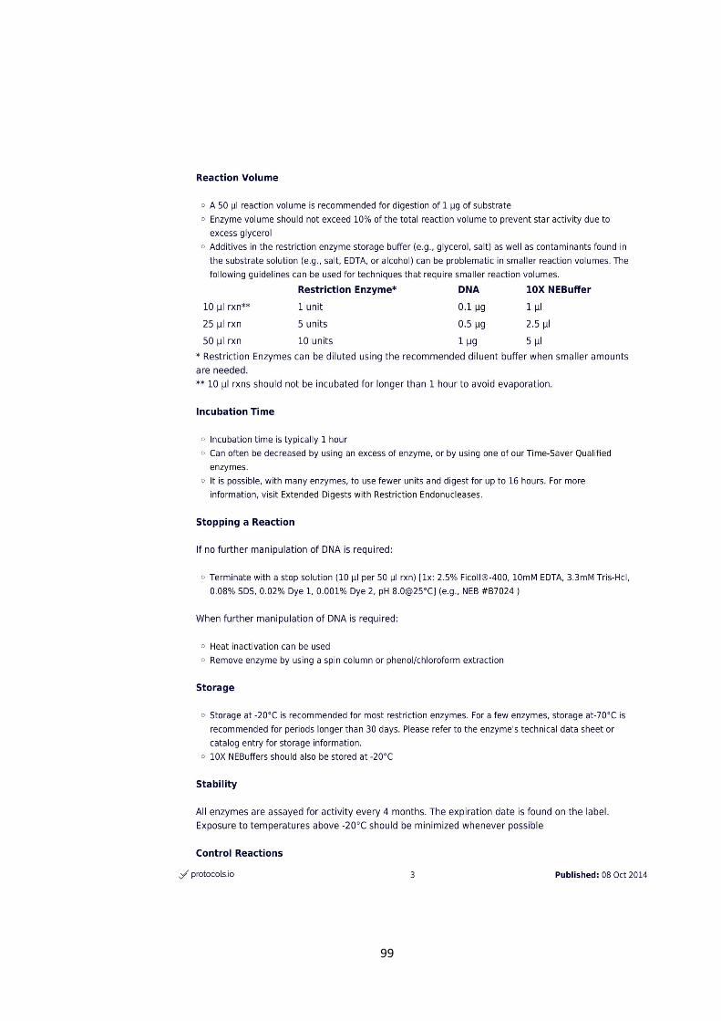
99
100
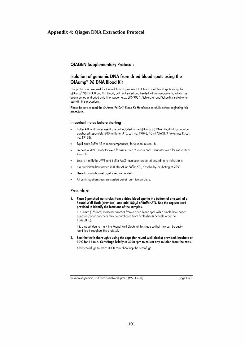
101
Appendix 4: Qiagen DNA Extraction Protocol
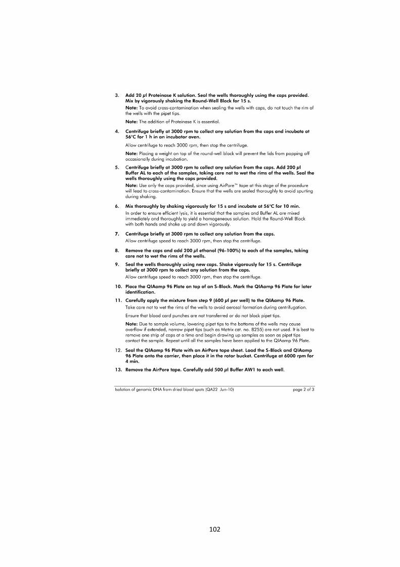
102
103