Embed Size (px)
Citation preview
An introduction to neurologyfor ages 8-13
Reproducible student booklet
Creative and mulitsensory activities for all types of learners
Internet-linked features
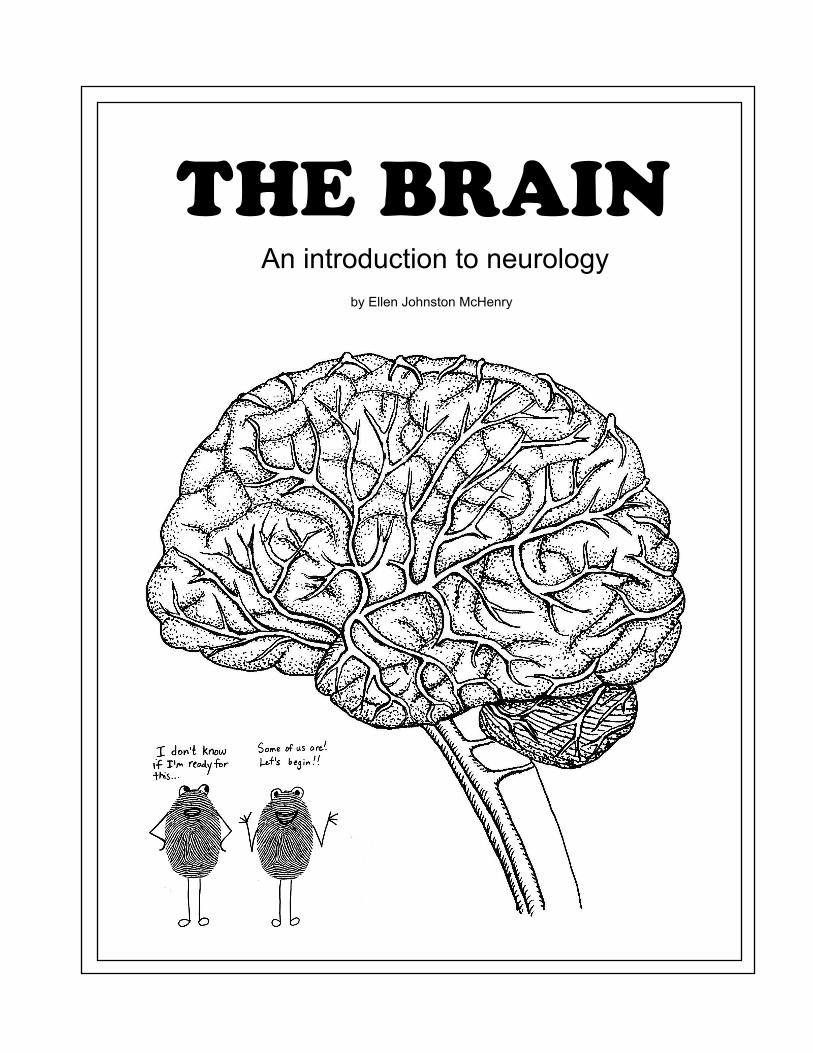
by Ellen Johnston McHenry
THE BRAINAn introduction to neurology
Copyright 2007Ellen Johnston McHenry
All rights reserved.The author gives permission for this student booklet to be photocopied for use with a
homeschool or with a single classroom. If you would like to use any part of this in your own publication, contact me at 532 Pike St., State College, PA 16801.
PLEASE NOTE:I read numerous books and web pages about the brain in preparation for writing this
curriculum, and have tried to present the most current information available. However, the science of neurology is growing and expanding rapidly. I encourage the reader not to rely on this booklet alone, but to use other sources of information, also.
This booklet was updated (and the Internet links checked) in February, 2011.
Nothing in this booklet should be used to diagnose any illness or syndrome.
Questions or comments about the curriculum may be directed to:[email protected]
REPRODUCIBLE
STUDENT BOOKLET
This booklet has all the basic information the students need to know, plus some activities they can do on their own. It is designed for use with multiple ages and/or abilities. You may use just the basic chapters, or you may add the extra “.5” chapters. (You can even pick and choose which “.5” chapters to add.) You can have some students doing one option, and some the other, because the supplemental, in-class activities are valid for either option. If you are using this curriculum in a school or co-op setting, we recommend assigning the student booklet as “homework” for the student to read before coming to class, and using classroom time to do the activities in the teacher’s section. See the “Additional Resources” page for additional reading material that can be used with students who want to know even more.
PLEASE NOTE: Some of the activites in the student booklet are Internet-linked. All websites have been screened ahead of time for suitability. However, we still advise in the instructions to the student that adult supervision is needed for using the Internet.
Here is a list of the topics each chapter addresses. The “.5” chapters have the same titles, with the addition of “More About...,” (except for chapter 10.5).
CHAPTER 1: A Very Brief History of Brain ResearchCHAPTER 2: Basic Brain AnatomyCHAPTER 3: Left Brain/Right BrainCHAPTER 4: Deep Inside the BrainCHAPTER 5: Brain CellsCHAPTER 6: Networks of NeuronsCHAPTER 7: Learning and MemoryCHAPTER 8: How the Brain Connects to the BodyCHAPTER 9: SleepCHAPTER 10: Brain DoctorsCHAPTER 10.5: Brain Problems
ANSWER KEY: You may choose whether or not to include this in your students’ booklets
ABOUT PRINTING AND BINDING: The easiest and least expensive way to assemble the student booklet is to three-hole punch the copied pages and put them into an inexpensive (paper cover) binder. You will find that the booklet is too thick for regular stapling, but heavy-duty staples might work. Please note that there are two options for the cover design. If expense is not an issue, comb-binding is very nice. For stapling or comb-binding, make sure to copy the cover onto heavy card stock, and add a blank sheet of card stock at the end as a back cover.

An introduction to neurology
by Ellen Johnston McHenry
An introduction to neurology
Copyright 2007Ellen Johnston McHenry
All rights reserved.The author gives permission for this student booklet to be photocopied for use with a
homeschool or with a single classroom. If you would like to use any part of this in your own publication, contact me at 532 Pike St., State College, PA 16801.
PLEASE NOTE:The science of neurology is growing and expanding rapidly. I read numerous books and web pages about the brain in preparation for writing this curriculum, and have tried to present the most current information available as of the time of publication. However, it is possible that by the time you use
this curriculum, new discoveries will have been made.
(Concerning the Internet links: If you find that some of the Internet links are no longer valid, try using key words to search for similar resources.)
NOTE: Nothing in this booklet should be used to diagnose any illness or syndrome.
CHAPTER 1
A VERY BRIEF HISTORY OF BRAIN RESEARCH
Even before reading this book, you already know more about the brain than the doctors and scientists of the ancient world did, up until about 300 B.C. If you’ve read any books about the brain, you probably know more about the brain that anyone in the Medieval world did. Brain research did not really begin until the late 1800s, and progressed slowly until the inven-tion of scanning devices in the late 1900s. Even today, in the 2000s, with all our advanced medical technology, there is a lot we don’t know about the brain.
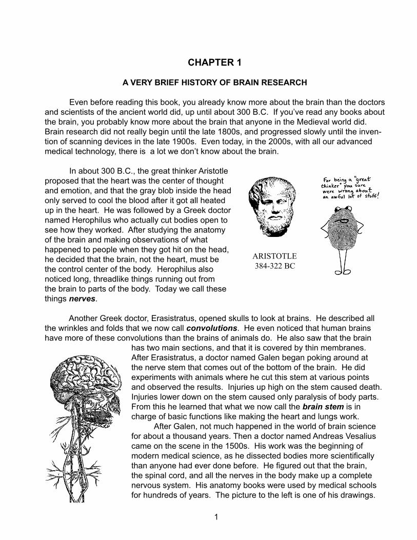
In about 300 B.C., the great thinker Aristotle proposed that the heart was the center of thought and emotion, and that the gray blob inside the head only served to cool the blood after it got all heated up in the heart. He was followed by a Greek doctor named Herophilus who actually cut bodies open to see how they worked. After studying the anatomy of the brain and making observations of what happened to people when they got hit on the head, he decided that the brain, not the heart, must be the control center of the body. Herophilus also noticed long, threadlike things running out from the brain to parts of the body. Today we call these things nerves.
Another Greek doctor, Erasistratus, opened skulls to look at brains. He described all the wrinkles and folds that we now call convolutions. He even noticed that human brains have more of these convolutions than the brains of animals do. He also saw that the brain
has two main sections, and that it is covered by thin membranes. After Erasistratus, a doctor named Galen began poking around at the nerve stem that comes out of the bottom of the brain. He did experiments with animals where he cut this stem at various points and observed the results. Injuries up high on the stem caused death. Injuries lower down on the stem caused only paralysis of body parts. From this he learned that what we now call the brain stem is in charge of basic functions like making the heart and lungs work. After Galen, not much happened in the world of brain science for about a thousand years. Then a doctor named Andreas Vesalius came on the scene in the 1500s. His work was the beginning of modern medical science, as he dissected bodies more scientifically than anyone had ever done before. He figured out that the brain, the spinal cord, and all the nerves in the body make up a complete nervous system. His anatomy books were used by medical schools for hundreds of years. The picture to the left is one of his drawings.
ARISTOTLE 384-322 BC
1
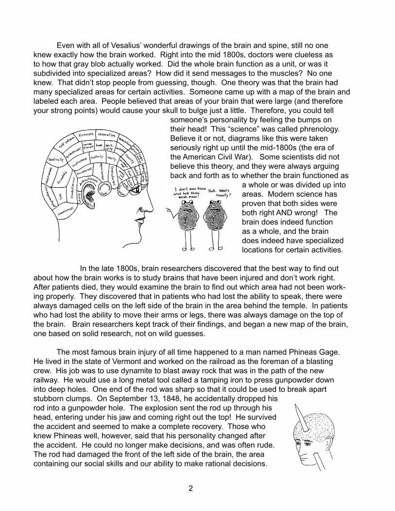
Even with all of Vesalius’ wonderful drawings of the brain and spine, still no one knew exactly how the brain worked. Right into the mid 1800s, doctors were clueless as to how that gray blob actually worked. Did the whole brain function as a unit, or was it subdivided into specialized areas? How did it send messages to the muscles? No one knew. That didn’t stop people from guessing, though. One theory was that the brain had many specialized areas for certain activities. Someone came up with a map of the brain and labeled each area. People believed that areas of your brain that were large (and therefore your strong points) would cause your skull to bulge just a little. Therefore, you could tell
someone’s personality by feeling the bumps on their head! This “science” was called phrenology. Believe it or not, diagrams like this were taken seriously right up until the mid-1800s (the era of the American Civil War). Some scientists did not believe this theory, and they were always arguing back and forth as to whether the brain functioned as
a whole or was divided up into areas. Modern science has proven that both sides were both right AND wrong! The brain does indeed function as a whole, and the brain does indeed have specialized locations for certain activities.
In the late 1800s, brain researchers discovered that the best way to find out about how the brain works is to study brains that have been injured and don’t work right. After patients died, they would examine the brain to find out which area had not been work-ing properly. They discovered that in patients who had lost the ability to speak, there were always damaged cells on the left side of the brain in the area behind the temple. In patients who had lost the ability to move their arms or legs, there was always damage on the top of the brain. Brain researchers kept track of their findings, and began a new map of the brain, one based on solid research, not on wild guesses.
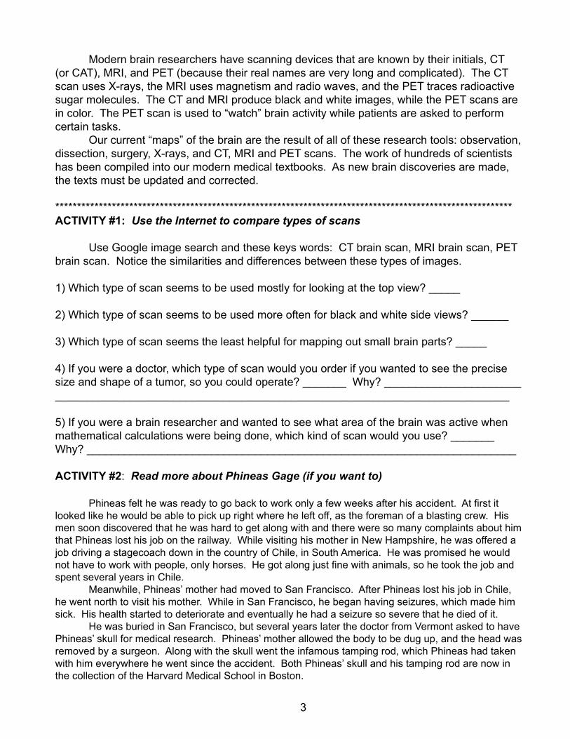
The most famous brain injury of all time happened to a man named Phineas Gage. He lived in the state of Vermont and worked on the railroad as the foreman of a blasting crew. His job was to use dynamite to blast away rock that was in the path of the new railway. He would use a long metal tool called a tamping iron to press gunpowder down into deep holes. One end of the rod was sharp so that it could be used to break apart stubborn clumps. On September 13, 1848, he accidentally dropped his rod into a gunpowder hole. The explosion sent the rod up through his head, entering under his jaw and coming right out the top! He survived the accident and seemed to make a complete recovery. Those who knew Phineas well, however, said that his personality changed after the accident. He could no longer make decisions, and was often rude. The rod had damaged the front of the left side of the brain, the area containing our social skills and our ability to make rational decisions.
2
Modern brain researchers have scanning devices that are known by their initials, CT (or CAT), MRI, and PET (because their real names are very long and complicated). The CT scan uses X-rays, the MRI uses magnetism and radio waves, and the PET traces radioactive sugar molecules. The CT and MRI produce black and white images, while the PET scans are in color. The PET scan is used to “watch” brain activity while patients are asked to perform certain tasks. Our current “maps” of the brain are the result of all of these research tools: observation, dissection, surgery, X-rays, and CT, MRI and PET scans. The work of hundreds of scientists has been compiled into our modern medical textbooks. As new brain discoveries are made, the texts must be updated and corrected.
*********************************************************************************************************ACTIVITY #1: Use the Internet to compare types of scans
Use Google image search and these keys words: CT brain scan, MRI brain scan, PET brain scan. Notice the similarities and differences between these types of images.
1) Which type of scan seems to be used mostly for looking at the top view? _____
2) Which type of scan seems to be used more often for black and white side views? ______
3) Which type of scan seems the least helpful for mapping out small brain parts? _____
4) If you were a doctor, which type of scan would you order if you wanted to see the precise size and shape of a tumor, so you could operate? _______ Why? _______________________________________________________________________________________________
5) If you were a brain researcher and wanted to see what area of the brain was active when mathematical calculations were being done, which kind of scan would you use? _______ Why? _____________________________________________________________________
ACTIVITY #2: Read more about Phineas Gage (if you want to)
Phineas felt he was ready to go back to work only a few weeks after his accident. At first it looked like he would be able to pick up right where he left off, as the foreman of a blasting crew. His men soon discovered that he was hard to get along with and there were so many complaints about him that Phineas lost his job on the railway. While visiting his mother in New Hampshire, he was offered a job driving a stagecoach down in the country of Chile, in South America. He was promised he would not have to work with people, only horses. He got along just fine with animals, so he took the job and spent several years in Chile. Meanwhile, Phineas’ mother had moved to San Francisco. After Phineas lost his job in Chile, he went north to visit his mother. While in San Francisco, he began having seizures, which made him sick. His health started to deteriorate and eventually he had a seizure so severe that he died of it. He was buried in San Francisco, but several years later the doctor from Vermont asked to have Phineas’ skull for medical research. Phineas’ mother allowed the body to be dug up, and the head was removed by a surgeon. Along with the skull went the infamous tamping rod, which Phineas had taken with him everywhere he went since the accident. Both Phineas’ skull and his tamping rod are now in the collection of the Harvard Medical School in Boston.
3
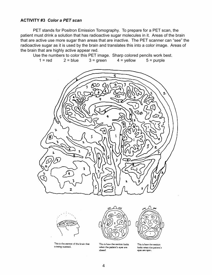
ACTIVITY #3 Color a PET scan
PET stands for Positron Emission Tomography. To prepare for a PET scan, the patient must drink a solution that has radioactive sugar molecules in it. Areas of the brain that are active use more sugar than areas that are inactive. The PET scanner can “see” the radioactive sugar as it is used by the brain and translates this into a color image. Areas of the brain that are highly active appear red. Use the numbers to color this PET image. Sharp colored pencils work best. 1 = red 2 = blue 3 = green 4 = yellow 5 = purple
4
CHAPTER 1.5
MORE ABOUT BRAIN RESEARCH
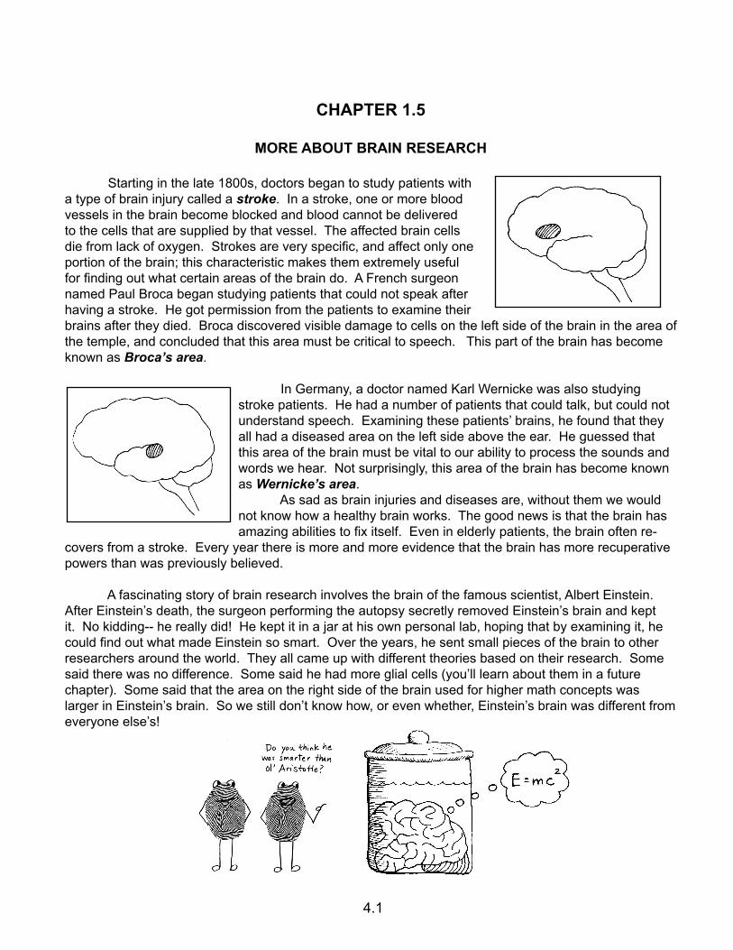
Starting in the late 1800s, doctors began to study patients with a type of brain injury called a stroke. In a stroke, one or more blood vessels in the brain become blocked and blood cannot be delivered to the cells that are supplied by that vessel. The affected brain cells die from lack of oxygen. Strokes are very specific, and affect only one portion of the brain; this characteristic makes them extremely useful for finding out what certain areas of the brain do. A French surgeon named Paul Broca began studying patients that could not speak after having a stroke. He got permission from the patients to examine their brains after they died. Broca discovered visible damage to cells on the left side of the brain in the area of the temple, and concluded that this area must be critical to speech. This part of the brain has become known as Broca’s area.
In Germany, a doctor named Karl Wernicke was also studying stroke patients. He had a number of patients that could talk, but could not understand speech. Examining these patients’ brains, he found that they all had a diseased area on the left side above the ear. He guessed that this area of the brain must be vital to our ability to process the sounds and words we hear. Not surprisingly, this area of the brain has become known as Wernicke’s area. As sad as brain injuries and diseases are, without them we would not know how a healthy brain works. The good news is that the brain has amazing abilities to fix itself. Even in elderly patients, the brain often re-
covers from a stroke. Every year there is more and more evidence that the brain has more recuperative powers than was previously believed.
A fascinating story of brain research involves the brain of the famous scientist, Albert Einstein. After Einstein’s death, the surgeon performing the autopsy secretly removed Einstein’s brain and kept it. No kidding-- he really did! He kept it in a jar at his own personal lab, hoping that by examining it, he could find out what made Einstein so smart. Over the years, he sent small pieces of the brain to other researchers around the world. They all came up with different theories based on their research. Some said there was no difference. Some said he had more glial cells (you’ll learn about them in a future chapter). Some said that the area on the right side of the brain used for higher math concepts was larger in Einstein’s brain. So we still don’t know how, or even whether, Einstein’s brain was different from everyone else’s!
4.1
ACTIVITY #1 Find out more about MRI and PET
Use Google search, or go to howstuffworks.com, to find out more about how MRI and PET scans work. Answer the following questions about each.
MRI:
1) What does MRI stand for? _________________________________________________ 2) How big is an MRI machine? _________________________________________________ 3) Does it really have a big magnet in it? ________ 4) Besides scanning the brain, what else is MRI good for? __________________________________ _________________________________________________________________________________ 5) Briefly describe how an MRI machine produces an image: _______________________________ _________________________________________________________________________________ _________________________________________________________________________________ ___________________________________________________________________________________________________________________________________________________________________________________________________________________________________________________ _________________________________________________________________________________
PET:
1) What does PET stand for? _________________________________________________2) What must a person receive before getting a PET scan? ____________________________ 3) What does the PET scan “see”? _____________________________________________________ 4) Why is the PET scan in color and what do the colors represent? ____________________________ _________________________________________________________________________________ _________________________________________________________________________________ 5) Besides brain imaging, what else is PET used for? ______________________________________ _________________________________________________________________________________
ACTIVITY #2 Find out more about Einstein’s brain
If you find Einstein’s brain a fascinating subject, there is more you can learn via the Internet. You can also see actual pictures of his brain, as well as a picture of the doctor who stole it. Just use Google image search for “Einstein’s brain.” (As always, make sure you have adult permission to access You-Tube and/or the Internet!)
ACTIVITY #3 Watch two short videos about Phineas Gage
Here are two very good videos about Phineas Gage. This first one features drawings done by kids and has excellent narration. It’s about 6 1/2 minutes long. http://www.youtube.com/watch?v=X4fGlny5cPg This second video takes you to Harvard Medical School where Phineas’ skull (yes, the actual skull) and the metal rod are on display. (Don’t worry, it’s not gross at all.) The interviewer, Alan Alda, poses questions to a brain scientist about which areas of Phineas’ brain were affected and how this helped scientists to learn about the funtion of the frontal lobe. The video is slightly under 6 minutes. http://www.youtube.com/watch?v=yXiM-nDYzX0&feature=related
4.2
CHAPTER 2
BASIC BRAIN ANATOMY
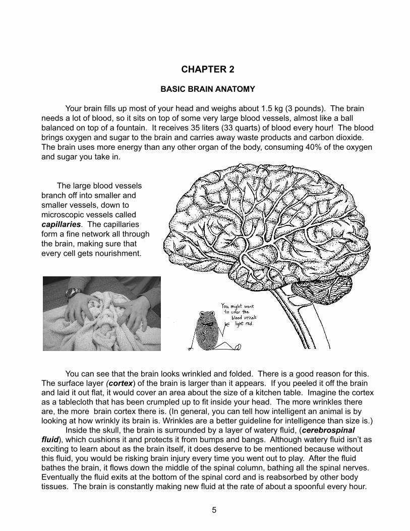
Your brain fills up most of your head and weighs about 1.5 kg (3 pounds). The brain needs a lot of blood, so it sits on top of some very large blood vessels, almost like a ball balanced on top of a fountain. It receives 35 liters (33 quarts) of blood every hour! The blood brings oxygen and sugar to the brain and carries away waste products and carbon dioxide. The brain uses more energy than any other organ of the body, consuming 40% of the oxygen and sugar you take in.
The large blood vessels branch off into smaller and smaller vessels, down to microscopic vessels called capillaries. The capillaries form a fine network all through the brain, making sure that every cell gets nourishment.
You can see that the brain looks wrinkled and folded. There is a good reason for this. The surface layer (cortex) of the brain is larger than it appears. If you peeled it off the brain and laid it out flat, it would cover an area about the size of a kitchen table. Imagine the cortex as a tablecloth that has been crumpled up to fit inside your head. The more wrinkles there are, the more brain cortex there is. (In general, you can tell how intelligent an animal is by looking at how wrinkly its brain is. Wrinkles are a better guideline for intelligence than size is.) Inside the skull, the brain is surrounded by a layer of watery fluid, (cerebrospinal fluid), which cushions it and protects it from bumps and bangs. Although watery fluid isn’t as exciting to learn about as the brain itself, it does deserve to be mentioned because without this fluid, you would be risking brain injury every time you went out to play. After the fluid bathes the brain, it flows down the middle of the spinal column, bathing all the spinal nerves. Eventually the fluid exits at the bottom of the spinal cord and is reabsorbed by other body tissues. The brain is constantly making new fluid at the rate of about a spoonful every hour.
5
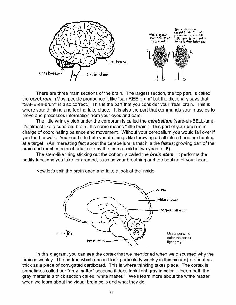
There are three main sections of the brain. The largest section, the top part, is called the cerebrum. (Most people pronounce it like “sah-REE-brum” but the dictionary says that “SARE-eh-brum” is also correct.) This is the part that you consider your “real” brain. This is where your thinking and feeling take place. It is also the part that commands your muscles to move and processes information from your eyes and ears. The little wrinkly blob under the cerebrum is called the cerebellum (sare-eh-BELL-um). It’s almost like a separate brain. It’s name means “little brain.” This part of your brain is in charge of coordinating balance and movement. Without your cerebellum you would fall over if you tried to walk. You need it to help you do things like throwing a ball into a hoop or shooting at a target. (An interesting fact about the cerebellum is that it is the fastest growing part of the brain and reaches almost adult size by the time a child is two years old!) The stem-like thing sticking out the bottom is called the brain stem. It performs the bodily functions you take for granted, such as your breathing and the beating of your heart.
Now let’s split the brain open and take a look at the inside.
In this diagram, you can see the cortex that we mentioned when we discussed why the brain is wrinkly. The cortex (which doesn’t look particularly wrinkly in this picture) is about as thick as a piece of corrugated cardboard. This is where thinking takes place. The cortex is sometimes called our “gray matter” because it does look light gray in color. Underneath the gray matter is a thick section called “white matter.” We’ll learn more about the white matter when we learn about individual brain cells and what they do.
Use a pencil to color the cortex light gray.
6
The sideways C-shaped thing in the middle, the corpus callosum, is a connecting bridge between the right and left sides of the brain. If you look at the outside of the brain you can see that there is a split down the middle. The cerebrum is in two halves, connected to each other by the corpus callosum. It is believed that the thicker the corpus callosum, the better the connection between the two hemispheres.
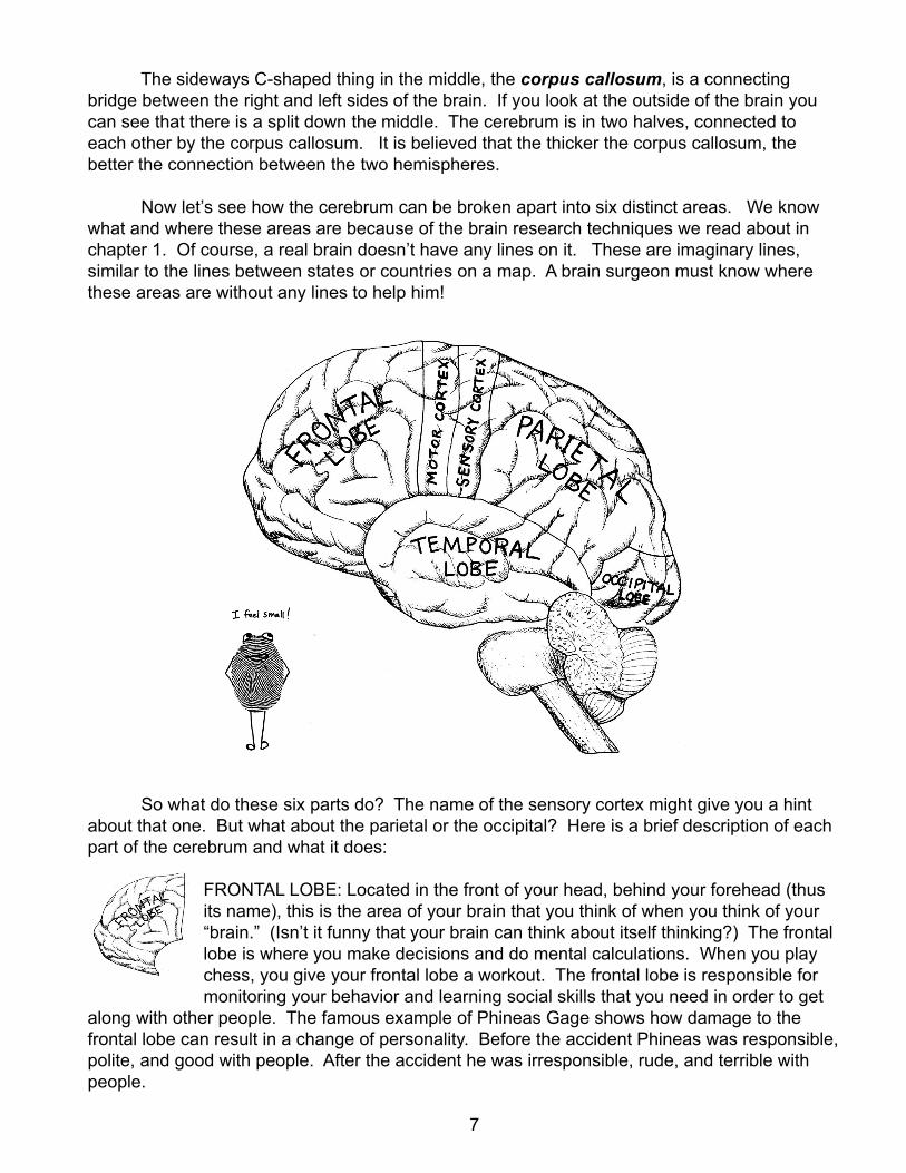
Now let’s see how the cerebrum can be broken apart into six distinct areas. We know what and where these areas are because of the brain research techniques we read about in chapter 1. Of course, a real brain doesn’t have any lines on it. These are imaginary lines, similar to the lines between states or countries on a map. A brain surgeon must know where these areas are without any lines to help him!
So what do these six parts do? The name of the sensory cortex might give you a hint about that one. But what about the parietal or the occipital? Here is a brief description of each part of the cerebrum and what it does:
FRONTAL LOBE: Located in the front of your head, behind your forehead (thus its name), this is the area of your brain that you think of when you think of your “brain.” (Isn’t it funny that your brain can think about itself thinking?) The frontal lobe is where you make decisions and do mental calculations. When you play chess, you give your frontal lobe a workout. The frontal lobe is responsible for monitoring your behavior and learning social skills that you need in order to get
along with other people. The famous example of Phineas Gage shows how damage to the frontal lobe can result in a change of personality. Before the accident Phineas was responsible, polite, and good with people. After the accident he was irresponsible, rude, and terrible with people.
7
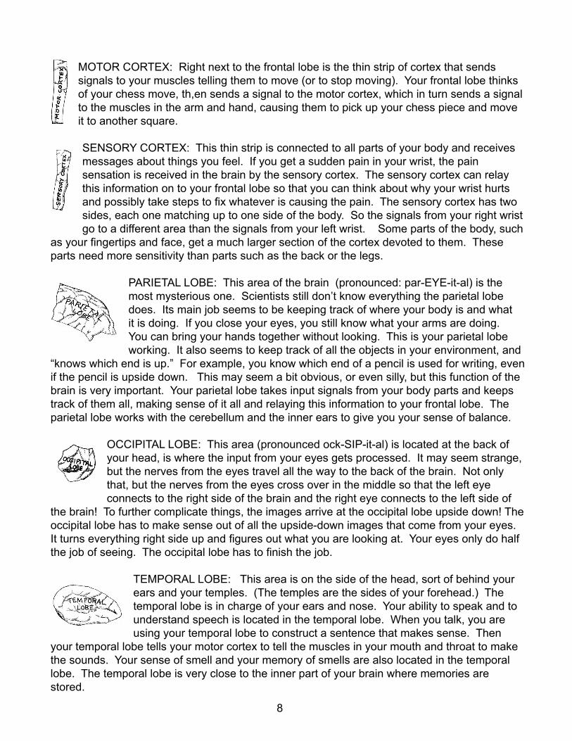
MOTOR CORTEX: Right next to the frontal lobe is the thin strip of cortex that sends signals to your muscles telling them to move (or to stop moving). Your frontal lobe thinks of your chess move, th,en sends a signal to the motor cortex, which in turn sends a signal to the muscles in the arm and hand, causing them to pick up your chess piece and move it to another square.
SENSORY CORTEX: This thin strip is connected to all parts of your body and receives messages about things you feel. If you get a sudden pain in your wrist, the pain sensation is received in the brain by the sensory cortex. The sensory cortex can relay this information on to your frontal lobe so that you can think about why your wrist hurts and possibly take steps to fix whatever is causing the pain. The sensory cortex has two sides, each one matching up to one side of the body. So the signals from your right wrist go to a different area than the signals from your left wrist. Some parts of the body, such
as your fingertips and face, get a much larger section of the cortex devoted to them. These parts need more sensitivity than parts such as the back or the legs.
PARIETAL LOBE: This area of the brain (pronounced: par-EYE-it-al) is the most mysterious one. Scientists still don’t know everything the parietal lobe does. Its main job seems to be keeping track of where your body is and what it is doing. If you close your eyes, you still know what your arms are doing. You can bring your hands together without looking. This is your parietal lobe working. It also seems to keep track of all the objects in your environment, and
“knows which end is up.” For example, you know which end of a pencil is used for writing, even if the pencil is upside down. This may seem a bit obvious, or even silly, but this function of the brain is very important. Your parietal lobe takes input signals from your body parts and keeps track of them all, making sense of it all and relaying this information to your frontal lobe. The parietal lobe works with the cerebellum and the inner ears to give you your sense of balance.
OCCIPITAL LOBE: This area (pronounced ock-SIP-it-al) is located at the back of your head, is where the input from your eyes gets processed. It may seem strange, but the nerves from the eyes travel all the way to the back of the brain. Not only that, but the nerves from the eyes cross over in the middle so that the left eye connects to the right side of the brain and the right eye connects to the left side of
the brain! To further complicate things, the images arrive at the occipital lobe upside down! The occipital lobe has to make sense out of all the upside-down images that come from your eyes. It turns everything right side up and figures out what you are looking at. Your eyes only do half the job of seeing. The occipital lobe has to finish the job.
TEMPORAL LOBE: This area is on the side of the head, sort of behind your ears and your temples. (The temples are the sides of your forehead.) The temporal lobe is in charge of your ears and nose. Your ability to speak and to understand speech is located in the temporal lobe. When you talk, you are using your temporal lobe to construct a sentence that makes sense. Then
your temporal lobe tells your motor cortex to tell the muscles in your mouth and throat to make the sounds. Your sense of smell and your memory of smells are also located in the temporal lobe. The temporal lobe is very close to the inner part of your brain where memories are stored.
8
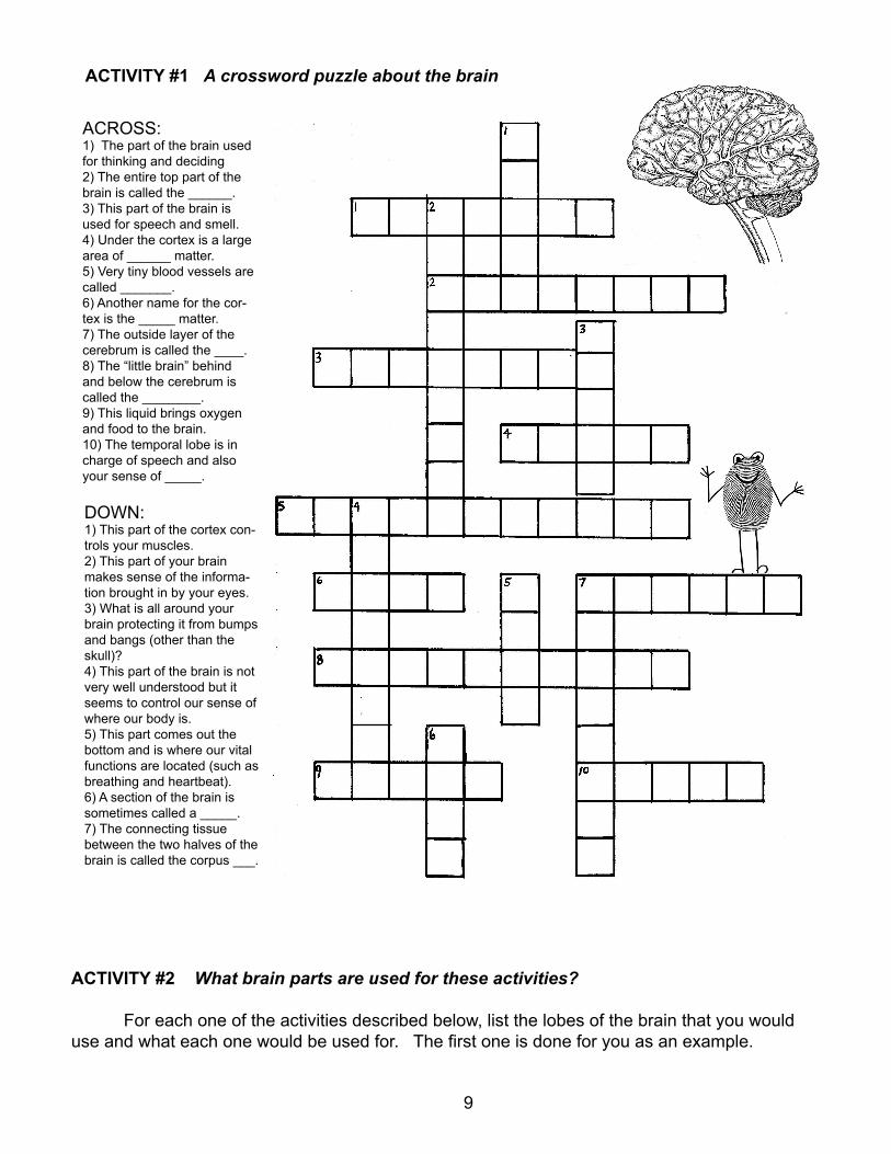
ACTIVITY #1 A crossword puzzle about the brain
ACTIVITY #2 What brain parts are used for these activities?
For each one of the activities described below, list the lobes of the brain that you would use and what each one would be used for. The first one is done for you as an example.
ACROSS:1) The part of the brain used for thinking and deciding2) The entire top part of the brain is called the ______.3) This part of the brain is used for speech and smell.4) Under the cortex is a large area of ______ matter.5) Very tiny blood vessels are called _______.6) Another name for the cor-tex is the _____ matter.7) The outside layer of the cerebrum is called the ____.8) The “little brain” behind and below the cerebrum is called the ________.9) This liquid brings oxygen and food to the brain.10) The temporal lobe is in charge of speech and also your sense of _____.
DOWN:1) This part of the cortex con-trols your muscles.2) This part of your brain makes sense of the informa-tion brought in by your eyes.3) What is all around your brain protecting it from bumps and bangs (other than the skull)?4) This part of the brain is not very well understood but it seems to control our sense of where our body is.5) This part comes out the bottom and is where our vital functions are located (such as breathing and heartbeat).6) A section of the brain is sometimes called a _____.7) The connecting tissue between the two halves of the brain is called the corpus ___.
9
1) Brushing your teeth: FRONTAL LOBE: decides to brush teeth and sends signals to motor cortexMOTOR CORTEX: tells muscles to move toothbrush around in the mouthSENSORY CORTEX: Feels the brush in your hand, feels the scrubbing on your gums and teethPARIETAL LOBE: Senses that the hands are raised to the mouth, also keeps you balancedOCCIPITAL LOBE: Makes sense of what your eyes are seeing in the mirrorTEMPORAL LOBE: Smells the toothpaste
2) Playing the pianoFRONTAL LOBE: ______________________________________________________________________________ MOTOR CORTEX: _____________________________________________________________________________ SENSORY CORTEX: __________________________________________________________________________ PARIETAL LOBE: ______________________________________________________________________________ OCCIPITAL LOBE: _____________________________________________________________________________ TEMPORAL LOBE: _____________________________________________________________________________
3) Riding a bicycleFRONTAL LOBE: ______________________________________________________________________________ MOTOR CORTEX: _____________________________________________________________________________ SENSORY CORTEX: __________________________________________________________________________ PARIETAL LOBE: ______________________________________________________________________________ OCCIPITAL LOBE: _____________________________________________________________________________ TEMPORAL LOBE: _____________________________________________________________________________
4) Talking on the telephoneFRONTAL LOBE: ______________________________________________________________________________ MOTOR CORTEX: _____________________________________________________________________________ SENSORY CORTEX: __________________________________________________________________________ PARIETAL LOBE: ______________________________________________________________________________ OCCIPITAL LOBE: _____________________________________________________________________________ TEMPORAL LOBE: _____________________________________________________________________________
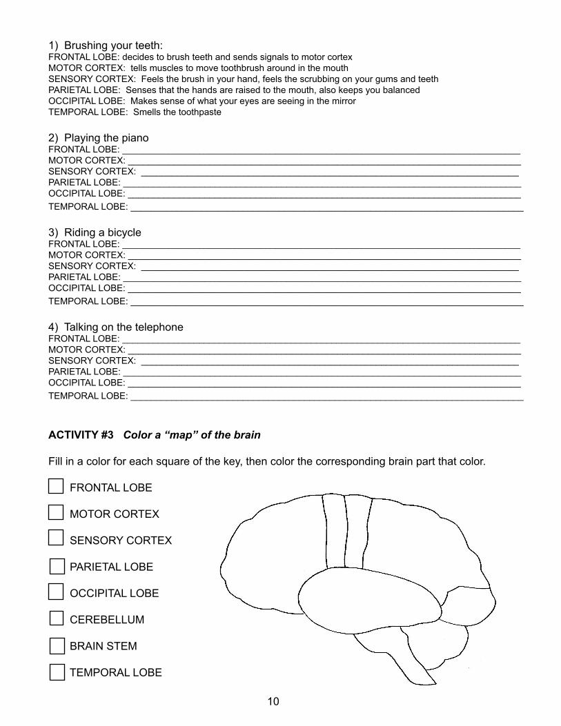
ACTIVITY #3 Color a “map” of the brain
Fill in a color for each square of the key, then color the corresponding brain part that color.
FRONTAL LOBE
MOTOR CORTEX
SENSORY CORTEX
PARIETAL LOBE
OCCIPITAL LOBE
CEREBELLUM
BRAIN STEM
TEMPORAL LOBE
10
CHAPTER 2.5
MORE ABOUT BASIC BRAIN ANATOMY
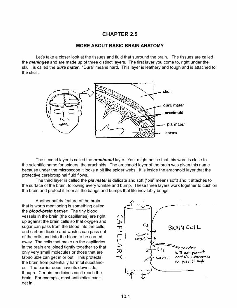
Let’s take a closer look at the tissues and fluid that surround the brain. The tissues are called the meninges and are made up of three distinct layers. The first layer you come to, right under the skull, is called the dura mater. “Dura” means hard. This layer is leathery and tough and is attached to the skull.
The second layer is called the arachnoid layer. You might notice that this word is close to the scientific name for spiders: the arachnids. The arachnoid layer of the brain was given this name because under the microscope it looks a bit like spider webs. It is inside the arachnoid layer that the protective cerebrospinal fluid flows. The third layer is called the pia mater is delicate and soft (“pia” means soft) and it attaches to the surface of the brain, following every wrinkle and bump. These three layers work together to cushion the brain and protect if from all the bangs and bumps that life inevitably brings.
Another safety feature of the brain that is worth mentioning is something called the blood-brain barrier. The tiny blood vessels in the brain (the capillaries) are right up against the brain cells so that oxygen and sugar can pass from the blood into the cells, and carbon dioxide and wastes can pass out of the cells and into the blood to be carried away. The cells that make up the capillaries in the brain are joined tightly together so that only very small molecules or those that are fat-soluble can get in or out. This protects the brain from potentially harmful substanc-es. The barrier does have its downside, though. Certain medicines can’t reach the brain. For example, most antibiotics can’t get in.
10.1
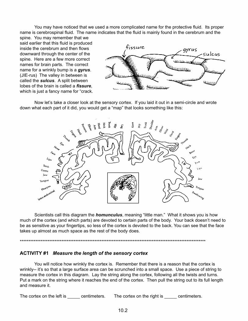
You may have noticed that we used a more complicated name for the protective fluid. Its proper name is cerebrospinal fluid. The name indicates that the fluid is mainly found in the cerebrum and the spine. You may remember that we said earlier that this fluid is produced inside the cerebrum and then flows downward through the center of the spine. Here are a few more correct names for brain parts. The correct name for a wrinkly bump is a gyrus. (JIE-rus) The valley in between is called the sulcus. A split between lobes of the brain is called a fissure, which is just a fancy name for “crack.
Now let’s take a closer look at the sensory cortex. If you laid it out in a semi-circle and wrote down what each part of it did, you would get a “map” that looks something like this:
Scientists call this diagram the homunculus, meaning “little man.” What it shows you is how much of the cortex (and which parts) are devoted to certain parts of the body. Your back doesn’t need to be as sensitive as your fingertips, so less of the cortex is devoted to the back. You can see that the face takes up almost as much space as the rest of the body does.
***********************************************************************************************************
ACTIVITY #1 Measure the length of the sensory cortex
You will notice how wrinkly the cortex is. Remember that there is a reason that the cortex is wrinkly-- it’s so that a large surface area can be scrunched into a small space. Use a piece of string to measure the cortex in this diagram. Lay the string along the cortex, following all the twists and turns. Put a mark on the string where it reaches the end of the cortex. Then pull the string out to its full length and measure it.
The cortex on the left is _____ centimeters. The cortex on the right is _____ centimeters.
10.2
ACTIVITY #2 Read more about the blood-brain barrier
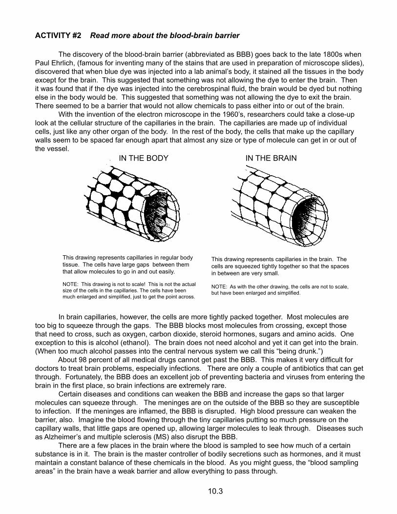
The discovery of the blood-brain barrier (abbreviated as BBB) goes back to the late 1800s when Paul Ehrlich, (famous for inventing many of the stains that are used in preparation of microscope slides), discovered that when blue dye was injected into a lab animal’s body, it stained all the tissues in the body except for the brain. This suggested that something was not allowing the dye to enter the brain. Then it was found that if the dye was injected into the cerebrospinal fluid, the brain would be dyed but nothing else in the body would be. This suggested that something was not allowing the dye to exit the brain. There seemed to be a barrier that would not allow chemicals to pass either into or out of the brain. With the invention of the electron microscope in the 1960’s, researchers could take a close-up look at the cellular structure of the capillaries in the brain. The capillaries are made up of individual cells, just like any other organ of the body. In the rest of the body, the cells that make up the capillary walls seem to be spaced far enough apart that almost any size or type of molecule can get in or out of the vessel.
In brain capillaries, however, the cells are more tightly packed together. Most molecules are too big to squeeze through the gaps. The BBB blocks most molecules from crossing, except those that need to cross, such as oxygen, carbon dioxide, steroid hormones, sugars and amino acids. One exception to this is alcohol (ethanol). The brain does not need alcohol and yet it can get into the brain. (When too much alcohol passes into the central nervous system we call this “being drunk.”) About 98 percent of all medical drugs cannot get past the BBB. This makes it very difficult for doctors to treat brain problems, especially infections. There are only a couple of antibiotics that can get through. Fortunately, the BBB does an excellent job of preventing bacteria and viruses from entering the brain in the first place, so brain infections are extremely rare. Certain diseases and conditions can weaken the BBB and increase the gaps so that larger molecules can squeeze through. The meninges are on the outside of the BBB so they are susceptible to infection. If the meninges are inflamed, the BBB is disrupted. High blood pressure can weaken the barrier, also. Imagine the blood flowing through the tiny capillaries putting so much pressure on the capillary walls, that little gaps are opened up, allowing larger molecules to leak through. Diseases such as Alzheimer’s and multiple sclerosis (MS) also disrupt the BBB. There are a few places in the brain where the blood is sampled to see how much of a certain substance is in it. The brain is the master controller of bodily secretions such as hormones, and it must maintain a constant balance of these chemicals in the blood. As you might guess, the “blood sampling areas” in the brain have a weak barrier and allow everything to pass through.
This drawing represents capillaries in regular body tissue. The cells have large gaps between them that allow molecules to go in and out easily.
NOTE: This drawing is not to scale! This is not the actual size of the cells in the capillaries. The cells have been much enlarged and simplified, just to get the point across.
IN THE BODY IN THE BRAIN
This drawing represents capillaries in the brain. The cells are squeezed tightly together so that the spaces in between are very small.
NOTE: As with the other drawing, the cells are not to scale, but have been enlarged and simplified.
10.3
ACTIVITY #3 Look at animal brains and compare them to the human brain
Check out brainmuseum.org. Click on “brain sections” in the menu bar on the left. Then scroll down and click on the name of any animal to see a picture (photograph) of its brain. The charts will show you different views of the brain: left, right, bottom, top. How do the sizes of the cerebrums compare to the total brain size? Remember that the cerebrum is where thinking takes place. Also, remember that wrinkles indicate a large surface area crunched into a small space. No wrinkles means less surface area, and, therefore, less thinking power. How does a rabbit brain compare to a pig? Which one would be capable of learning tricks? Compare the manatee brain with the dolphin brain. Both are sea mammals, but which one is smarter? Can you spot any differences between a human brain and a chimp brain? Notice in many of the animal brains that there is a large bulbous part coming out in front of the frontal lobe. These are sensory areas, likely connected to the nose. How much of the lion’s brain is devoted to the sense of smell?
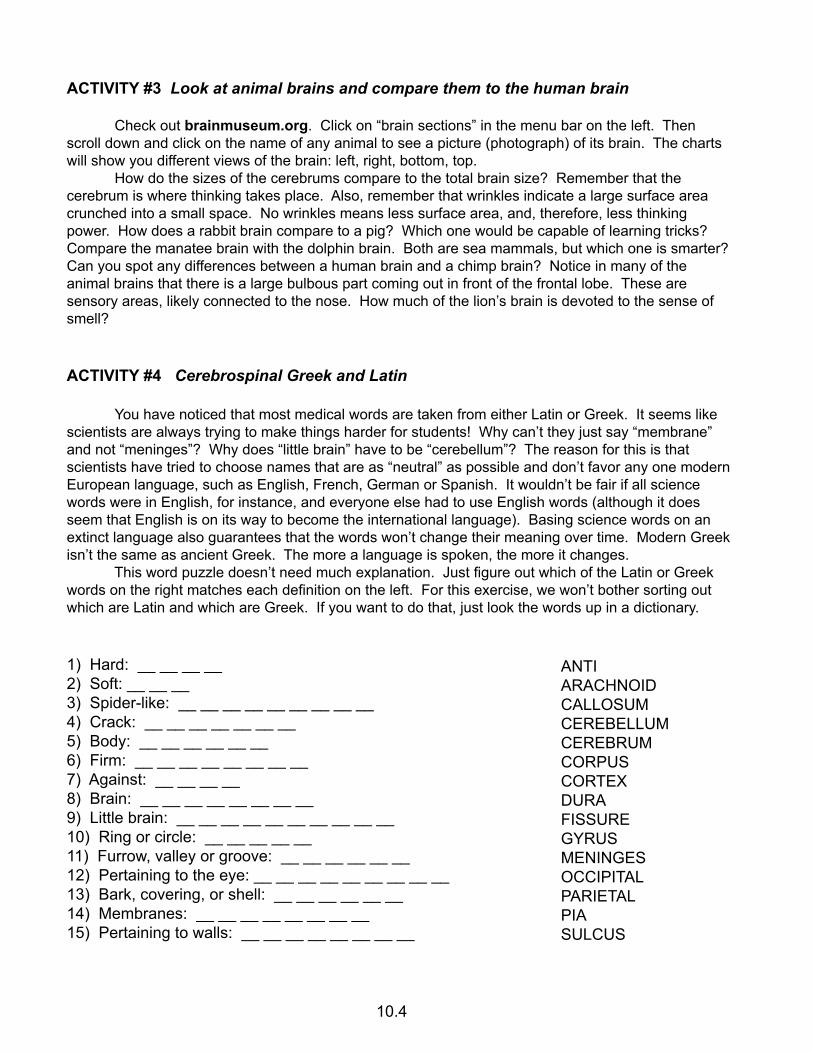
ACTIVITY #4 Cerebrospinal Greek and Latin
You have noticed that most medical words are taken from either Latin or Greek. It seems like scientists are always trying to make things harder for students! Why can’t they just say “membrane” and not “meninges”? Why does “little brain” have to be “cerebellum”? The reason for this is that scientists have tried to choose names that are as “neutral” as possible and don’t favor any one modern European language, such as English, French, German or Spanish. It wouldn’t be fair if all science words were in English, for instance, and everyone else had to use English words (although it does seem that English is on its way to become the international language). Basing science words on an extinct language also guarantees that the words won’t change their meaning over time. Modern Greek isn’t the same as ancient Greek. The more a language is spoken, the more it changes. This word puzzle doesn’t need much explanation. Just figure out which of the Latin or Greek words on the right matches each definition on the left. For this exercise, we won’t bother sorting out which are Latin and which are Greek. If you want to do that, just look the words up in a dictionary.
1) Hard: __ __ __ __2) Soft: __ __ __ 3) Spider-like: __ __ __ __ __ __ __ __ __4) Crack: __ __ __ __ __ __ __5) Body: __ __ __ __ __ __6) Firm: __ __ __ __ __ __ __ __7) Against: __ __ __ __8) Brain: __ __ __ __ __ __ __ __9) Little brain: __ __ __ __ __ __ __ __ __ __10) Ring or circle: __ __ __ __ __11) Furrow, valley or groove: __ __ __ __ __ __12) Pertaining to the eye: __ __ __ __ __ __ __ __ __13) Bark, covering, or shell: __ __ __ __ __ __14) Membranes: __ __ __ __ __ __ __ __ 15) Pertaining to walls: __ __ __ __ __ __ __ __
ANTIARACHNOIDCALLOSUMCEREBELLUMCEREBRUMCORPUSCORTEXDURAFISSUREGYRUSMENINGESOCCIPITALPARIETALPIASULCUS
10.4
TEACHER’SSECTION
Additional resources and
In-class activities
All patterns in this section are reproducible and may be photocopied for student use.

RECOMMENDED RESOURCES
BOOKS
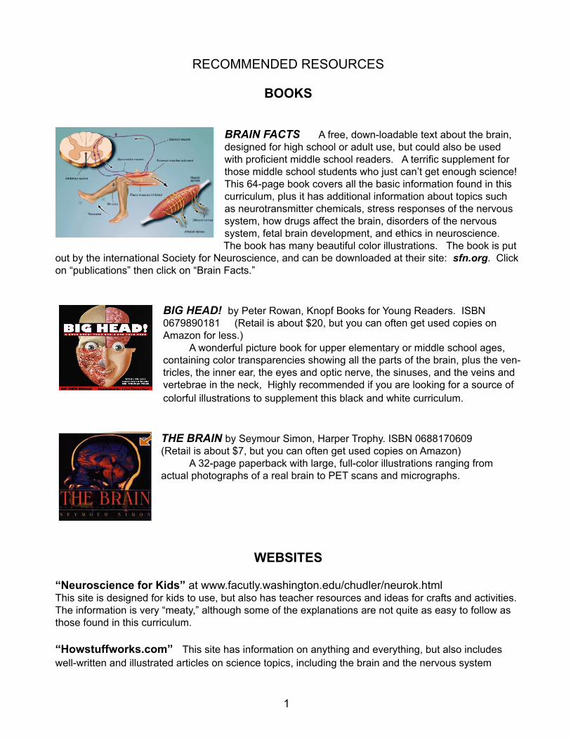
BRAIN FACTS A free, down-loadable text about the brain, designed for high school or adult use, but could also be used with proficient middle school readers. A terrific supplement for those middle school students who just can’t get enough science! This 64-page book covers all the basic information found in this curriculum, plus it has additional information about topics such as neurotransmitter chemicals, stress responses of the nervous system, how drugs affect the brain, disorders of the nervous system, fetal brain development, and ethics in neuroscience. The book has many beautiful color illustrations. The book is put
out by the international Society for Neuroscience, and can be downloaded at their site: sfn.org. Click on “publications” then click on “Brain Facts.”
BIG HEAD! by Peter Rowan, Knopf Books for Young Readers. ISBN 0679890181 (Retail is about $20, but you can often get used copies on Amazon for less.) A wonderful picture book for upper elementary or middle school ages, containing color transparencies showing all the parts of the brain, plus the ven-tricles, the inner ear, the eyes and optic nerve, the sinuses, and the veins and vertebrae in the neck, Highly recommended if you are looking for a source of colorful illustrations to supplement this black and white curriculum.
THE BRAIN by Seymour Simon, Harper Trophy. ISBN 0688170609(Retail is about $7, but you can often get used copies on Amazon) A 32-page paperback with large, full-color illustrations ranging from actual photographs of a real brain to PET scans and micrographs.
WEBSITES
“Neuroscience for Kids” at www.facutly.washington.edu/chudler/neurok.htmlThis site is designed for kids to use, but also has teacher resources and ideas for crafts and activities. The information is very “meaty,” although some of the explanations are not quite as easy to follow as those found in this curriculum.
“Howstuffworks.com” This site has information on anything and everything, but also includes well-written and illustrated articles on science topics, including the brain and the nervous system
1

ADDITIONAL ACTIVITY IDEAS
CHAPTER 1
1) Do an “MRI” of an orange
You will need an orange (the big, not-too-juicy kind is best), a knife, a cutting board, and some-thing on which to lay out all the slices. Optional: some raisins to represent abnormal areas in the brain that you can locate with your “MRI” First, show the students enough MRI images so that they understand that an MRI is not just one image, but a whole series of images. (If you don’t have any actual MRI scans available, images can be printed from the Internet easily enough.) Explain that each image represents a very thin layer of the head. The first image is the top layer, and the last image is the bottom one, with the rest of the images exactly in order from top to bottom. Explain that you will simulate an MRI by using an orange for a head and actual slices instead of pictures. Make sure the students understand that real MRI’s don’t really slice your head! Then begin slicing the orange and lay out the slices in order as they are cut. Make the slices as thin as possible. You’ll end up with as many as 15-20 slices. If you want to have something interesting to discover inside the orange, you can prepare the orange ahead of time by poking a small hole and pushing a raisin into the interior. If you happen to have a hypodermic needle available, another interesting idea would be to put some food coloring into the hypodermic, insert the hypo into the orange, and squeeze a little coloring deep inside the orange. Finding the splotches of color would be an interesting event for the observing students.
CHAPTER 2
1) Take a “hands-on” tour of the cerebrum
The teacher or parent leads this activity, demonstrating where the students are to put their hands while they listen to the teacher/parent tell them the information about each lobe.
FRONTAL LOBE: Make one hand cover the whole forehead. Underneath your hand is your frontal lobe. If you see someone in this position it looks like they are thinking, or like they forgot something. This is how you can remember that the frontal lobe is where your thinking and deciding takes place. This is the lobe that is in charge. It’s got a lot of responsibility! Isn’t it funny that when this lobe gets stressed, we put our hand on it?!
MOTOR CORTEX: Put each hand into a fist except for the first (pointer) finger sticking up. Then touch the tips of the fingers together, like they are pointing at each other. Keeping them together, put them on top of your head so they form a band right over the top (a little like a hair band). The strip of brain underneath your fingers is called the motor cortex. The word “motor” makes you think of movement. The motor cortex is the place in the brain that sends the signals to your muscles telling them to move. Notice that it is right next to the frontal lobe. This is handy, because the frontal lobe is what tells the motor cortex to send out its signals. Your frontal lobe decides the body needs to move, then the motor cortex actually sends out the signals.
3
SENSORY CORTEX: Move your finger “hair band” back about one centimeter (half an inch). The thin strip of brain running underneath the place where your fingers are is called the sensory cortex. Make those two fingers rub or scratch your head. This is a clue as to what the sensory cortex does. The sensory cortex is where your brain processes the “touch” sensations coming in from all parts of your body. When something scratches or touches your skin, the signals are sent to the sensory cortex.
PARIETAL LOBE: Take one hand and put it over the crown of your head, right over the place where your hair sprouts out. Underneath your hand is your parietal lobe. The name of this lobe comes from the Latin word for wall. That doesn’t make much sense, does it? Actually, the lobe is named after the part of the skull that covers it, the parietal bone. The parietal lobe is not very well understood, and remains somewhat of a mystery, but we do know the parietal lobe helps you to know where your body parts are, even if you can’t see them. Try this: Put your arms out to the sides, close your eyes, and stick out your index (pointer) fingers. Can you bring your index fingers together without opening your eyes? You probably can, or very nearly so. How can you know where those fingers are without looking at them? It is your parietal lobe that keeps track of where they are. The parietal lobe also works with your cerebellum and inner ear to give you your sense of balance.
OCCIPITAL LOBE: Put your hands behind your head, with fingers “knitted” together, as though you are lying in a grassy field watching the clouds roll by. Behind your hands is your occipital lobe (ok-SIP-it-al). This is the lobe where your vision is located. It’s what actually “sees” those clouds floating by. The images from your eyes are sent to the occipital lobe for processing. When the images come in, they are upside down and your brain must learn to turn them right-side up. A brain researcher once tried an experiment where he asked someone to wear special glasses that made everything look upside down. The result was that the occipital lobe compensated for what the glasses were doing and reversed the image a second time, making everything appear right-side up again!
TEMPORAL LOBE: Put your fingers on your temples. Behind your fingers and over your ears is the temporal lobe. This part of the brain is connected to your ears, which is not surprising, but is also where your sense of smell is. In addition to these functions, the temporal lobe is where your speech center is located. Right behind your temple is the area that forms words and sentences (then relays this to the motor cortex, which makes your lips and tongue move). Right above your ear is the area that understands words and sentences.
3) Brain cookies
You will need a batch of standard sugar cookie dough and some food coloring. Divide the dough into at least four parts. Use food coloring to color each a different color. (The more colors the better.) Use the outline drawing shown in activity #3 in chapter 2 as your guide to make a flat cookie where each lobe is a different color.
4
4) Draw brain parts on a real head
You will need a tight-fitting swim cap and a marker. Put the cap on a volunteer, then draw in the lobes on the cap. (This activity is guaranteed to produce giggles.)
5) Cerebrospinal fluid experiment You will need at least two raw eggs, at least one small container (just slightly larger than the egg), water, and rubber bands or duct tape to keep the lid on the container. Put a raw egg inside the container. The egg will represent the brain and the container will represent the skull. Secure the lid. Shake or drop the egg and see if it breaks. This simulates what would happen if you had no cerebrospinal fluid around your brain. Now fill the container with water and secure the lid. Shake and drop the container again, and see what happens. Does the water around the egg protect it from breaking? NOTE: Some plastic containers will be more brittle than they appear and will crack when dropped. We recommend either a soft plastic, such as a yogurt container, or an extremely heavy-duty plastic, such as indestructible “Tupperware.” You may want to drop it into a large container that will catch any water that leaks out.
6) Observe a thin membrane similar to the meninges in the brain
You will need: a raw egg and something to crack it into. Crack the egg and dump out the contents. Carefully crush the side of one of the empty egg shells, so that you have some pieces that are being held together by a thin membrane. Notice that the membrane is attached to the inside of the egg shell. In the brain, the outer layer of the meninges (the dura mater) is attached to the skull. Now gently pull the cracked pieces away from the rest of the shell so that the membrane slowly peels off the inside of the shell. It’s not too hard to do. You should be able to get a piece of membrane at least a centimeter wide and two centimeters long, possibly larger. (If it doesn’t seem to be working, try a different egg.) Gently pull and stretch the membrane to test its strength. It’s surprisingly tough. You might want to see what happens if you leave the membrane out to dry. How does lack of water affect its strength?
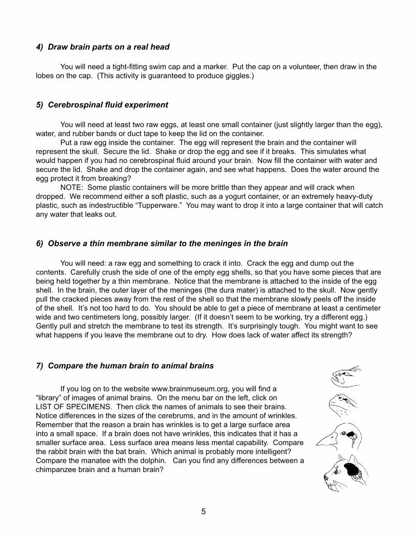
7) Compare the human brain to animal brains
If you log on to the website www.brainmuseum.org, you will find a “library” of images of animal brains. On the menu bar on the left, click on LIST OF SPECIMENS. Then click the names of animals to see their brains. Notice differences in the sizes of the cerebrums, and in the amount of wrinkles. Remember that the reason a brain has wrinkles is to get a large surface area into a small space. If a brain does not have wrinkles, this indicates that it has a smaller surface area. Less surface area means less mental capability. Compare the rabbit brain with the bat brain. Which animal is probably more intelligent? Compare the manatee with the dolphin. Can you find any differences between a chimpanzee brain and a human brain?
5