Embed Size (px)
Citation preview
An Integrated Diagnosis Strategy for CongenitalMyopathiesJohann Bohm1,2,3,4,5., Nasim Vasli1,2,3,4,5., Edoardo Malfatti6,7,8,9, Stephanie Le Gras1,2,3,4,10,
Claire Feger1,2,3,4,5, Bernard Jost1,2,3,4,10, Nicole Monnier11, Julie Brocard12, Hatice Karasoy13,
Marion Gerard14, Maggie C. Walter15, Peter Reilich15, Valerie Biancalana1,2,3,4,5,16, Christine Kretz1,2,3,4,5,
Nadia Messaddeq1,2,3,4,17, Isabelle Marty12, Joel Lunardi11, Norma B. Romero6,8,9, Jocelyn Laporte1,2,3,4,5*
1 Department of Translational Medicine and Neurogenetics, Institut de Genetique et de Biologie Moleculaire et Cellulaire, Illkirch, France, 2 Institut National de la Sante et
de la Recherche Medicale, U964, Illkirch, France, 3 Centre national de la recherche scientifique, UMR7104, Illkirch, France, 4 Universite de Strasbourg, Illkirch, France,
5 College de France, chaire de genetique humaine, Illkirch, France, 6 Institut de Myologie, Unite de Morphologie Neuromusculaire, GHU La Pitie-Salpetrere, Paris, France,
7 Department of Neurological, Neurosurgical, and Behavioral Sciences, University of Siena, Italy, 8 Universite Paris 6 UR76, Institut National de la Sante et de la Recherche
Medicale UMR 974, Centre national de la recherche scientifique UMR 7215, Institut de Myologie, GHU La Pitie-Salpetriere, Paris, France, 9 Centre de reference de
pathologie neuromusculaire Paris-Est, Institut de Myologie, GHU La Pitie-Salpetriere, Assistance Publique-Hopitaux de Paris, Paris, France, 10 DNA microarrays and
Sequencing platform, Institut de Genetique et de Biologie Moleculaire et Cellulaire, Illkirch, France, 11 Laboratoire de Biochimie Genetique et Moleculaire, CHU de
Grenoble, Grenoble, France, 12 Institut des Neurosciences, Inserm U836, La Tronche, France, 13 Department of Neurology, Ege University School of Medicine, Izmir,
Turkey, 14 Service de Genetique, CHU de Caen, Caen, France, 15 Friedrich-Baur-Institute, Department of Neurology, Ludwig-Maximilians University, Munich, Germany,
16 Faculte de Medecine, Laboratoire de Diagnostic Genetique, Nouvel Hopital Civil, Strasbourg, France, 17 Imaging Center, Institut de Genetique et de Biologie
Moleculaire et Cellulaire, Illkirch, France
Abstract
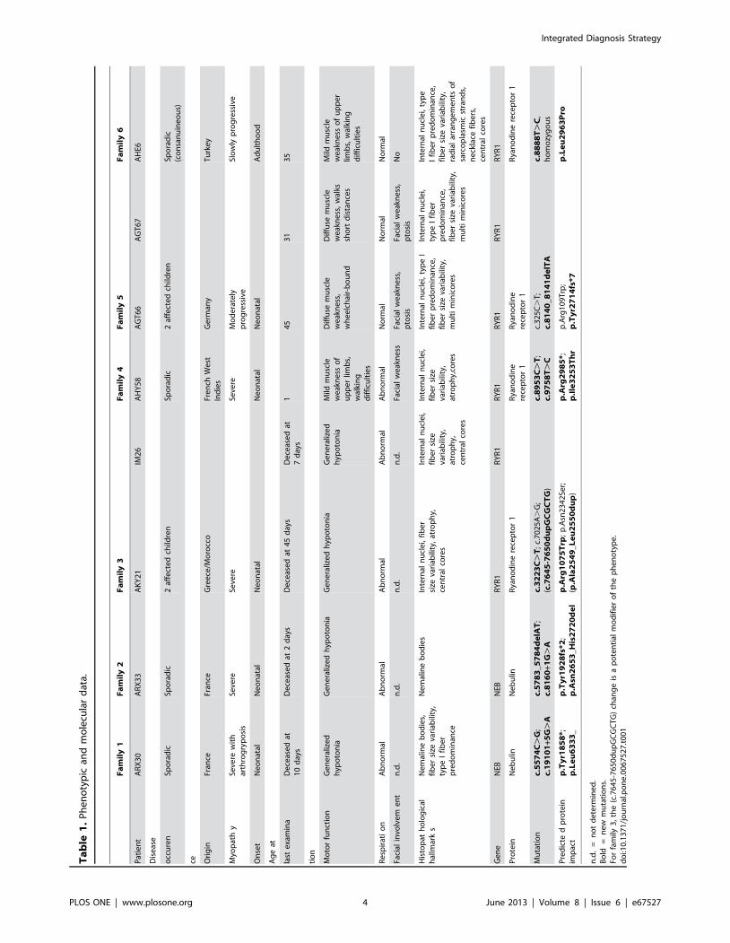
Congenital myopathies are severe muscle disorders affecting adults as well as children in all populations. The diagnosis ofcongenital myopathies is constrained by strong clinical and genetic heterogeneity. Moreover, the majority of patientspresent with unspecific histological features, precluding purposive molecular diagnosis and demonstrating the need for analternative and more efficient diagnostic approach. We used exome sequencing complemented by histological andultrastructural analysis of muscle biopsies to identify the causative mutations in eight patients with clinically differentskeletal muscle pathologies, ranging from a fatal neonatal myopathy to a mild and slowly progressive myopathy with adultonset. We identified RYR1 (ryanodine receptor) mutations in six patients and NEB (nebulin) mutations in two patients. Wefound novel missense and nonsense mutations, unraveled small insertions/deletions and confirmed their impact on splicingand mRNA/protein stability. Histological and ultrastructural findings of the muscle biopsies of the patients validated theexome sequencing results. We provide the evidence that an integrated strategy combining exome sequencing with clinicaland histopathological investigations overcomes the limitations of the individual approaches to allow a fast and efficientdiagnosis, accelerating the patient’s access to a better healthcare and disease management. This is of particular interest forthe diagnosis of congenital myopathies, which involve very large genes like RYR1 and NEB as well as genetic and phenotypicheterogeneity.
Citation: Bohm J, Vasli N, Malfatti E, Le Gras S, Feger C, et al. (2013) An Integrated Diagnosis Strategy for Congenital Myopathies. PLoS ONE 8(6): e67527.doi:10.1371/journal.pone.0067527
Editor: Gisela Nogales-Gadea, University Hospital Vall d’Hebron, Spain
Received November 16, 2012; Accepted May 21, 2013; Published June 24, 2013
Copyright: � 2013 Bohm et al. This is an open-access article distributed under the terms of the Creative Commons Attribution License, which permitsunrestricted use, distribution, and reproduction in any medium, provided the original author and source are credited.
Funding: This work was supported by Institut National de la Sante et de la Recherche Medicale (INSERM), Centre national de la recherche scientifique (CNRS),University of Strasbourg, College de France and grants from ANR, GIS Institute for rare diseases and IBiSA, Association Francaise contre les myopathies, MuscularDystrophy Association (United States of America) and the Myotubular Trust. This work was supported by the INSERM, the CNRS, University of Strasbourg, Collegede France and grants from the Agence Nationale de la Recherche (ANR, grant CM-WES), Muscular Dystrophy Association (MDA, grant 2010-52655) and MyotubularTrust. The funders had no role in study design, data collection and analysis, decision to publish, or preparation of the manuscript.
Competing Interests: The authors have declared that no competing interests exist.
* E-mail: [email protected]
. These authors contributed equally to this work.
Introduction
Congenital myopathies (CM) are rare disorders characterized
by early-onset muscle weakness and classified based on the
predominance of particular histological anomalies on muscle
biopsies. They have an estimated prevalence of about 1:25 000
and are usually associated with neonatal or childhood onset,
progressive or non-progressive muscle weakness, breathing diffi-
culties and delayed motor milestones [1,2]. The main congenital
myopathy subgroups are protein aggregate myopathies (primarily
nemaline myopathy), core myopathies and centronuclear myop-
athies (CNM), respectively characterized by rod-like protein
accumulations, focal myofibrillar disorganization, and nuclear
centralization on muscle biopsies [3]. Other congenital myopathy
subgroups have been reported with different structural hallmarks
[4]. As congenital myopathies are usually severe with a high
recurrence risk in affected families, molecular diagnosis is
important to provide an adequate healthcare and genetic
counseling.
PLOS ONE | www.plosone.org 1 June 2013 | Volume 8 | Issue 6 | e67527
Although many genes have been associated with congenital
myopathies in the past years, a recent study reported that only 16
out of 46 US patients were molecularly diagnosed [5]. This is due
to the fact that despite clinical and histological examinations, the
majority of the patients presented with unspecific features.
Especially for the neonatal cases, a reliable diagnosis is often
challenging. Another reason is the genetic heterogeneity in
congenital myopathies with the implication of more than 20
known genes [4], opposing efficient molecular diagnosis. In
addition, some of the genes implicated in congenital myopathies
belong to the largest genes of the human genome, as TTN (363
exons; MIM#188840) mutated in congenital myopathy with fatal
cardiomyopathy, NEB (183 exons; MIM#161650) mutated in
nemaline myopathy, or RYR1 (106 exons; MIM#180901) mutated
in different pathologies. The aim of this study was to propose and
validate an integrated approach including exome sequencing for
the diagnosis of congenital myopathies with neonatal and adult
onset. The next generation sequencing technology has become an
effective strategy for massively parallel analysis of a large number
of genes and has led to the successful identification of several
Mendelian disease genes [6]. This approach is however uncom-
monly used in routine molecular diagnosis despite its potential
synergy with clinical and histological investigations. Sanger
sequencing of single genes remains the major technique for
monogenetic pathologies with characteristic clinical manifesta-
tions. It is time-consuming and not centralized, demonstrating the
need for a more efficient diagnostic approach.
Here we used an integrated exome sequencing strategy to
identify the causative mutations in eight patients from six families
with clinically different neonatal or adult-onset congenital
myopathies. We found pathogenic mutations in the large RYR1
and NEB genes, and histopathological and ultrastructural analysis
of the muscle biopsies of the patients confirmed and validated the
exome sequencing results. In conclusion, we provide the evidence
that exome sequencing in combination with histological analyses is
a fast, efficient and reliable method to identify disease-causing
mutations in unsolved myopathy cases. Our integrated approach is
particularly relevant for disease groups with genetic and pheno-
typic heterogeneity.
Materials and Methods
PatientsPatients originated from France (Families 1 and 2), Greece/
Morocco (Family 3), French West Indies (Family 4), Germany
(Family 5), and Turkey (Family 6). Sample collection was
performed with written informed consent from the patients or
their legal guardians according to the declaration of Helsinki and
experimentation was approved by the INSERM institutional
review board (‘‘Comite de protection des personnes Est IV’’).
Muscle biopsies were obtained from deltoid (ARX30, AKY21,
IM26, AHY58, AHE6), biceps brachii (ATG66, AGT67) or
tibialis anterior ARX33).
Linkage analysisFor whole-genome analysis, the genomic DNA of patient AHE6
was hybridized on Affymetrix SNP array 6.0 according to the
manufacturer’s instructions. Loss of heterozygosity was analyzed
with GeneChip DNA Analysis and Chromosome Copy Number
Analysis softwares (Affymetrix, Santa Clara, CA, USA).
Exome sequencingGenomic DNA was prepared from peripheral blood by routine
procedures and quality-controlled. DNA was sheared using the
Covaris E210 (KBioscience, Herts, UK) followed by automatic
library preparation with the SPRI-TE machine (Beckman Coulter
Inc., Brea, CA, USA). Exon capture was performed with the
Agilent SureSelect Array v1 (families 3–6) or the refined Agilent
SureSelect Human all Exon 50 Mb Kit (targeting all NEB and
TTN exons; Families 1 and 2) (Agilent Technologies, Santa Clara,
CA, USA). Enriched DNA fragments were sequenced on an
Illumina Genome Analyzer IIx to generate 72nt single reads for
AHE6 and paired-end reads for AGT66, AGT67, AHY58,
AKY21 and IM26. For patients ARX30 and ARX33 we
performed ‘‘trio sequencing’’, i.e. exome sequencing with 72 nt
paired-end reads of the patient and both healthy parents.
Bioinformatic analysisSequence data were analyzed using Illumina Pipeline RTA
(Real-Time Analysis) version 1.7 and aligned to the reference
genome GRCh37/hg19 using BWA [7]. Variant calling and
filtering of reads sharing the same start position and strand was
done with Samtools [8]. Variants were considered as heterozygous
when present in 20 – 80 % of the reads and as homozygous when
present in $ 80 % of the reads. For SNP/indel annotation and
filtering SVA, Ensembl60, dbSNP134, 1000 genomes, and
NHLBI exome variant server were used. Impact of variations
were predicted using SIFT [9] (maximum pathogenic score: 0),
PolyPhen V2 [10] (maximum pathogenic score: 1), NNSPLICE
[11] and Human Splicing Finder [12] (Table S5).
Mutation characterizationMutation confirmation and segregation analysis were performed
by PCR and Sanger sequencing of the RYR1/NEB exons
harboring the mutations and the adjacent exon-intron boundaries.
Primer sequences and PCR conditions are listed in Table S2. The
mutations were numbered according to GenBank NM_000540.2
and NP_000531.2 (RYR1) and NM_001164507.1 and
NP_001157979.1 (NEB). Nucleotide position reflects cDNA
numbering with +1 corresponding to the A of the ATG translation
initiation codon.
Muscle histologyFor histochemical analyses, transverse sections (10 mm) of the
muscle biopsies were stained with hematoxylin-eosin, Gomori
trichrome, NADH tetrazolium reductase (NADH-TR), periodic
acid-Schiff and ATPase and assessed for nuclei position, fiber
morphology, fiber type distribution, cores and accumulations/
infiltrations.
Electron microscopyMuscle sections were fixed in 2.5% paraformaldehyde, 2.5%
glutaraldehyde, and 50 mM CaCl2 in 0.1 M cacodylate buffer
(pH 7.4), and post-fixed with 2% OsO4, 0.8% K3Fe(CN)6 in
0.1 M cacodylate buffer (pH 7.4) for 2 h at 4uC and incubated
with 5% uranyl acetate for 2 h at 4uC. Samples were dehydrated
in graded series of ethanol and embedded in epon resin 812.
Ultrathin sections (70 nm) were contrasted with uranyl and lead
citrate and viewed at 70 kv with a Morgagni 268D electron
microscope and a Mega View III camera (Soft Imaging System,
Munster, Germany).
RNARNA was extracted from the deltoid muscle biopsy of patients
ARX30, IM26 and AHE6 and from a tibialis anterior biopsy of
patient ARX33 using Tri reagent (Molecular Research Center
Inc., Cincinnati, OH, USA), and reverse transcribed using the
Integrated Diagnosis Strategy
PLOS ONE | www.plosone.org 2 June 2013 | Volume 8 | Issue 6 | e67527
SuperScriptH III kit (Invitrogen, Carlsbad, CA, USA). Sequencing
of the entire RYR1 cDNA of IM26 and AHE6 confirmed the
presence of the RYR1 mutations identified by exome sequencing
and excluded further sequence aberrations. The impact of the
NEB mutations on splicing in patients ARX30 and ARX33 was
assessed using NEB specific primers.
ProteinWestern blot was performed using routine protocols on a deltoid
muscle biopsies from AHE6 and a healthy age-matched control
using a home-made rabbit anti-RYR1 [13] and a DE-R-11 mouse
anti-Desmin antibody (Dakocytomation, Trappes, France) for
normalization. Signals were detected with a chemiluminescent
HRP substrate and quantified using a ChemiDoc XRS apparatus
(Biorad, Marnes la Coquette, France) and the Quantity One
software (Biorad). The quantification was repeated 6 times.
Results
An integrated diagnosis approach for congenitalmyopathies
We studied eight patients from six families with different clinical
and histological features suggestive of congenital myopathies. The
neonatal forms ranged from fatal shortly after birth to moderately
progressive, and the adult form was mild and slowly progressive
(Table 1). Using Sanger sequencing, the patients were previously
excluded for several genes implicated in nemaline or centronuclear
myopathies, including ACTA1, TPM2, TPM3, TNNT1 for families
1 and 2, MTM1 and BIN1 for families 3 to 6, and DNM2 for
families 3, 4 and 6. These inconclusive preliminary analyses
underscore the necessity of an alternative and more efficient
molecular diagnosis. We therefore used an integrated approach,
combining exome sequencing and histological investigations.
Exome sequencing was performed for all patients and for the
parents of families 1 and 2 (trio sequencing) and pointed - in
combination with histopathology - unambiguously to single
myopathy genes. A statistical overview of the sequencing results
is shown in Tables 2, 3, 4, 5 and the coverage of each exon of the
known genes implicated in congenital myopathies is specified in
Tables S3 and S4. The inheritance pattern was taken into account
for the selection of candidate genes. For the sporadic cases in
families 1 and 2, genes with two heterozygous variants segregating
from each parent were selected as the analysis for de novo
mutations was not conclusive. For the non-consanguineous
families 3 and 5 with two affected members each, genes with
two common heterozygous variations were selected. For family 4,
we verified dominant and recessive scenarios. For patient AHE6
from the consanguineous family 6, we focused on homozygous
variants mapped in large homozygous regions, determined by
homozygosity-by-descent (Table S1). The impact of potential
mutations was predicted with SIFT and PolyPhen for amino acid
changes and with NNSPLICE and Human Splicing Finder for
changes potentially affecting splicing. All identified mutations were
verified by Sanger sequencing in the starting genomic DNA and
also in the cDNA for families 1, 2, 3 and 6. Histological analysis of
muscle biopsies was performed for all patients. For five patients
(ARX30, AKY21, IM26, AHY58 and AHE6), we additionally
analyzed the muscle biopsy by electron microscopy.
Clinical and histological findings and mutation detectionby exome sequencing
For patient ARX30 from family 1, polyhydramnios and fetal
akinesia were diagnosed during pregnancy and the patient
presented with severe neonatal hypotonia, respiratory distress,
arthrogryposis, hip hyperlaxity, club feet and dysmorphic features.
Through exome sequencing, we identified two heterozygous
mutations in NEB: the c.5574C.G (exon 45; p.Tyr1858*)
nonsense mutation on the maternal allele and the
c.19101+5G.A (intron 122) mutation on the paternal allele,
confirming autosomal recessive inheritance (Figure 1). A deltoid
muscle biopsy revealed nemaline rods, marked fiber size variability
and type I fiber predominance (Figure 2). Ultrastructural analysis
confirmed the presence of numerous nemaline bodies, and
revealed mild disorganization of the myofibrillar structure and
Z-band streaming (Figure 3). These findings were consistent with
the mutations in NEB found by exome sequencing. We performed
additional molecular analysis to confirm the impact of the
mutations. The paternal mutation was predicted to impair splicing
of exon 122 and RNA reverse transcription and cDNA sequencing
confirmed major skipping of this in-frame exon (Figure 4). The
nonsense mutation was not seen on the cDNA sequence,
suggesting degradation of the maternal allele by nonsense-
mediated mRNA decay (NMD).
Patient ARX33 from family 2 presented with a severe muscle
weakness at birth and deceased in the following days due to
cardio-respiratory arrest. We identified one NEB mutation on the
paternal allele (c.5783_5784delAT) in exon 46, involving a
frameshift and a premature stop codon (p.Tyr1928fs*2), and one
NEB donor splice site mutation on the maternal allele
(c.8160+1G.A, intron 58) (Figure 1). Gomori trichrome staining
of a tibialis anterior muscle biopsy revealed nemaline rods (Figure
S1), electron microscopy data were not available. The impact of
the splice mutation was verified by muscle mRNA analysis and
demonstrated complete skipping of the in-frame exon 58. The 2
nucleotides deletion was not seen on the cDNA sequence,
suggesting nonsense-mediated mRNA decay of the paternal allele
(Figure 4).
AKY21 and IM26 from family 3 deceased from a severe muscle
disorder shortly after birth. Pregnancy was normal and both had
low Apgar scores, generalized hypotonia and respiratory distress at
birth. Cardiac examinations were normal. We detected three
heterozygous RYR1 mutations in exons 25 (c.3223C.T;
p.Arg1075Trp), 43 (c.7025A.G; p.Asn2342Ser), and 48
(c.7645_7650dupGCGCTG; p.Ala2549_Leu2550dup). A DNA
sample of the father was not available and sequencing of the
maternal DNA revealed the presence of the c.3223C.T mutation
in the absence of the other sequence aberrations, suggesting
autosomal recessive inheritance with compound heterozygosity
(Figure 1). The c.3223C.T (p.Arg1075Trp) missense mutation on
the maternal allele affects a conserved amino acid (Figure S2) and
is predicted to be highly damaging by Polyphen (score 1.000) and
SIFT (Score 0.000). The c.7025A.G (p.Asn2342Ser) missense on
the paternal allele has been associated with malignant hyperther-
mia susceptibility [14] (rs147213895), known to result from
heterozygous RYR1 mutations. Both missense mutations were
found with equal intensity in the cDNA obtained by reverse
transcription of the RNA from the patient muscle biopsy,
suggesting that both alleles are equally expressed and that the
mutations do not impact on mRNA stability. The deltoid muscle
biopsies of both patients revealed nuclear internalization, atrophy,
fiber size variability, and areas devoid of oxidative enzyme activity
(Figure 2). Ultrastructural analysis of the biopsy of the patients
showed that the disorganized myofibrillar areas around the
internalized nuclei cover a large part of the fiber diameter. The
longitudinal muscle section revealed prominent Z-band streaming
(Figure 3). These findings were suggestive of a myopathy with
cores. As RYR1 mutations mostly involve cores, the histopatho-
Integrated Diagnosis Strategy
PLOS ONE | www.plosone.org 3 June 2013 | Volume 8 | Issue 6 | e67527
Ta
ble
1.
Ph
en
oty
pic
and
mo
lecu
lar
dat
a.
Fa
mil
y1
Fa
mil
y2
Fa
mil
y3
Fa
mil
y4
Fa
mil
y5
Fa
mil
y6
Pat
ien
tA
RX
30
AR
X3
3A
KY
21
IM2
6A
HY
58
AG
T6
6A
GT
67
AH
E6
Dis
eas
e
occ
ure
nSp
ora
dic
Spo
rad
ic2
affe
cte
dch
ildre
nSp
ora
dic
2af
fect
ed
child
ren
Spo
rad
ic(c
on
san
uin
eo
us)
ce Ori
gin
Fran
ceFr
ance
Gre
ece
/Mo
rocc
oFr
en
chW
est
Ind
ies
Ge
rman
yT
urk
ey
Myo
pat
hy
Seve
rew
ith
arth
rog
ryp
osi
sSe
vere
Seve
reSe
vere
Mo
de
rate
lyp
rog
ress
ive
Slo
wly
pro
gre
ssiv
e
On
set
Ne
on
atal
Ne
on
atal
Ne
on
atal
Ne
on
atal
Ne
on
atal
Ad
ult
ho
od
Ag
eat
last
exa
min
aD
ece
ase
dat
10
day
sD
ece
ase
dat
2d
ays
De
ceas
ed
at4
5d
ays
De
ceas
ed
at7
day
s1
45
31
35
tio
n
Mo
tor
fun
ctio
nG
en
era
lize
dh
ypo
ton
iaG
en
era
lize
dh
ypo
ton
iaG
en
era
lize
dh
ypo
ton
iaG
en
era
lize
dh
ypo
ton
iaM
ildm
usc
lew
eak
ne
sso
fu
pp
er
limb
s,w
alki
ng
dif
ficu
ltie
s
Dif
fuse
mu
scle
we
akn
ess
,w
he
elc
hai
r-b
ou
nd
Dif
fuse
mu
scle
we
akn
ess
,w
alks
sho
rtd
ista
nce
s
Mild
mu
scle
we
akn
ess
of
up
pe
rlim
bs,
wal
kin
gd
iffi
cult
ies
Re
spir
ati
on
Ab
no
rmal
Ab
no
rmal
Ab
no
rmal
Ab
no
rmal
Ab
no
rmal
No
rmal
No
rmal
No
rmal
Faci
alin
volv
em
en
tn
.d.
n.d
.n
.d.
n.d
.Fa
cial
we
akn
ess
Faci
alw
eak
ne
ss,
pto
sis
Faci
alw
eak
ne
ss,
pto
sis
No
His
top
ath
olo
gic
alh
allm
ark
sN
em
alin
eb
od
ies,
fib
er
size
vari
abili
ty,
typ
eI
fib
er
pre
do
min
ance
Ne
mal
ine
bo
die
sIn
tern
aln
ucl
ei,
fib
er
size
vari
abili
ty,
atro
ph
y,ce
ntr
alco
res
Inte
rnal
nu
cle
i,fi
be
rsi
zeva
riab
ility
,at
rop
hy,
cen
tral
core
s
Inte
rnal
nu
cle
i,fi
be
rsi
zeva
riab
ility
,at
rop
hy,
core
s
Inte
rnal
nu
cle
i,ty
pe
Ifi
be
rp
red
om
inan
ce,
fib
er
size
vari
abili
ty,
mu
lti
min
ico
res
Inte
rnal
nu
cle
i,ty
pe
Ifi
be
rp
red
om
inan
ce,
fib
er
size
vari
abili
ty,
mu
lti
min
ico
res
Inte
rnal
nu
cle
i,ty
pe
Ifi
be
rp
red
om
inan
ce,
fib
er
size
vari
abili
ty,
rad
ial
arra
ng
em
en
tso
fsa
rco
pla
smic
stra
nd
s,n
eck
lace
fib
ers
,ce
ntr
alco
res
Ge
ne
NEB
NEB
RY
R1
RY
R1
RY
R1
RY
R1
RY
R1
RY
R1
Pro
tein
Ne
bu
linN
eb
ulin
Rya
no
din
ere
cep
tor
1R
yan
od
ine
rece
pto
r1
Rya
no
din
ere
cep
tor
1R
yan
od
ine
rece
pto
r1
Mu
tati
on
c.5
57
4C
.G
;c.
19
10
1+5
G.
Ac.
57
83
_5
78
4d
elA
T;
c.8
16
0+1
G.
Ac.
32
23
C.
T;
c.7
02
5A
.G
;(c
.76
45
-76
50
du
pG
CG
CT
G)
c.8
95
3C
.T
;c.
97
58
T.
Cc.
32
5C
.T
;c.
81
40
_8
14
1d
elT
Ac.
88
88
T.
C,
ho
mo
zyg
ou
s
Pre
dic
ted
pro
tein
imp
act
p.T
yr1
85
8*;
p.L
eu
63
33
_p
.Ty
r19
28
fs*2
;p
.Asn
26
53
_H
is2
72
0d
el
p.A
rg1
07
5T
rp;
p.A
sn2
34
2Se
r;(p
.Ala
25
49
_L
eu
25
50
du
p)
p.A
rg2
98
5*;
p.I
le3
25
3T
hr
p.A
rg1
09
Trp
;p
.Ty
r27
14
fs*7
p.L
eu
29
63
Pro
n.d
.=
no
td
ete
rmin
ed
.B
old
=n
ew
mu
tati
on
s.Fo
rfa
mily
3,
the
(c.7
64
5-7
65
0d
up
GC
GC
TG
)ch
ang
eis
ap
ote
nti
alm
od
ifie
ro
fth
ep
he
no
typ
e.
do
i:10
.13
71
/jo
urn
al.p
on
e.0
06
75
27
.t0
01
Integrated Diagnosis Strategy
PLOS ONE | www.plosone.org 4 June 2013 | Volume 8 | Issue 6 | e67527
logical findings and the exome sequencing results are in
agreement.
AHY58 from family 4 was born with general amyotrophy, axial
and peripheral hypotonia and arthrogryposis after normal
pregnancy. Although the health status slightly improved, the
patient had temporal respiratory distress and deglutition problems.
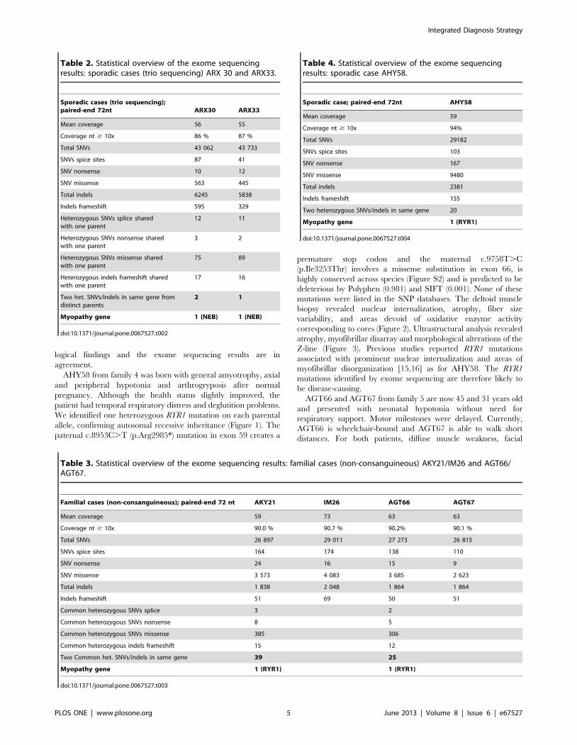
We identified one heterozygous RYR1 mutation on each parental
allele, confirming autosomal recessive inheritance (Figure 1). The
paternal c.8953C.T (p.Arg2985*) mutation in exon 59 creates a
premature stop codon and the maternal c.9758T.C
(p.Ile3253Thr) involves a missense substitution in exon 66, is
highly conserved across species (Figure S2) and is predicted to be
deleterious by Polyphen (0.981) and SIFT (0.001). None of these
mutations were listed in the SNP databases. The deltoid muscle
biopsy revealed nuclear internalization, atrophy, fiber size
variability, and areas devoid of oxidative enzyme activity
corresponding to cores (Figure 2). Ultrastructural analysis revealed
atrophy, myofibrillar disarray and morphological alterations of the
Z-line (Figure 3). Previous studies reported RYR1 mutations
associated with prominent nuclear internalization and areas of
myofibrillar disorganization [15,16] as for AHY58. The RYR1
mutations identified by exome sequencing are therefore likely to
be disease-causing.
AGT66 and AGT67 from family 5 are now 45 and 31 years old
and presented with neonatal hypotonia without need for
respiratory support. Motor milestones were delayed. Currently,
AGT66 is wheelchair-bound and AGT67 is able to walk short
distances. For both patients, diffuse muscle weakness, facial
Table 2. Statistical overview of the exome sequencingresults: sporadic cases (trio sequencing) ARX 30 and ARX33.
Sporadic cases (trio sequencing);paired-end 72nt ARX30 ARX33
Mean coverage 56 55
Coverage nt $ 10x 86 % 87 %
Total SNVs 43 062 43 733
SNVs spice sites 87 41
SNV nonsense 10 12
SNV missense 563 445
Total indels 6245 5838
Indels frameshift 595 329
Heterozygous SNVs splice sharedwith one parent
12 11
Heterozygous SNVs nonsense sharedwith one parent
3 2
Heterozygous SNVs missense sharedwith one parent
75 89
Heterozygous indels frameshift sharedwith one parent
17 16
Two het. SNVs/indels in same gene fromdistinct parents
2 1
Myopathy gene 1 (NEB) 1 (NEB)
doi:10.1371/journal.pone.0067527.t002
Table 3. Statistical overview of the exome sequencing results: familial cases (non-consanguineous) AKY21/IM26 and AGT66/AGT67.
Familial cases (non-consanguineous); paired-end 72 nt AKY21 IM26 AGT66 AGT67
Mean coverage 59 73 63 63
Coverage nt $ 10x 90.0 % 90.7 % 90.2% 90.1 %
Total SNVs 26 897 29 011 27 273 26 815
SNVs spice sites 164 174 138 110
SNV nonsense 24 16 15 9
SNV missense 3 573 4 083 3 685 2 623
Total indels 1 838 2 048 1 864 1 864
Indels frameshift 51 69 50 51
Common heterozygous SNVs splice 3 2
Common heterozygous SNVs nonsense 8 5
Common heterozygous SNVs missense 385 306
Common heterozygous indels frameshift 15 12
Two Common het. SNVs/indels in same gene 39 25
Myopathy gene 1 (RYR1) 1 (RYR1)
doi:10.1371/journal.pone.0067527.t003
Table 4. Statistical overview of the exome sequencingresults: sporadic case AHY58.
Sporadic case; paired-end 72nt AHY58
Mean coverage 59
Coverage nt $ 10x 94%
Total SNVs 29182
SNVs spice sites 103
SNV nonsense 167
SNV missense 9480
Total indels 2381
Indels frameshift 155
Two heterozygous SNVs/indels in same gene 20
Myopathy gene 1 (RYR1)
doi:10.1371/journal.pone.0067527.t004
Integrated Diagnosis Strategy
PLOS ONE | www.plosone.org 5 June 2013 | Volume 8 | Issue 6 | e67527
weakness and ptosis were diagnosed. They have contractures and
absent reflexes. By comparing the exome sequencing data of both
patients, we identified common compound heterozygous RYR1
mutations, confirming autosomal recessive inheritance (Figure 1).
The c.325C.T (p.Arg109Trp) missense mutation in exon 4 on
the maternal allele has been reported to be associated (in
combination with a second RYR1 mutation) with multi-minicore
and central core disease [17,18]. The c.8140_8141delTA deletion
in exon 51 on the paternal allele has never been described and is
predicted to induce a frameshift and a premature TAA stop codon
(p.Tyr2714fs*7). The biceps brachii biopsies of patients AGT66
and AGT67 revealed nuclear internalization, fiber size variability,
type I fiber predominance, and multiple dot-like areas devoid of
oxidative enzyme activity, suggestive of multiminicore disease
(MmD) (Figure 2). MmD is a recessive disorder linked to mutations
in RYR1, the exome sequencing results are therefore in agreement
with the histological data.
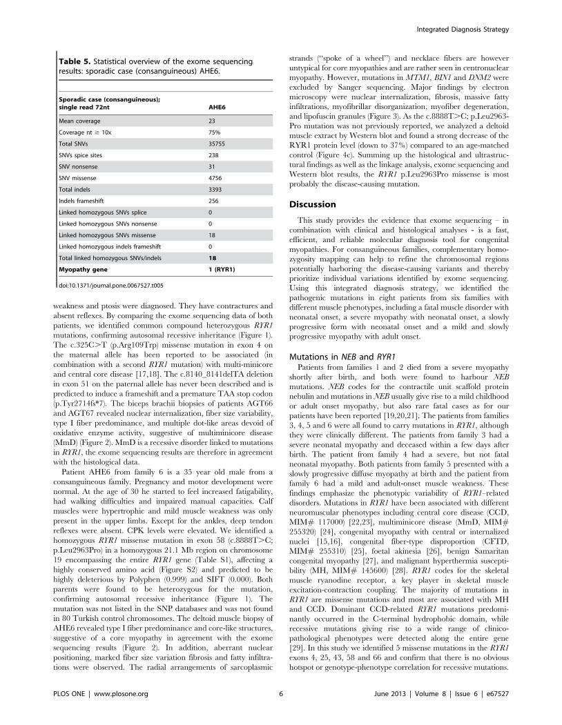
Patient AHE6 from family 6 is a 35 year old male from a
consanguineous family. Pregnancy and motor development were
normal. At the age of 30 he started to feel increased fatigability,
had walking difficulties and impaired manual capacities. Calf
muscles were hypertrophic and mild muscle weakness was only
present in the upper limbs. Except for the ankles, deep tendon
reflexes were absent. CPK levels were elevated. We identified a
homozygous RYR1 missense mutation in exon 58 (c.8888T.C;
p.Leu2963Pro) in a homozygous 21.1 Mb region on chromosome
19 encompassing the entire RYR1 gene (Table S1), affecting a
highly conserved amino acid (Figure S2) and predicted to be
highly deleterious by Polyphen (0.999) and SIFT (0.000). Both
parents were found to be heterozygous for the mutation,
confirming autosomal recessive inheritance (Figure 1). The
mutation was not listed in the SNP databases and was not found
in 80 Turkish control chromosomes. The deltoid muscle biopsy of
AHE6 revealed type I fiber predominance and core-like structures,
suggestive of a core myopathy in agreement with the exome
sequencing results (Figure 2). In addition, aberrant nuclear
positioning, marked fiber size variation fibrosis and fatty infiltra-
tions were observed. The radial arrangements of sarcoplasmic
strands (‘‘spoke of a wheel’’) and necklace fibers are however
untypical for core myopathies and are rather seen in centronuclear
myopathy. However, mutations in MTM1, BIN1 and DNM2 were
excluded by Sanger sequencing. Major findings by electron
microscopy were nuclear internalization, fibrosis, massive fatty
infiltrations, myofibrillar disorganization, myofiber degeneration,
and lipofuscin granules (Figure 3). As the c.8888T.C; p.Leu2963-
Pro mutation was not previously reported, we analyzed a deltoid
muscle extract by Western blot and found a strong decrease of the
RYR1 protein level (down to 37%) compared to an age-matched
control (Figure 4c). Summing up the histological and ultrastruc-
tural findings as well as the linkage analysis, exome sequencing and
Western blot results, the RYR1 p.Leu2963Pro missense is most
probably the disease-causing mutation.
Discussion
This study provides the evidence that exome sequencing – in
combination with clinical and histological analyses - is a fast,
efficient, and reliable molecular diagnosis tool for congenital
myopathies. For consanguineous families, complementary homo-
zygosity mapping can help to refine the chromosomal regions
potentially harboring the disease-causing variants and thereby
prioritize individual variations identified by exome sequencing.
Using this integrated diagnosis strategy, we identified the
pathogenic mutations in eight patients from six families with
different muscle phenotypes, including a fatal muscle disorder with
neonatal onset, a severe myopathy with neonatal onset, a slowly
progressive form with neonatal onset and a mild and slowly
progressive myopathy with adult onset.
Mutations in NEB and RYR1Patients from families 1 and 2 died from a severe myopathy
shortly after birth, and both were found to harbour NEB
mutations. NEB codes for the contractile unit scaffold protein
nebulin and mutations in NEB usually give rise to a mild childhood
or adult onset myopathy, but also rare fatal cases as for our
patients have been reported [19,20,21]. The patients from families
3, 4, 5 and 6 were all found to carry mutations in RYR1, although
they were clinically different. The patients from family 3 had a
severe neonatal myopathy and deceased within a few days after
birth. The patient from family 4 had a severe, but not fatal
neonatal myopathy. Both patients from family 5 presented with a
slowly progressive diffuse myopathy at birth and the patient from
family 6 had a mild and adult-onset muscle weakness. These
findings emphasize the phenotypic variability of RYR1–related
disorders. Mutations in RYR1 have been associated with different
neuromuscular phenotypes including central core disease (CCD,
MIM# 117000) [22,23], multiminicore disease (MmD, MIM#255320) [24], congenital myopathy with central or internalized
nuclei [15,16], congenital fiber-type disproportion (CFTD,
MIM# 255310) [25], foetal akinesia [26], benign Samaritan
congenital myopathy [27], and malignant hyperthermia suscepti-
bility (MH, MIM# 145600) [28]. RYR1 codes for the skeletal
muscle ryanodine receptor, a key player in skeletal muscle
excitation-contraction coupling. The majority of mutations in
RYR1 are missense mutations and most are associated with MH
and CCD. Dominant CCD-related RYR1 mutations predomi-
nantly occurred in the C-terminal hydrophobic domain, while
recessive mutations giving rise to a wide range of clinico-
pathological phenotypes were detected along the entire gene
[29]. In this study we identified 5 missense mutations in the RYR1
exons 4, 25, 43, 58 and 66 and confirm that there is no obvious
hotspot or genotype-phenotype correlation for recessive mutations.
Table 5. Statistical overview of the exome sequencingresults: sporadic case (consanguineous) AHE6.
Sporadic case (consanguineous);single read 72nt AHE6
Mean coverage 23
Coverage nt $ 10x 75%
Total SNVs 35755
SNVs spice sites 238
SNV nonsense 31
SNV missense 4756
Total indels 3393
Indels frameshift 256
Linked homozygous SNVs splice 0
Linked homozygous SNVs nonsense 0
Linked homozygous SNVs missense 18
Linked homozygous indels frameshift 0
Total linked homozygous SNVs/indels 18
Myopathy gene 1 (RYR1)
doi:10.1371/journal.pone.0067527.t005
Integrated Diagnosis Strategy
PLOS ONE | www.plosone.org 6 June 2013 | Volume 8 | Issue 6 | e67527
Advantages of the integrated diagnosis strategyThe integrated diagnosis strategy combining exome sequencing
with clinical and histological investigations is suitable for
congenital myopathies for several reasons. First, congenital
myopathies are clinically heterogeneous and our and other studies
have demonstrated that especially the neonatal forms are often not
clinically consistent, so that several candidate genes can be
considered. Second, congenital myopathies are also genetically
heterogeneous and mutations in several large genes account for a
large number of cases. Protein aggregate myopathy for instance is
linked to mutations in 7 different genes to date (ACTA1, TPM2,
TPM3, TNN1, NEB, CFL2, KBTBD13). Third, congenital myop-
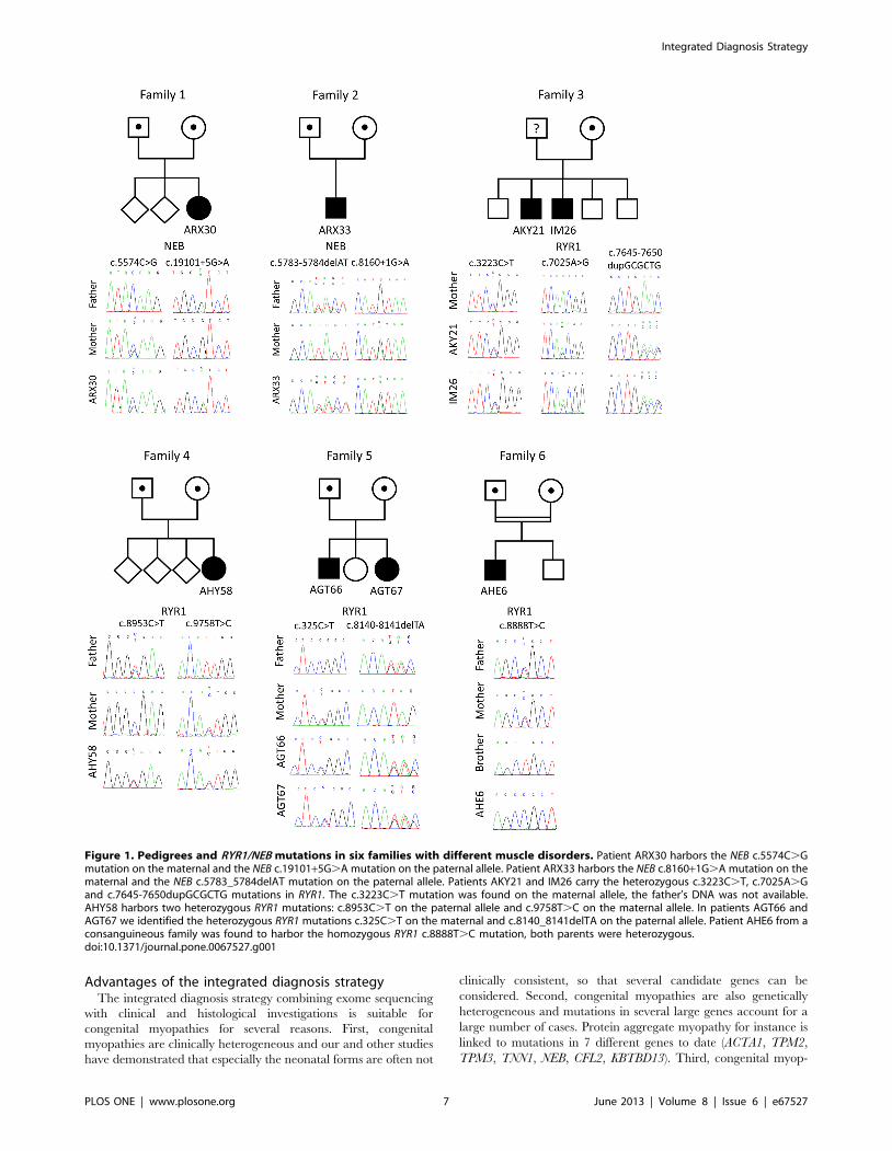
Figure 1. Pedigrees and RYR1/NEB mutations in six families with different muscle disorders. Patient ARX30 harbors the NEB c.5574C.Gmutation on the maternal and the NEB c.19101+5G.A mutation on the paternal allele. Patient ARX33 harbors the NEB c.8160+1G.A mutation on thematernal and the NEB c.5783_5784delAT mutation on the paternal allele. Patients AKY21 and IM26 carry the heterozygous c.3223C.T, c.7025A.Gand c.7645-7650dupGCGCTG mutations in RYR1. The c.3223C.T mutation was found on the maternal allele, the father’s DNA was not available.AHY58 harbors two heterozygous RYR1 mutations: c.8953C.T on the paternal allele and c.9758T.C on the maternal allele. In patients AGT66 andAGT67 we identified the heterozygous RYR1 mutations c.325C.T on the maternal and c.8140_8141delTA on the paternal allele. Patient AHE6 from aconsanguineous family was found to harbor the homozygous RYR1 c.8888T.C mutation, both parents were heterozygous.doi:10.1371/journal.pone.0067527.g001
Integrated Diagnosis Strategy
PLOS ONE | www.plosone.org 7 June 2013 | Volume 8 | Issue 6 | e67527
athies are classified based on histological hallmarks, but the
majority of patients present non-specific features [5]. Some patient
biopsies display a mix of several histological hallmarks as it is the
case for our patient from family 6, increasing the number of
candidate genes. Forth, the analysis can be performed in a unique
laboratory, while Sanger sequencing is mostly performed in
different diagnostic centers specialized for single or a small number
of genes.
Exome sequencing and histological analyses arecomplementary
Massively parallel sequencing allows a fast testing of all genes
previously linked to a given disease, including the large genes [30].
Importantly, exome sequencing also covers new myopathy genes
that will be discovered in the future. The dropping costs of this
technique pave the way for a routine use in molecular diagnosis
and are already far below the estimated expenses for classical
Sanger sequencing of all exons of large genes as RYR1 or NEB.
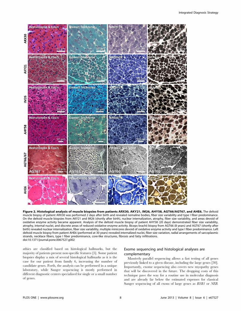
Figure 2. Histological analysis of muscle biopsies from patients ARX30, AKY21, IM26, AHY58, AGT66/AGT67, and AHE6. The deltoidmuscle biopsy of patient ARX30 was performed 2 days after birth and revealed nemaline bodies, fiber size variability and type I fiber predominance.On the deltoid muscle biopsies from AKY21 and IM26 (shortly after birth), nuclear internalization, atrophy, fiber size variability, and areas devoid ofoxidative enzyme activity became apparent. Analysis of the deltoid muscle biopsy of patient AHY58 (20 days) demonstrated fiber size variability,atrophy, internal nuclei, and discrete areas of reduced oxidative enzyme activity. Biceps brachii biopsy from AGT66 (8 years) and AGT67 (shortly afterbirth) revealed nuclear internalization, fiber size variability, multiple minicores devoid of oxidative enzyme activity and type I fiber predominance. Leftdeltoid muscle biopsy from patient AHE6 (performed at 30 years) revealed internalized nuclei, fiber size variation, radial arrangements of sarcoplasmicstrands, necklace fibers, type I fiber predominance, core-like structures, fibrosis and fatty infiltrations.doi:10.1371/journal.pone.0067527.g002
Integrated Diagnosis Strategy
PLOS ONE | www.plosone.org 8 June 2013 | Volume 8 | Issue 6 | e67527
However, exome sequencing does not detect intronic mutations
and may generate a large list of variants of uncertain significance.
The validation of the disease-causing mutations therefore requires
the synergistic combination of the exome sequencing data with
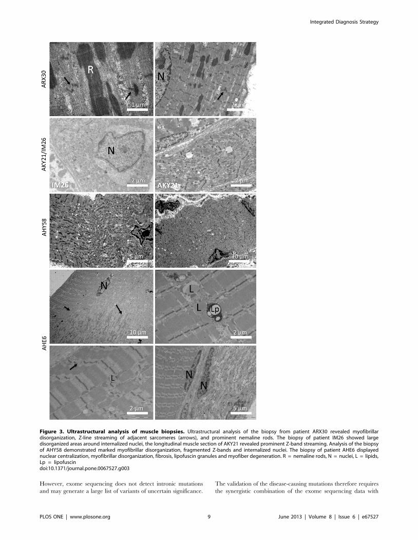
Figure 3. Ultrastructural analysis of muscle biopsies. Ultrastructural analysis of the biopsy from patient ARX30 revealed myofibrillardisorganization, Z-line streaming of adjacent sarcomeres (arrows), and prominent nemaline rods. The biopsy of patient IM26 showed largedisorganized areas around internalized nuclei, the longitudinal muscle section of AKY21 revealed prominent Z-band streaming. Analysis of the biopsyof AHY58 demonstrated marked myofibrillar disorganization, fragmented Z-bands and internalized nuclei. The biopsy of patient AHE6 displayednuclear centralization, myofibrillar disorganization, fibrosis, lipofuscin granules and myofiber degeneration. R = nemaline rods, N = nuclei, L = lipids,Lp = lipofuscindoi:10.1371/journal.pone.0067527.g003
Integrated Diagnosis Strategy
PLOS ONE | www.plosone.org 9 June 2013 | Volume 8 | Issue 6 | e67527
clinical and histological analyses. Exome sequencing and histology
can be performed in parallel and the results need to be evaluated
by specialized diagnostic centers and associated research labora-
tories with an expertise on the underlying pathophysiological
mechanisms.
Conclusion
Exome sequencing or targeted massively parallel sequencing
improves molecular diagnosis of myopathies when combined with
histopathology and molecular validation, and can accelerate the
patient’s access to a better healthcare and disease management.
Supporting Information
Figure S1 Gomori trichrome staining of a tibialisanterior muscle section from patient ARX33 revealedthe presence of nemaline rods.
(TIF)
Figure S2 The novel RYR1 mutations c.3223C.T (Fam-ily 3), c.8888T.C (Family 6) and c.9758T.C (Family 5)affect the conserved residues Arg1075, Leu2963 andIle3253, respectively. Protein alignment demonstrates that
Arg1075 is conserved throughout the listed species. Leu2963 is
replaced by a chemically similar residue in chicken and Ile3253 is
replaced by the chemically similar valine in drosophila and leucine
in the nematode.
(TIF)
Table S1 Homozygosity mapping for AHE6.
(DOCX)
Table S2 Primer sequences and PCR conditions.
(DOCX)
Table S3 Coverage of the congenital myopathy genesfor families 1 and 2.
(DOCX)
Table S4 Coverage of the congenital myopathy genesfor families 3-6.
(DOCX)
Table S5 Web resources.
(DOCX)
Acknowledgments
We thank the families for their cooperation and interest in this study, and
Muriel Philipps, Serge Vicaire, Christelle Thibault-Carpentier, Jean-Luc
Weickert and Linda Manere for technical assistance.
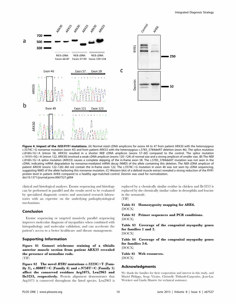
Figure 4. Impact of the NEB/RYR1 mutations. (A) Normal sized cDNA amplicons for exons 44 to 47 from patient ARX30 with the heterozygousc.5574C.G nonsense mutation (exon 45) and from patient ARX33 with the heterozygous c.5783_5784delAT deletion (exon 46). The splice mutationc.8160+1G.A (intron 58, ARX33) resulted in a shorter NEB cDNA amplicon (exons 57–60) compared to the control. The splice mutationc.19101+5G.A (intron 122, ARX30) involved a weak cDNA amplicon (exons 120–124) of normal size and a strong amplicon of smaller size. (B) The NEBc.8160+1G.A splice mutation (ARX33) causes a complete skipping of the in-frame exon 58. The c.5783_5784delAT mutation was not seen in thecDNA, indicating mRNA degradation by nonsense-mediated mRNA decay (NMD) of the allele containing this deletion. The NEB cDNA amplicon ofpatient ARX30 (exons 122–126) did not contain the in-frame exon 122. The c.5574C.G mutation in exon 46 was not seen by cDNA sequencing,suggesting NMD of the allele harboring this nonsense mutation. (C) Western blot of a deltoid muscle extract revealed a strong reduction of the RYR1protein level in patient AHE6 compared to a healthy age-matched control. Desmin was used for normalization.doi:10.1371/journal.pone.0067527.g004
Integrated Diagnosis Strategy
PLOS ONE | www.plosone.org 10 June 2013 | Volume 8 | Issue 6 | e67527
Author Contributions
Conceived and designed the experiments: J. Bohm NBR J. Laporte.
Performed the experiments: J. Bohm NV CF NM J. Brocard VB CK IM
NM. Analyzed the data: J. Bohm NV EM SLG NM IM. Contributed
reagents/materials/analysis tools: BJ HK MG MCW PR J. Lunardi.
Wrote the paper: J. Bohm J. Laporte.
References
1. North K (2008) What’s new in congenital myopathies? Neuromuscul Disord 18:
433–442.2. Wang CH, Dowling JJ, North K, Schroth MK, Sejersen T, et al. (2012)
Consensus statement on standard of care for congenital myopathies. J ChildNeurol 27: 363–382.
3. Nance JR, Dowling JJ, Gibbs EM, Bonnemann CG (2012) Congenital
myopathies: an update. Curr Neurol Neurosci Rep 12: 165–174.4. Sewry CA (2008) Pathological defects in congenital myopathies. J Muscle Res
Cell Motil 29: 231–238.5. Amburgey K, McNamara N, Bennett LR, McCormick ME, Acsadi G, et al.
(2011) Prevalence of congenital myopathies in a representative pediatric united
states population. Ann Neurol 70: 662–665.6. Ng SB, Nickerson DA, Bamshad MJ, Shendure J (2010) Massively parallel
sequencing and rare disease. Hum Mol Genet 19: R119–124.7. Li H, Durbin R (2009) Fast and accurate short read alignment with Burrows-
Wheeler transform. Bioinformatics 25: 1754–1760.8. Li H, Handsaker B, Wysoker A, Fennell T, Ruan J, et al. (2009) The Sequence
Alignment/Map format and SAMtools. Bioinformatics 25: 2078–2079.
9. Ng PC, Henikoff S (2003) SIFT: Predicting amino acid changes that affectprotein function. Nucleic Acids Res 31: 3812–3814.
10. Ramensky V, Bork P, Sunyaev S (2002) Human non-synonymous SNPs: serverand survey. Nucleic Acids Res 30: 3894–3900.
11. Reese MG, Eeckman FH, Kulp D, Haussler D (1997) Improved splice site
detection in Genie. J Comput Biol 4: 311–323.12. Desmet FO, Hamroun D, Lalande M, Collod-Beroud G, Claustres M, et al.
(2009) Human Splicing Finder: an online bioinformatics tool to predict splicingsignals. Nucleic Acids Res 37: e67.
13. Marty I, Robert M, Villaz M, De Jongh K, Lai Y, et al. (1994) Biochemical
evidence for a complex involving dihydropyridine receptor and ryanodinereceptor in triad junctions of skeletal muscle. Proc Natl Acad Sci U S A 91:
2270–2274.14. Robinson R, Carpenter D, Shaw MA, Halsall J, Hopkins P (2006) Mutations in
RYR1 in malignant hyperthermia and central core disease. Hum Mutat 27:977–989.
15. Bevilacqua JA, Monnier N, Bitoun M, Eymard B, Ferreiro A, et al. (2011)
Recessive RYR1 mutations cause unusual congenital myopathy with prominentnuclear internalization and large areas of myofibrillar disorganization.
Neuropathol Appl Neurobiol 37: 271–284.16. Wilmshurst JM, Lillis S, Zhou H, Pillay K, Henderson H, et al. (2010) RYR1
mutations are a common cause of congenital myopathies with central nuclei.
Ann Neurol 68: 717–726.
17. Jungbluth H, Zhou H, Hartley L, Halliger-Keller B, Messina S, et al. (2005)
Minicore myopathy with ophthalmoplegia caused by mutations in the ryanodinereceptor type 1 gene. Neurology 65: 1930–1935.
18. Zhou H, Yamaguchi N, Xu L, Wang Y, Sewry C, et al. (2006) Characterizationof recessive RYR1 mutations in core myopathies. Hum Mol Genet 15: 2791–
2803.
19. Wallgren-Pettersson C, Donner K, Sewry C, Bijlsma E, Lammens M, et al.(2002) Mutations in the nebulin gene can cause severe congenital nemaline
myopathy. Neuromuscul Disord 12: 674–679.20. Pelin K, Hilpela P, Donner K, Sewry C, Akkari PA, et al. (1999) Mutations in
the nebulin gene associated with autosomal recessive nemaline myopathy. Proc
Natl Acad Sci U S A 96: 2305–2310.21. Lawlor MW, Ottenheijm CA, Lehtokari VL, Cho K, Pelin K, et al. (2011) Novel
mutations in NEB cause abnormal nebulin expression and markedly impairedmuscle force generation in severe nemaline myopathy. Skelet Muscle 1: 23.
22. Quane KA, Healy JM, Keating KE, Manning BM, Couch FJ, et al. (1993)Mutations in the ryanodine receptor gene in central core disease and malignant
hyperthermia. Nat Genet 5: 51–55.
23. Zhang Y, Chen HS, Khanna VK, De Leon S, Phillips MS, et al. (1993) Amutation in the human ryanodine receptor gene associated with central core
disease. Nat Genet 5: 46–50.24. Monnier N, Ferreiro A, Marty I, Labarre-Vila A, Mezin P, et al. (2003) A
homozygous splicing mutation causing a depletion of skeletal muscle RYR1 is
associated with multi-minicore disease congenital myopathy with ophthalmo-plegia. Hum Mol Genet 12: 1171–1178.
25. Clarke NF, Waddell LB, Cooper ST, Perry M, Smith RL, et al. (2010) Recessivemutations in RYR1 are a common cause of congenital fiber type disproportion.
Hum Mutat 31: E1544–1550.
26. Romero NB, Monnier N, Viollet L, Cortey A, Chevallay M, et al. (2003)Dominant and recessive central core disease associated with RYR1 mutations
and fetal akinesia. Brain 126: 2341–2349.27. Bohm J, Leshinsky-Silver E, Vassilopoulos S, Le Gras S, Lerman-Sagie T, et al.
(2012) Samaritan myopathy, an ultimately benign congenital myopathy, iscaused by a RYR1 mutation. Acta Neuropathol.
28. Gillard EF, Otsu K, Fujii J, Khanna VK, de Leon S, et al. (1991) A substitution
of cysteine for arginine 614 in the ryanodine receptor is potentially causative ofhuman malignant hyperthermia. Genomics 11: 751–755.
29. Jungbluth H, Sewry CA, Muntoni F (2011) Core myopathies. Semin PediatrNeurol 18: 239–249.
30. Vasli N, Laporte J (2013) Impacts of massively parallel sequencing for genetic
diagnosis of neuromuscular disorders. Acta Neuropathol 125: 173–185.
Integrated Diagnosis Strategy
PLOS ONE | www.plosone.org 11 June 2013 | Volume 8 | Issue 6 | e67527