Embed Size (px)
Citation preview
Montclair State University Montclair State University
Montclair State University Digital Montclair State University Digital
Commons Commons
Theses, Dissertations and Culminating Projects
5-2018
An Eye-Tracking Investigation of Facial Affect Recognition in An Eye-Tracking Investigation of Facial Affect Recognition in
Traumatic Brain Injury and Healthy Individuals Traumatic Brain Injury and Healthy Individuals
Joseph Walter DeAngelis Montclair State University
Follow this and additional works at: https://digitalcommons.montclair.edu/etd
Part of the Psychology Commons
Recommended Citation Recommended Citation DeAngelis, Joseph Walter, "An Eye-Tracking Investigation of Facial Affect Recognition in Traumatic Brain Injury and Healthy Individuals" (2018). Theses, Dissertations and Culminating Projects. 126. https://digitalcommons.montclair.edu/etd/126
This Thesis is brought to you for free and open access by Montclair State University Digital Commons. It has been accepted for inclusion in Theses, Dissertations and Culminating Projects by an authorized administrator of Montclair State University Digital Commons. For more information, please contact [email protected].
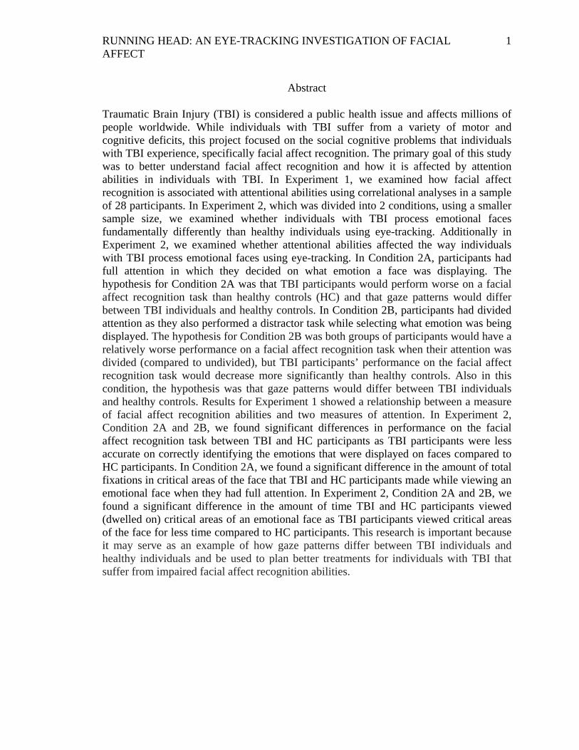
RUNNING HEAD: AN EYE-TRACKING INVESTIGATION OF FACIAL AFFECT
1
Abstract
Traumatic Brain Injury (TBI) is considered a public health issue and affects millions of people worldwide. While individuals with TBI suffer from a variety of motor and cognitive deficits, this project focused on the social cognitive problems that individuals with TBI experience, specifically facial affect recognition. The primary goal of this study was to better understand facial affect recognition and how it is affected by attention abilities in individuals with TBI. In Experiment 1, we examined how facial affect recognition is associated with attentional abilities using correlational analyses in a sample of 28 participants. In Experiment 2, which was divided into 2 conditions, using a smaller sample size, we examined whether individuals with TBI process emotional faces fundamentally differently than healthy individuals using eye-tracking. Additionally in Experiment 2, we examined whether attentional abilities affected the way individuals with TBI process emotional faces using eye-tracking. In Condition 2A, participants had full attention in which they decided on what emotion a face was displaying. The hypothesis for Condition 2A was that TBI participants would perform worse on a facial affect recognition task than healthy controls (HC) and that gaze patterns would differ between TBI individuals and healthy controls. In Condition 2B, participants had divided attention as they also performed a distractor task while selecting what emotion was being displayed. The hypothesis for Condition 2B was both groups of participants would have a relatively worse performance on a facial affect recognition task when their attention was divided (compared to undivided), but TBI participants’ performance on the facial affect recognition task would decrease more significantly than healthy controls. Also in this condition, the hypothesis was that gaze patterns would differ between TBI individuals and healthy controls. Results for Experiment 1 showed a relationship between a measure of facial affect recognition abilities and two measures of attention. In Experiment 2, Condition 2A and 2B, we found significant differences in performance on the facial affect recognition task between TBI and HC participants as TBI participants were less accurate on correctly identifying the emotions that were displayed on faces compared to HC participants. In Condition 2A, we found a significant difference in the amount of total fixations in critical areas of the face that TBI and HC participants made while viewing an emotional face when they had full attention. In Experiment 2, Condition 2A and 2B, we found a significant difference in the amount of time TBI and HC participants viewed (dwelled on) critical areas of an emotional face as TBI participants viewed critical areas of the face for less time compared to HC participants. This research is important because it may serve as an example of how gaze patterns differ between TBI individuals and healthy individuals and be used to plan better treatments for individuals with TBI that suffer from impaired facial affect recognition abilities.
AN EYE-TRACKING INVESTIGATION OF FACIAL AFFECT 3
AN EYE-TRACKING INVESTIGATION OF FACIAL AFFECT RECOGNITION IN TRAUMATIC BRAIN INJURY AND HEALTHY INDIVIDUALS
A THESIS
Submitted in partial fulfillment of the requirements
For the degree of Master of Arts
By
JOSEPH WALTER DEANGELIS
Montclair State University
Montclair, NJ
2018

AN EYE-TRACKING INVESTIGATION OF FACIAL AFFECT 4
Copyright © 2018 by Joseph Walter DeAngelis. All rights reserved.

AN EYE-TRACKING INVESTIGATION OF FACIAL AFFECT 6
Table of Contents Page
Abstract ................................................................................................................................1 Signature Page .....................................................................................................................2 Title Page .............................................................................................................................3 Copyright Page.....................................................................................................................4 Acknowledgements ..............................................................................................................5 Table of Contents ...........................................................................................................6 – 8 List of Tables .......................................................................................................................9 List of Figures ....................................................................................................................10 List of Supplemental Figures & Illustrations .....................................................................11 Introduction ................................................................................................................12 – 25 Order of Thesis .........................................................................................................12 What is Traumatic Brain Injury ........................................................................12 – 13 Prevalence of TBI ............................................................................................13 – 14 Causes and Symptoms of TBI..........................................................................14 – 17 What is Social Cognition? .......................................................................................17 Why Study Social Cognition? ..........................................................................18 – 20 Studying Facial Affect Recognition................................................................20 – 21 Eye – Tracking ................................................................................................21 – 22 How Healthy Individuals View a Face ...........................................................22 – 23 Current Study ..................................................................................................23 – 24 Aims & Hypotheses ........................................................................................24 – 25 Experiment 1 – Methods ............................................................................................25 – 28
AN EYE-TRACKING INVESTIGATION OF FACIAL AFFECT 7
Participants ......................................................................................................25 – 26 Measures .........................................................................................................26 – 28 Experiment 1 – Results ..............................................................................................28 – 29 Relationship Between Facial Affect Recognition Ability & Attention .................28 Experiment 1 – Summary of Findings ...................................................................29 Experiment 2 – Methods ............................................................................................29 – 35 Participants .............................................................................................................29 Eye – Tracker .........................................................................................................30 Stimuli ............................................................................................................30 – 31 ROI Creation ..................................................................................................31 – 32 Experiment 2 – Design ...........................................................................................33 Procedure .......................................................................................................33 – 34 Statistical Analyses ........................................................................................34 – 35 Experiment 2 – Results ..............................................................................................35 – 44 Accuracy Rates on Condition 2A vs. Condition 2B ..............................................35
Accuracy Rates on Each Emotion in Condition 2A .......................................35 – 36
Accuracy Rates on Each Emotion in Condition 2B ...............................................36
Interaction Between Participant Group and Attention ...................................37 – 38
Eye – Tracking Analysis ........................................................................................38
Number of Fixations Inside and Outside of the ROIs in Condition 2A .................38
Number of Fixations Inside and Outside of the ROIs in Condition 2B .................39
Fixations in Specific ROIs in Condition 2A ..........................................................39
Fixations in Specific ROIs in Condition 2B ..........................................................40
AN EYE-TRACKING INVESTIGATION OF FACIAL AFFECT 8
Dwell Time Inside & Outside of ROIs in Condition 2A .................................40 – 41
Dwell Time Inside & Outside of ROIs in Condition 2B .................................41 – 42
Dwell Time Inside & Outside of Specific ROIs in Condition 2A ...........................42
Dwell Time Inside & Outside of Specific ROIs in Condition 2B ...................42 – 43
Experiment 2 – Summary of Findings .............................................................43 – 44
Discussion ..................................................................................................................45 – 55
Correlations Between Attention and Facial Affect Recognition .......................46 – 47
Behavioral ..........................................................................................................47 – 50
Eye – Tracking ...................................................................................................50 – 53
Limitations .................................................................................................................54
Future Directions ...............................................................................................54 – 55
References ..........................................................................................................56 – 60
Supplemental Figures & Illustrations ................................................................................61
AN EYE-TRACKING INVESTIGATION OF FACIAL AFFECT 9
List of Tables
Table Page 1. Participant information in Experiment 2.............................................................29 2. Emotion accuracy rates in Condition 2A ................................................................. 35 3. Emotion accuracy rates in Condition 2B .............................................................36 4. Number of fixations inside and outside of the ROIs in Condition 2A ................38 5. Number of fixations inside & outside of the ROIs in Condition 2B ..................39 6. Number of fixations on the eyes, nose, and mouth ROIs in Condition 2A……39 7. Number of fixations on the eyes, nose, and mouth ROIs in Condition 2B .........40 8. Dwell time inside and outside of ROIs in Condition 2A ....................................40 9. Dwell time inside and outside of ROIs in Condition 2B ....................................41 10. Dwell time inside and outside of specific ROIs in Condition 2A .......................42 11. Dwell time inside and outside of specific ROIs in Condition 2B .......................43
AN EYE-TRACKING INVESTIGATION OF FACIAL AFFECT 10
List of Figures
Figure Page 1. Example of each emotion ......................................................................................30 2. Example of eyes, nose, and mouth ROIs ..............................................................32 3. Column chart depicting mean accuracy on the facial affect recognition task .......35 4. Column chart depicting mean accuracy rates on each emotion in Condition 2A ..36 5. Column chart depicting mean accuracy rates on each emotion in Condition 2B ..36 6. Line chart depicting performance on the facial affect recognition task in the full and divided attention conditions for both groups of participants ......................................37 7. Total number of fixations made on the faces throughout the facial affect recognition task in Condition 2A .......................................................................................38 8. Total number of fixations made on the faces throughout the facial affect recognition task in Condition 2B .......................................................................................39 9. Fixations made in the eyes, nose, and mouth ROIs in Condition 2A .....................39 10. Fixations made in the eyes, nose, and mouth ROIs in Condition 2B .....................40 11. Dwell time inside and outside of the ROIs in Condition 2A ..................................40 12. Dwell time inside and outside of the ROIs in Condition 2B ..................................41 13. Dwell time percentage in the eyes, nose, mouth, and non – ROIs in Condition 2A. .....................................................................................................................42 14. Dwell time percentage in the eyes, nose, mouth, and non – ROIs in Condition 2B ......................................................................................................................43

AN EYE-TRACKING INVESTIGATION OF FACIAL AFFECT 11
List of Supplemental Figures and Illustrations
Illustration Page 1. Bivariate correlation matrix between the TOFER, Digit Span, and SDMT ..................61
AN EYE-TRACKING INVESTIGATION OF FACIAL AFFECT 12
Introduction
Order of Thesis
This thesis is constructed in the following ways: in the section of the introduction,
it presents a background of Traumatic Brain Injury (TBI), why it is necessary to study it,
how it occurs and its symptoms, and the research methods used in studying it. Next, the
thesis will focus on social cognition by giving a description of it, its importance to study,
and methods used in studying it. Then the thesis will focus on the method used in the
current study to examine social cognition in TBI: eye-tracking. The introduction will then
focus on the study, hypotheses, and specific aims and goals of the experiment. In the
second section, the experiment, methods, and results will be presented. In the third
section, the thesis will conclude with a general discussion of the experiment.
What is Traumatic Brain Injury?
TBI can be defined as an alteration in brain function caused by an injury/impact to
the head or brain. It may manifest into seizure, coma, confusion, altered levels of
consciousness, sensory or motor neurological deficits, and more (Bruns & Hauser, 2003).
It is important to understand the distinction between TBI and Head Injury (HI). HI is a
nonspecific term that includes external injuries to the head, face and scalp. It may also
include contusions, lacerations, abrasions, and fractures and may or may not be
associated with TBI (Bruns & Hauser, 2003). TBI, however is damage to the brain or the
tissues in the brain (Bruns & Hauser, 2003). Acquired Brain Injury (ABI) is an umbrella
term that includes TBI but TBI refers to an acquired, sudden-onset, non-progressive, and
non-degenerative condition while ABI includes all brain injuries (Bruns & Hauser, 2003).
AN EYE-TRACKING INVESTIGATION OF FACIAL AFFECT 13
There are 3 main classifications for TBI severity: mild, moderate, and severe. The
Glasgow Coma Scale is currently the most widely used clinical assessment tool in
classifying TBI severity. It is based on an individual’s responses of eye opening, verbal
function, and motor function to different stimuli. Traditionally, a score of 13 to 15 is
considered mild, 9 to 12 is considered moderate, and < 9 is considered severe (Teasdale
et al., 1979).
Prevalence of TBI
TBI affects millions of people worldwide and is considered a public health
problem (Stocchetti & Zanier, 2016). Due to more people aging in the Western
hemisphere, falls in the elderly increase the incidence of TBI (Roozenbeek et al., 2013).
Individuals with TBI require prolonged hospital care, require long-term rehabilitation,
and may suffer from cognitive, physical, and mental disabilities that affect them
throughout their lifetime. Globally, TBI survivors generally have a lower life expectancy
than the general population (Rosenfeld et al., 2012). This may be due to the difficulties
that TBI individuals experience including motor and cognitive complications. A motor or
cognitive difficulty may affect an individual’s life expectancy because an individual with
a motor difficulty may not be able to perform tasks that require motor skills, like walking
or exercising. Individuals with cognitive difficulties may not be able to make logical or
healthy choices in their life, which may lead them to make poor/risky decisions regarding
their health.
The consequences of TBI also present economic costs to individuals that suffer
from severe TBI. In the United States of America, the total lifetime cost of severe TBI
per case is approximately $400,000.00 and this figure is attributed to lost productivity
AN EYE-TRACKING INVESTIGATION OF FACIAL AFFECT 14
and disability of the individual affected (Rosenfeld et al., 2012). With TBI as a public
health problem and the obvious economic consequences of TBI, researchers should invest
more time into studying TBI.
Causes and Symptoms of TBI
TBI usually occurs when some form of external or mechanical force acts on the
head or body causing brain dysfunction. The most common causes of TBI in the USA
are: falls, motor vehicle accidents, assault, or being struck by another individual or an
object (Thurman et al., 1999). This section will first provide an understanding of the
biology behind TBI and then present the symptomology.
Individuals that suffer from TBI are affected by a variety of brain dysfunctions
that include white matter degradation and protein misfolding. When TBI occurs, it may
cause alterations or disruptions in the axonal cytoskeleton and possibly impair axonal
transport. TBI may damage structural networks in the brain and damage communication
between neurons (Rodriguez-Paez et al., 2005). Individuals with TBI may be at risk for
developing other neurodegenerative disorders including dementia and Alzheimer’s
disease (Johnson et al., 2010). It has been shown that individuals with TBI accumulate
amyloid- β peptides and have defective tau proteins, both of which are associated with
Alzheimer’s disease (Johnson et al., 2010).
Individuals affected by TBI may have physical disabilities, cognitive
impairments, a higher rate of developing other psychiatric disorders, and impairments in
social functioning. In terms of physical disabilities, people with TBI may have motor
deficits that include balance and gait issues and spasticity problems. Individuals with TBI
may experience difficulties balancing, standing, and walking at a normal rate (Basford et
AN EYE-TRACKING INVESTIGATION OF FACIAL AFFECT 15
al., 2003). Individuals with TBI may also experience muscle over-activity, muscle
rigidity, muscle tremors, and motor weakness (Bergfeldt et al., 2006).
In terms of cognitive issues, TBI may cause deficits in learning, attention,
memory, information processing speed, and other high-level cognitive functions
(Stocchetti & Zanier, 2016). TBI individuals may experience prolonged memory loss
after TBI and may also have difficulties in short-term and working memory (Lyeth et al.,
1990). People affected by TBI may experience difficulties in executive functioning. This
can include difficulties in planning and motivation. For example, an individual with TBI
may find it difficult to plan their day and have motivation to perform the tasks of their
day (Cicerone et al., 2006). Also, individuals affected by TBI may be at higher risk for
developing psychiatric disorders including, anxiety, depression, psychosis, and other
disruptive behaviors and personality changes. The comorbidity of TBI with these
disorders makes an individual at greater risk for substance abuse (Zgaljardic et al., 2015).
One issue that individuals with TBI experience and one of the main focuses of
this project is attention. Individuals with TBI experience difficulties in goal oriented
behavior because this behavior depends on sustaining attention (Bonnelle et al., 2011). It
is also known that individuals with TBI have difficulty in divided attention, or
performing more than one thing/paying attention to more than one thing at a time. Many
studies show that individuals with TBI consistently perform worse than healthy controls
on tasks that require divided attention and this might be due to the fact that individuals
with TBI are not be able to sustain the required cognitive resources to pay attention to
more than one thing for long periods of time (Azouvi et al., 2004). In Azouvi et al.,
(2004), participants with moderate to severe TBI and healthy controls completed an
AN EYE-TRACKING INVESTIGATION OF FACIAL AFFECT 16
experimental task and a distractor task simultaneously and were measured on the speed
and accuracy of their responses. The experimental task was a visual go-no go task and the
distractor task was a random number generation test. The visual go-no go task consisted
of a cross and a circle presented on a computer screen. Participants were instructed to
respond by pressing a button on the computer keyboard whenever the cross appeared and
to not respond whenever the circle appeared. The distractor task was a random number
generation test in which participants had to randomly say a number aloud between 1 and
10. Participants were instructed to avoid patterns (i.e., saying 1,2,3,4 and 2,4,6,8) while
saying the numbers aloud. The study found that TBI individuals rated both tasks as more
difficult and responded less frequently and less accurately than the healthy controls did in
the go-no go task (Azouvi et al., 2004).
The combination of cognitive, physical, and emotional processing difficulties that
an individual with TBI may experience may lead to difficulties reintegrating into their
communities and may affect their overall quality of life (QoL). Individuals with TBI may
have poor conversation abilities that include making poor or crude jokes, suddenly
changing topics, focusing too much on oneself, making uninhibited remarks or unwanted
advances, and over disclosing personal information (McDonald et al., 2003).
It has been shown that individuals with TBI experience difficulties in social
cognition, a set of skills which includes recognizing emotions on faces, Theory of Mind
(ToM), and interpreting social cues (Croker & McDonald, 2005, Babbage et al., 2011).
Furthermore, individuals with TBI experience interpersonal problems including
difficulties in social communication (effectively communicating their thoughts, opinions,
and/or desires to their friends, family, and peers) and difficulties in maintaining social
AN EYE-TRACKING INVESTIGATION OF FACIAL AFFECT 17
and romantic relationships (Struchen et al., 2011). These problems in interpersonal
communication are important to study because of the social nature of today’s society.
People need to communicate effectively with others in order to achieve their goals. Often,
the first step in effective communication is recognizing and understanding the emotions
an individual is displaying on their face (facial affect recognition) (Crocker & McDonald,
2005). If an individual with TBI cannot effectively recognize and interpret emotion being
displayed on someone’s face, this may lead to a negative social interaction. Since
individuals with TBI experience interpersonal problems, this thesis focuses on social
cognition impairments, specifically in facial affect recognition, how which individuals
with TBI suffer.
What is Social Cognition?
Social cognition is a broad term used to describe the way social information is
processed. This includes the ability to detect what emotions people are feeling (or
showing) and appropriately respond to these emotions (Henry et al., 2015). Two main
components of social cognition are facial affect recognition and Theory of Mind (ToM).
Facial affect recognition refers to an individual’s ability to accurately recognize the
emotion displayed on someone’s face and ToM refers to one’s ability to attribute mental
states (beliefs, desires, intents, etc.) to themselves and others. It also describes the ability
to understand that other people have different perspectives and intentions from their own.
This thesis will focus on facial affect recognition.
Why Study Social Cognition
While many studies focus on motor and physical problems of individuals with
TBI, it is also important to focus on the social deficits of individuals with TBI. This is an
AN EYE-TRACKING INVESTIGATION OF FACIAL AFFECT 18
important aspect to study because people live social lives. People live in a social
environment in which they express their feelings and emotions. Often, individuals with
TBI rely on caretakers, family, and friends for help accessing/providing medical services
and social care. For example, an individual with TBI may need to have a friend drive
them to the doctor’s office. This involves social planning as both individuals have to
communicate about the pick-up time, the appointment time, and the drop off time. If an
individual with TBI cannot successfully communicate with others then this may lead to
social isolation and trouble reintegrating into society after the brain injury. If an
individual with TBI becomes socially isolated due to impairments in social cognitive
abilities, then this may lead to negative thoughts and emotions because they are isolated
(e.g., nobody to talk to/spend time with, nobody to express feelings to, etc.). For this
reason, it is important to understand how individuals with TBI function socially.
Individuals with TBI have difficulty in facial affect recognition (recognizing
emotions displayed on faces). A meta-analysis of 296 adults with moderate to severe TBI
from 13 different studies conducted by Babbage et al., (2011), showed that up to 39% of
individuals with severe TBI have difficulty in recognizing emotions from static
presentations of facial expressions. It is also known that recognition of emotional
expression in voice is impaired following TBI (Dimoska et al., 2010). This is important
due to the role attention may play in facial affect recognition. An individual with TBI
may have to focus on what someone is saying and their facial expression at the same
time.
While Individuals with TBI have difficulty recognizing all emotions presented on
a face compared to healthy controls, and they have particular difficulties in recognizing
AN EYE-TRACKING INVESTIGATION OF FACIAL AFFECT 19
negative emotions. One study found that individuals with TBI experience a greater deficit
in recognizing negative emotions (e.g., sadness, anger, and disgust) than positive
emotions (e.g., happiness and surprise) (Croker & McDonald, 2005). This is important
because individuals with TBI may observe negative emotions often. For example, an
individual with TBI’s caretaker or family member may be upset about a particular issue.
An individual with TBI may mistake the sadness on their caretaker’s or family member’s
face as anger and assume the caretaker or family member is angry with them. This could
result in a possible argument or a strained relationship between them. The primary goal
of this study was to understand how individuals with TBI view different facial/emotional
expressions.
This project also sought to understand how attention contributes to facial affect
recognition. In order for an individual with TBI to recognize the emotion being displayed
on a face, they must first pay attention to that face. In terms of divided attention, an
individual with TBI may experience situations in which they are talking to/discerning the
facial expression of someone in a noisy or loud environment (e.g., a party or another
social setting like the mall, grocery store, etc.) or may have to recognize the facial
expressions of two people at the same time. Further, an individual with TBI may have to
recognize the facial expression of someone whose facial expression does not match what
he or she is saying. For example, someone might say they are happy but their facial
expression shows anger. An individual with TBI may have to divide their attention to
what that person is saying and their facial expression while saying it. A good example of
this is sarcasm and lying. It has been shown that individuals with TBI experience
difficulties in understanding when someone is being sarcastic and determining if
AN EYE-TRACKING INVESTIGATION OF FACIAL AFFECT 20
someone is lying (Honan et al., 2016). This is important because sarcasm is used by
people in daily conversations and is used to make jokes. If an individual with TBI does
not understand that someone is being sarcastic, then this could lead to a negative social
engagement. If an individual with TBI does not understand if/when someone is lying,
then this could also lead to a negative social engagement.
Studying Facial Affect Recognition
The most common way to assess social cognition, specifically facial affect
recognition abilities, is by displaying static images of faces showing different emotions to
the participants and having the participants state what emotion is being shown on the face
(Henry et al., 2015). Many studies have used the standardized Ekman & Friesen (1971)
stimuli set. These stimuli consist of different black and white photographs of actors
displaying 6 basic emotions (happiness, sadness, disgust, fear, anger, surprise, and a
neutral facial expression). While studies that use static images are helpful, they do
present some disadvantages. One of the main disadvantages of using static stimuli is that
they are not ecologically valid. In a more real-world setting, people are usually
interacting with each other in a noisy environment with sounds and other distractions
taking place. This may make it more difficult for an individual with TBI to focus and
recognize the emotion presented on someone’s face. Another main disadvantage of the
studies mentioned before is they do not provide an explanation of how an individual with
TBI processes facial expressions. These studies show that TBI individuals have difficulty
in facial affect recognition but do not explain how they experience these difficulties or
how they are making the mistakes leading up to incorrectly identifying a specific emotion
presented on a face.
AN EYE-TRACKING INVESTIGATION OF FACIAL AFFECT 21
Eye-Tracking
Eye-tracking is used to investigate gaze behavior and can provide insight into
social cognition. Eye-tracking studies are generally conducted by illuminating the eye
with an infrared beam and then capturing the reflected image on a video camera. The
cornea and the pupil are two parts of the eye that are captured from the reflected image.
This gives one enough information to determine what and where on a screen/image a
participant is looking (Boraston & Blakemore, 2007).
The current project utilized eye-tracking as one of the main paradigms because
understanding how an individual with TBI views facial expression is important. Studies
show that individuals have difficulties in facial affect recognition but not how these
difficulties occur. This study is different than many studies examining facial affect
recognition in individuals with TBI because it investigated how individuals with TBI
viewed facial expression. Understanding how these difficulties occur is important
because it can help develop treatments/interventions for individuals suffering from TBI.
For example, if an individual is focusing on a part of the face for too long/too short then
this information can be integrated into treatments/interventions designed to improve
facial affect recognition abilities.
This project sought to understand if TBI individuals view a face fundamentally
different than healthy individuals (e.g., TBI individuals may fixate on a part of a face for
too long or too short a time period compared to healthy controls). This project also
investigated full vs. divided attention of facial affect recognition in individuals with TBI.
Specifically, how facial affect recognition performance changes when participants have
full attention or divided attention on the task. This is important because this study sought
AN EYE-TRACKING INVESTIGATION OF FACIAL AFFECT 22
to achieve higher ecological validity than other studies investigating facial affect
recognition. This study may serve as an example for other researchers investigating social
cognition in TBI using eye-tracking and be used to plan better treatments for individuals
with TBI that suffer from impaired facial affect recognition abilities. For example, if an
individual with TBI has difficulty recognizing the emotion sadness on a face, then a
treatment/intervention could be developed to remedy this. Specifically, eye-tracking
helps plan better treatments because it allows researchers and other individuals to
understand where a socially impaired individual is looking at on a facial expression and
for how long. It is not enough to just know that an individual is impaired on recognizing
an emotion on a face. Eye-tracking allows researchers to understand whether an
individual with TBI has difficulty recognizing emotion on a face is viewing the mouth,
nose, or eyes for too long or too short a time period compared to healthy individuals. This
information can then be used as feedback for that individual and this information can be
used to initiate changes in the individual’s gaze behavior.
How Healthy Individuals View a Face
Since this project focuses on how TBI individuals and healthy individuals view a
face, it is important to understand the ways healthy individuals view a face. In a study
conducted by (Dalton et al., 2005), autistic and healthy children viewed emotional facial
expressions and non-emotional facial expressions while their eyes were being tracked.
Healthy children viewed the eyes and mouth of an emotional and non-emotional face for
a significantly longer amount of time than the autistic children. In adult studies, it has
been shown that healthy adults fixate mainly on the eyes, the nose, and the mouth (the
“core features” of a face). Healthy adults first fixate on the eyes of an emotional facial
AN EYE-TRACKING INVESTIGATION OF FACIAL AFFECT 23
expression then the nose, and then the mouth with the eyes being the most fixated on area
(Boraston & Blakemore, 2007). Most normal adults spend different amounts of time
fixating on different areas of a face for each emotion (Boraston & Blakemore, 2007). For
example, a healthy participant may view the mouth region of a surprised face for a longer
amount of time compared to other areas on the face if that face has an open mouth, which
may indicate the feeling of surprise. A healthy participant may view the nose region of an
angry face for a longer amount of time compared to other areas on the face if that face
has a scrunched nose, which may indicate the feeling of anger (Boraston & Blakemore,
2007).
Current Study
The current study focused on facial affect recognition and consisted of two
experiments. Experiment 1 sought to understand how facial affect recognition was
associated with attentional abilities. This was done by correlating measures of attention
and a measure of facial affect recognition abilities. This would lead to the understanding
if attention positively or negatively affects facial affect recognition abilities. In
Experiment 2, there were 2 conditions, which both utilized eye-tracking. The first
condition (Condition 2A), sought to understand how TBI and healthy individuals’
performance differs in identifying emotions and how participants’ gaze patterns differ.
This was conducted using eye-tracking and, in this condition, participants had full
attention on the task. The second condition (Condition 2B), also sought to understand
how TBI and healthy individuals’ performance differs on identifying emotions and how
participants’ gaze patterns differed when their attention was divided. This was also
conducted using eye-tracking. In this condition, participants had divided attention on the
AN EYE-TRACKING INVESTIGATION OF FACIAL AFFECT 24
task as they engaged in a distractor task while simultaneously completing the facial affect
identification task.
Aims and Hypotheses
The question that this project sought to answer is why social cognitive deficits
occur in individuals with TBI. In experiment 1, the hypothesis was that there would be a
positive relationship between attention and facial affect recognition (i.e., if a participant
scores high on a measure of attention, then they will also score high on a measure of
facial affect recognition). This might be because an individual needs to pay attention to
the expressions on a face in order to correctly identify the emotion being displayed.
In Experiment 2, in condition 2A, the hypothesis was that TBI participants would
perform worse on the facial affect recognition task than healthy controls. In terms of eye-
tracking in condition 2A, the hypothesis was that gaze patterns would differ between TBI
individuals and healthy controls. Specifically, the prediction was that TBI individuals
would fixate more on non-regions of interest (ROI) than the main ROIs compared to
healthy controls. ROIs are regions or areas on a face that individuals can view in order to
gain information on a face. In experiment 2, the main ROIs were the eyes, nose, and the
mouth and the non-ROIs were the ears, hair, and other facial area. The eyes, nose, and
mouth were determined as the main ROIs because they have been shown to provide more
information about emotion compared to the non-ROIs, in both adult and child studies
(Boraston & Blakemore, 2007; Dalton et al., 2005). For example, a surprised individual
may have their mouth open when they express surprise. Thus, someone viewing the
mouth may be viewing it in order to distinguish what emotion is being displayed.
AN EYE-TRACKING INVESTIGATION OF FACIAL AFFECT 25
In condition 2B, the hypothesis was that both groups of participants would have
relatively low performance on the facial affect recognition task when their attention was
divided (compared to undivided), but TBI participants’ performance on the facial affect
recognition task would reduce more significantly than healthy controls. In the eye
tracking aspect of condition 2B, the hypothesis was also that gaze patterns would differ
between TBI individuals and healthy controls. Specifically, TBI participants would lose
focus on the facial affect recognition task and may make fewer fixations of the main
ROIs of an emotional facial expression compared to healthy controls when they are asked
to simultaneously view a face and complete a distractor task. This is because an
individual with TBI may not be able to fixate on the main ROIs of an emotional face as
often as healthy controls due to issues in divided attention.
Experiment 1 - Methods
Participants
Data was drawn from a randomized clinical trial that examined social cognitive
deficits in individuals with TBI conducted by Kessler Foundation. Participants were
recruited from Kessler Foundation’s participant information database. There were 28 TBI
participants (23 males and 5 females). The TBI participants met the criteria of the current
study, which were:
(1) Age 25 – 65 years
(2) Had sustained a moderate or severe TBI as determined by the Glasgow Coma
Scale score less than or equal to 12 or post-traumatic amnesia or loss of
consciousness of at least 24 hours.
(3) Age 18 or older at the time of injury.
AN EYE-TRACKING INVESTIGATION OF FACIAL AFFECT 26
(4) At least one year after injury.
Participants were excluded from the study if they possessed impaired vision or
hearing, had pre-injury psychiatric history, and/or had substance dependence. The
mean age of TBI participants was 45 years (SD = 12.3) and they were on average 9.5
years post injury (SD = 11). The mean length of education was 14 years (SD = 2.19).
Participants sustained mild TBI (4%), moderate TBI (11% of participants), and severe
TBI (46% of participants). 39% of TBI participants in this sample had an injury
severity that was unknown.
Measures
These 3 assessments were utilized in order to evaluate the relationship between
facial affect recognition ability and attention.
Digit Span: Each segment of this test (forward and backward) consisted of seven
pairs of random number sequences that the examiner read aloud at the rate of one
per second. Both segments depended upon auditory attention and working
memory to be performed effectively. In the digit span forward segment, the
participant was instructed to repeat the string of digits in the same order in which
they were presented by the examiner. Conversely, in the digit span backward
segment, the subject was instructed to repeat the string of digits in the reverse
order. The Digit Span test has also shown high internal consistency reliability
(r=.90) (Wechsler, 1997).
Symbol Digit Modalities Test (SDMT): The SDMT involved the conversion of a
set of simple geometric designs into a written response. It has been demonstrated
to be sensitive to the presence of brain damage in numerous studies. The SDMT
AN EYE-TRACKING INVESTIGATION OF FACIAL AFFECT 27
required the examinee to substitute a number for a randomized presentation of a
geometric figure. The appropriate number was shown in a key containing the
Arabic numbers 1 through 9, each with a different geometric figure. The SDMT
has shown good test-retest (r=.76) and alternate forms (r=.82, r=.84) reliability.
The sensitivity of the SDMT to the cognitive effects of a number of neurological
illnesses and injuries has been demonstrated repeatedly (Smith, 1982).
Task of Facial Emotion Recognition (TOFER): The TOFER consisted of 36 black
and white images that are of faces expressing one of the following 6 emotions:
happiness, fear, anger, sadness, surprise, or disgust. The images were taken from
The Karolinska Directed Emotional Faces (KDEF)—a database of 4900 pictures
of humans expressing different emotions at different angles (Goeleven et al.,
2008). All of the faces faced directly toward the camera or screen, and 6 images
of each emotion were presented. Participants taking part in a study utilizing the
TOFER are asked to “select the emotion that best fits the actor’s facial
expression,” and to “respond as quickly as possible.” A total score on the TOFER
is the sum of the number of correct responses, and each subscore is the sum of
correct responses within a particular emotion. The psychometric properties of the
KDEF database have been examined to ensure that the stimuli are valid (Goeleven
et al., 2008). To ensure that the emotions portrayed by the stimuli are accurately
identified at a rate higher than chance; chance proportion scores were calculated
for each emotion separately. Analyses suggested that selection of the intended
emotion was far above chance level for every emotion (p<.0001) (Goeleven et al.,
2008). Test-retest reliability was high for the KDEF pictures: 87.96% of the
AN EYE-TRACKING INVESTIGATION OF FACIAL AFFECT 28
emotions were rated the same at time point 1 and time point 2 (separated by 1
week) (Goeleven et al., 2008).
We conducted Pearson correlations (one-tailed) between the TOFER and the two
measures of attention (Digit Span and SDMT). This was conducted in order to measure if
facial affect recognition ability is associated with attention.
Experiment 1 - Results
Relationship Between Facial Affect Recognition Ability & Attention
Pearson correlations were conducted between the one measure of facial affect
recognition ability (TOFER) and the two measures of attention (Digit Span and SDMT)
(See Illustration 1 in the “Supplemental Figures & Tables” section for all correlations).
The correlations were based on the one-tailed level because we had a specific hypothesis
of the direction of the relationship between facial affect recognition ability and attention
(i.e., an individual must utilize attention in order to accurately identify an emotion on a
face). There was a significant positive correlation between the TOFER and the Digit Span
(forward version) r (26) = .44, p = .011. There was a significant positive correlation
between the TOFER and the Digit Span (backward version) r (26) = .36, p = .034. There
was a significant positive relationship between the TOFER and the total score of the Digit
Span r (26) = .41, p = .017. There was a significant positive correlation between the
TOFER and the SDMT r(26) = .57, p = .001. These significant positive correlations
indicate that attention has a role in the ability to identify emotions on faces.
Experiment 1 – Summary of Findings
In Experiment 1, we found that there was a significant positive correlation
between the TOFER and the Digit Span and the TOFER and the SDMT. This suggests
AN EYE-TRACKING INVESTIGATION OF FACIAL AFFECT 29
that there is a link between facial affect recognition abilities and attention. Our hypothesis
for Experiment 1 was confirmed.
Experiment 2 – Methods
Participants
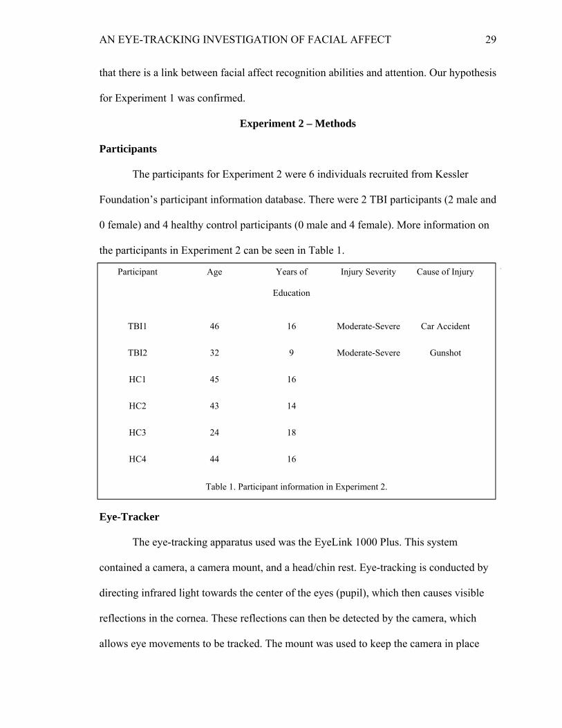
The participants for Experiment 2 were 6 individuals recruited from Kessler
Foundation’s participant information database. There were 2 TBI participants (2 male and
0 female) and 4 healthy control participants (0 male and 4 female). More information on
the participants in Experiment 2 can be seen in Table 1.
Eye-Tracker
The eye-tracking apparatus used was the EyeLink 1000 Plus. This system
contained a camera, a camera mount, and a head/chin rest. Eye-tracking is conducted by
directing infrared light towards the center of the eyes (pupil), which then causes visible
reflections in the cornea. These reflections can then be detected by the camera, which
allows eye movements to be tracked. The mount was used to keep the camera in place
Participant Age Years of
Education
Injury Severity Cause of Injury Y
TBI1 46 16 Moderate-Severe Car Accident
TBI2 32 9 Moderate-Severe Gunshot
HC1 45 16
HC2 43 14
HC3 24 18
HC4 44 16
Table 1. Participant information in Experiment 2.
AN EYE-TRACKING INVESTIGATION OF FACIAL AFFECT 30
and steady. The head/chin rest was used to stabilize a participant’s head so that eye
movements could be tracked efficiently.
Stimuli
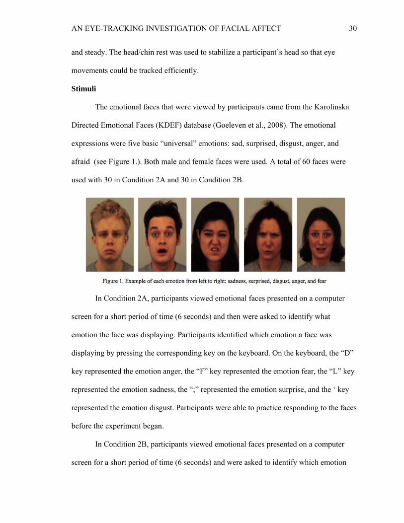
The emotional faces that were viewed by participants came from the Karolinska
Directed Emotional Faces (KDEF) database (Goeleven et al., 2008). The emotional
expressions were five basic “universal” emotions: sad, surprised, disgust, anger, and
afraid (see Figure 1.). Both male and female faces were used. A total of 60 faces were
used with 30 in Condition 2A and 30 in Condition 2B.
In Condition 2A, participants viewed emotional faces presented on a computer
screen for a short period of time (6 seconds) and then were asked to identify what
emotion the face was displaying. Participants identified which emotion a face was
displaying by pressing the corresponding key on the keyboard. On the keyboard, the “D”
key represented the emotion anger, the “F” key represented the emotion fear, the “L” key
represented the emotion sadness, the “;” represented the emotion surprise, and the ‘ key
represented the emotion disgust. Participants were able to practice responding to the faces
before the experiment began.
In Condition 2B, participants viewed emotional faces presented on a computer
screen for a short period of time (6 seconds) and were asked to identify which emotion
AN EYE-TRACKING INVESTIGATION OF FACIAL AFFECT 31
the face was displaying while simultaneously completing a distractor task. The distractor
task for condition 2B consisted of listening to and responding to low, medium, and high
tones while viewing an emotional face. The low tone played at a frequency of 100 Hz, the
medium tone played at a frequency of 150 Hz, and the high tone played at a frequency of
200Hz. The tones were delivered via external speakers, were randomly presented to
participants and were played for no longer than 3 seconds. Participants responded to the
emotional faces in the same manner as in Condition 2A. Participants responded to the
tones by pressing the corresponding key on the keyboard. On the keyboard, the “L” key
represented the low tone, the “;” key represented the medium tone, and the “‘” key
represented the high tone. Participants would view the face while simultaneously
listening to and responding to the tones, and then they would be asked to identify the
emotion expressed on the face shown. Participants were able to practice the distractor
task by itself and were also able to practice the distractor task with the facial affect
recognition task simultaneously before the experiment began.
ROI Creation
ROIs help to separate and distinguish different areas of the face from each other.
For example, if an individual is viewing the eyes on a face, it is important to understand
what part of the face constitutes the eye region, where the eye region starts, and where the
eye region ends. In this project, all ROIs were free-drawn using the EyeLink Data Viewer
program. The guidelines for drawing the ROIs were based off of (Wells et al., 2016 &
Arizpe et al., 2016) and were adapted to fit the faces used in this project. Two ROIs made
up the eyes, one ROI on the left eye and one ROI on the right eye. The ROIs began right
above the top of the eyebrows, continued to the outer most part of the eyebrows, and
AN EYE-TRACKING INVESTIGATION OF FACIAL AFFECT 32
ended at the molar fold (the groove in the skin where the upper cheek muscles meet the
eye sockets). For the nose, the ROI began at the bottom of the eyes, continued to the
edges of the bulbs of the nostrils, and ended at the bottom of the nose. For the mouth, the
ROI began at the philtrum (the vertical groove in the middle of the upper lip), continued
to the outer most part of the mouth muscles, and ended at the labiomedial crease (the
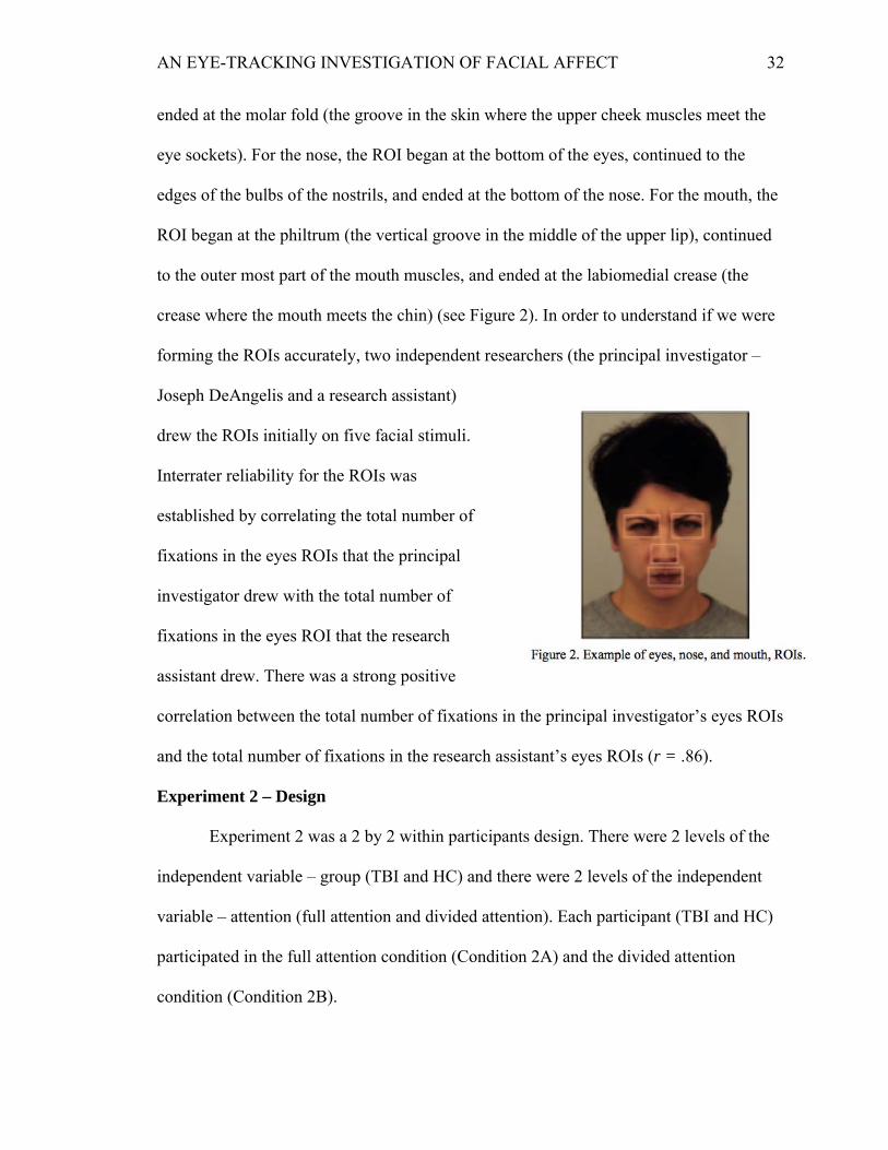
crease where the mouth meets the chin) (see Figure 2). In order to understand if we were
forming the ROIs accurately, two independent researchers (the principal investigator –
Joseph DeAngelis and a research assistant)
drew the ROIs initially on five facial stimuli.
Interrater reliability for the ROIs was
established by correlating the total number of
fixations in the eyes ROIs that the principal
investigator drew with the total number of
fixations in the eyes ROI that the research
assistant drew. There was a strong positive
correlation between the total number of fixations in the principal investigator’s eyes ROIs
and the total number of fixations in the research assistant’s eyes ROIs (r = .86).
Experiment 2 – Design
Experiment 2 was a 2 by 2 within participants design. There were 2 levels of the
independent variable – group (TBI and HC) and there were 2 levels of the independent
variable – attention (full attention and divided attention). Each participant (TBI and HC)
participated in the full attention condition (Condition 2A) and the divided attention
condition (Condition 2B).
AN EYE-TRACKING INVESTIGATION OF FACIAL AFFECT 33
Procedure
Participants entered the experiment room and were given informed consent. The
experiment room was sound reduced and contained the eye-tracker. Participants were
then familiarized with the eye-tracker and they put their head and chin on the head and
chin rest. Once participants were comfortable, they engaged in practice trials for both
conditions 2A and 2B. Participants’ eyes were not tracked during the practice and was
conducted in order to familiarize participants with the task. After the practice trials,
participants’ eyes movements were calibrated and validated. This was done in order to
ensure the highest level of eye-tracking accuracy. Once this was done, participants either
began condition 2A or 2B as the order of the conditions was randomly assigned.
In condition 2A (Full Attention Condition), participants viewed each face for 6
seconds for a total of 30 trials. After 15 trials, participants received a short break and eye
movements were recalibrated and revalidated. In order to determine what emotion was
being displayed, participants hit the corresponding key on the keyboard that aligned with
the emotion being displayed. Before starting the other experiment, participants received a
break and calibration and validation was conducted again.
In condition 2B (Divided Attention Condition), participants responded to the tone
that was being played (approximately 3 or 4 tones per trial) and then responded to the
emotional face for a total of 30 trials with 6 seconds viewing each face. Participants
selected the tones and the emotion displayed on the face by selecting the corresponding
key on the keyboard. After 15 trials, participants were given a short break and eye
movements were recalibrated and revalidated. Once this was done, participants finished
AN EYE-TRACKING INVESTIGATION OF FACIAL AFFECT 34
the remaining 15 trials. After both experiments were completed, participants were
debriefed and exited the experiment room.
Statistical Analyses
Data analysis was performed using the Statistical Package for the Social Sciences
(SPSS). Statistical significance was set at an alpha level of 0.05. For the behavioral data,
participants’ performance on the facial affect recognition task in both the full attention
and divided attention conditions was examined. Participants’ accuracy on correctly
identifying each specific emotion in both the full and divided attention conditions (i.e.,
how accurate participants were on correctly identifying the emotion sadness when the
emotional face was displaying sadness) was also examined. These variables were
compared across groups using a Mann – Whitney U test and an independent samples t-
test. An ANOVA was not conducted because the small sample size would affect the
power of the test. A Mann-Whitney U test was conducted because this study had a small
sample size and because we believed the behavioral data would not be normally
distributed (i.e., TBI participants would be less accurate on the facial affect recognition
task compared to HC participants). For the eye-tracking data, the amount of time both
groups of participants in both conditions spent fixating on the main ROIs (left eye, right
eye, mouth, and nose) compared to the amount of time spent fixating on the
supplemental/non-ROIs was examined. These variables were compared across groups
using an independent samples t-test.
AN EYE-TRACKING INVESTIGATION OF FACIAL AFFECT 35
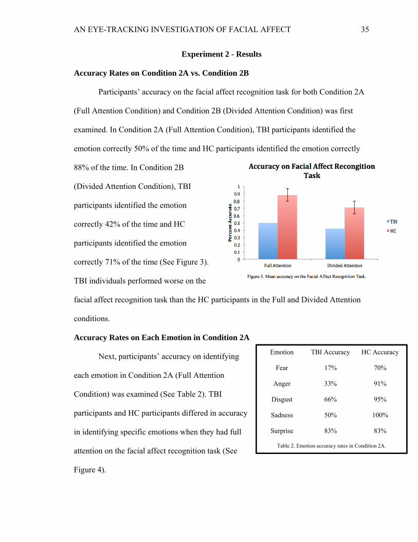
Experiment 2 - Results
Accuracy Rates on Condition 2A vs. Condition 2B
Participants’ accuracy on the facial affect recognition task for both Condition 2A
(Full Attention Condition) and Condition 2B (Divided Attention Condition) was first
examined. In Condition 2A (Full Attention Condition), TBI participants identified the
emotion correctly 50% of the time and HC participants identified the emotion correctly
88% of the time. In Condition 2B
(Divided Attention Condition), TBI
participants identified the emotion
correctly 42% of the time and HC
participants identified the emotion
correctly 71% of the time (See Figure 3).
TBI individuals performed worse on the
facial affect recognition task than the HC participants in the Full and Divided Attention
conditions.
Accuracy Rates on Each Emotion in Condition 2A
Next, participants’ accuracy on identifying
each emotion in Condition 2A (Full Attention
Condition) was examined (See Table 2). TBI
participants and HC participants differed in accuracy
in identifying specific emotions when they had full
attention on the facial affect recognition task (See
Figure 4).
Emotion TBI Accuracy HC Accuracy
Fear 17% 70%
Anger 33% 91%
Disgust 66% 95%
Sadness 50% 100%
Surprise 83% 83%
Table 2. Emotion accuracy rates in Condition 2A.
AN EYE-TRACKING INVESTIGATION OF FACIAL AFFECT 36
Accuracy Rates on Each Emotion in Condition 2B
Participants’ accuracy on identifying each emotion in Condition 2B (Divided
Attention Condition) was examined (See Table 3). TBI participants and HC participants
differed in accuracy in identifying specific emotions when their attention was divided on
the facial affect recognition task (see Figure 5). In this condition, there were no
differences in accuracy on the emotion afraid.
Emotion TBI Accuracy HC Accuracy
Afraid 41% 41%
Anger 25% 75%
Disgust 33% 75%
Sadness 33% 70%
Surprise 75% 91%
Table 3. Emotion accuracy rates in Condition 2B.
AN EYE-TRACKING INVESTIGATION OF FACIAL AFFECT 37
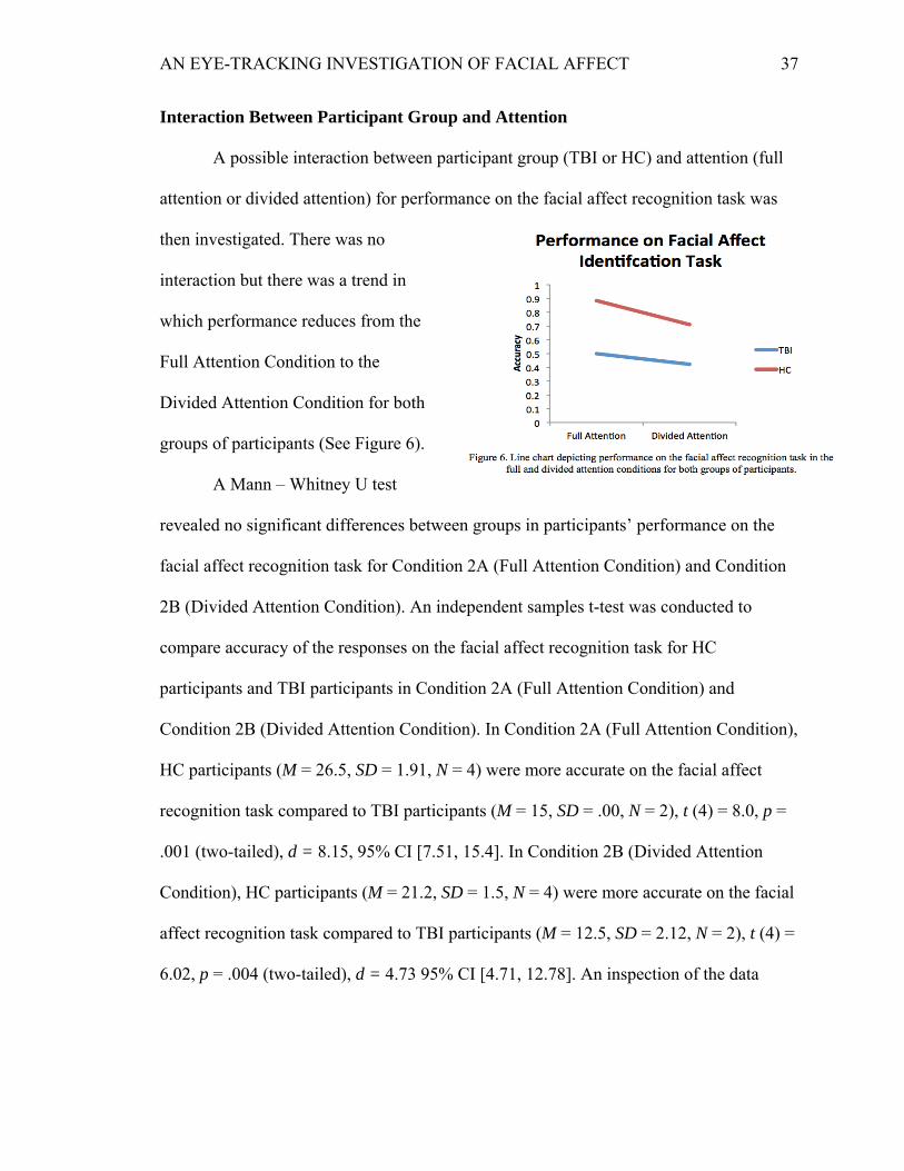
Interaction Between Participant Group and Attention
A possible interaction between participant group (TBI or HC) and attention (full
attention or divided attention) for performance on the facial affect recognition task was
then investigated. There was no
interaction but there was a trend in
which performance reduces from the
Full Attention Condition to the
Divided Attention Condition for both
groups of participants (See Figure 6).
A Mann – Whitney U test
revealed no significant differences between groups in participants’ performance on the
facial affect recognition task for Condition 2A (Full Attention Condition) and Condition
2B (Divided Attention Condition). An independent samples t-test was conducted to
compare accuracy of the responses on the facial affect recognition task for HC
participants and TBI participants in Condition 2A (Full Attention Condition) and
Condition 2B (Divided Attention Condition). In Condition 2A (Full Attention Condition),
HC participants (M = 26.5, SD = 1.91, N = 4) were more accurate on the facial affect
recognition task compared to TBI participants (M = 15, SD = .00, N = 2), t (4) = 8.0, p =
.001 (two-tailed), d = 8.15, 95% CI [7.51, 15.4]. In Condition 2B (Divided Attention
Condition), HC participants (M = 21.2, SD = 1.5, N = 4) were more accurate on the facial
affect recognition task compared to TBI participants (M = 12.5, SD = 2.12, N = 2), t (4) =
6.02, p = .004 (two-tailed), d = 4.73 95% CI [4.71, 12.78]. An inspection of the data
AN EYE-TRACKING INVESTIGATION OF FACIAL AFFECT 38
revealed that both groups of participants were responding to the tones at least 50% of the
time in Condition 2B (Divided Attention Condition).
Eye-Tracking Analysis
The data analysis for the eye-tracking portion of Experiment 2 contained 5
participants (2 TBI and 3 HC) as 1 participant was removed from the analysis due to
unreliable data. An ANOVA was not conducted because the small sample size would
affect the power.
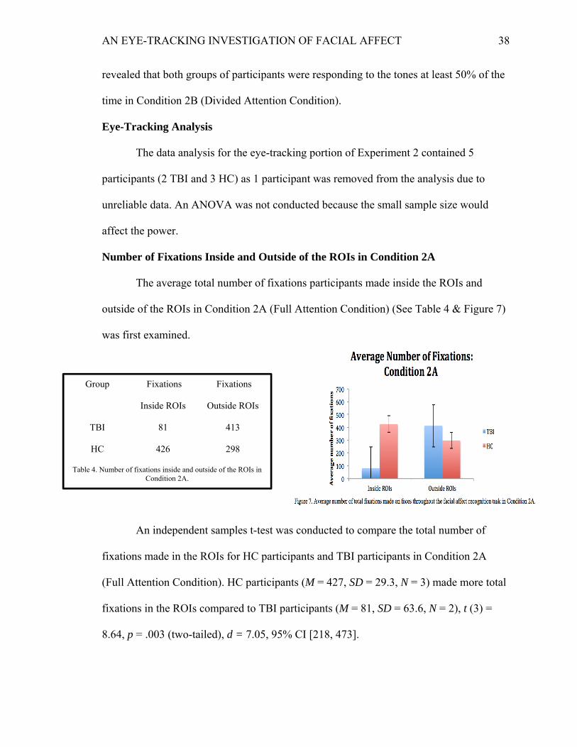
Number of Fixations Inside and Outside of the ROIs in Condition 2A
The average total number of fixations participants made inside the ROIs and
outside of the ROIs in Condition 2A (Full Attention Condition) (See Table 4 & Figure 7)
was first examined.
An independent samples t-test was conducted to compare the total number of
fixations made in the ROIs for HC participants and TBI participants in Condition 2A
(Full Attention Condition). HC participants (M = 427, SD = 29.3, N = 3) made more total
fixations in the ROIs compared to TBI participants (M = 81, SD = 63.6, N = 2), t (3) =
8.64, p = .003 (two-tailed), d = 7.05, 95% CI [218, 473].
Group Fixations
Inside ROIs
Fixations
Outside ROIs
TBI 81 413
HC 426 298
Table 4. Number of fixations inside and outside of the ROIs in Condition 2A.
AN EYE-TRACKING INVESTIGATION OF FACIAL AFFECT 39
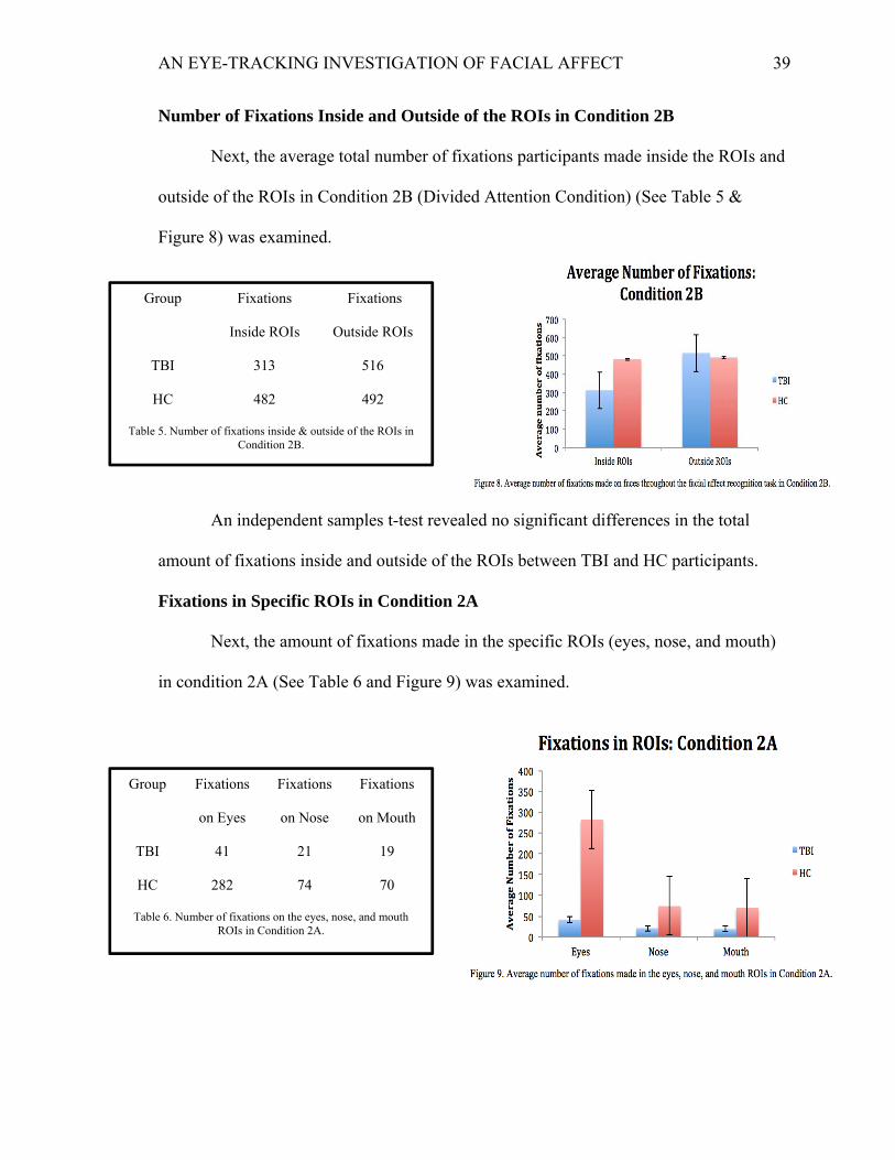
Number of Fixations Inside and Outside of the ROIs in Condition 2B
Next, the average total number of fixations participants made inside the ROIs and
outside of the ROIs in Condition 2B (Divided Attention Condition) (See Table 5 &
Figure 8) was examined.
An independent samples t-test revealed no significant differences in the total
amount of fixations inside and outside of the ROIs between TBI and HC participants.
Fixations in Specific ROIs in Condition 2A
Next, the amount of fixations made in the specific ROIs (eyes, nose, and mouth)
in condition 2A (See Table 6 and Figure 9) was examined.
Group Fixations
Inside ROIs
Fixations
Outside ROIs
TBI 313 516
HC 482 492
Table 5. Number of fixations inside & outside of the ROIs in Condition 2B.
Group Fixations
on Eyes
Fixations
on Nose
Fixations
on Mouth
TBI 41 21 19
HC 282 74 70
Table 6. Number of fixations on the eyes, nose, and mouth ROIs in Condition 2A.
AN EYE-TRACKING INVESTIGATION OF FACIAL AFFECT 40
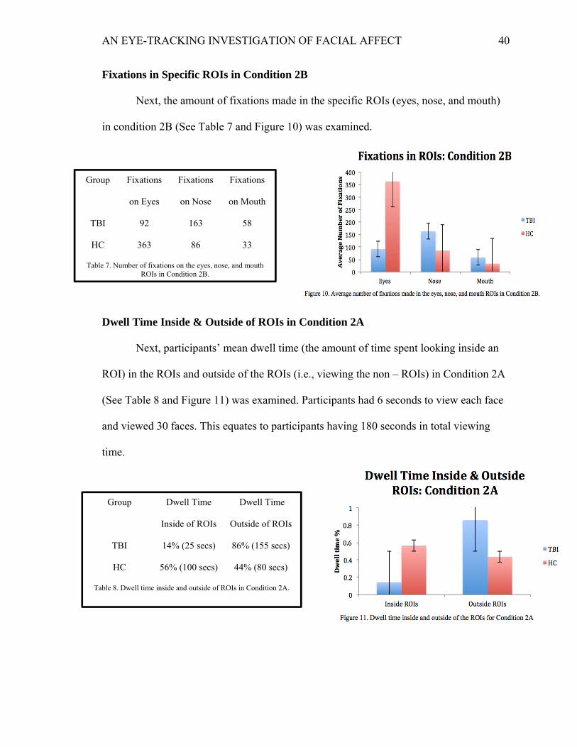
Fixations in Specific ROIs in Condition 2B
Next, the amount of fixations made in the specific ROIs (eyes, nose, and mouth)
in condition 2B (See Table 7 and Figure 10) was examined.
Dwell Time Inside & Outside of ROIs in Condition 2A
Next, participants’ mean dwell time (the amount of time spent looking inside an
ROI) in the ROIs and outside of the ROIs (i.e., viewing the non – ROIs) in Condition 2A
(See Table 8 and Figure 11) was examined. Participants had 6 seconds to view each face
and viewed 30 faces. This equates to participants having 180 seconds in total viewing
time.
Group Fixations
on Eyes
Fixations
on Nose
Fixations
on Mouth
TBI 92 163 58
HC 363 86 33
Table 7. Number of fixations on the eyes, nose, and mouth ROIs in Condition 2B.
Group Dwell Time
Inside of ROIs
Dwell Time
Outside of ROIs
TBI 14% (25 secs) 86% (155 secs)
HC 56% (100 secs) 44% (80 secs)
Table 8. Dwell time inside and outside of ROIs in Condition 2A.
AN EYE-TRACKING INVESTIGATION OF FACIAL AFFECT 41
An independent samples t-test was conducted to compare the dwell time (in
seconds) in the ROIs for HC and TBI participants in Condition 2A (Full Attention
Condition). HC participants (M = 101, SD = 26.6, N = 3) spent more time viewing the
ROIs compared to TBI participants (M = 25, SD = 26, N = 2), t (3) = 3.12, p = .05 (two-
tailed), d = 2.8, 95% CI [-1.3, 152.62]. An independent samples t-test was conducted to
compare the dwell time (in seconds) outside of the ROIs (the non-ROIs) for HC and TBI
participants in Condition 2A (Full Attention Condition). HC participants (M = 78.85, SD
= 26.69, N = 3) spent less time viewing the non-ROIs compared to TBI participants (M =
98.72, SD = 18.19, N = 2), t (3) = -3.12, p = .05 (two-tailed), d = -2.8, 95% CI [-152.26,
1.3].
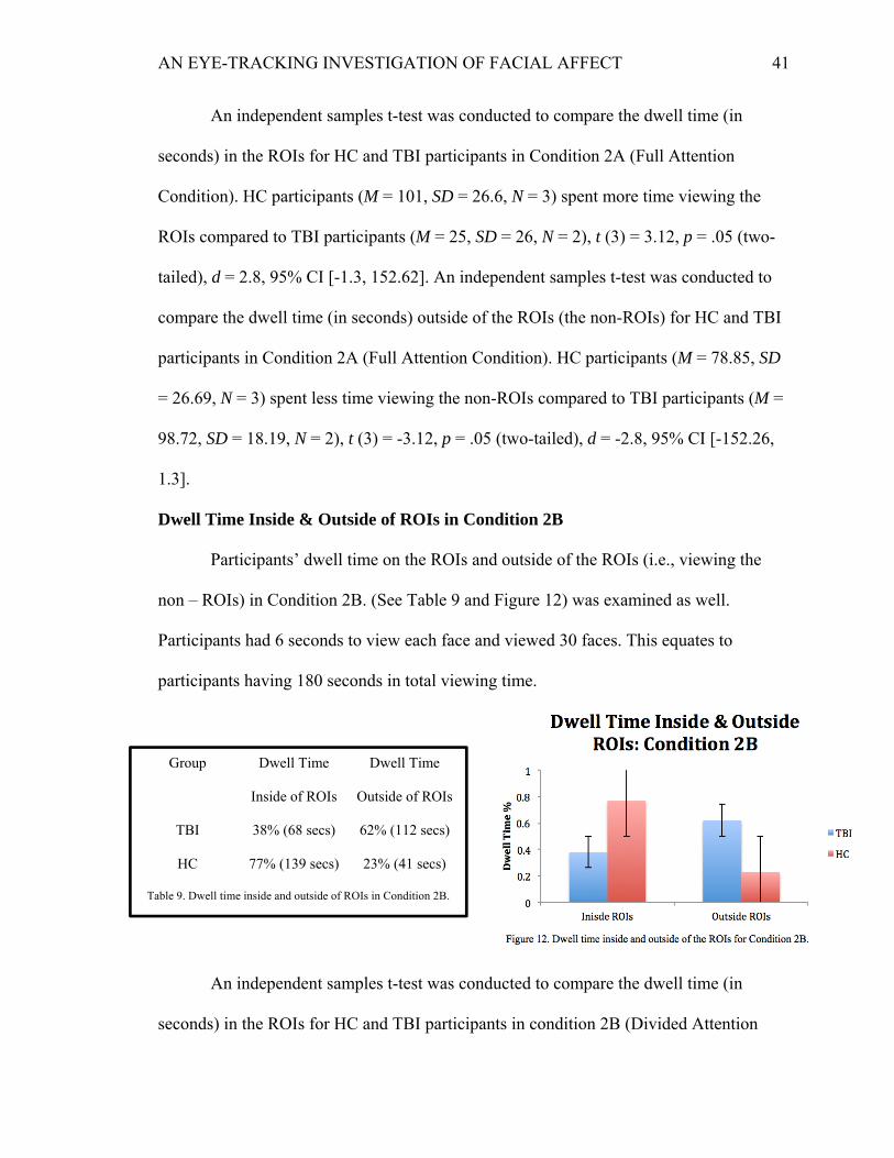
Dwell Time Inside & Outside of ROIs in Condition 2B
Participants’ dwell time on the ROIs and outside of the ROIs (i.e., viewing the
non – ROIs) in Condition 2B. (See Table 9 and Figure 12) was examined as well.
Participants had 6 seconds to view each face and viewed 30 faces. This equates to
participants having 180 seconds in total viewing time.
An independent samples t-test was conducted to compare the dwell time (in
seconds) in the ROIs for HC and TBI participants in condition 2B (Divided Attention
Group Dwell Time
Inside of ROIs
Dwell Time
Outside of ROIs
TBI 38% (68 secs) 62% (112 secs)
HC 77% (139 secs) 23% (41 secs)
Table 9. Dwell time inside and outside of ROIs in Condition 2B.
AN EYE-TRACKING INVESTIGATION OF FACIAL AFFECT 42
Condition). HC participants (M = 138.41, SD = 12.44, N = 3) spent more time viewing
the ROIs compared to TBI participants (M = 98.72, SD = 18.19, N = 2), t (3) = 2.97, p =
.05 (two-tailed), d = 2.54, 95% CI [-2.77, 82.15]. An independent samples t-test was
conducted to compare the dwell time (in seconds) outside of the ROIs (the non-ROIs) for
HC and TBI participants in Condition 2B (Divided Attention Condition). HC participants
(M = 41.58, SD = 12.44, N = 3) spent less time viewing the non – ROIs compared to TBI
participants (M = 81.27. SD = 18.19, N = 2), t (3) = -2.97, p = .05 (two-tailed), d = -2.54,
95% CI [-82.15, 2.77].
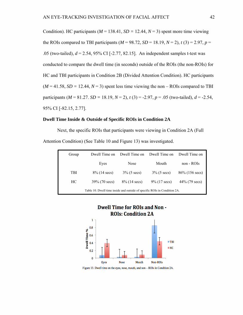
Dwell Time Inside & Outside of Specific ROIs in Condition 2A
Next, the specific ROIs that participants were viewing in Condition 2A (Full
Attention Condition) (See Table 10 and Figure 13) was investigated.
Group Dwell Time on
Eyes
Dwell Time on
Nose
Dwell Time on
Mouth
Dwell Time on
non - ROIs
TBI 8% (14 secs) 3% (5 secs) 3% (5 secs) 86% (156 secs)
HC 39% (70 secs) 8% (14 secs) 9% (17 secs) 44% (79 secs)
Table 10. Dwell time inside and outside of specific ROIs in Condition 2A.
AN EYE-TRACKING INVESTIGATION OF FACIAL AFFECT 43
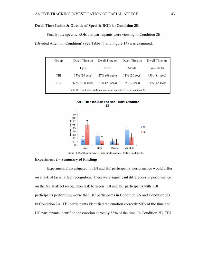
Dwell Time Inside & Outside of Specific ROIs in Condition 2B
Finally, the specific ROIs that participants were viewing in Condition 2B
(Divided Attention Condition) (See Table 11 and Figure 14) was examined.
Experiment 2 – Summary of Findings
Experiment 2 investigated if TBI and HC participants’ performance would differ
on a task of facial affect recognition. There were significant differences in performance
on the facial affect recognition task between TBI and HC participants with TBI
participants performing worse than HC participants in Condition 2A and Condition 2B.
In Condition 2A, TBI participants identified the emotion correctly 50% of the time and
HC participants identified the emotion correctly 88% of the time. In Condition 2B, TBI
Group Dwell Time on
Eyes
Dwell Time on
Nose
Dwell Time on
Mouth
Dwell Time on
non - ROIs
TBI 17% (30 secs) 27% (49 secs) 11% (20 secs) 45% (81 secs)
HC 60% (108 secs) 13% (23 secs) 4% (7 secs) 23% (42 secs)
Table 11. Dwell time inside and outside of specific ROIs in Condition 2B.
AN EYE-TRACKING INVESTIGATION OF FACIAL AFFECT 44
participants identified the emotion correctly 42% of the time and HC participants
identified the emotion correctly 71% of the time (See Figure 3).
In terms of eye-tracking, the investigation was if participants’ gaze patterns
would differ while viewing emotional facial expressions. There was a significant
difference in the amount of total fixations that TBI and HC participants made inside and
outside of the ROIs while viewing an emotional face when they had full attention.
Compared to HC participants, TBI participants made fewer fixations inside of the ROIs
and made more fixations outside of the ROIs (viewing the non-ROIs) when they had full
attention during the facial affect recognition task (See Figure 7). Interestingly, there was
no significant difference in the amount of total fixations that TBI and HC participants
made inside the ROIs and outside of the ROIs while viewing a face when their attention
was divided (See Figure 8). Additionally, there was a significant difference in the amount
of time TBI and HC participants viewed (dwelled on) inside the ROIs and outside of the
ROIs of an emotional face when they had full attention and when their attention was
divided during the facial affect recognition task. Compared to HC participants, TBI
participants spent more time viewing the non-ROIs (outside of the ROIs) of an emotional
face and spent less time viewing the ROIs of an emotional face when they had full
attention and when their attention was divided on the facial affect recognition task (See
Figures 11 & 12).
There was also differences between the amounts of fixations that TBI participants
and HC participants made on specific ROIs (i.e., the eyes nose and mouth ROIs) and
found differences in the amount of time TBI participants and HC participants spent
viewing specific ROIs. Compared to HC participants, TBI participants made fewer
AN EYE-TRACKING INVESTIGATION OF FACIAL AFFECT 45
fixations on the eyes, nose, and mouth ROIs when they had full attention on the facial
affect recognition task (See Figure 9) but made more fixations on the nose and mouth
ROIs when their attention was divided during the facial affect recognition task (See
Figure 10). Compared to HC participants, participants with TBI spent less time viewing
the eyes, nose, and mouth ROIs when they had full attention on the facial affect
recognition task (See Figure 13) but spent more time viewing the nose and mouth ROIs
when their attention was divided during the facial affect recognition task (See Figure 14).
Discussion
The overall goal of this project was to investigate why social cognitive
difficulties, specifically difficulties in facial affect recognition, exist in individuals with
TBI. Experiment 1 investigated the relationship between attention and facial affect
recognition abilities. In order to investigate this relationship, we conducted correlations
between a measure of facial affect recognition ability and two measures of attention. We
found significant positive correlations between the measure of facial affect recognition
ability and the two measures of attention. In other words, individuals that performed high
on the measure of facial affect recognition ability also performed high on the two
measures of attention. Experiment 2 investigated differences in TBI and HC participants’
performance on a facial affect recognition task and how participants’ gaze patterns
differed using eye-tracking. In terms of behavior, we found significant differences on
both groups of participants’ performance on the facial affect recognition task for
Condition 2A (Full Attention Condition) and Condition 2B (Divided Attention
Condition). TBI participants were less accurate on the facial affect recognition task
compared to HC participants when they had full attention and when their attention was
AN EYE-TRACKING INVESTIGATION OF FACIAL AFFECT 46
divided on the facial affect recognition task. In terms of eye-tracking, we found that TBI
participants made fewer fixations inside of the ROIs and made more fixations outside of
the ROIs (viewing the non-ROIs) when they had full attention during the facial affect
recognition task compared to HC participants.
Additionally, we found that, compared to HC participants, TBI participants spent
more time viewing the non-ROIs (outside of the ROIs) of an emotional face and spent
less time viewing the ROIs of an emotional face when they had full attention and when
their attention was divided on the facial affect recognition task. We also found
differences between the amounts of fixations that TBI participants and HC participants
made on specific ROIs (i.e., the eyes nose and mouth ROIs) and found differences in the
amount of time TBI participants and HC participants spent viewing specific ROIs.
Compared to HC participants, TBI participants made fewer fixations on the eyes, nose,
and mouth ROIs when they had full attention on the facial affect recognition task but
made more fixations on the nose and mouth ROIs when their attention was divided
during the facial affect recognition task. In Condition 2A, participants with TBI spent less
time viewing the eyes, nose, and mouth ROIs on the facial affect recognition task
compared to HC participants. In Condition 2B, TBI participants spent more time viewing
the nose and mouth ROIs on the facial affect recognition task compared to HC
participants.
Correlations Between Attention and Facial Affect Recognition
There was a significant positive correlation between a task of facial affect
recognition (TOFER) and a task of attention (the Digit Span) and there was a significant
positive correlation between the TOFER and the SDMT (a task of attention and
AN EYE-TRACKING INVESTIGATION OF FACIAL AFFECT 47
processing speed). These results mean that individuals with TBI that perform well on a
measure of facial affect recognition also perform high on measures of attention. This
indicates that attention may play a role in facial affect recognition abilities. This is an
important link because individuals with TBI have difficulties focusing and paying
attention (Bonnelle et al., 2011). These findings are consistent with prior research as
individuals that have impaired Theory of Mind (ToM) abilities/impaired social cognitive
abilities also have impairments in executive functioning, working memory, verbal
memory, and visual memory (Kim et al., 2011). In the current study, the TOFER can be
considered a measure of facial affect recognition/social cognitive ability as it requires
participants to identify emotions (Goeleven et al., 2008). The Digit Span and the SDMT
are measures of attention and processing speed but they are also related to executive
functioning, working memory, and verbal memory (Wechsler, 1997; Smith, 1982). This
is important because if an individual with TBI can not pay attention to an emotional face,
then they may misidentify the emotion on that face. Due to the strong positive
relationships that Experiment 1 showed, it may be possible to develop therapies and
interventions that first focus on improving skills in executive functioning, working
memory, and verbal memory, which may in time, improve facial affect recognition
abilities.
Behavioral
For the behavioral data, TBI participants were less accurate on the facial affect
recognition task compared to HC participants when they had full attention and when their
attention was divided. TBI participants were less accurate identifying emotions on the
facial affect recognition task (compared to HC participants) when they had full attention
AN EYE-TRACKING INVESTIGATION OF FACIAL AFFECT 48
because TBI participants have impaired facial affect recognition abilities (Babbage et al.,
2011; Croker & McDonald, 2005). TBI participants were less accurate identifying
emotions on the facial affect recognition task (compared to HC participants) when their
attention was divided because individuals with TBI experience difficulties in
focusing/attending to something when their attention is divided (Azouvi et al., 2004). The
behavioral data findings are consistent with prior research by Azouvi et al., (2004) and
Park et al., (1999), which found that individuals with TBI experiences difficulties in task-
switching and score less highly (compared to HC participants) on measures of working
memory when their attention was divided.
When each emotion was examined separately, we found that TBI and HC
participants also differed on accuracy rates for each emotion for the full and divided
attention conditions. In Condition 2A, TBI participants experienced the most difficulty
correctly identifying the emotions fear (17% correct), anger (33% correct), and sadness
(50% correct) compared to HC participants. In Condition 2B, TBI participants
experienced the most difficulty correctly identifying the emotions fear (45% correct),
anger (25% correct), disgust (33% correct) and sadness (33% correct). Both groups of
participants in both conditions experienced little difficulties in correctly identifying the
emotion surprise. These findings are consistent with previous research as individuals with
TBI experience difficulties in correctly identifying negative emotions such as fear, anger,
and sadness (Croker & McDonald, 2005; Genova et al., 2017). It may be possible that
individuals with TBI experience difficulties in correctly identifying these emotions
because their friends/family/caregivers may not want to expose them to these negative
emotions. Receiving a TBI can be a life changing experience and many individuals close
AN EYE-TRACKING INVESTIGATION OF FACIAL AFFECT 49
to someone with a TBI may not want their loved one to experience any negativity or
hostility after such a traumatic event. In doing so, friends/family/caregivers of individuals
with TBI might do their best to not express negative emotions around an individual with
TBI.
This study is innovative because it is the first study to examine how divided
attention affects facial affect recognition. This is important because individuals with TBI
may experience situations in which their attention is divided quite frequently. An
individual with TBI may have to divide their attention while talking to/interacting with a
family member or a caretaker while simultaneously focusing on the environment around
them (i.e., in the home, at a grocery store, at a party, etc.). If an individual with TBI can
not successfully divided their attention in a situation like this, then they may risk
incorrectly identifying the emotion displayed on their family
member’s/friend’s/caretaker’s face and may trigger the individual with TBI to respond in
a way that is not congruent with the emotion displayed (i.e., responding to a happy
emotional expression with an angry or sad response). This may lead to negative social
engagements, which could lead to social isolation in individuals with TBI.
Other factors may have influenced the findings of the current study. For example,
cognitive fatigue (i.e., a lack of mental energy that is perceived by the individual during
common and usual activities) may have played a role in why TBI participants performed
worse than HC participants on the task of facial affect recognition when their attention
was divided. In a study conducted by Kohl et al., (2009), functional magnetic resonance
imaging (fMRI) was used to assess cognitive fatigue in individuals with TBI. TBI
participants and HC participants completed a modified SDMT while having their brain
AN EYE-TRACKING INVESTIGATION OF FACIAL AFFECT 50
activity monitored by fMRI. They found that TBI participants exhibited increased brain
activity, which represented increased cerebral effort. This increased cerebral effort may
have manifested as cognitive fatigue. Our findings relate to Kohl et al., (2009) because
TBI participants may have experienced an increase in brain activity during Condition 2B
(Divided Attention Condition) as they had to identify the emotion on the face presented
while simultaneously identifying the tone being played. These two tasks combined may
have resulted in an increase in brain activity for TBI participants and may have led them
to become cognitively fatigued in Condition 2B (Divided Attention Condition) and may
have made it extremely difficult to focus and correctly identify the emotions displayed on
the faces during the facial affect recognition task.
Eye – Tracking
TBI participants made fewer fixations inside of the ROIs and made more fixations
outside of the ROIs (viewing the non-ROIs) when they had full attention during the facial
affect recognition task compared to HC participants. Also, compared to HC participants,
TBI participants spent more time viewing the non-ROIs (outside of the ROIs) of an
emotional face and spent less time viewing the ROIs of an emotional face when they had
full attention and when their attention was divided on the facial affect recognition task.
These findings are consistent with another specialized population – Autism Spectrum
Disorder (ASD). Individuals with ASD tend to make more fixations and spend more time
viewing non-critical regions of the face (ears, forehead, hair, other facial area, etc.) and
tend to make fewer fixations and spend less time viewing the critical regions of the face
(i.e., eyes, nose, and mouth) compared to HC participants (Boraston & Blakemore, 2007;
Dalton et al., 2005).
AN EYE-TRACKING INVESTIGATION OF FACIAL AFFECT 51
We found differences between the number of fixations that TBI participants and
HC participants made on specific ROIs (i.e., the eyes nose and mouth ROIs) in the full
and divided attention conditions and found differences in the amount of time TBI
participants and HC participants spent viewing specific ROIs in both the full and divided
attention conditions. Both TBI and HC participants in Condition 2A fixated on more
frequently and spent more time viewing the eyes ROI compared to the other ROIs (nose
and mouth). This result is consistent with other research because HC individuals and TBI
individuals tend to view the eyes of others during conversations (Turkstra, 2005). There
are other tests that measure social cognitive abilities/facial affect recognition abilities.
One test in particular, the Reading the Mind in the Eyes test, is a measure of social
cognitive abilities and facial affect recognition abilities. In this test, participants view
static images of just the eyes region and identify which emotion a full emotional face
would be displaying based on the information provided by the eyes region (Baker et al.,
2014). Previous research shows that individuals with TBI perform worse on this test
compared to HC participants (Baker et al., 2014). In this study, both TBI and HC
participants fixated on the eyes ROI more often and viewed the eyes ROI for a longer
period of time compared to the other critical ROIs (i.e., nose and mouth). However, TBI
participants fixated on the eyes ROI less often and viewed the eyes ROI for a lesser
amount of time compared to HC participants. This suggests that the eyes may contain key
information that is useful when attempting to correctly identify an emotional face, but
TBI participants may not retain/utilize this key information as well as HC participants do
while attempting to correctly identify an emotion displayed on a face.
AN EYE-TRACKING INVESTIGATION OF FACIAL AFFECT 52
These results are important because, until this study was conducted, we did not
know exactly where and for how long participants with TBI fixated on an emotional face.
Many studies that investigate facial affect recognition only informed us that participants
with TBI have difficulties correctly identifying emotions. This study is the first of its kind
to investigate exactly where (i.e., eyes, nose, mouth, or non-ROIs) and for how long that
TBI participants fixated on an emotional face when they had full attention on a facial
affect recognition task and when their attention was divided on a facial affect recognition
task. The information gained from this study can be used to inform/develop treatments
and interventions for individuals with TBI that suffer from poor facial affect recognition
abilities. By using eye-tracking, we can understand where and for how long an individual
with TBI is fixating on an emotional face, and then try to correctly alter TBI individuals’
gaze patterns. For example, if an individual with TBI is fixating on a non-critical ROI
(ears, hair, forehead, other facial area, etc.) for too long of a time period, then we can
inform the individual of this and attempt to switch their focus to a critical ROI on the face
(eyes, nose, mouth). By consistently “training” individuals with TBI to change their
fixation patterns from non-critical ROIS to critical ROIs, we may be able to improve
facial affect recognition abilities in TBI individuals.
One finding that stood out was how participants’ number of fixations on the ROIs
and outside the ROIs (viewing the non-ROIs) did not significantly differ in Condition 2B
(Divided Attention Condition) but their total dwell time significantly differed in
Condition 2B (Divided Attention Condition). This could be explained by the idea that
dwelling on an emotional face (i.e., viewing it) may be different than fixating (i.e.,
concentrated viewing) on an emotional face. This difference could also be explained
AN EYE-TRACKING INVESTIGATION OF FACIAL AFFECT 53
through attention. TBI participants may have been able to dwell on the ROIs of an
emotional face but having their attention divided made it more difficult for them to
actually fixate and concentrate on the specific ROIs and the information gained from
viewing them. For example, an individual with TBI may have been able to view the ROIs
of an emotional face but they may not have been able to gain information from the ROIs
(i.e., they may have looked at a face’s open mouth but not associated it with the emotion
surprise). This suggests that there may be some area in the brain/process that occurs in
the brain that enables individuals to switch from just viewing an area on the face, to
fixating on that area and gaining information from it. In individuals with TBI, this brain
area/brain process may be damaged or may not function as efficiently as a healthy
individual’s area/process. This also suggests that this area/process is interfered with when
attention is divided, even more with an individual with TBI.
Another interesting finding was that, in Condition 2B, TBI participants fixated
more frequently and spent more time viewing the nose and mouth ROIs on the facial
affect recognition task compared to HC participants. This might have occurred because
the division of attention in Condition 2B led TBI participants to focus on an ROI that
may not have contained the most informative facial information. For example, the eyes
ROI may have contained the best facial information for participants to view and utilize in
order to correctly identify an emotion on a face. With divided attention, TBI participants
may have lost/shifted their focus from the eyes (which may have contained the most
important facial information to correctly identify an emotion on a face) to the nose and
mouth (which may have contained less useful facial information to correctly identify an
emotion on a face).
AN EYE-TRACKING INVESTIGATION OF FACIAL AFFECT 54
Limitations
Experiment 2 had a small sample size (6 participants for behavioral and 5
participants for eye-tracking). We still believe that the results are valid but it is important
to understand that the results, especially significance, may be due to a lack of
participants. The lack of participants was due to difficulties in recruiting as many
potential participants were over the age range (over 50 years old). The sample also
consisted of 2 males with TBI and 4 female HC participants. Gender may have been a
reason there were significant differences but we did not expect men and women to differ
on a test of facial affect recognition abilities as previous research states that there are no
differences (Hoffmann et al., 2010). Another limitation is the stimuli in this study may
have lacked ecological validity. The stimuli in this study were static images of faces
displaying different emotions. In more real-world situations, an individual is usually
talking to or interacting with another person while trying to correctly identify the emotion
they are displaying on their face. The static images we used in this study may not have
best represented this situation.
Future Directions
This experiment will be conducted again with an adequate sample size, with
participants matched across gender, and with the addition of participants with Multiple
Sclerosis (MS). Adding another specialized population to this experiment will inform us
on how another specialized population compares to individuals with TBI in terms of eye-
tracking and behavior. Adding MS participants will enable us to determine if TBI
individuals and MS individuals have the same or different gaze patterns. Additionally,
adding MS participants will allow us to understand if they fixate longer/shorter on one
AN EYE-TRACKING INVESTIGATION OF FACIAL AFFECT 55
area of the face compared to TBI participants. Finally, adding MS participants will enable
us to understand what emotions they misidentify compared the TBI participants. This
experiment will also add Theory of Mind (ToM) measures in order to investigate if
participants that are impaired on facial affect recognition also have impaired ToM
abilities.
Future experiments will utilize measures with greater ecological validity than
static images of faces displaying emotions. In order to increase the ecological validity of
the study we will use Virtual Reality Technology (VR). VR is a computer-generated
scenario that simulates a realistic experience. VR will be used to simulate talking to and
interacting with someone while simultaneously trying to correctly identify what emotion
the individual is displaying on his or her face. We will also add different levels of
distraction (low distraction, medium distraction, and high distraction) in order to
determine what level of distraction and when the level of distraction negatively
influences a participant’s performance on the facial affect recognition task. This will
enable us to understand if one group of participants has more difficulty identifying
emotions compared to another group of participants on a certain level of distraction (i.e.,
TBI participants may perform worse than MS participants on the facial affect recognition
task during the medium distraction condition but MS participants may perform better
than TBI participants on the facial affect recognition task during the low distraction
condition).
AN EYE-TRACKING INVESTIGATION OF FACIAL AFFECT 56
References
Arizpe, J., Kravitz, D. J., Walsh, V., Yovel, G., & Baker, C. I. (2016). Differences in looking at
own-and other-race faces are subtle and analysis-dependent: An account of discrepant
reports. PLoS One, 11(2), e0148253.
Azouvi, P., Couillet, J., Leclercq, M., Martin, Y., Asloun, S., & Rousseaux, M. (2004). Divided
attention and mental effort after severe traumatic brain injury. Neuropsychologia, 42(9),
1260-1268.
Babbage, D. R., Yim, J., Zupan, B., Neumann, D., Tomita, M. R., & Willer, B. (2011). Meta-
analysis of facial affect recognition difficulties after traumatic brain injury.
Baker, C. A., Peterson, E., Pulos, S., & Kirkland, R. A. (2014). Eyes and IQ: A meta-analysis of
the relationship between intelligence and “Reading the Mind in the
Eyes”. Intelligence, 44, 78-92.
Basford, J. R., Chou, L. S., Kaufman, K. R., Brey, R. H., Walker, A., Malec, J. F., ... & Brown,
A. W. (2003). An assessment of gait and balance deficits after traumatic brain
injury. Archives of physical medicine and rehabilitation, 84(3), 343-349.
Bergfeldt, U., Borg, K., Kullander, K., & Julin, P. (2006). Focal spasticity therapy with
botulinum toxin: effects on function, activities of daily living and pain in 100 adult
patients. Journal of Rehabilitation Medicine, 38(3), 166-171.
Bonnelle, V., Leech, R., Kinnunen, K. M., Ham, T. E., Beckmann, C. F., De Boissezon, X., ... &
Sharp, D. J. (2011). Default mode network connectivity predicts sustained attention
deficits after traumatic brain injury. Journal of Neuroscience, 31(38), 13442-13451.
Boraston, Z., & Blakemore, S. J. (2007). The application of eye�tracking technology in the study
of autism. The Journal of physiology, 581(3), 893-898.
AN EYE-TRACKING INVESTIGATION OF FACIAL AFFECT 57
Bruns, J., & Hauser, W. A. (2003). The epidemiology of traumatic brain injury: a
review. Epilepsia, 44(s10), 2-10.
Cicerone, K., Levin, H., Malec, J., Stuss, D., & Whyte, J. (2006). Cognitive rehabilitation
interventions for executive function: moving from bench to bedside in patients with
traumatic brain injury. Journal of cognitive neuroscience, 18(7), 1212-1222.
Croker, V., & McDonald, S. (2005). Recognition of emotion from facial expression following
traumatic brain injury. Brain Injury, 19(10), 787-799.
Dalton, K. M., Nacewicz, B. M., Johnstone, T., Schaefer, H. S., Gernsbacher, M. A., Goldsmith,
H. H., ... & Davidson, R. J. (2005). Gaze fixation and the neural circuitry of face
processing in autism. Nature neuroscience, 8(4), 519.
Dimoska, A., McDonald, S., Pell, M. C., Tate, R. L., & James, C. M. (2010). Recognizing vocal
expressions of emotion in patients with social skills deficits following traumatic brain
injury. Journal of the international neuropsychological Society, 16(2), 369-382.
Ekman, P., & Friesen, W. V. (1971). Constants across cultures in the face and emotion. Journal
of personality and social psychology, 17(2), 124.
Genova, H. M., Genualdi, A., Goverover, Y., Chiaravalloti, N. D., Marino, C., & Lengenfelder,
J. (2017). An investigation of the impact of facial affect recognition impairments in
moderate to severe TBI on fatigue, depression, and quality of life. Social
neuroscience, 12(3), 303-307.
Goeleven, E., De Raedt, R., Leyman, L., & Verschuere, B. (2008). The Karolinska directed
emotional faces: a validation study. Cognition and emotion, 22(6), 1094-1118.
Henry, J. D., Cowan, D. G., Lee, T., & Sachdev, P. S. (2015). Recent trends in testing social
cognition. Current opinion in psychiatry, 28(2), 133-140.
AN EYE-TRACKING INVESTIGATION OF FACIAL AFFECT 58
Hoffmann, H., Kessler, H., Eppel, T., Rukavina, S., & Traue, H. C. (2010). Expression intensity,
gender and facial emotion recognition: Women recognize only subtle facial emotions
better than men. Acta psychologica, 135(3), 278-283.
Honan, C. A., McDonald, S., Sufani, C., Hine, D. W., & Kumfor, F. (2016). The awareness of
social inference test: Development of a shortened version for use in adults with acquired
brain injury. Clinical Neuropsychologist, 30(2), 243–264.
Johnson, V. E., Stewart, W., & Smith, D. H. (2010). Traumatic brain injury and amyloid-β
pathology: a link to Alzheimer's disease? Nature Reviews Neuroscience, 11(5), 361-370.
Kim, H. S., Shin, N. Y., Jang, J. H., Kim, E., Shim, G., Park, H. Y., ... & Kwon, J. S. (2011).
Social cognition and neurocognition as predictors of conversion to psychosis in
individuals at ultra-high risk. Schizophrenia research, 130(1), 170-175.
Kohl, A. D., Wylie, G. R., Genova, H. M., Hillary, F. G., & Deluca, J. (2009). The neural
correlates of cognitive fatigue in traumatic brain injury using functional MRI. Brain
injury, 23(5), 420-432.
Lyeth, B. G., Jenkins, L. W., Hamm, R. J., Dixon, C. E., Phillips, L. L., Clifton, G. L., ... &
Hayes, R. L. (1990). Prolonged memory impairment in the absence of hippocampal cell
death following traumatic brain injury in the rat. Brain research, 526(2), 249-258.
McDonald, S., Flanagan, S., Rollins, J., & Kinch, J. (2003). TASIT: A new clinical tool for
assessing social perception after traumatic brain injury. The Journal of head trauma
rehabilitation, 18(3), 219-238.
Park, N. W., Moscovitch, M., & Robertson, I. H. (1999). Divided attention impairments after
traumatic brain injury. Neuropsychologia, 37(10), 1119-1133.
AN EYE-TRACKING INVESTIGATION OF FACIAL AFFECT 59
Rodriguez-Paez, A. C., Brunschwig, J. P., & Bramlett, H. M. (2005). Light and electron
microscopic assessment of progressive atrophy following moderate traumatic brain injury
in the rat. Acta neuropathologica, 109(6), 603-616.
Roozenbeek, B., Maas, A. I., & Menon, D. K. (2013). Changing patterns in the epidemiology of
traumatic brain injury. Nature Reviews Neurology, 9(4), 231-236.
Rosenfeld, J. V., Maas, A. I., Bragge, P., Morganti-Kossmann, M. C., Manley, G. T., & Gruen,
R. L. (2012). Early management of severe traumatic brain injury. The Lancet, 380(9847),
1088-1098.
Smith A; Symbol Digit Modalities Test Manual. Los Angeles, CA: Western Psychological
Services; 1982.
Stocchetti, N., & Zanier, E. R. (2016). Chronic impact of traumatic brain injury on outcome and
quality of life: a narrative review. Critical Care, 20(1), 148.
Struchen, M. A., Pappadis, M. R., Sander, A. M., Burrows, C. S., & Myszka, K. A. (2011).
Examining the contribution of social communication abilities and affective/behavioral
functioning to social integration outcomes for adults with traumatic brain injury. The
Journal of head trauma rehabilitation, 26(1), 30-42.
Teasdale, G., Murray, G., Parker, L., & Jennett, B. (1979). Adding up the Glasgow coma score.
In Proceedings of the 6th European Congress of Neurosurgery (pp. 13-16). Springer
Vienna
Thurman, D. J., Alverson, C., Dunn, K. A., Guerrero, J., & Sniezek, J. E. (1999). Traumatic
brain injury in the United States: a public health perspective. The Journal of head trauma
rehabilitation, 14(6), 602-615.
AN EYE-TRACKING INVESTIGATION OF FACIAL AFFECT 60
Turkstra, L. S. (2005). Looking while listening and speaking: Eye-to-face gaze in adolescents
with and without traumatic brain injury. Journal of Speech, Language, and Hearing
Research, 48(6), 1429-1441.
Wechsler D; Wechsler Memory Scale - Third Edition. San Antonio, TX: The Psychological
Corporation; 1997.
Wells, L. J., Gillespie, S. M., & Rotshtein, P. (2016). Identification of emotional facial
expressions: effects of expression, intensity, and sex on eye gaze. PloS one, 11(12),
e0168307.
Zgaljardic, D. J., Seale, G. S., Schaefer, L. A., Temple, R. O., Foreman, J., & Elliott, T. R.
(2015). Psychiatric disease and post-acute traumatic brain injury. Journal of
neurotrauma, 32(23), 1911-1925.
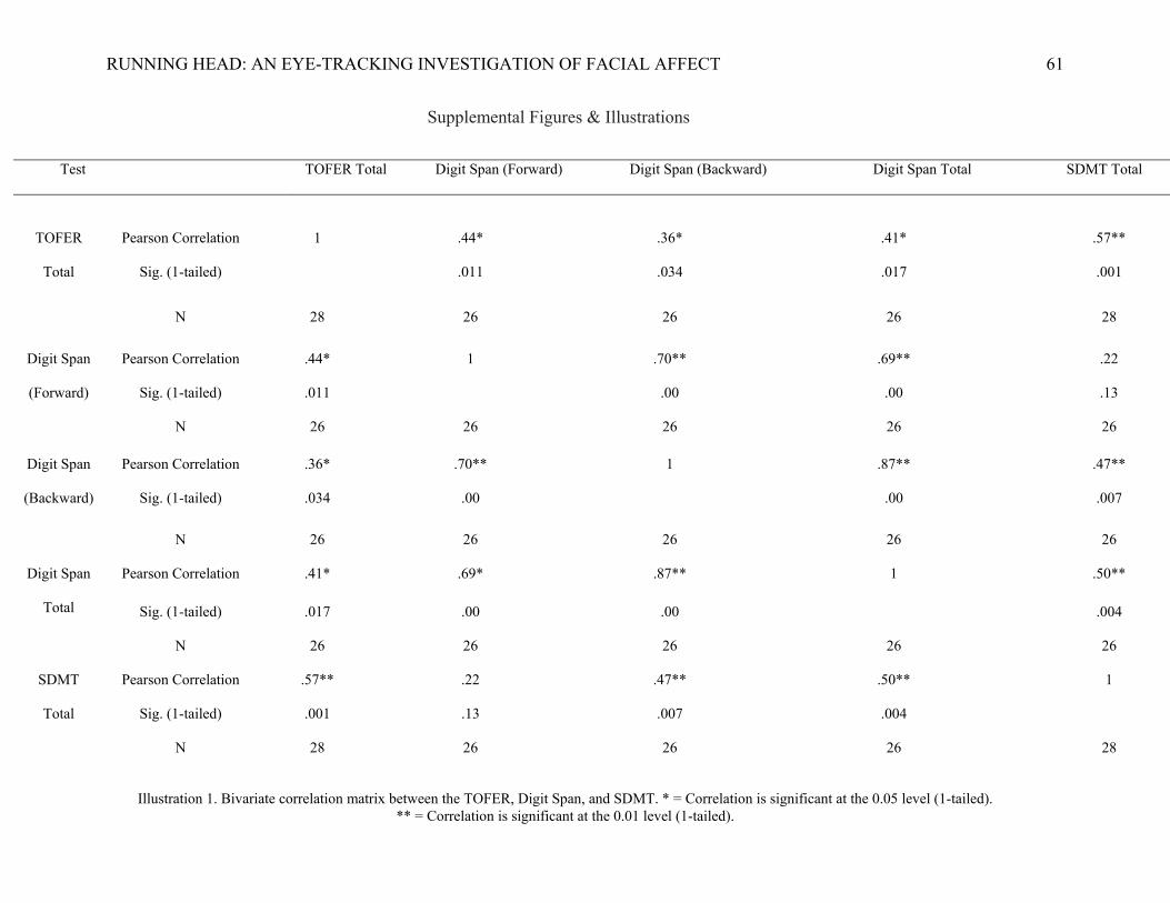
RUNNING HEAD: AN EYE-TRACKING INVESTIGATION OF FACIAL AFFECT 61
Supplemental Figures & Illustrations
Test TOFER Total Digit Span (Forward) Digit Span (Backward) Digit Span Total SDMT Total
TOFER
Total
Pearson Correlation 1 .44* .36* .41* .57**
Sig. (1-tailed) .011 .034 .017 .001
N 28 26 26 26 28
Digit Span
(Forward)
Pearson Correlation .44* 1 .70** .69** .22
Sig. (1-tailed) .011 .00 .00 .13
N 26 26 26 26 26
Digit Span
(Backward)
Pearson Correlation .36* .70** 1 .87** .47**
Sig. (1-tailed) .034 .00 .00 .007
N 26 26 26 26 26
Digit Span
Total
Pearson Correlation .41* .69* .87** 1 .50**
Sig. (1-tailed) .017 .00 .00 .004
N 26 26 26 26 26
SDMT
Total
Pearson Correlation .57** .22 .47** .50** 1
Sig. (1-tailed) .001 .13 .007 .004
N 28 26 26 26 28
Illustration 1. Bivariate correlation matrix between the TOFER, Digit Span, and SDMT. * = Correlation is significant at the 0.05 level (1-tailed). ** = Correlation is significant at the 0.01 level (1-tailed).