Embed Size (px)
Citation preview

Original Article
An Experimental Study of Histopathologic Effects of Hemostatic Agents Used in Spinal
SurgeryIdiris Altun
-OBJECTIVE: To evaluate and compare the histopatho-logic effects of various hemostatic agents used in spinalsurgery on an experimental laminectomy model in rats.
-METHODS: There were 110 rats randomly allocated into11 groups, including sham, control, AnkaferdBlood Stopper(herbal hemostatic), SeraSeal (agar and bovine factor pro-teins), FLOSEAL (gelatin granules and thrombin), SURGIFLO(gelatin paste), HELITENE (absorbable collagen), Beriplast(fibrin sealant containing fibrinogen, factor XIII, andthrombin), TISSEEL (fibrin sealant), BLOODCARE (hemostaticpowder), and SURGICEL (oxidized cellulose polymer) groups.Hemostatic agents were applied on the epidural region afterlaminectomy was performed until the identification of duramater and nerve root. After a follow-up period of 12 weeks,rats were sacrificed, and histologic sections were per-formed proximal and distal to laminectomy zone. Groupswere histopathologically compared in terms of chronicinflammation, fibrosis, and vascularization.
-RESULTS: There was no difference between groups interms of acute inflammation (P [ 0.159). Chronic inflam-mation was more remarkable in the herbal hemostaticgroup (P [ 0.036), and there was severe fibrosis inabsorbable collagen hemostatic, fibrin sealant, and powderhemostatic agent groups (P < 0.001). Vascularity was moreobvious in herbal hemostatic; fibrin sealant; absorbablecollagen; fibrin sealant containing fibrinogen, factor XIII,and thrombin; hemostatic powder; and oxidized cellulosepolymer groups (P < 0.001).
-CONCLUSIONS: Hemostatic agents can cause notablehistopathologic alterations, including inflammation,
Key words- Epidural- Fibrosis- Hemostatic- Histopathology- Inflammation- Lumbar- Vascularization
Abbreviations and AcronymsABS: AnkaferdBlood stopper
WORLD NEUROSURGERY 90: 147-153, JUNE 2016
fibrosis, and vascularity. In this context, flowable hemo-stats such as gelatin granules and thrombin or gelatinpaste seem to provide more promising results in spinalsurgery.
INTRODUCTION
oagulation has been the mainstay of hemostasis duringsurgery in the last century. However, problems such as
Ccarbonization of the tissue or adhesions to the electrodemay be seen as a result of high temperature.1,2 In this context,hazards associated with thermal injury constitute an importantlimitation for use of electrocautery in surgical practice. However,failure to achieve hemostasis during surgery can lead to excessivebleeding, which complicates the procedure and causes a sub-stantial risk for morbidity and mortality.3
In spinal surgery, these problems can be more obvious, andnerve injury and dural tears may exist as well as increasedoperative time and need for postoperative blood transfusions.4,5
If bleeding cannot be controlled by conventional methods,topical hemostatic agents can be used. These topical hemostaticagents are classified into active and passive groups. Passivehemostatic agents consist of collagens, cellulose, and gelatins,and they function by contact activation and enhance aggregationof platelets. Active hemostatic agents, such as fibrin sealants,may include thrombin, and they act biologically on the clottingmechanism.1,4
These hemostatic agents may cause significant histopathologicalterations, such as inflammation, adhesion, and fibrosis. Thesechanges may facilitate extradural compression of the nervousstructures, and traction of nerve root by epidural adhesion
Department of Neurosurgery, Kahramanmaras Sutcu Imam University Medical Faculty,Kahramanmaras, Turkey
To whom correspondence should be addressed: Idiris Altun, M.D.[E-mail: [email protected]]
Citation: World Neurosurg. (2016) 90:147-153.http://dx.doi.org/10.1016/j.wneu.2016.02.052
Journal homepage: www.WORLDNEUROSURGERY.org
Available online: www.sciencedirect.com
1878-8750/$ - see front matter ª 2016 Elsevier Inc. All rights reserved.
www.WORLDNEUROSURGERY.org 147

ORIGINAL ARTICLE
IDIRIS ALTUN HISTOPATHOLOGICAL EFFECTS OF HEMOSTATIC AGENTS
and fibrosis not only impairs arterial supply but also mayblock axoplasmic transport, resulting in painful radiculopathy.1
Furthermore, the presence of scar tissue in the operative areaaugments the risks for nerve root lesions and dural tears. To thebest of our knowledge, the histopathologic effects of hemostaticagents have not been extensively studied in the medicalliterature. The objective of the present study was to assess andcompare the histopathologic changes attributed to the use ofvarious topical hemostatic agents on the epidural region in anexperimental laminectomy model on lumbar spines.
MATERIALS AND METHODS
Study DesignThis experimental study was carried out in the experimentalresearch laboratory of our university following approval of thelocal research ethics committee of experimental animal studies(2014/01-48). In this study, 110 male adult Wistar rats (weighing400e450 g) were used. Access to food and water was provided adlibitum, and a diurnal cycle for 12:12 hours of light/darkness wasprovided. All experimental procedures were implemented inaccordance with the protocols of the institutional animal careand use committee. Rats were allocated into 11 groups accordingto the topical hemostatic agent to be applied on the epiduralregion of the laminectomy site at the level of L4.
Surgical ProcedureThe surgical procedure of lumbar laminectomy was performed asdescribed previously in the literature.5 General anesthesia wasinduced by application of pentobarbital (10 mL/kg). Aftershaving the hair around L4, the naked skin was sterilized withpovidone-iodine solution. Under aseptic conditions, visualizationwas provided under a surgical microscope, and a conventionalmidline approach was used for access to the L4 area. Afterdissection of the fascia and paraspinal muscles, L4 laminar wasremoved using a burr. A laminectomy defect of 7 mm � 4 mm wasformed using a high-speed drill, and complete exposure to dura
Figure 1. Under a surgical microscope (A) after dissection of tdefect was formed using a high-speed drill and complete exp
148 www.SCIENCEDIRECT.com WORLD NEU
mater and the nerve root was provided (Figure 1). The length ofdissection of a single block was 6 mm. Nothing was done to thecontrol group (Figure 2). In the sham group, laminectomy wasperformed (Figure 3), and 0.1 mL saline was administeredtopically on the epidural region at the site of laminectomy for 5minutes. Liquid hemostatic agents (AnkaferdBlood stopper[ABS; Ankaferd _Ilac Kozmetik AS, Istanbul, Turkey], SeraSeal[Wortham Laboratories, Chattanooga, Tennessee, USA],FLOSEAL Hemostatic Matrix [Baxter Healthcare Corporation,Deerfield, Illinois, USA], SURGIFLO Hemostatic Matrix Kit withthrombin [Ethicon Inc., Somerville, New Jersey, USA], BeriplastP Combi-Set 3 mL [Aventis Pharma Limited, Mumbai, India],TISSEEL fibrin sealant [Baxter Healthcare Corporation]) 0.1 mL,powder hemostatic agent (HELITENE [Integra NeuroSciences,Plainsboro, New Jersey, USA]) 0.1 gr, and matrix hemostaticagents (SURGICEL original absorbable hemostat [Ethicon, Inc.,Somerville, New Jersey, USA], BLOODCARE Matrix [Life Line pluss.r.o, Brno, Czech Republic]) 0.1 cm3 were administered topicallyand covered on the epidural region at the site of laminectomy for 5minutes. After 5 minutes, the laminectomy sites were irrigatedimmediately with saline solution to eliminate the surplus relevanthemostatic agent. The wound was closed in layers using the samesuture material in each animal. No complications or adverse re-actions were encountered during or after surgery.
Hemostatic AgentsAdvanced hemostatic agents administered topically in the cur-rent study included FLOSEAL Hemostatic Matrix, SURGIFLOHemostatic Matrix Kit with thrombin, ABS (100 mL), SeraSeal,HELITENE, Beriplast P Combi-Set 3 mL, TISSEEL fibrin sealant,BLOODCARE Matrix, and SURGICEL original absorbable he-mostat. After a follow-up period of 12 weeks, rats were sacrificedby intracardiac perfusion of potassium (0.5 mL/100 g bodyweight). Tissue specimens were resected on the epidural planeproximal and distal to the sites of laminectomy. The section oftissue specimen was performed in the axial direction of thespinal column. After dehydration of tissue specimens, formalin-
he fascia and paraspinal muscles, (B) a laminectomyosure to dura mater and the nerve root.
ROSURGERY, http://dx.doi.org/10.1016/j.wneu.2016.02.052
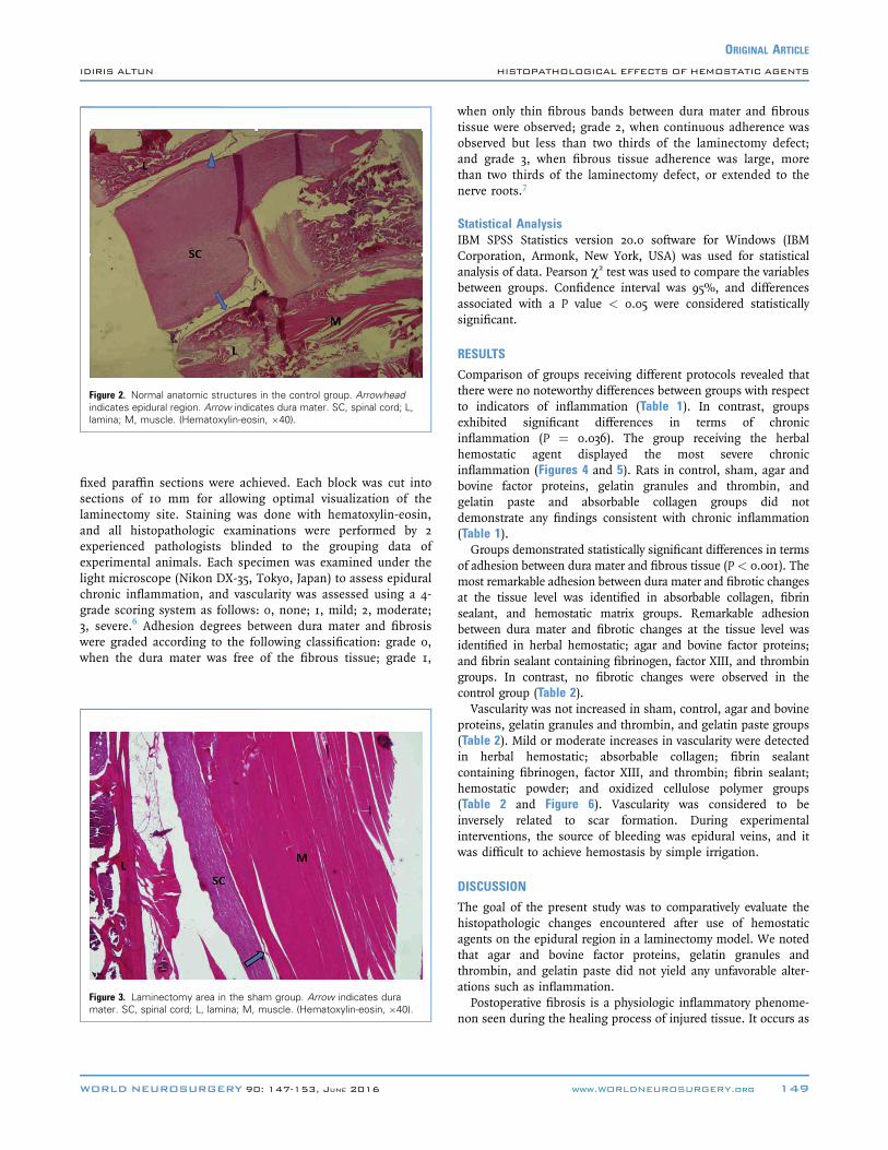
Figure 2. Normal anatomic structures in the control group. Arrowheadindicates epidural region. Arrow indicates dura mater. SC, spinal cord; L,lamina; M, muscle. (Hematoxylin-eosin, �40).
ORIGINAL ARTICLE
IDIRIS ALTUN HISTOPATHOLOGICAL EFFECTS OF HEMOSTATIC AGENTS
fixed paraffin sections were achieved. Each block was cut intosections of 10 mm for allowing optimal visualization of thelaminectomy site. Staining was done with hematoxylin-eosin,and all histopathologic examinations were performed by 2experienced pathologists blinded to the grouping data ofexperimental animals. Each specimen was examined under thelight microscope (Nikon DX-35, Tokyo, Japan) to assess epiduralchronic inflammation, and vascularity was assessed using a 4-grade scoring system as follows: 0, none; 1, mild; 2, moderate;3, severe.6 Adhesion degrees between dura mater and fibrosiswere graded according to the following classification: grade 0,when the dura mater was free of the fibrous tissue; grade 1,
Figure 3. Laminectomy area in the sham group. Arrow indicates duramater. SC, spinal cord; L, lamina; M, muscle. (Hematoxylin-eosin, �40).
WORLD NEUROSURGERY 90: 147-153, JUNE 2016
when only thin fibrous bands between dura mater and fibroustissue were observed; grade 2, when continuous adherence wasobserved but less than two thirds of the laminectomy defect;and grade 3, when fibrous tissue adherence was large, morethan two thirds of the laminectomy defect, or extended to thenerve roots.7
Statistical AnalysisIBM SPSS Statistics version 20.0 software for Windows (IBMCorporation, Armonk, New York, USA) was used for statisticalanalysis of data. Pearson c2 test was used to compare the variablesbetween groups. Confidence interval was 95%, and differencesassociated with a P value < 0.05 were considered statisticallysignificant.
RESULTS
Comparison of groups receiving different protocols revealed thatthere were no noteworthy differences between groups with respectto indicators of inflammation (Table 1). In contrast, groupsexhibited significant differences in terms of chronicinflammation (P ¼ 0.036). The group receiving the herbalhemostatic agent displayed the most severe chronicinflammation (Figures 4 and 5). Rats in control, sham, agar andbovine factor proteins, gelatin granules and thrombin, andgelatin paste and absorbable collagen groups did notdemonstrate any findings consistent with chronic inflammation(Table 1).Groups demonstrated statistically significant differences in terms
of adhesion between dura mater and fibrous tissue (P < 0.001). Themost remarkable adhesion between dura mater and fibrotic changesat the tissue level was identified in absorbable collagen, fibrinsealant, and hemostatic matrix groups. Remarkable adhesionbetween dura mater and fibrotic changes at the tissue level wasidentified in herbal hemostatic; agar and bovine factor proteins;and fibrin sealant containing fibrinogen, factor XIII, and thrombingroups. In contrast, no fibrotic changes were observed in thecontrol group (Table 2).Vascularity was not increased in sham, control, agar and bovine
proteins, gelatin granules and thrombin, and gelatin paste groups(Table 2). Mild or moderate increases in vascularity were detectedin herbal hemostatic; absorbable collagen; fibrin sealantcontaining fibrinogen, factor XIII, and thrombin; fibrin sealant;hemostatic powder; and oxidized cellulose polymer groups(Table 2 and Figure 6). Vascularity was considered to beinversely related to scar formation. During experimentalinterventions, the source of bleeding was epidural veins, and itwas difficult to achieve hemostasis by simple irrigation.
DISCUSSION
The goal of the present study was to comparatively evaluate thehistopathologic changes encountered after use of hemostaticagents on the epidural region in a laminectomy model. We notedthat agar and bovine factor proteins, gelatin granules andthrombin, and gelatin paste did not yield any unfavorable alter-ations such as inflammation.Postoperative fibrosis is a physiologic inflammatory phenome-
non seen during the healing process of injured tissue. It occurs as
www.WORLDNEUROSURGERY.org 149

Table 1. Analysis of Acute and Chronic Inflammatory Changes After Application of Topical Hemostatic Agents in Control, Sham, andStudy Groups
Group
Variable
P Value
Variable
P Value
Acute Inflammation Chronic Inflammation
None Mild Moderate Severe None Mild Moderate Severe
Control 10 (100%) 0 0 0 0.159 10 (100%) 0 0 0 0.036*
Sham 10 (100%) 0 0 0 10 (100%) 0 0 0
Herbal hemostaticy 7 (70%) 2 (20%) 1 (10%) 0 7 (70%) 0 2 (20%) 1 (10%)
Agar and bovine factor proteinsz 10 (100%) 0 0 0 10 (100%) 0 0 0
Gelatin granules and thrombinx 10 (100%) 0 0 0 10 (100%) 0 0 0
Gelatin pastek 10 (100%) 0 0 0 10 (100%) 0 0 0
Absorbable collagen{ 10 (100%) 0 0 0 10 (100%) 0 0 0
Fibrin sealant containing fibrinogen,factor XIII, and thrombin#
10 (100%) 0 0 0 5 (50%) 3 (30%) 2 (20%) 0
Fibrin sealant** 10 (100%) 0 0 0 8 (80%) 1 (10%) 1 (10%) 0
Hemostatic powderyy 10 (100%) 0 0 0 8 (80%) 1 (10%) 1 (10%) 0
Oxidized cellulose polymerzz 9 (90%) 1 (10%) 0 0 7 (70%) 3 (30%) 0 0
Control group underwent no procedures at all, whereas sham group received topical isotonic saline after laminectomy.*Statistically significant.yAnkaferdBlood Stopper.zSeraSeal.xFLOSEAL.kSURGIFLO.{HELITENE.#Beriplast.**TISSEEL.yyBLOODCARE.zzSURGICEL.
Figure 4. Widespread tissue fibrosis and chronic inflammatory cellsbetween dura mater. SC, spinal cord; F, fibrosis; I, inflammatory cells; L,lamina. (Hematoxylin-eosin, �40).
Figure 5. Grade III fibrosis between dura mater. The epidural fibrosis wasadhered to the underlying dura mater and spinal cord. SC, spinal cord; F,fibrosis; I, inflammatory cells. (Hematoxylin-eosin, �40).
150 www.SCIENCEDIRECT.com WORLD NEUROSURGERY, http://dx.doi.org/10.1016/j.wneu.2016.02.052
ORIGINAL ARTICLE
IDIRIS ALTUN HISTOPATHOLOGICAL EFFECTS OF HEMOSTATIC AGENTS

Table 2. Analysis of Fibrosis and Vascularity After Application of Topical Hemostatic Agents in Control, Sham, and Study Groups
Group
Variable
P Value
Variable
P Value
Fibrosis Vascularity
None Mild Moderate Severe None Mild Moderate Severe
Control 10 (100%) 0 0 0 < 0.001* 10 (100%) 0 0 0 < 0.001*
Sham 2 (20%) 3 (30%) 3 (30%) 2 (20%) 10 (100%) 0 0 0
Herbal hemostaticy 0 1 (10%) 4 (40%) 5 (50%) 7 (70%) 0 3 (30%) 0
Agar and bovine factor proteinsz 0 1 (10%) 4 (40%) 5 (50%) 10 (100%) 0 0 0
Gelatin granules and thrombinx 0 3 (30%) 4 (40%) 3 (30%) 10 (100%) 0 0 0
Gelatin pastek 0 3 (30%) 4 (40%) 3 (30%) 10 (100%) 0 0 0
Absorbable collagen{ 0 0 3 (30%) 7 (70%) 9 (90%) 0 1 (10%) 0
Fibrin sealant containing fibrinogen,factor XIII, and thrombin#
0 0 5 (50%) 5 (50%) 5 (50%) 5 (50%) 0 0
Fibrin sealant** 0 0 3 (30%) 7 (70%) 5 (50%) 2 (20%) 3 (30%) 0
Hemostatic powderyy 0 0 3 (30%) 7 (70%) 6 (60%) 4 (40%) 0 0
Oxidized cellulose polymerzz 0 0 3 (40%) 7 (70%) 6 (60%) 3 (30%) 1 (10%) 0
Control group underwent no procedures at all, whereas sham group received topical isotonic saline after laminectomy.*Statistically significant.yAnkaferdBlood Stopper.zSeraSeal.xFLOSEAL.kSURGIFLO.{HELITENE.#Beriplast.**TISSEEL.yyBLOODCARE.zzSURGICEL.
ORIGINAL ARTICLE
IDIRIS ALTUN HISTOPATHOLOGICAL EFFECTS OF HEMOSTATIC AGENTS
a result of the organization of the fibrin matrix in the damagedtissue together with the exudation of fibrinogen and proliferationof fibroblasts.8,9 Nevertheless, excessive fibrosis in surroundingtissues may cause unintended consequences. In particular, fibrosisaround the dura mater and nerve roots would trigger tethering ofnerves, neurologic symptoms, and increased risks of nerve injuriesafter laminectomy of the spine. Furthermore, fibrosis causes asubstantial risk of nerve root injury, and reduction of theinflammatory reaction, inhibition of fibroblastic proliferation, andrelease of exudate may decrease these risks.9 Therefore,knowledge of the histopathologic reactions associated withhemostatic agents is necessary for selection of the appropriateoption during surgery.Fibrin sealants are not only used for strengthening repairs of
elective cerebrospinal fluid fistulas in spinal surgery, but they alsoprovide hemostasis and reduce scar formation.9 Additionalbenefits of fibrin sealants include intraoperative hemostasis withreduced postoperative drainage, reduced transfusionrequirements, and reduced cost and scar formation.8,10
Cekinmez et al.6 investigated the efficacy of different steroiddoses, steroids combined with fibrin sealants, and fibrinsealants alone for limiting epidural fibrosis after spinal
WORLD NEUROSURGERY 90: 147-153, JUNE 2016
procedures in a rat model. There were 100 L4-5 laminectomiesperformed in 100 Sprague-Dawley rats, and histologic evaluationsperformed 1, 2, 4, and 6 weeks after the procedure demonstratedcomparable frequencies of “epidural fibrosis, inflammation,necrosis and abscess formation.”6 These authors concluded thatno treatment arm proved beneficial.6
Richards et al.8 used fibrin sealants with ADCON-L gel (Glia-tech, Cleveland, Ohio, USA), a carbohydrate polymer gel shown toinhibit postsurgical adhesions, as a medicated adhesion barrier toreduce posterior spinal epidural adhesions after laminectomies ina sheep model. Laminectomy defects were treated with fibrinsealants alone or ADCON-L gel, or no treatment was performed.Epidural fibrosis and adhesions were assessed 12 weeks later withmagnetic resonance imaging, peel-off testing, and histology.These authors concluded that the ADCON-L gel preparationeffectively reduced epidural adhesions in this sheep model.8
In 2014, Epstein11 applied TISSEEL in 22 of 39 patientsundergoing laminectomy who exhibited increased intraoperativebleeding. Patients undergoing laminectomy who did notdemonstrate increased bleeding did not receive TISSEEL.Additionally, Wu et al.12 assessed 82 consecutive patientsundergoing posterior lumbar fusion or posterior lumbar
www.WORLDNEUROSURGERY.org 151

Figure 6. Vascularization (arrow) in severe fibrosis between dura mater.SC, spinal cord; F, fibrosis. (Hematoxylin-eosin, �100).
ORIGINAL ARTICLE
IDIRIS ALTUN HISTOPATHOLOGICAL EFFECTS OF HEMOSTATIC AGENTS
interbody fusion who were randomly assigned to receiveabsorbable gelatin sponge versus no sponge. They found thatpatients receiving the gelatin sponge versus no sponge showedreduced average drainage, reduced perioperative bloodtransfusion requirements, and reduced length of stay withoutadverse sequelae.12
ABS is a hemostatic agent that consists of a mixture of 5 plants:Thymus vulgaris, Glycyrrhiza glabra, Vitis vinifera, Alpinia officinarum,and Urtica dioica. Each of these plants has effects on endothelialcells, blood cells, angiogenesis, cell proliferation, vascular dy-namics, and cell mediators.13 The main mechanism of ABSinvolves the formation of encapsulated protein bonds, which arethe foci of erythrocyte aggregation. ABS has been successfullyused in many clinical cases as a hemostatic agent.13 ABS can bea safe and promising hemostatic agent in neurosurgery. Once itsadvantages and disadvantages are documented and clarified,ABS can achieve worldwide popularity beyond its local use.Commonly used gelatin-thrombinebased flowable advanced
topical hemostatic agents offer important advantages in surgerybecause they can conform to wound contours and fill deeplesions.2,14 These products have been labeled as hemostaticmatrices and exhibit passive and active mechanisms of action inthe blood-clotting cascade via contact activation and active bio-logic agents, including thrombin.14
The present study yielded important clinical clues for selectionof suitable hemostatic agents in spinal surgery. First, we notedthat inflammation was similar in all groups receiving variousagents. These findings indicate that the inflammatory process,likely to be linked with fibrosis or vascularity, is more likely to beshaped in the chronic phase. Therefore, efforts must be made tomodify and attenuate the chronic inflammation to accomplishmore favorable clinical outcomes. Second, agar and bovine factorproteins, gelatin granules with thrombin, and gelatin paste didnot result in either remarkable chronic inflammatory changes orfibrosis. Moreover, vascularity was similar with sham and controlgroups for these 3 hemostatic agents. Consequently, we advocate
152 www.SCIENCEDIRECT.com WORLD NEU
the use of these 3 hemostatic agents during spinal surgery in alaminectomy model in rats because they yielded the most favor-able histopathologic results.During surgery, advanced hemostatic agents possess advantages,
such as diminution of blood loss, reduction of the need for bloodtransfusion, avoidance of the need for systemic hemostatic drugs,and shorter duration of stay in the operating room.15 These benefitscan potentially reduce length of stay in the hospital and decrease thecost of care. It has been postulated that clinical efficacy of anadvanced flowable hemostatic matrix compared with anonflowable topical hemostat (e.g., Gelfoam or SURGICEL) wasassociated with a more significant reduction in time to hemostasisand greater control of bleeding by reducing patient blood loss.16,17
None of the hemostasis materials available for neurosurgery,such as gelatins, collagens, oxidized cellulosis, fibrin, thrombin,polysaccharides, and hydrogel-based hemostats or combinationsof the aforementioned materials, is 100% complication-free, andthey may cause swelling or other histopathologic changes whenleft in situ. Swelling in a confined space such as the spinal canalmay be dangerous because vital neural structures may becompromised.18 Richter et al.19 reported that ADCON-L gel pro-vided no additional benefit in terms of scar formation for patientswho underwent 1-level lumbar microdiscectomy.Consistent with these data, results of the present study indi-
cated that flowable hemostatic matrix resulted in more acceptablehistopathologic changes compared with nonflowable hemostaticmatrix. Therefore, along with individualized and careful assess-ment of each patient, choice of an active flowable hemostaticmatrix can be a more effective tool to address a broad range ofactive bleeding in spinal surgery. This hemostatic matrix isespecially important in spinal surgery because visualization iscritical to efficient and successful surgery. Despite the widespreaduse of advanced hemostatic matrices in surgery, trials focusing oncomparing a variety of hemostatic agents have been limited. Wehope that our results will provide new insights for rational use ofhemostatic agents during spinal surgery. Hemostatic agents canoffer additional advantages for the surgeon particularly duringprocedures around the nerve root.The main limitations of this study are experimental design and
challenges in extrapolation of our data to humans. Other limita-tions are lack of information on the amount of bleeding indifferent groups and variability of pressure during application ofhemostatic agents. Although maximal effort was expended to haveclear-cut cross sections and identical histologic views, variabilitymay have occurred between groups. Further randomized, multi-center trials on larger series are necessary to elucidate the histo-pathologic impact of hemostatic agents used in spinal surgery.
CONCLUSIONS
The results of the present study show that hemostatic agents cancause notable histopathologic alterations, such as inflammation,fibrosis, and vascularity. In this context, flowable hemostats, suchas gelatin granules with thrombin or gelatin paste, seem to pro-vide more promising results in spinal surgery.
ACKNOWLEDGMENTS
The author thanks Harun Çıralık, M.D., for assistance with thehistopathologic evaluation.
ROSURGERY, http://dx.doi.org/10.1016/j.wneu.2016.02.052

ORIGINAL ARTICLE
IDIRIS ALTUN HISTOPATHOLOGICAL EFFECTS OF HEMOSTATIC AGENTS
REFERENCESnonsteroidal anti-inflPa 1976). 1995;20:557-
1. Nishida K, Kakutani K, Maeno K, Takada T,Yurube T, Kuroda R, et al. Efficacy of hemostasisfor epidural venous plexus and safety for neuralstructure using soft coagulation system in spinalsurgery: a laboratory investigation using a porcinemodel. J Spinal Disord Tech. 2013;26:E281-E285.
2. Price JS, Tackett S, Patel V. Observational evalu-ation of outcomes and resource utilization fromhemostatic matrices in spine surgery. J Med Econ.2015;18:777-786.
3. Renkens KL Jr, Payner TD, Leipzig TJ, Feuer H,Morone MA, Koers JM, et al. A multicenter, pro-spective, randomized trial evaluating a newhemostatic agent for spine surgery. Spine (Phila Pa1976). 2001;26:1645-1650.
4. Peters A, Verma K, Slobodyanyuk K, Cheriyan T,Hoelscher C, Schwab F, et al. Antifibrinolyticsreduce blood loss in adult spinal deformity sur-gery: a prospective, randomized controlled trial.Spine (Phila Pa 1976). 2015;40:E443-E449.
5. Lee JY, Stenzel W, Ebel H, Wedekind C,Ernestus RI, Klug N. Mitomycin C in preventingspinal epidural fibrosis in a laminectomy model inrats. J Neurosurg. 2004;100(1 suppl):52-55.
6. Cekinmez M, Sen O, Atalay B, Erdogan B,Bavbek M, Caner H, et al. Effects of methylprednisolone acetate, fibrin glue and combinationof methyl prednisolone acetate and fibrin glue inprevention of epidural fibrosis in a rat model.Neurol Res. 2010;32:700-705.
7. He Y, Revel M, Loty B. A quantitative model ofpostlaminectomy scar formation. Effects of a
WORLD NEUROSURGERY 90: 147-153, J
ammatory drug. Spine (Phila563.
8. Richards PJ, Turner AS, Gisler SM, Kraft S,Nuss K, Mark S, et al. Reduction in post-laminectomy epidural adhesions in sheep using afibrin sealant-based medicated adhesion barrier.J Biomed Mater Res B Appl Biomater. 2010;92:439-446.
9. Yu CH, Lee JH, Baek HR, Nam H. The effective-ness of poloxamer 407-based new anti-adhesivematerial in a laminectomy model in rats. EurSpine J. 2012;21:971-979.
10. Epstein NE. Hemostasis and other benefits offibrin sealants/glues in spine surgery beyondcerebrospinal fluid leak repairs. Surg Neurol Int.2014;5(suppl 7):S304-S314.
11. Epstein NE. Tisseel utilized as hemostatic in spinesurgery impacts time to drain removal and lengthof stay. Surg Neurol Int. 2014;5(suppl 7):S354-S361.
12. Wu J, Jin Y, Zhang J, Shao H, Yang D, Chen J.Hemostatic techniques following multilevel pos-terior lumbar spine surgery: a randomized controltrial. J Spinal Disord Tech. 2014;27:442-446.
13. Yuce S, Candirli C, Yenidunya S, Muslu B. Newhemostatic agent: the effect of Ankaferd BloodStopper on healing wounds in experimental skinincision model. Turk J Med Sci. 2014;44:288-294.
14. Gazzeri R, Galarza M, Alfier A. Safety biocom-patibility of gelatin hemostatic matrix (Floseal andSurgiflo) in neurosurgical procedures. Surg TechnolInt. 2012;22:49-54.
15. David G, Lim S, Gunnarsson C, Kocharian R,Roy S. Similar patient outcomes yet different
UNE 2016 ww
hospital costs between flowable hemostaticagents. J Med Econ. 2015;18:735-745.
16. Nasso G, Piancone F, Bonifazi R, Romano V,Visicchio G, De Filippo CM, et al. Prospectiverandomized clinical trial of the FloSeal matrixsealant in cardiac surgery. Ann Thorac Surg. 2009;88:1520-1526.
17. Weaver FA, Hood DB, Zatina M, Messina L,Badduke B. Gelatin-thrombin based hemostaticsealant for intraoperative bleeding in vascularsurgery. Ann Vasc Surg. 2002;16:286-293.
18. Menovsky T, Plazier M, Rasschaert R, Maas AI,Parizel PM, Verbeke S. Massive swelling ofSurgicel� Fibrillar� hemostat after spinal sur-gery. Case report and a review of the literature.Minim Invasive Neurosurg. 2011;54:257-259.
19. Richter HP, Kast E, Tomczak R, Besenfelder W,Gaus W. Results of applying ADCON-L gel afterlumbar discectomy: the German ADCON-L study.J Neurosurg. 2001;95:179-189.
Conflict of interest statement: This study was supported byKahramanmaras Sutcu Imam University Research Foundation(2014/3-40 M).
Received 19 December 2015; accepted 9 February 2016
Citation: World Neurosurg. (2016) 90:147-153.http://dx.doi.org/10.1016/j.wneu.2016.02.052
Journal homepage: www.WORLDNEUROSURGERY.org
Available online: www.sciencedirect.com
1878-8750/$ - see front matter ª 2016 Elsevier Inc.All rights reserved.
w.WORLDNEUROSURGERY.org 153