Embed Size (px)
Citation preview

An Essay toward a Dynamic Morphology of
the Mammalian Nephron*
JEAN OLIVER, M.D.
Brooklyn, .New York
“We shall have occasion to suggest the analysis of physical entities into structures of events, and even events, as I shall try to show, may be regarded with advantage as having structure.”
B. Russell “We may consider, therefore, organic form as an expression of a pattern of processes
of an ordered system of forces. This point of view can be called dynamic morphology.” L. von Bertala&yY
T HUS from Parnassian heights speak the philosophers, and in laboratory and clinic the busy practitioners,
“in perplexity and doubtful dilemma” be- fore the enigma of structure and function that daily confronts them, take heart in their seemly endless effort of translating physical substance into dynamic activity. Of one thing they are sure: Neither the practical accomplishments nor the intellectual satis- factions so richly inherent in the method they follow can come to full accomplishment unless this dichotomy of structure and func- tion is for the moment at least solved. There can be no “functional” answer to their prob- lems standing apart from the “structural” explanation; like stereographic images the two must be fused for clear vision. T Since this is so, fortunate is he who has chosen the field of renal activity as his garden to cultivate, for no other organ offers as does the kidney in the complexities of its activity and its
t The suggestion that the nervous system offers even more remarkable opportunities for correlation of struc- ture and function will be admitted by all but to some will appear a counsel of perfection since its complexities seem at present to transcend resolution. It is an interesting point that both these biologic systems, cerebral and renal, are concerned with similar fundamental problems of living, namely, the adjustment of the organism to its
form so much for admiration and wonder- ment that yet remains reasonably within the capabilities of present day technics.
It will be apparent then that in a con- sideration of the activity of the renal tubule, although a morphologist may be helpful in various ways, first and foremost it is his part to determine if possible what it is that func- tions. The functionalist knows only what as blood goes into the conceptual area he calls a “kidney” and what as blood and urine comes out; the morphologist describes the physical aspects of the interim. On the framework of his description are fitted by inference observed dynamic events and this conceptual pattern must then be confirmed empirically by the observation of some specific and appropriate structural change. Such is the ideal accomplishment and in certain instances we shall see that it has been closely approached in achievement.
In simpler terms, the morphologist is to “localize function.” In this endeavor there opens before him a series of isolable struc- tures of infinitely regressing magnitude the
environments and that they resolve it in converse ways. The nervous system modifies the organism and its behavior to fit the external environment; the kidney alters the internal environment to fit the organism.
* From the Department of Pathology, the College of Medicine, State University Medical Center at New York City, Brooklyn, N. Y. The author’s work herein described is supported by grants-in-aid from the Life Insurance Medical Research Fund, The Commonwealth Fund, and the American and New York Heart Associations.
88 AMERICAN JOURNAL OF MEDICINE

Morphology of Mammalian Nephron-Oliver 89
end of which in the light of his increasing technical ability is not in sight. Thus he localizes renal activity in the nephron and proceeds to glomerular filter and to tubule. In the latter he must distinguish between parts of tubule and, within any given part, differing segments. Passing to the cells of the tubule wall he comes upon membranes, both an internal (and one of these, “the brush border” of the proximal convolution, most remarkably differentiated in structure) and an external, the membrana propria, of simpler configuration. Within the cell he finds a whole hierarchy of cytoplasmic particulate bodies, batonets, “droplets,” mitochondria and microsomes, again a series of decreasing magnitudes that seems limited in its members only by his ability to spin them out of his cytoplasmic sus- pensions, for the microsomes approach the limits of ordinary visibility and are not much larger than certain polymolecular complexes. All this variety of structure the morphologist can observe “before and after” the functional experiment and so point to physical alterations that have occurred for correlation with measured activity. But let us look at some examples for specific illustration.
One of the earliest of accurate and objec- tive localizations was the demonstration in 1912 by Susuki3 that dyes such as trypan blue or carmine are absorbed by the proximal convolution and stored in its epithelial cells. Susuki moreover noticed that the amount of dye absorbed and stored decreased as one departed from the origin of the tubule at the glomerulus; by means of the number of granules of dye seen in sections of its convolutions he divided this portion of the nephron into four segments. In 1915 dissections of vitally stained neph- rons4q5 confirmed the general inferences he had drawn from his study of sections; but when the proximal convolution was seen in its entirety, it was perfectly obvious that the distribution of dye indicated not that the proximal convolution is composed of seg- ments but that in its handling of the dye it is a continuous functional unit in which
JULY, 1350
exists a simple decreasing gradient of ab- sorption. One might suppose from the appearance of this gradient that the whole length of the convolution was lined by functionally equivalent cells and that each had removed the same proportion of the total concentration of the constantly de- creasing material that passed over its surface.
Another gradient of tubule activity de- monstrable in the proximal convolution is similar in its distribution to the one just described for the dye. It has been quanti- tatively measured by a joint effort of func- tionalists and morphologists,6,7 the former removing samples of tubule fluid from the proximal convolutions of small mammals and determining their sugar and creatinine content and the latter dissecting out the tapped nephrons, identifying the point of puncture and measuring its distance from the beginning and the end of the convolu- tion. (Fig. 1.) It will be noticed that ab- sorption by the tubule cells results in an evenly falling concentration of sugar so that most of it is removed by the time the first half of the convolution has been passed; similarly it can be shown that the greater part of the water of the original filtrate has been absorbed. Under physiologic condi- tions it is therefore apparent that the lumen surface of the cells of the terminal portion of the proximal convolution does not come in contact with any considerable concentra- tion of sugar. If phlorhizin is given, the gradient of concentration of sugar in the tubule fluid is reversed and the terminal portion is flooded with a high concentration. (Fig. 2.)
It would appear therefore that there is a certain degree of similarity in the behavior of the cells of the proximal convolution in the handling of two substances as different as dye and sugar since under normal condi- tions the activity of the first part of the con- volution is chiefly concerned with the taking up of the two absorbed materials. The conditions to which the cells of the terminal portion of the convolution are exposed in the two instances are, however, very

90 Morphology of Mammalian Nephron--Oliver
different. In the case of sugar, little if any question is not open to direct examination remains for absorption by the time the in the experimental animal for this portion tubule fluid reaches them, while in the case of the tubule lies too deep in the substance of the dye, which is continuously appearing of the kidney to be reached by a micro- in the urine, the cells are exposed to a pipette. A morphologic approach can, certain concentration of it but do not take however, give us at least indirect evidence
ABSORPTION OF SUGAR
FIG. 1. The absorption of sugar by the mammalian nephron. The arrow indicates the point of puncture of the tubule; the black spots in the tubule are oil droplets injected to prevent retrograde flow of tubule fluid; X 14. (Am. J. Physiol., 134: 562, 1941.)
it up. As a matter of experimental fact it is impossible to fill these cells of the terminal segment by repeated injections of dye and its consequent excretion in the urine; it continues to heap up and accumulate in the upper part of the convolution until cell damage is evident but the terminal part remains, except for a few scattered granules, unstained.
The cells of the terminal portion of the proximal convolution therefore differ func- tionally from those of its upper portion in that they are unable to absorb the dye. The question arises, can they absorb sugar? The
for we have an opportunity to observe such a situation in human diabetes in which persistent glycosuria indicates that not only the terminal portion of the proximal con- volution but also the whole tubule is being flooded with sugar solution.
It has been known since the late 1880’s that great accumulations of glycogen may be found in the cells of the tubules of the kidney in diabetes. The significance of their occurrence, however, remained obscure since there was no unanimity of opinion as to just where in the course of the renal tubule they were situated. Microdissection
AMERICAN JOURNAL OF MEDICINE

Morphology of Mammalian Nephron-Oliver
of such nephrons has localized these accu- mulations quite accurately; they are found only in the terminal portion of the proximal convolution. 8 All degrees of accumulation may be observed from that which produces only a slight irregularity in the contour of
FIG. 2. The action of phlorhizin on sugar absorption;
X 14. (Am. 3. Physiol., 134: 562, 1941.)
the tubule to the occurrence of great masses of vacuolar cells which distort its con- figuration into the most bizarre of patterns. Even in the most extreme examples, how- ever, the upper portion of the proximal con- volution remains undisturbed. (Fig. 3.)
Our modern knowledge of glucose-gly- cogen metabolism allows us, with the exercise of that considerable amount of inference which scientific custom tolerates in such quandaries, now to answer the question as to whether the distal segment of the proximal convolution can absorb sugar if it is available. Apparently it can for there is no other likely source of glycogen except the glucose of the tubule fluid. *
* There is no other likely source unless one wishes to
propose that for which there is no evidence and what
,,ri,.u, 1950
3‘
_” i: .
FIG. 3. Photomicrographic montage of proxi-
mal convolution from human kidney in dia-
betes showing the storage of glycogen in the
terminal segment; iron hematoxylin stain;
original magnification 150 X reduced to
28 X. (Haruey Led., 40: 102, 1945.)
However, this conclusion does not mean that the function of the two segments even in the handling of sugar is identical. As I have stated, the upper convolution pre- sumably continues to absorb great amounts in the diabetic and handles the excess in a manner at least compatible with its normal structure. The sugar absorbed by the lower segment is not passed on to the blood but is at least in part synthesized to glycogen and
would complicate our hypothesis beyond any reasonably
credible limit, namely, that thr cells of the upper
proximal convolution absorb sugar from the tubule fluid
and those of the lower segment secrete it from the blood.

Morphology of Mammalian Nephron-Oliver
accumulates within the cell to the point of its structural disruption. So the morpho- logic evidence strongly suggests a further functional differential localization, namely, the occurrence of two carbohydrate enzyme systems in the proximal convolution, the one in its upper portion having to do only with the absorption of sugar, the second in its terminal portion concerned with both absorption of glucose and glycogen synthesis.
The pattern of the gradients of absorption so far described seem reasonably compre- hensible in that they are concerned with the continuous removal of a substance from a fluid which is passing down a tube. Absorp- tion of another group of substances (namely, proteins) produces a less easily explicable pattern.
If a protein of moderate molecular weight, 70,000 or less, is injected intra- venously or intraperitoneally, it appears in the urine and can under appropriate con- ditions be identified and so distinguished from the animal’s own plasma proteins which may also be escaping at the same time into the tubule fluid. The glomerulus of the various species differs considerably in its permeability; and since that of the rat is so readily permeable that even his own proteins normally leak through and produce proteinuria, this animal is especially con- venient for experimental study. *
If, therefore, a mixture of small proteins (e.g., egg white) is injected into a rat,
* The question of how permeable the human glo- merulus is or, as it is usually put, whether the “normal” glomerular membrane of man leaks protein, comes down in the last analysis to a definition of what are to be con- sidered the conditions of normal activity. Since a brisk walk may produce a slight proteinuria, it hardly seems that such can be considered frankly “pathologic.” Moreover, as Addis has shown, the slightest protein leakage of the order of 5 mg./lOO cc. filtrate at the
“proteinuria” of almost pure egg white proteins occurs and the cells of the con- voluted tubules are found filled with droplets. y Similar observations of droplets in the renal tubule after injection of vari- ous proteins have been made previously by many investigators (Gerard and Cor- dier, lo Smetana and Johnson, l1 Randerath l2 Rather13) but our particular problem is the exact localization of the process, first in relation to the proximal convolution and secondly to the structural organization of the absorbing cell.
In the isolated proximal convolution of a rat so treated is found a gradient quite different from any we have so far described. The droplets appear in moderate numbers at the origin of the tubule, then increase to a maximum and finally disappear before the terminal portion of the convolution is reached. The same type of gradient of absorbed protein droplets in the same portion of the convolution is found in the proteinuria following the repeated injection of the animal’s own serum. (Fig. 4.) It would seem that a complex gradient of this sort must be due to the pattern of distribu- tion of the absorbing cells for it is difficult to see how variation in the concentration of the material in the tubule fluid could produce it.
So far the gradients described have had to do with a fairly well defined physiologic process, namely, tubular absorption;t other
glomerulus (and no other mammallian vascular mem- brane is so impermeable as this) would produce in man a loss of 9 gm. of protein in the urine in twenty-four hours unless it were absorbed by the tubule to the trace that is found in the urine. It would seem of necessity, therefore, that under every-day conditions of life protein is both filtered at the glomerulus and absorbed in the tubule and that the question as to whether these condi- tions are “normal” will concern only the idealist.
t It is common practice to distinguish sharply a great number of semantic abstractions in describing cellular activity in the renal tubule. The taking up and storage of dye or protein is “athrocytosis,” sugar entering the cell from the tubule fluid is “absorbed” while urea “diffuses” back through the tubule wall. Phenol red is “secreted” from blood to urine, but the passage of neutral red in the same direction is by some mechanism apparently so inconsequential as to deserve no name at all. Doubtless all these concepts can in theory be accurately defined and justified, but it is certain that at best they constitute descriptions of a series of cellular activities in which various processes, such as the rate of transport through the cell and degree of metabolic alteration within the cell of the transported material, differ either quantitatively or qualitatively. An analogy drawn between the mechanisms of “athrocytosis” of protein and “absorp- tion” of sugar would seem obviously forced. But how should the taking up from the tubule lumen of an amino acid be classified between such categories as athrocytosis and absorption? In the present uncertain state of our knowledge of renal tubule activity the morphologist will admit these terms have heuristic but not argumentative value.
AMERICAN JO”RNAr. OF MEDICINk

Morphology of Mammalian Nephron-Oliver 93
gradients may be observed in the proximal convolution concerning which we are less well informed as to mechanisms and mean- ing. Fat, for example, occurs in the form of visible droplets in the cells of the normal
FIG. 4. Camera lucida drawing of rat nep- hron showing droplets of absorbed rat serum in the middle third of the proximal convolution. X 28. (3. Mt. Sinai Hosp., 15: 175, 1948.)
proximal convolutions of some species. It seems likely in certain instances that this fat has passed from the blood capillaries to the renal cells as it does in other tissues and that the renal deposit is therefore part
JULY, 1950
FIG. 5. Photomicrographic montage of a complete proximal convolution from a normal cat kidney stained with Sudan III. The fat droplets appear black and are concentrated in the middle third of the con- volution; original magnification 100 X reduced to 30 x.
of the general processes of normal fat metabolism. On the other hand, it is well known that under some experimental con- ditions lipids artifically introduced into the blood stream may pass through the glom- erular filter and be absorbed by the renal cells from the tubule fluid, so that our inter- pretation of the mechanism of deposit in the normal animal is at best tentative.
Whatever its provenance a remarkable amount of fat is found in the proximal con- volution of the normal cat, and in dissected specimens it is easily seen that the distribu- tion of the droplets is similar to the gradient observed in the absorption of protein for its maximum of intensity is situated well down toward the middle third of the convolution and the terminal portion is regularly free of visible droplets. (Fig. 5.) In the normal dog much less fat is commonly present and this is uniformly limited to the terminal

94 Morphology of Mammalian Nephron-Oliver
segment of the convolution14 and so resem- bles in its location the site of glycogen formation in man. The comparison of gradients similar in distribution affecting such different metabolic processes is men- tioned since they are factual. What signifi- cance there may be in these coincidental localizations of protein-fat and carbo- hydrate-fat accumulation is problematic to say the least.
Very little is known concerning the local- ization of the fat deposition that is seen under abnormal conditions in the tubule of the human nephron. Only a few examples of “fatty kidneys” have been dissected; in these no uniformity of accumulation in any particular part of the nephron has been observed nor any deposit which from the regularity of its pattern might be considered a gradient. This does not perhaps seem surprising in those instances of renal damage in which the fatty change may be explained by the anoxia of ischemia but it was remark- able in one quite “genuine” case of “lipoid nephrosis,” a condition in which some general pattern of metabolic or pathogenic disturbance is tacitly assumed, to find no morphologic pattern at all in the accumula- tions of the tubular fat.* A complete ran- domness of deposit was observed, however; the proximal convolutions were indeed most severely affected, but short stretches of tubule in all parts of nephron filled with fat droplets alternated with segments that were entirely free. Obviously we need more exact information concerning the “dy- namic morphology” of the metabolism of fat in the nephron.
After this description of varying complex gradients in the proximal convolution it is perhaps well to close with an example which is in comparison relatively simple in its pattern. Such a one is that which is ob- served in the accumulation of minerals in the nephron, a process that may be very accurately and completely studied by the method of micro-incineration. The dis- sected nephrons are mounted on a glass slide and placed in a combustion furnace for 30 minutes at a temperature of 500°C.
All organic material is destroyed and only the ash of non-volatile minerals, Na, K, Mg, Ca, remains adherent to the slide in the exact and recognizable pattern of the original nephron. The preparation is ob- served with dark field illumination.
In spodograms of dissected nephrons from the normal rat it is evident that it is the proximal convolution that contains the greatest amount of mineral. It is, of course, difficult in such preparations to judge accurately the concentration of mineral in the various portions of the nephron as the amount of tubular substance is greater in the thick proximal convolution than in the thin ascending limb of Henle’s loop, al- though this source of error is less marked in the moderately thick distal convolution. However, when the concentration of min- eral, as determined by chemical analysis, is greatly increased by the administration of vitamin D or parathyroid hormone, it is easily seen that there has been no significant increase in the amount of ash in any part of the nephron except the proximal con- volution, In this segment there is no evi- dence whatever of any gradient of distribu- tion; from its origin at the glomerulus to its terminal tip the spodogram is solidly caked with masses of mineral oxide. (Figs. 6 and 7.)
In the metabolism of water, carbo- hydrate, protein, fat and mineral it has been shown that the greatest intensity of the processes, at least as far as can be demon- strated by the methods of histochemical morphology, is located in the proximal convolution. A summary in diagrammatic and non-quantitative form of the various gradients is shown in Figure 8. This leaves the loop .of Henle with its thin and broad portion, the distal convolution and the connecting tubule unaccounted for in our functional-structural synthesis, and these portions comprise the greater part of the total length of the nephron.
The morphologist at least can say nothing much about this considerable stretch of tubule for the simple reason that he does not see much happening in it. The func-
AMERICAN JOURNAL OF MEDICINE

Morphology of Mammalian Nephron--Oliue~ 95
FIG. 6. Spodograms (montage) of proxihal convolution (left) and ascending limb and distal convolution (right) from normal rat kidney; original magnification 100 X reduced to 25 X.
tionalists have the negative evidence, arising largely from inability of the morphologist to contribute any positive information, that a certain process has not been localized in the proximal convolution; so they conclude that it must occur distal to this segment of the nephron. This would seem an exceed- ingly conservative use of the logical method but unfortunately the word “distal” has in our problem a double meaning. Besides its general usage of “further on” it also is the name of a specific portion of the nephron, the distal convolution or tubule. One is therefore at times uncertain when he is told that facultative water absorption or acidifi- cation of the tubule fluid occurs in the “distal tubule” whether some distal tubule or the distal tubule is meant.
In the case of the acidification of the tubule fluid we are fortunate since the color
JULY, 1950
of indicators in the tubule fluid has been seen to change in the upper part of the ascending limb of Henle’s loop and in the distal convolution of mammals in various sorts of experimental procedure. 15. ’ 6 This objective information along with certain indirect evidence drawn from pathologic material to be submitted later would seem to fulfill our requirement that both the functional and the morphologic aspect of a phenomenon be demonstrated before’a con- clusion is reached.
As to the absorption of the 20 or so per cent of water that must still be removed from the tubule fluid after it has left the proximal convolution before it becomes urine, the situation is not so clear. The loop of Henle since early times has been sug- gested as the important locus of absorption of water. The morphologist must admit
Morphology of Mammalian Nephron-Oliver
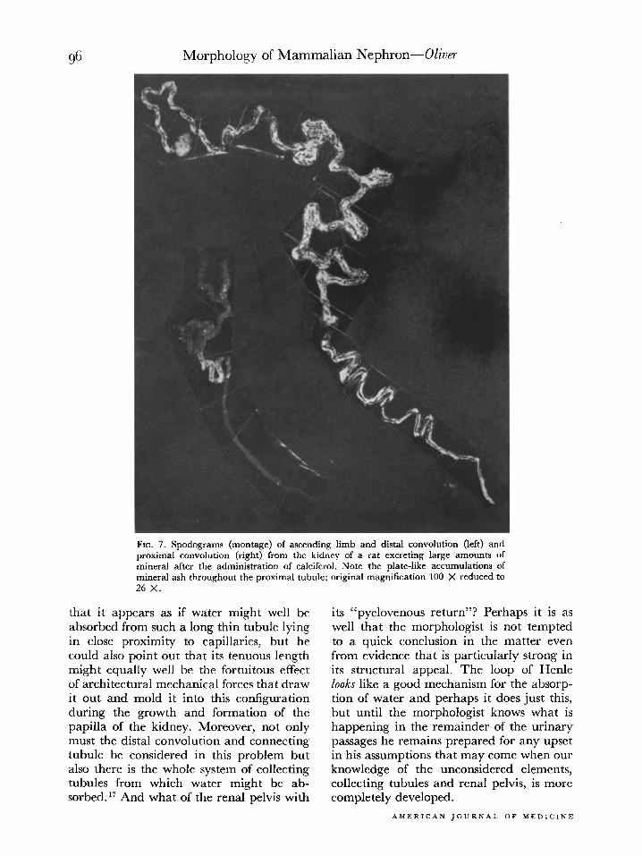
FIG. 7. Spodograms (montage) of ascending limb and distal convolution (left) and proximal convolution (right) from the kidney of a rat excreting large amounts of mineral after the administration of calciferol. Note the plate-like accumulations of mineral ash throughout the proximal tubule; original magnification 100 X reduced to 26 x.
that it appears as if water might well be absorbed from such a long thin tubule lying in close proximity to capillaries, but he could also point out that its tenuous length might equally well be the fortuitous effect of architectural mechanical forces that draw it out and mold it into this configuration during the growth and formation of the papilla of the kidney. Moreover, not only must the distal convolution and connecting tubule be considered in this problem but also there is the whole system of collecting tubules from which water might be ab- sorbed.” And what of the renal pelvis with
its “pyelovenous return”? Perhaps it is as well that the morphologist is not tempted to a quick conclusion in the matter even from evidence that is particularly strong in its structural appeal. The loop of Henle looks like a good mechanism for the absorp- tion of water and perhaps it does just this, but until the morphologist knows what is happening in the remainder of the urinary passages he remains prepared for any upset in his assumptions that may come when our knowledge of the unconsidered elements, collecting tubules and renal pelvis, is more completely developed.
AMERICAN JOURNAL OF MEDICINE

Morphology of Mammalian Nephron-Oliver 97
We have seen so far the localization of various metabolic processes in tubules whose structural characteristics are normal; the morphologist as pathologist can also con- tribute information from instances in which frankly abnormal lesions have developed in specific parts of the nephron. Again it is the proximal convolution that stands out above all other segments as the one which reacts to the administration of toxic substances by the development of regressive change and cell death. This localization doubtless is determined by the fact that the functional mechanism produces the lesion, namely, by the absorption or, less likely, by the secretion of the toxic substance in this part of the nephron which so much evidence has marked as its most active part. *
It was Susuki3 who first described the remarkable differences in the damage pro- duced by the three heavy metals, chromium, uranium and mercury. He showed in histologic sections that potassium bichro- mate damages the first third of the proximal convolution, uranium nitrate its middle third and sublimate its terminal portion. These findings have been confirmed by dissection.“‘ls
While speaking of toxic damage that occurs in the proximal convolution reference might well be made to the cellular change noted in the renal tubules which was
designated by the older pathologists as “parenchymatous degeneration.” ‘Cloudy swelling” and “hyaline droplet” formation are commonly described as varieties of this pathologic process, the first being char- acterized by the development of fine granules in the cytoplasm of the renal epithelium and the latter by the accumu- lation of large “protein droplets.” Roth types of change are found in human kidneys at autopsy under conditions where various sorts of clinical evidence, often proteinuria associated with the presence of some renal toxic factor either known or suspected, indicate the renal lesion commonly desig- nated by that etymologically absurd and conceptually obfuscatory term, nephrosis. t
The first of the “degenerations,” cloudy swelling, in which the tubule cell is swollen and its cytoplasm finely granular, may no doubt occur as a bona-fide cellular lesion in various intoxications; but since it can be shown experimentally that its changes are accurately duplicated by postmortem alter- ations in as short a time as fifteen minutes, the practical pathologist quite properly tends to discount its significance in human postmortem material. Under such condi- tions it may be found in any part of the tubule of the nephron.
The occurrence of large “hyaline drop- lets” is quite another matter although the
* There are descriptions in the literature of acute toxic damage in all parts of the nephron, and it is common to find the ascending limb almost automatically included by pathologists when they describe in histologic sections a lesion in the proximal convolution. But the identification in histologic sections of these portions of the nephron is difficult enough under normal circumstances and quite impossible when pathologic lesions have altered their characteristic structure. Although no systematic study has been made in our laboratory of all the described lesions of toxic damage, in those that have been examined by dissection the structural lesions are, for all practical purposes, limited to the proximal convolution, provided there has not been a vascular disturbance associated with or part of the toxic action. This is frequently the case, especially when the toxic action is severe, and under these conditions damage may result by &hernia to any part of the nephron. This problem of ischemic damage and its relation to and differentiation from direct toxic action will be considered in more detail in a study of the lesions which develop in the nephron in conditions of shock that is now being prepared for publication.
t This curious barbarism was introduced by the clinician. Friedrich Miiller, apparently because to his ear “osis” had the proper antithetical ring to “itis” and so seemed appropriate as a sort of counter-term to nephritis. The suffix “osis” had at the time an accepted meaning, “to be full of,” as in lipoidosis or carcinomatosis, so that by all the custom and usage of medical nomenclature nephrosis means “full of kidney.” On this etymologically nonsensical basis an elaborate superstructure of vague and varied conceptual meaning has developed, not surprisingly, so that in the end the word has come to be a mere label appropriate to its user’s ignorance of what he is talking about. Thus proteinuria of unknown mechanism associated with edema, the complex structural damage that occurs in the kidney in shock, an obscure disturbance of calcium metabolism showing excess mineral excretion or the entirely normal absorption and storage of hemoglobin by the renal cells are all “nephroses.” If all that is desired is to say that one is uncertain but suspects something is wrong with renal structure or activity, nephropathy is a word that at least is not per se ridiculous. Yet clinical authorities, agreeing entirely with these animadversions, insist that the term nephrosis is too well “estab- lished” to be discarded. At least one should realize that he can use the word nephrosis in only one of two manners. either cynically or naively, and neither attitude seems appropriate to the intellectual dignity of the science or the art of medicine.
JULY, 1950

98 Morphology of Mammalian Nephron-Oliver
older teaching of Fahr’” described them as a second stage or “chronic” form of the small granules of cloudy swelling. They are the result, not of “degeneration” if by that term one means a lessening or perversion of activity, but of the normal protein ab-
TOXIC ACTION FIG. 8. The Iocation and distribution of gradients in the proximal convolution.
sorptive and storing capacity of the cells of the proximal convolution that we have previously described. Unpublished experi- mental evidence has shown that such factors as time and protein concentration are required for the formation of visible drop- lets in the absorbing cells. Their absence therefore does not mean that protein is not being absorbed. Their presence is direct evidence of a functional integrity of a sort in the renal cells, and indirectly, in most instances at least, they indicate that an increased permeability of the glomerular membrane exists.
The long-continued and excessive ab- sorption and storage of protein can lead, however, to the development of cellular damage and even to what properly may be called a chronic tubular nephropathy. This is a very common, spontaneous lesion in old ratszO for these animals have a normally permeable glomerular membrane and a consequent physiologic proteinuria. 21 Se- vere tubular lesions with marked archi- tectural change in the nephrons also result from the “nephrotic episodes” of glomerular nephritis or as a result of the proteinuria of the amyloid kidney. In all these conditions the fundamental mechanism of the tubular
change is an overloading of the cells of the proximal convolution with protein, a result- ing intracellular “indigestion” and conse- quent death and desquamation of the renal cells which, carried by the current of the tubule fluid, lodge in the distal convolution. We shall return later to the resulting archi- tectural changes that result in this portion of the nephron.
A more detailed consideration of the hyaline droplets of absorbed “protein,” whether normal or abnormal, seems war- ranted at this time for a closer examination of them shows that these objects are much more complex in their chemical structure than has previously been suspected. They, as well as the other particulate bodies of the cytoplasm of the cells of the proximal con- volution, the mitochondrial rodlets and the smaller microsomes, may be isolated and subjected to biochemical analysis by grind- ing up the renal cells and preparing pure suspensions of the various particles by differential centrifugalization and other procedures. Work in progress9 has shown that the intracellular droplet of “absorbed protein,” egg white for example, is in fact a complex structure that contains only a relatively moderate amount of egg white proteins and a greater amount of ribonucleic acid and other cytoplasmic constituents. Morphologic evidence shows these sub- stances to be derived in large part from the dissolution of the mitochondrial batonets of the renal cells. Similar cytologic changes are found in the absorption of amino acids by the renal cells of the proximal convolution.
Apparently, therefore, what is being seen by the morphologist in “droplet” formation is the structural aspect of the mechanism by which the enzyme-containing and energy- rich cytoplasmic elements of the renal cells combine with and metabolize absorbed proteins and amino acids, A great amount of biochemical evidence indicates that processes involving both synthesis and degradation of these substances occur in the kidney, so that the morphologist as he sees the mitochondrial rodlets dissolve and the “protein droplets” form, then dis- integrate and finally disappear can perhaps
AMERICAN JOURNAL OF MEDICINE

belie\ Te that he has at least localized if not
Morphology of Mammalian Nephron-Oliver
FIG. 9. A hypertrophied nephron from chronic glomerular nephritis. Note that in comparison with the normal nephron of Figure 10 the greater part of the hypertrophy is located in the proximal convolution; X 15. (Courtesy of Paul B. Hoeber-Harper Bras.)
elucidated the metabolic process in the most minute of the cytologic elements of the renal cell.
To return to the localization in the tubule of the nephron of pathologic change that is dependent on the function of the involved part, another example is found in the mechanism of cast formation. The classic description of the location of casts in the lumen of the tubule as they are seen
J”‘.‘, 1950
FIG. 10. A normal nephron from the human kidney; X 15. (Courtesy of Paul B. Hoebrr- Harper Bros.)
in histologic sections gives the impression that they may form in any part of the nephron. In dissected material it is evident that although disorganized debris and desquamated epithelial or red blood cells may be found throughout the tubular system from the glomerulus to the papilla? rarely if ever are formed consolidated casts seen except in the distal convolution, the connecting tubule and in the collecting tubules.22 There is one exception to this rule, namely, the occurrence of casts of Bence Jones protein which may form as high in the nephron tubule as the proximal convolution. 8
The coagulation of proteins is the funda- mental mechanism in the formation of casts, and by this coagulation debris, fatty material and even desquamated epithelial or red blood cells may be incorporated into

100 Morphology of Mammalian Nephron-Oliver
the cast structure. The iso-electric point of the protein. the pH of the tubule fluid, its electrolyte content and the dispersive effect of urea are well known factors that influence protein coagulation, and it is in the distal convolution that the proper equilibrium
GROWTN OF PARTS IN RELATION TO TOTAL NEPHRON GROWTH
VI TOT.NEMlON a6 91.1 174 ao22l
FIG. 11. Heterogonic normal growth of proximal con- volution in rat kidney. The volume of the various por- tions of the nephron are plotted logarithmically against the volume of the entire nephron. Only the proximal convolution (P/Z) fulfills the straight-line require- ment of heterogonic growth.
for its occurrence is attained. That Bence Jones body coagulates in the proximal con- volution becomes explicable, therefore, by the fact of its high iso-electric point. How- ever, another substance, first described by Moernerz3 in normal urine, later found by Addis in hyaline casts and then demon- strated in dissected normal nephrons to be concentrated in the tubule cells of the distal nephron8 seems essential to coagulation and definitive cast formation. As to what this substance may be, the morphologist can state only that it is metachromatic in its reaction to toluidin blue as he sees it in both renal cells and casts and that Moerner considered it to be chondroitin-sulfuric acid.
However uncertain the details of our knowledge of the exact mechanism of cast formation, enough is clear to explain why
these structures are found only in the distal convolution and the collecting tubules. Those which form in the lower collecting system may flush out into the urine and therefore be both innocuous to the patient and helpful to the physician; those in the distal convolution are more likely to stick and it is to the result of this blockage that a great part of the architectural change of chronic renal disease is due.22
A final example of structural change in the tubule of the nephron occurring under pathologic conditions may be cited by the morphologist in explanation of his inability to demonstrate as elaborate a picture of structural-functional correlation in the dis- tal portions of the nephron as he could in the proximal convolution. It will be recalled that he has repeatedly implied that this might be due to the essential facts of the situation, a lack of structural reaction truly correlating with a corresponding functional mediocrity. Such a conclusion is consider- ably strengthened by what he observes happening when the full load of renal activity is thrown upon a reduced number of nephrons. The functional result of the “compensatory” hypertrophy that develops may be entirely adequate either when nephrons have been destroyed by chronic renal disease or after surgical or experi- mental nephrectomy. The tubular mass of the kidney, using the term in the old- fashioned newtonian sense, increases, but this increase cannot be truly measured by the weight of the kidney or even by that of a single nephron if one were isolated and weighed for all parts of the tubule have not grown in response to the work stimulus to the same degree. Figures 9 and 10 show a hypertrophied nephron from a kidney of chronic glomerular nephritis and a nephron from a normal kidney; it is evident that the increase in mass is much greater in the proximal convolution than in the remainder of the nephron. Similar results are obtained in experimental compensatory hypertro- phy26 and the conclusion is thereby forced on a morphologist that the greater part of the work of the kidney is performed by that part which has preferentially responded to
AMERICAN JOURNAL OF MEDICINE

Morphology of Mammalian Nephron-Oliver 101
the growth stimulus. In fact, the normal growth of the individual by increasing the metabolic load on the nephrons as he in- creases in size performs the same experi- ment for it has been shown8 that the nephron under normal conditions grows irregularly in its parts. The loops of Henle and the distal convolution increase by the ordinary laws of growth increment, but the proximal convolution shows that peculiar form of exponential increase commonly found in hyperactive parts and which Huxley 26 has termed heterogonic growth. (Fig. 11.)
The examples of structural-functional correlation that have been cited will be sufficient to indicate that although mor- phologist and functionalist are perhaps separated by that ineluctable barrier which seems to cut through all aspects of human thought and endeavor with a resulting dis- rupting duality of expression and concept, nevertheless it is not a wall that stands between them but rather a transparent or at least translucent screen through which each can dimly see what his neighbor is doing. Nowhere as in the study of renal activity are the possibilities of such a meet- ing of minds more clearly promised for in the quite untranslatable words of Fritz Kiihn,27 “Die alten Anatomen nannten die Niere das viscus elegantissimum, das ele- ganteste Eingeweide, und das ist es in der Tat. Von Busseren Anblick bis in die letzten mikroskopischen Feinheit iibertrumpft die Niere die andere Organe durch ihre Fiille an Sisthetischen Motiven und durch die Eleganz, mit der in ihr die konstruktiven Probleme gel&t sind.”
Such terms will not seem extravagant to those who know the form and beauty, redundant words, of structure and function in this viscus elegantissimum.
REFERENCES
I. RUSSELL, B. Human Knowledge, Its Scope and Limits. New York, 1948. Simon & Schuster.
2. VUN BERTAI.ANPFY, L. The theory of open systems in physics and biology. Science, 111: 23, 1950.
3. SUSWKI, T. Zur Morphologie der Nierensekretion. Jena, 1912. Fischer.
4. OLIVER, J. The hiitogenesis of chronic uranium
JULY, 1950
5.
6.
7.
8.
9.
10.
11.
12.
13.
14.
15.
16.
17.
18.
19.
20.
21.
22.
23.
24.
2.5.
26.
27.
nephritis with especial reference to epithelial regeneration. 3. Exper. Med., 24: 425, 1915,
VON M~LLENDORF, W. Die Dispersitlt der Farbstoffe, ihre Beziehungen zu Ausscheidung und Speicher- ung in der Niere. Ein Beitrag zur Histophysiologie der Niere. Anat. Heffe, 53: 81, 1915.
WALKER, A. M. and OLIVER, J. Methods for the collection of fluid from single glomeruli and tubules of the mammalian kidney. Am. 3. Phyriol., 134: 562, 1941.
WALKER, A. M., BOTT, P. A., OLIVER, J. and MACDOWEL.L, M. C. The collection and analysis of fluid from single nephrons of the mammalian kidney. Am. 3. Physiol., 134: 580, 1941.
OLIVER, J. New directions in renal morphology. Harvey Lect., 40: 102, 1945.
OI.IVER, J. The structure of the metabolic process in the nephron. Janeway Lecture. 3. Mt. Sinai Hosp., 15: 175, 1948.
GERARD, P. and CORDIER, R. Esquisse d’une histo- physiologie comparCe du rein des verttbres. Biol. Rev., 9: 110, 1934.
SMETANA, H. and JOHNSON, F. R. The origin of colloid and lipoid droplets in the epithelial cells of the renal tubules. Am. 3. Path., 109: 1029,1942.
RANDERATH, E. Zur pathologischen Anatomie der sog. Amyloidenephrose. Virchows Arch. J’. path. Amt., 314: 388, 1947.
RATHER, I,. J. Renal athrocytosis and intracellular digestion of intraperitoneally injected hemoglobin in rats. J. Exjxr. Med., 87: 163, 1948.
FOOT, J. F. and GRAFLIN. A. L. Quantitative meas- urements of the fat-laden and fat-free segments of the proximal tubule in the nephron of the cat and dog. Amt. Rec., 72: 169, 1938.
STIEGLITZ, E. J. Histologic hydrogen-ion studies of the kidney. Arch. Int. Med.. 33: 483. 1924.
MCMASTER; P. D. and EL&AN, R.’ The relative reaction within living mammalian tissues. 3. Exuper. Med., 47: 797, 1928.
HOWELL, A. B. and GERSH, I. Conservation of water by the rodent dipodmys. 3. Mammal., 16: 1, 1935.
EDWARDS, J. G. The renal tubule (nephron) as affected by mercury. Am. 3. Path., 18: 1011, 1942.
VOLHARD, F. and FAHR, T. Die Brightsche Nieren- krankheit. Berlin, 1914. Springer.
SAXTON, J. A., JR. and KIMBALL, G. C. Relation of nephrosis and other diseases of albino rats to agr and to modifications of diet. Arch. Path., 32: 951. 1941.
GILSON, S. B. Studies on proteinuria in the rat. Sot. E‘rp~r. Biol. Med., 72: 608, 1949.
OLIVER, J. The Architecture of the Kidney in Chronic Bright’s Disease. New York, 1939. Paul Hoeber-Harper Bros.
MOERNER, K. A. H. Untersuchungen iiber die Proteinstoffe und die eiweissfgllenden Substanzen des normalen Menschenharns. Skandinav. Arch. f. Physiol., 6: 332, 1895.
.bDIS, T. The renal lesion in Bright’s disease. Harvey Lect.. 23: 222, 1928.
OLIVER, J. The regulation of renal activity. .4rch. Int. Med., 34: 258. 1924.
HUXL.EY, J. S. Problems of Relative Growth. New York, 1932. Dial Press.
KUHN, F. Der Mensch. Ziirich and Leipsig, 1939. A. Muller Verlag.