Embed Size (px)
Citation preview
P/N 600119-001
Amplatz® Canine Duct Occluder
_________________________________________________________________________
Instruction Manual
Distributed exclusively by:
www.infinitimedical.com
Sales: (917) 558-0548 or [email protected]
2
Table of Contents _________________________________________________________________________
1. Brief Device Description ................................................3 2. How Supplied ..................................................................4 3. Indications and Usage....................................................5 4. Contraindications ...........................................................5 5. Warnings .........................................................................6 6. Precautions .....................................................................6
- Potential Adverse Events ...................................................... 7 7. Clinical Use Information.................................................7
- Materials Required ................................................................. 8 - Ductus Evaluation/Aortography ........................................... 8 - Device Selection..................................................................... 9 - Delivery Catheter Selection................................................... 9 - Delivery Catheter Placement............................................... 10 - Device Preparation and Loading ........................................ 11 - Device Delivery..................................................................... 11
3
Amplatz® Canine Duct Occluder Distributed exclusively by Infiniti Medical™ Instructions for Use
Caution: Federal law (USA) restricts this device to sale by or on the order of a veterinarian. This device is intended to be used in animals only. It is illegal to sell or utilize this device to treat humans.
_____________________________________________________________
1. Brief Device Description The Amplatz Canine Duct Occluder is a self-expanding device made from a nitinol wire mesh and is used to close patent ductus arteriosus (PDA) in dogs. A distal flat disk on the pulmonary artery side of the ductal ostium provides secure positioning in the pulmonary artery. As the device is implanted, the proximal cupped disk expands to conform to the shape of the ductal ampulla. The waist of the device spans the pulmonic ostium of the ductus and the dense nitinol mesh occludes the communication. The Amplatz Canine Duct Occluder is sterile and packaged in a loader. The therapeutic device is attached to a 110, 120, or 135 cm long delivery cable and vise that is placed within a hoop dispenser. The Amplatz Canine Duct Occluder is available in several waist diameters as listed in Table 1. The device should be sized relative to the true diameter of the pulmonic ostium of the ductus as measured by arteriography in a ratio between 1.5 and 2. Appropriate sizing is crucial to the success of the procedure and can be accomplished only by arteriography.
4
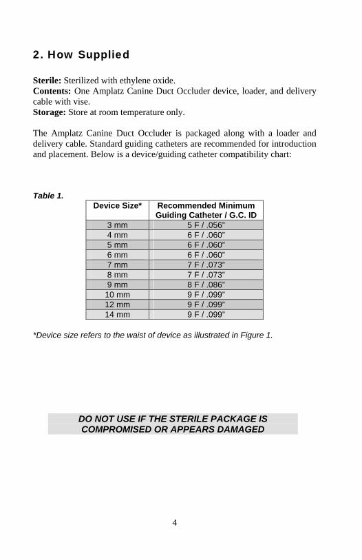
2. How Supplied Sterile: Sterilized with ethylene oxide. Contents: One Amplatz Canine Duct Occluder device, loader, and delivery cable with vise. Storage: Store at room temperature only. The Amplatz Canine Duct Occluder is packaged along with a loader and delivery cable. Standard guiding catheters are recommended for introduction and placement. Below is a device/guiding catheter compatibility chart: Table 1.
Device Size* Recommended Minimum Guiding Catheter / G.C. ID
3 mm 5 F / .056” 4 mm 6 F / .060” 5 mm 6 F / .060” 6 mm 6 F / .060” 7 mm 7 F / .073” 8 mm 7 F / .073” 9 mm 8 F / .086” 10 mm 9 F / .099” 12 mm 9 F / .099” 14 mm 9 F / .099”
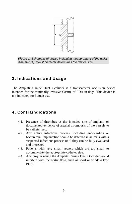
*Device size refers to the waist of device as illustrated in Figure 1.
DO NOT USE IF THE STERILE PACKAGE IS COMPROMISED OR APPEARS DAMAGED
5
Figure 1. Schematic of device indicating measurement of the waist diameter (A). Waist diameter determines the device size.
3. Indications and Usage The Amplatz Canine Duct Occluder is a transcatheter occlusion device intended for the minimally invasive closure of PDA in dogs. This device is not indicated for human use.
4. Contraindications
4.1. Presence of thrombus at the intended site of implant, or documented evidence of arterial thrombosis of the vessels to be catheterized.
4.2. Any active infectious process, including endocarditis or bacteremia. Implantation should be deferred in animals with a suspected infectious process until they can be fully evaluated and or treated.
4.3. Patients with very small vessels which are too small to accommodate the appropriate catheter size.
4.4. Anatomy in which the Amplatz Canine Duct Occluder would interfere with the aortic flow, such as short or window type PDA.
6
5. Warnings
5.1. Do not use if the sterile barrier has been compromised in any way.
5.2. This device is intended for single-use only; do not reuse. Do not resterilize.
5.3. Note the product “Use By” date specified on the package. 5.4. Only veterinarians familiar with the complications, side
effects, and hazards commonly associated with transcatheter closure of PDA should use this device.
5.5. Do not release the Amplatz Canine Duct Occluder from the delivery cable if the device does not conform to its original configuration (Figure 9) or if the device position is unstable. Recapture the device and redeploy. If still unsatisfactory, recapture the device and replace with a new device or re-evaluate the anatomy of the ductus for proper sizing or candidacy for transcatheter closure.
5.6. Veterinarians should be prepared to deal with urgent situations which require removal of embolized devices that result in critical hemodynamic compromise. This includes the availability of an on-site surgeon.
5.7. Access site complications may occur that require surgical intervention.
6. Precautions
6.1. Handling The Amplatz Canine Duct Occluder is for single use only. Do not reuse or resterilize.
6.2. Sizing Accurate sizing of the ductus is crucial and mandatory for Amplatz Canine Duct Occluder device selection. Potential errors due to magnification should be considered when sizing the device.
6.3. Post Implantation • The risk of bacterial seeding of the device during subsequent surgical, dental or other procedures is not known. Antibiotic prophylaxis may be employed if the patient is to undergo such a procedure within 6 months of implantation. This is in accordance with the recommendation of the
7
American Heart Association (in humans). There is no evidence based data regarding this scenario in the veterinary population and therefore antibiotic prophylaxis is at the discretion of the veterinarian. • Any patient who has a residual shunt should undergo periodic echocardiography to guide further management.
Potential Adverse Events Placement of the Amplatz Canine Duct Occluder involves standard interventional cardiac catheterization techniques that have known risks. The following are potential adverse events that may occur:
Air embolus Fever Allergic contrast reaction Allergic drug reaction Hyper/Hypotension Anesthesia reactions Apnea Perforation of vessel Arrhythmia Peripheral Embolism Bacterial endocarditis Bleeding Thrombus formation Delivery system failure Vascular access site complications Hematoma at access / puncture site Embolization Death
7. Clinical Use Information Inspect all products prior to use. Do not use if the package is open or damaged, or if the product is damaged. Avoid unnecessary handling, which may kink or damage the delivery system.
8
Materials Required*
One Amplatz Canine Duct Occluder .035” exchange wire Non-selective diagnostic catheter (i.e. with end- and side-holes)
with vessel sizing marker bands Introducer sheath Guiding catheter or delivery sheath Sterile procedure pack Iodinated contrast Normal saline Two to three 10-20cc syringes Small surgical set Access needle Hemostasis valve
*Depending on the size of the animal and other considerations, more equipment may be needed.
Ductus Evaluation/Aortography Precise sizing of the ductus requires aortography, which should be performed using standard endovascular techniques. A cut down is usually performed to gain access to the femoral artery, although percutaneous access may be a reasonable alternative in certain cases. The right side is commonly chosen. A vascular sheath is placed and the animal is positioned such that the ductus can be imaged in profile (lateral). A non-selective catheter is advanced over a guidewire and positioned in the thoracic aorta, above the ductus communication. To assist in sizing, a marker catheter should be used. An aortogram is performed with an injection rate that is appropriate for the intrinsic rate of flow. The images should be interrogated for the following information:
Suitability of the anatomy of the ductus for transcatheter occlusion. Size of ductus (see Figure 2).
9
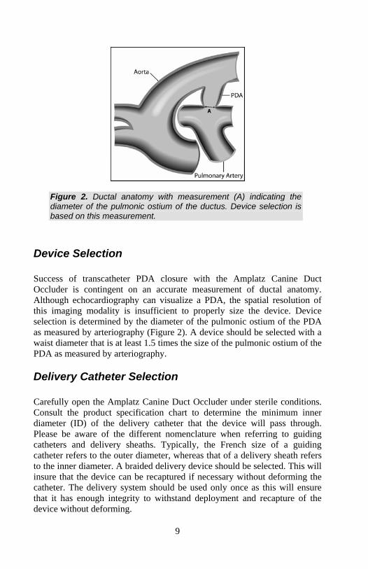
Figure 2. Ductal anatomy with measurement (A) indicating the diameter of the pulmonic ostium of the ductus. Device selection is based on this measurement.
Device Selection Success of transcatheter PDA closure with the Amplatz Canine Duct Occluder is contingent on an accurate measurement of ductal anatomy. Although echocardiography can visualize a PDA, the spatial resolution of this imaging modality is insufficient to properly size the device. Device selection is determined by the diameter of the pulmonic ostium of the PDA as measured by arteriography (Figure 2). A device should be selected with a waist diameter that is at least 1.5 times the size of the pulmonic ostium of the PDA as measured by arteriography.
Delivery Catheter Selection Carefully open the Amplatz Canine Duct Occluder under sterile conditions. Consult the product specification chart to determine the minimum inner diameter (ID) of the delivery catheter that the device will pass through. Please be aware of the different nomenclature when referring to guiding catheters and delivery sheaths. Typically, the French size of a guiding catheter refers to the outer diameter, whereas that of a delivery sheath refers to the inner diameter. A braided delivery device should be selected. This will insure that the device can be recaptured if necessary without deforming the catheter. The delivery system should be used only once as this will ensure that it has enough integrity to withstand deployment and recapture of the device without deforming.

10
Delivery Catheter Placement Following aortography and with the vascular access sheath in place, an exchange wire is placed across the PDA (Figure 3). The use of an angle-tipped selective (i.e. end-hole) catheter may facilitate passage of the exchange wire across the PDA. The guiding catheter or delivery sheath with tapered dilator in place is then passed over the exchange wire and across the PDA under fluoroscopic visualization (Figure 4). The dilator and exchange wire are then removed, and a hemostasis valve is connected to the end of the delivery system. In small animals, it may be difficult to place a vascular access sheath that is large enough to accept the guiding catheter or delivery sheath. In these cases, the short vascular sheath may be removed over the exchange wire and the delivery system with tapered dilator is placed directly over the exchange wire.
Figure 3. The tip of the exchange wire should be placed in a stable position in the pulmonary artery. A selective catheter may be helpful in crossing the ductus.
11
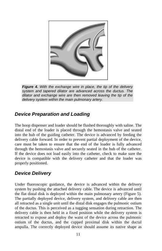
Figure 4. With the exchange wire in place, the tip of the delivery system and tapered dilator are advanced across the ductus. The dilator and exchange wire are then removed leaving the tip of the delivery system within the main pulmonary artery.
Device Preparation and Loading The hoop dispenser and loader should be flushed thoroughly with saline. The distal end of the loader is placed through the hemostasis valve and seated into the hub of the guiding catheter. The device is advanced by feeding the delivery cable forward. In order to prevent partial deployment of the device, care must be taken to ensure that the end of the loader is fully advanced through the hemostasis valve and securely seated in the hub of the catheter. If the device does not load easily into the catheter, check to make sure the device is compatible with the delivery catheter and that the loader was properly positioned.
Device Delivery Under fluoroscopic guidance, the device is advanced within the delivery system by pushing the attached delivery cable. The device is advanced until the flat distal disk is deployed within the main pulmonary artery (Figure 5). The partially deployed device, delivery system, and delivery cable are then all retracted as a single unit until the distal disk engages the pulmonic ostium of the ductus. This is perceived as a tugging sensation during retraction. The delivery cable is then held in a fixed position while the delivery system is retracted to expose and deploy the waist of the device across the pulmonic ostium of the ductus, and the cupped proximal disk within the ductal ampulla. The correctly deployed device should assume its native shape as
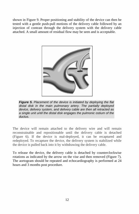
12
shown in Figure 9. Proper positioning and stability of the device can then be tested with a gentle push-pull motions of the delivery cable followed by an injection of contrast through the delivery system with the delivery cable attached. A small amount of residual flow may be seen and is acceptable.
Figure 5. Placement of the device is initiated by deploying the flat distal disk in the main pulmonary artery. The partially deployed device, delivery system, and delivery cable are then all retracted as a single unit until the distal disk engages the pulmonic ostium of the ductus.
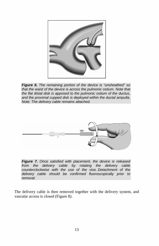
The device will remain attached to the delivery wire and will remain reconstrainable and repositionable until the delivery cable is detached (Figure 6). If the device is mal-deployed, it can be recaptured and redeployed. To recapture the device, the delivery system is stabilized while the device is pulled back into it by withdrawing the delivery cable. To release the device, the delivery cable is detached by counterclockwise rotations as indicated by the arrow on the vise and then removed (Figure 7). The aortogram should be repeated and echocardiography is performed at 24 hours and 3 months post procedure.
13
Figure 6. The remaining portion of the device is “unsheathed” so that the waist of the device is across the pulmonic ostium. Note that the flat distal disk is apposed to the pulmonic ostium of the ductus, and the proximal cupped disk is deployed within the ductal ampulla. Note: The delivery cable remains attached.
Figure 7. Once satisfied with placement, the device is released from the delivery cable by rotating the delivery cable counterclockwise with the use of the vise. Detachment of the delivery cable should be confirmed fluoroscopically prior to removal.
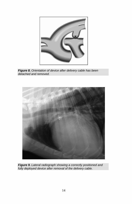
The delivery cable is then removed together with the delivery system, and vascular access is closed (Figure 8).
14
Figure 8. Orientation of device after delivery cable has been detached and removed.
Figure 9. Lateral radiograph showing a correctly positioned and fully deployed device after removal of the delivery cable.

15
_____________________________________________________________
This product is distributed exclusively by Infiniti MedicalTM. Technical support is available at
(917) 558-0549. _____________________________________________________________
Patent Protected