Embed Size (px)
Citation preview

Plant Physiol. (1986) 81, 36-400032-0889/86/8 1/0036/05/$0 1.00/0
Amino Acid Transport in Suspension-Cultured Plant Cells'VI. INFLUENCE OF pH BUFFERS, CALCIUM, AND PREINCUBATION MEDIA ON L-LEUCINEUPTAKE
Received for publication September 17, 1985 and in revised form December 11, 1985
MARK A. SCHNEEGURT2 AND CARL N. MCDANIEL*Department ofBiology, Rensselaer Polytechnic Institute, Troy, New York 12180-3590
ABSTRACT
The rate at which L-leucine was transported into suspension-culturedNicotiana tabacum cv Wisconsin 38 cells increased more than 2-foldover a period of hours when the cells were preincubated in a 1% sucrosesolution. This increase in uptake rate was eliminated if certain tris bufferswere included in the preincubation solution while other buffers had littleeffect. Calcium could reverse the effect of the inhibitory buffers only ifthe buffer and calcium were present together from the beginning of thepreincubation period. It was the amine group of the inhibitory bufferswhich was responsible for the inhibition. Preincubation in a completeculture medium (EM Linsmaier, F Skoog 1965 Physiol Plant 18: 100-127) led to minimal changes in L-leucine uptake rate over a 10 hourpreincubation period indicating that the uptake rate was stabilized bythis medium. The complete medium stabilized the L-leucine uptake rateas a result of its ionic composition and not because of its osmolarity.Most of the increased uptake rate observed after preincubation in a 1%sucrose solution could be inhibited by 2,4-dinitrophenol or carbonylcyanide m-chlorophenyl hydrazone, or high concentrations of L-phenyl-alanine or L-leucine. Therefore much of the increase could be accountedfor by an increase in active transport of L-leucine.
Increases in the rate of solute uptake have been induced inplant cells and tissues by a procedure of washing and preincu-bation (8, 11, 15, 17, 18). This effect has been observed using anumber of species and for a broad range ofsubstrates: phosphate,potassium, chloride, glycine, glucose, and guanosine monophos-phate (11); sulfate (10); L-serine (18); malate, glycerate-3-P, ura-cil, and several amino acids (8); and others. Since Epstein's (5)early report that calcium is required for the integrity of selectiveion transport mechanisms, most transport studies have employedmedia which contain calcium. It has been reported that the highrates of solute uptake which are observed after preincubation aredependent upon calcium in the medium (2, 7, 8, 10, 18). How-ever, other researchers have demonstrated that an enhanceduptake rate can be induced without the addition of calcium tothe preincubation solution (6, 11, 15). Transport workers alsodiffer in their use ofpH buffers in preincubation and incubationsolutions. Some routinely employ pH buffers (2, 7, 8, 10, 15, 18)while others do not (11, 13, 14, 19). Reinhold's laboratoryinitially employed pH buffers (17), but because buffers wereshown to influence the amino acid uptake process, they discon-tinued the use of buffers (1, 16).
'Supported in part by National Science Foundation grant (DCB84-09709) to C. N. M.
2Current address: Box G, Brown University, Providence, RI 02912.
Using suspension-cultured tobacco cells we have investigatedthe effect of several buffers, calcium, and various other mediumcomponents on the enhanced L-leucine uptake rate induced bypreincubation in a 1% sucrose solution. Results reported hereindicate that certain tris buffers inhibited this increase in uptakerate and that calcium could relieve the tris buffer inhibition ofthe enhanced uptake rate. Moreover, L-leucine uptake rate wasmore stable when cells were transferred into a complete culturemedium than when transferred into a solution of low ionicstrength.
MATERIALS AND METHODS
Cell Culture. Nicotiana tabacum cv Wisconsin 38 cells, origi-nally isolated from pith, were maintained in LS medium3 aspreviously described (3). Subculturing was performed every 7 dby adding 50 ml of fresh LS medium to a culture flask and thendividing this suspension between two 125-ml Erlenmeyer flasks.Experiments were conducted on the 3rd d after subculturingnear the end of the exponential growth phase (4).Uptake Assay. Under aseptic conditions, three or four flasks
ofcultured cells were combined, poured through a screen basket(mesh size of 1.5 mm) to remove clumps, and collected bycentrifugation (161Og for 1 min). Two to 3 ml of cells weretransferred to 50-ml tubes. The cells were washed three times byresuspending the cells in about 50 ml ofthe appropriate solution,shaking gently by hand for 2 min, letting the cells settle for about4 min, and decanting the wash solution. The same solution,except where noted, was used for washing, preincubating, andmeasuring uptake except, L-leucine was present in the uptakesolution at a concentration of 0.1 mM. After the third wash, cellswere resuspended in 50 ml of solution and transferred to 125-mlErlenmeyer flasks for preincubation under conditions identicalto those used for cell culture. After the preincubation period,cells were permitted to settle and the preincubation solutiondecanted. L-Leucine uptake was measured employing [3H]-L-leucine in a fresh solution identical to the preincubation solutionunless otherwise stated. Uptake rate was measured by collectingsamples after 6 and 12 min ofincubation as previously described
3Abbreviations: LS medium, Linsmaier and Skoog (12) medium(major salts: 20 mM NH4NO3, 20 mM KNO3, 2.5 mM CaCI2, 2.5 mMMgSO4; minor salts: 0.1 mm H3BO3, 0.1 mM MnSO4, 30 AM ZnSO4, 5Mm KI, 1 AM Na2MoO4, 100 nM CoCl2, 80 nM CuSO4; phosphate: I mmKH2PO4; iron: 0.1 mM FeSO4, 0.1 mm Na2 EDTA; organic: I AMthiamine, lMm inositol) supplemented with kinetin (0.1 mg/l), a-na-phthaleneactic acid (2.0 mg/l), and sucrose (4%); PEG, polyethyleneglycol (200 mol wt); BTP, 1,3-bis[tris(hydroxymethyl)methylamino]pro-pane; BIS-TRIS, bis(2-hydroxyethyl)imino-tris(hydroxymethyl)methane;TAPS, tris(hydroxymethyl)methylaminopropanesulphonic acid; DNP,dinitrophenol; CCCP, carbonyl cyanide m-chlorophenyl hydrazone.
36
Dow
nloaded from https://academ
ic.oup.com/plphys/article/81/1/36/6082136 by guest on 29 N
ovember 2021
AMINO ACID TRANSPORT IN SUSPENSION-CULTURED PLANT CELLS
(4, 14). All solutions were adjusted to a pH of 5.5 with NaOHor HCI prior to use. Uptake rate was expressed as nmol/(ml HV.min) where HV was the volume of cell debris which pelleted in1 min at 16 1Og after a sample of cells had been homogenized(3). This standardization parameter has been equated to otherstandardization parameters (e.g. fresh weight, packed cell vol-ume) (3).
Efflux. After a 6 h preincubation period as described above,cells were centrifuged and washed three times with 50 ml of theappropriate solution. Cells were resuspended in 20 ml of theappropriate solution containing [3H]-L-leucine and incubated for1 h. Cells were then washed three times with 50 ml of theappropriate solution. The loaded, washed cells were resuspendedin 20 ml of the appropriate solution and a 0.2 ml sample wastaken every 10 min for 1 h. Each 0.2 ml sample was immediatelycentrifuged to remove cells and 0.1 ml of the supernatant wascounted. The efflux rate constant was calculated as previouslydescribed (4).
Metabolic Inhibitors. After a 6 h preincubation, cells wereincubated in the presence of the metabolic inhibitor for 15 min.After the 15 min incubation, cells were centrifuged, the super-natant was decanted and uptake measured as described above inthe appropriate solution containing the metabolic inhibitor.
Chemicals. L-[4,5-3H (N)]Leucine was obtained from ICN;Liquiscint from National Diagnostics; 1,1,1 tris (hydroxymethyl)ethanol from Aldrich; and all other chemicals from Sigma orFisher Scientific. All chemicals were reagent grade or ACS certi-fied.
RESULTS
Uptake Rate Prior to Preincubation. The L-leucine uptake ratefor cells prior to preincubation was estimated by measuring theuptake rate of cells which had been washed three times as in thenormal preparation procedure or of cells which had been washedonly once (Table I). One wash in low ionic strength solutionsreduced the L-leucine uptake rate significantly when comparedto the rate in LS medium. However, the rate recovered duringthe time (approximately 45 min) it took to complete the threewash procedure except when the solution contained 1% sucroseplus BTP.Uptake Rate after Preincubation. L-Leucine uptake rate mark-
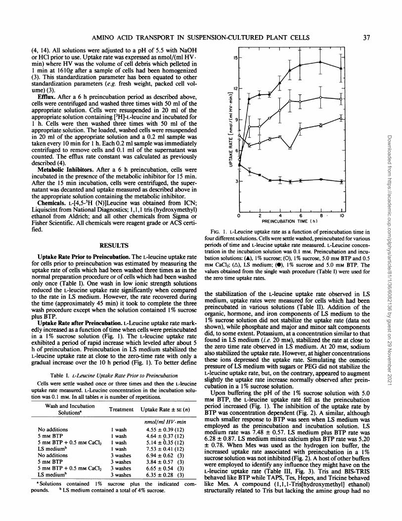
edly increased as a function oftime when cells were preincubatedin a 1% sucrose solution (Fig. 1). The L-leucine uptake rateexhibited a period of rapid increase which leveled after about 5h of preincubation. Preincubation in LS medium stabilized theL-leucine uptake rate at close to the zero-time rate with only agradual increase over the 10 h period (Fig. 1). To better define
Table I. L-Leucine Uptake Rate Prior to PreincubationCells were settle washed once or three times and then the L-leucine
uptake rate measured. L-Leucine concentration in the incubation solu-tion was 0.1 mM. In all tables n is number of repetitions.
Wash and Incubation Treatment Uptake Rate ± SE (n)Solutions'
nmol/ml HV.minNo additions I wash 4.55 ± 0.39 (12)5 mM BTP 1 wash 4.64 + 0.37 (12)5 mM BTP + 0.5 mm CaCI2 I wash 5.14 + 0.35 (12)LS medium' I wash 7.53 ± 0.41 (12)No additions 3 washes 6.94 ± 0.62 (3)5 mM BTP 3 washes 3.84 +0.57 (3)5 mM BTP + 0.5 mM CaCl2 3 washes 6.65 + 0.54 (3)LS mediumb 3 washes 6.35 ±0.28 (3)a Solutions contained 1% sucrose plus the indicated com-
pounds. b LS medium contained a total of4% sucrose.
EC
a.
3-
0 2 4 6 8 10PREINCUBATION TIME (h)
FIG. 1. L-Leucine uptake rate as a function of preincubation time infour different solutions. Cells were settle washed, preincubated for variousperiods of time and L-leucine uptake rate measured. L-Leucine concen-tration in the incubation solution was 0.1 mM. Preincubation and incu-bation solutions: (A), 1% sucrose; (0), 1% sucrose, 5.0 mm BTP and 0.5mM CaCl2; (A), LS medium; (0), 1% sucrose and 5.0 mM BTP. Thevalues obtained from the single wash procedure (Table I) were used forthe zero time uptake rates.
the stabilization of the L-leucine uptake rate observed in LSmedium, uptake rates were measured for cells which had beenpreincubated in various solutions (Table II). Addition of theorganic, hormone, and iron components of LS medium to the1% sucrose solution did not stabilize the uptake rate (data notshown), while phosphate and major and minor salt componentsdid, to some extent. Potassium, at a concentration similar to thatfound in LS medium (i.e. 20 mM), stabilized the rate at close tothe zero time rate observed in LS medium. At 20 mm, sodiumalso stabilized the uptake rate. However, at higher concentrationsthese ions depressed the uptake rate. Simulating the osmoticpressure of LS medium with sugars or PEG did not stabilize theL-leucine uptake rate, but, on the contrary, appeared to augmentslightly the uptake rate increase normally observed after prein-cubation in a 1% sucrose solution.Upon buffering the pH of the 1% sucrose solution with 5.0
mM BTP, the L-leucine uptake rate fell as the preincubationperiod increased (Fig. 1). The inhibition of the uptake rate byBTP was concentration dependent (Fig. 2). A similar, althoughmuch smaller response to BTP was seen when LS medium wasemployed as the preincubation and incubation solution. LSmedium rate was 7.48 ± 0.57. LS medium plus BTP rate was6.28 ± 0.87. LS medium minus calcium plus BTP rate was 5.20± 0.78. When Mes was used as the hydrogen ion buffer, theincreased uptake rate associated with preincubation in a 1%sucrose solution was not inhibited (Fig. 2). A host ofother bufferswere employed to identify any influence they might have on theL-leucine uptake rate (Table III, Fig. 3). Tris and BIS-TRISbehaved like BTP while TAPS, Tes, Hepes, and Tricine behavedlike Mes. A compound (1,1, l-Tris[hydroxymethyl] ethanol)structurally related to Tris but lacking the amine group had no
37
Dow
nloaded from https://academ
ic.oup.com/plphys/article/81/1/36/6082136 by guest on 29 N
ovember 2021

SCHNEEGURT AND McDANIEL
Table II. Effect ofthe Ionic Composition and Osmotic Pressure ofPreincubation and Incubation Solutionson L-Leucine Uptake Rate
Cells were settle washed, preincubated for 6 h, and L-leucine uptake rate measured. L-Leucine concentrationin the incubation solution was 0.1 mM.
Preincubation % of Rate % of Rateand Incubation Solutionsa in LS Medium in 1% Sucrose Uptake Rate ± SE (n)
nmol/ml HV- minLS mediumb C 100 53 7.65 ± 0.48 (23)No additions: 189 100 14.49 ± 0.52 (33)LS major salts 134 71 10.27 ± 0.68 (6)LS minor salts 146 77 11.16 ± 0.80 (6)1 mMKH2PO4 153 81 11.67± 0.51 (6)20 mM KNO3 84 44 6.40 ± 0.53 (6)40 mM KNO3 64 34 4.80 ± 0.07 (3)20mMKCI 97 51 7.43±0.19 (3)20mMNaC1 84 44 6.41 ± 1.11 (3)40 mM NaCi 42 22 3.23 ± 0.60 (3)Sucrose(3%)c 221 117 16.91 ±0.70(12)Sucrose (3.5%) + sorbitol (1.6%)c 198 105 15.18 ± 0.53 (6)PEG (4%)c 210 111 16.10 ± 0.93 (3)
a Solutions contained I% sucrose plus the indicated compounds. b LS medium contained a total of 4%sucrose. c These solutions had the following osmotic pressures: I% sucrose equaled 0.7 bar, 4% sucroseequaled 2.9 bar, 4% PEG equaled 5.0 bar, 4.5% sucrose plus 1.6% sorbitol equaled 5.3 bar, LS mediumequaled 5.4 bar.
SE
I
10
\
,, 5
0 5 10BUFFER CONCENTRATION (mM)
FIG. 2. L-Leucine uptake rate as a function of BTP or Mes concen-
tration. Cells were settle washed, preincubated for 6 h and L-leucineuptake rate measured. L-Leucine concentration in the incubation solu-tion was 0.1 mM. (0), BTP; (A), Mes.
effect on the enhancement of the uptake rate, while 1,3 diami-nopropane which lacks the tris (hydroxymethyl) methane groupsof BTP was somewhat more potent than BTP in inhibiting theL-leucine uptake rate.Calcium chloride in the preincubation solution counteracted
the inhibitory effect ofBTP, Tris, and BIS-TRIS thereby allowingfor an increase in uptake rate during preincubation (Fig. 1; TableIII). This calcium-BTP relationship was also observed using LSmedium (see preceding paragraph). The calcium effect was con-centration dependent with complete reversal of the effect of 5mM BTP at 1 mm calcium (data not presented). The calcium-BTP relationship was a calcium effect and not due to anions(Table IV). Magnesium was a moderately effective replacementof calcium. Lanthanum ions alone prevented the increase in L-leucine uptake rate and had only a small influence on the BTPinhibition. The calcium effect was only seen if the BTP andcalcium were initially present together indicating that calcium
could not readily reverse an established BTP effect (Table V).Efflux. After a standard 6 h preincubation, cells were loaded
with radioactively labeled L-leucine. The loaded cells werewashed and the release of label into the medium was measuredover a 1 h period. The efflux rate constants were 0.0 16 ± 0.001min-' and 0.015 ± 0.003 min-' for cells preincubated in 1%sucrose plus BTP and 1% sucrose, respectively.
Transport Inhibitors. After a standard 6 h preincubation,metabolic inhibitors reduced the L-leucine uptake rate of cellspreincubated in 1% sucrose or LS medium to the same extent(Table VI). High concentrations of L-phenylalanine or L-leucinesimilarly inhibited the uptake rate of 1% sucrose or LS mediumpreincubated cells. The absolute magnitudes ofthe inhibited anduninhibited rates were generally twice as great for 1% sucrose ascompared to LS medium preincubated cells. The presence ofcycloheximide in the preincubation solution also prevented anincrease in the L-leucine uptake rate.
DISCUSSIONThe data presented established that the L-leucine uptake rate
in suspension-cultured tobacco cells was increased more than 2-fold by preincubating the cells in solutions of low ionic strength.These uptake rate increases were not observed when the prein-cubation solution was a complete culture medium like LS me-dium. Thus, the complete medium had a stabilizing effect onthe L-leucine uptake rate. It was the ionic strength and not theosmotic pressure of the complete medium which was criticalsince preincubation in nonionic solutions which had an osmoticpressure similar to that ofLS medium also permitted the increasein uptake rate. The uptake rate increase in low ionic strengthsolutions was time dependent and was not dependent upon thepresence of calcium in the preincubation solution.Some researchers who use tris buffers in their preincubation
solutions have concluded that calcium is required in the prein-cubation solution in order to obtain uptake rate increases foramino acids (2, 8, 18). Others have reported that increases inuptake rate do not require the presence of calcium in the prein-cubation solution (11, 15). We have clearly demonstrated thatcalcium was only required for increases in the L-leucine uptakerate when certain tris buffers were included in the preincubation
38 Plant Physiol. Vol. 81, 1986
Dow
nloaded from https://academ
ic.oup.com/plphys/article/81/1/36/6082136 by guest on 29 N
ovember 2021

AMINO ACID TRANSPORT IN SUSPENSION-CULTURED PLANT CELLS
Table III. Effect ofPreincubation and Incubation with Various Buffers on L-Leucine Uptake RateCells were settle washed, preincubated for 6 h, and the L-leucine uptake rate measured. L-Leucine
concentration in the incubation solution was 0.1 mm.
Preincubation and % of Rate Uptake Rate ± SE (n)Incubation Solutions' in 1% Sucrose
nmol/ml HV- minNo additions 100 13.61 ± 0.41 (28)5 mM TES 96 13.03 ± 0.89 (6)5 mM Hepes 94 12.78 ± 0.68 (12)5 mM TAPS 91 12.41± 0.91 (6)5 mM Tricine 88 11.98 ± 1.11 (5)5 mM Tris 49 6.63 ± 0.41 (12)5 mMTris + 0.5 mm CaCl2 75 10.21 ± 0.54 (12)5 mM BIS-TRIS 50 6.82 ± 0.33 (6)5 mM BIS-TRIS + 0.5 mm CaCI2 64 8.69 ± 0.67 (6)5mM BTP 24 3.33±0.16(27)5 mM BTP + 0.5 mM CaC12 79 10.75 ± 0.54 (6)5 mM 1,1,1-Tris(hydroxymethyl)ethanol 112 15.22 ± 1.02 (5)5 mM 1,3-Diaminopropane 14 1.85 ± 0.28 (6)
aSolutions contained I % sucrose plus indicated compounds.
0
11TRIS-CH2-C-OH
TRICINE
0
TRIS-(CH2)2-S-OH110
TES
H2N-(CH2)3-NH2
DAP *
CH20HI
HOH2C-C-CH20H
CH20H
THME
FIG. 3. Chemical structures of tris buffers and related compounds.TRIS in a formula indicates that a molecule of TRIS is linked via thenitrogen atom of TRIS to the carbon atom indicated. DAP, 1,3 diami-nopropane. THME, 1,1, 1-Tris (hydroxymethyl) ethanol. (*), The com-
pounds which inhibited the increase in L-leucine uptake rate observedwhen cells were preincubated in a 1% sucrose solution.
solution. Thus, it appears that the reported "calcium effect" is arelease of the inhibition of the increase in uptake rate caused bycertain tris buffers.
All of the inhibitory buffers we tested have one structuralfeature in common, an amine group which is active in pHbuffering. Without this amine group, the inhibitory effect of Triswas absent (i.e. 1,1,l-tris[hydroxymethyllethanol). A moleculewith just the interior portion of BTP (i.e. 1,3-diaminopropane)was slightly more inhibitory than BTP. The inhibitory activityof the tris buffers was lost if a strong acid group is present inclose proximity to the amine as -is the case with TAPS, Tes, andTricine (Fig. 3). It is possible that interactions involving nucleo-philic attack by the amine nitrogen may be reduced by theinfluence of the acid group. Alternatively, the acid group mayprovide an intramolecular charge balance for the predominantlypositively charged amine group thereby altering its ionic prop-
Table IV. Effect ofCalcium Salts, MgCl2 and LaCl3 on BTP Inhibitionofthe L-leucine Uptake Rate
Cells were settle washed, preincubated for 6 h, and L-leucine uptakerate measured. L-Leucine concentration in the incubation solution was0.1 mM.
Preincubation and % of Rate Uptake RateIncubation Solutions' in I% Sucrose ± SE (n)
nmol/ml HV- minNo additions 100 15.40 ± 0.60 (9)5 mM BTP 22 3.40 ± 0.44 (9)5 mM BTP + 0.5 mM Ca (SO4)2 96 14.73 ± 0.96 (3)5 mM BTP + 0.5 mM Ca (NO3)2 85 13.13 ± 0.52 (3)5 mM BTP + 0.5 mm MgC12 54 8.33 ± 0.70 (6)5 mM BTP + 0.5 mM LaCl3 37 5.64 ± 0.68 (6)0.5 mM LaCl3 32 4.98 ± 0.26 (6)'Solutions contained 1 % sucrose plus indicated compounds.
erties. Incubation of bacteria in solutions containing Tris hasbeen reported to alter membrane structure and function (9).Although there was no significant change in efflux, it is possiblethat the inhibitory tris buffers were interacting with the plant cellmembrane and that calcium ions protected the membrane.
It is difficult to determine the mechanism of the tris buffereffect without a clear understanding of the mechanism of theuptake rate enhancement observed upon preincubation of plantcells and tissues in low ionic strength solutions. Such an under-standing is not available, but some parameters are known. Thedevelopment of the enhanced amino acid uptake rate is inhibitedby cycloheximide (1, 8, 11) implying that protein synthesis isimportant to this development. Calcium did not reverse the BTPeffect when added after BTP had acted, but BTP reversed theincrease in uptake rate when added midway in an 8 h preincu-bation. Thus BTP acted, directly or indirectly, to interfere withuptake processes, perhaps by disrupting mechanisms employedby cells to respond to their environment and regulate uptakerates.
It is clear from the work reported and cited here that plantcells regulate the rate of solute uptake in response to environ-mental factors. It would be of interest to know if the increase inL-leucine uptake rate observed after preincubation in solutionswith low ionic strength was energy dependent and carrier me-diated. If the total uptake rate increase is energy dependent, thenmetabolic inhibitors like DNP and CCCP should inhibit the
39
CH20H
H2N-C-CH20H
CH20H
TRIS *
TRIS-(CH2)3-TRIS
BTP *
, CH2-CH20HTRIS
CH2-CH20H
BIS TRIS *
011
TRIS-(CH2)3-S-OH
0
TAPS
Dow
nloaded from https://academ
ic.oup.com/plphys/article/81/1/36/6082136 by guest on 29 N
ovember 2021

40 SCHNEEGURT AND McDANIEL
Table V. Irreversibility ofthe BTP EffectCells were settle washed and initially preincubated for 4 or 9 h. Those
cells which were initially preincubated for 4 h were treated in one ofthree ways: (a) the L-leucine uptake rate was measured after 4 h, (b) thecells were settle washed after 4 h and preincubated for an additional 4 hin a second solution prior to measurement of L-leucine uptake rate, or(c) after the initial 4 h preincubation period, CaCl2 was added to makethe solution 0.5 mm CaCl2 and L-leucine uptake rate was measured 4 hlater. L-Leucine uptake rate was measured in an incubation solutionidentical to the last preincubation solution except for the presence of 0.1mM L-leucine.
First Preincubation Treatment Uptake Rate ± SE (n)Solutiona
nmol/ml HV. minNo additions Rate measured after 4 h 12.90 ± 0.85 (6)5 mM BTP Rate measured after 4 h 3.14 ± 0.14 (6)No additions Rate measured after 9 h 14.33 ± 0.74 (6)5 mM BTP Rate measured after 9 h 2.24 ± 0.19 (5)
SecondWash Preincubation
Solutiona
No additions Yes No additions 16.27 ± 0.68 (6)No additions Yes 5 mM BTP 2.94 ± 0.03 (6)5 mM BTP Yes No additions 4.07 ± 0.27 (6)5 mM BTP Yes 0.5 mM CaC12 2.31 ± 0.21 (6)No additions No Add CaC12 17.70 ± 0.44 (3)5 mM BTP No Add CaCl2 2.98 ± 0.12 (3)
a Solutions contained 1% sucrose plus indicated compounds.
Table VI. Effect of Transport Inhibitors on L-Leucine Uptake RateCells were settle washed, preincubated for 6 h, and the L-leucine
uptake rate measured. DNP or CCCP was administered 15 min beforeuptake was measured. Cycloheximide (10 mg/l) was present throughoutpreincubation and uptake periods. L-phenylalanine and L-leucine wereonly present during the uptake period. L-Leucine concentration in theincubation solution was 0.1 mM.
Preincubation % of Rate Uptake Rateand Incubation in 1% Sucrose or ± SE (n)
Solutions LS Medium
nmol/ml HV. minA. I% sucrose plus
No additions 100 16.73 ± 0.78 (11)0.1 mM DNP 28 4.65 ± 0.40 (6)0.01 mm CCCP 56 9.33 ± 0.67 (6)Cycloheximide 47 7.90 ± 0.48 (3)50mM L-leua 39 6.54 ± 1.28 (3)50 mM L-phe 36 6.06 ± 0.65 (3)
B. LS medium plusNo additions 100 8.29 ± 0.41 (9)0.1 mM DNP 25 2.06 ± 0.11 (3)0.01 mMCCCP 44 3.68 ± 0.28 (3)Cycloheximide 54 4.44 ± 0.18 (3)50 mM L-Leu' 30 2.50 ± 0.10 (3)50 mM L-phe 34 2.80 ± 0.17 (3)
a The concentration of L-leucine in these assays was considered to be0.1 mMm so that the inhibitory influence of the 50 mM L-leucine could beascertained.
Plant Physiol. Vol. 81, 1986
observed increase in uptake rate. If carriers are responsible, thenhigh concentrations ofcompeting amino acids should also inhibitthe increased portion of the uptake rate. DNP, CCCP, and highconcentrations of L-phenylalanine or L-leucine did inhibit mostof the increase in uptake rate observed. Therefore, much of theincreased uptake resulted from active transport. However, sinceall ofthe increase was not inhibited, it is possible that diffusionaluptake also increased during preincubation.The results reported here indicate that some tris buffers should
not be used in amino acid transport studies because of theirdeleterious effects on L-leucine uptake. Preincubation and meas-urement of uptake in low ionic strength solutions also signifi-cantly influenced the L-leucine uptake rate. Since a completemedium like LS stabilized the uptake rate, a complete mediumwould be the preferred solution for amino acid transport studies.
Acknowledgments-We thank Joan Gebhardt and Susan Singer for their assist-ance and critical comments throughout the duration of this project.
LITERATURE CITED
1. ABRAHAM G, L REINHOLD 1980 Mechanism of effect of aging on membranetransport in leaf strips of Centranthus ruber possible ethylene involvementin cutting shock. Planta 150: 380-384
2. BERRY SL, HM HARRINGTON, RL BERNSTEIN, RR HENKE 1981 Amino-acidtransport into cultured tobacco cells. Planta 153: 511-518
3. BLACKMAN MS, CN McDANIEL 1978 Amino acid transport in suspensioncultured plant cells. I. Methods and kinetics of L-leucine uptake. Plant SciLett 13: 27-34
4. BLACKMAN MS, CN McDANIEL 1980 Amino acid transport in suspension-cultured plant cells. II. Chracterization of L-leucine uptake. Plant Physiol66: 261-266
5. EPSTEIN E 1961 The essential role of calcium in selective cation transport byplant cells. Plant Physiol 36: 437-444
6. HANCOCK JG 1970 Properties and formation ofthe squash high-affinity glucosetransport system. Can J Bot 48: 15 15-1520
7. HARRINGTON HM, RR HENKE 1981 Amino acid transport into culturedtobacco cells. I. Lysine transport. Plant Physiol 67: 373-378
8. HARRINGTON HM, SL BERRY, RR HENKE 1981 Amino acid tranport intocultured tobacco cells. II. Effect of calcium. Plant Physiol 67: 379-384
9. IRVIN RT, TJ MACALIsTER, JW COSTERTON 1981 Tris(hydroxymethyl)aminomethane buffer modification of Escherichia coli outer membranepermeability. J Bact 145: 1397-1403
10. JONES SL, IK SMITH 1981 Sulfate transport in cultured tobacco cells. PlantPhysiol 67: 445-448
1 1. LEONARD RT, JB HANSON 1972 Induction and development of increased ionabsorption in corn root tissue. Plant Physiol 49: 430-435
12. LINSMAIER EM, F SKOOG 1965 Organic growth factor requirements of tobaccotissue cultures. Physiol Plant 18: 100-127
13. MARETZKI A, M THOM 1970 Arginine and lysine transport in sugarcane cellsuspension cultures. Biochemistry 9: 2731-2736
14. McDANIEL CN 1983 Transport of ions and organic molecules. In DA Evans,WR Sharp, PV Ammirato, Y Yamada, eds, Handbook of Plant Cell Culture,Vol I, Techniques for Propagation and Breeding. MacMillan, New York, pp696-7 14
15. RUBINSTEIN B, TA TATTAR 1980 Regulation of amino acid uptake into oatmesophyll cells: a comparison between protoplasts and leaf segments. J ExpBot 31: 269-279
16. SHTARKSHALL RA, L REINHOLD 1974 Multiphasic amino acid transport in leafcells. In U Zimmermann, J Dainty, eds, Membrane Transport in Plants.Springer-Verlag, Berlin, pp 338-342
17. SHTARKSHALL RA, L REINHOLD, H HAREL 1970 Transport of amino acids inbarley leaf tissue. J Exp Bot 21: 915-925
18. SMITH IK 1978 Role of calcium in serine transport into tobacco cells. PlantPhysiol 62: 941-948
19. SOLDAL T, P NISSEN 1978 Multiphasic uptake of amino acids by barley roots.Physiol Plant 43: 181-188
Dow
nloaded from https://academ
ic.oup.com/plphys/article/81/1/36/6082136 by guest on 29 N
ovember 2021


![Molecular Analysis of a Bifunctional Fatty Acid Conjugase ... FADX...rescence staining patterns in tobacco (Nicotiana taba-cum cv Bright-Yellow 2 [BY-2]) suspension-cultured cells](https://img.pdfslide.us/doc/110x75/6031740e5d062f421d0ff6e0/molecular-analysis-of-a-bifunctional-fatty-acid-conjugase-fadx-rescence-staining.jpg)