Embed Size (px)
Citation preview

GE HealthcareLife Sciences
Instructions 29-0142-86 AA
Amersham™ ECL™ Select WesternBlotting Detection ReagentRPN 2235

Table of Contents31 Description ...................................................................................................52 Important user information .................................................................63 Handling ........................................................................................................74 Required components ............................................................................95 Western blotting optimization ............................................................
126 Protocol .........................................................................................................187 Additional information ............................................................................218 Troubleshooting guide ............................................................................239 Related products .......................................................................................
2 29-0142-86 AA

1 DescriptionAmersham ECL Select Western blotting detection reagent fromGE Healthcare provides very high sensitivity for chemiluminescentdetection of immobilized specific antigens conjugated toHorseradishPeroxidase (HRP) labeled antibodies.
IntroductionChemiluminescence is defined as light emission produced in amultistep reaction whereby peroxidase catalyzes the oxidation ofluminol. In the presence of chemical enhancers and catalysts, thelight intensity and the duration of light emission is greatly increasedin a process known as enhanced chemiluminescence (ECL). ECLbased on horseradish peroxidase (HRP)-conjugated secondary anti-bodies is a sensitive detection method where the light emission isproportional to protein quantity. Themulti-step reaction is illustratedbelow.
29-0142-86 AA 3
Design and featuresAmersham ECL Select detection reagent is designed to provide veryhigh light output. This means that medium to very low proteinamounts (low picogram levels) can be detected using highly dilutedantibodies. This makes Amersham ECL Select suitable for the mostdemanding Western blots regarding sensitivity requirements. Thehigh signal intensity can fully utilize the advantages of CCD camerabased imaging equipment andmakes ECL Select optimal for detec-tionwith ImageQuant™LAS systems fromGEHealthcare. The outputlight can also be detected using X-ray film (Amersham Hyperfilm™product range).
CompatibilityAmersham ECL Select detection reagent is compatible with PVDFandnitrocellulosemembranes (suchasAmershamHybond™productrange), Amersham ECL Prime Blocking Agent, ECL Blocking Agentand other commonly used blocking agents such as BSA and non-fatdry milk.
4 29-0142-86 AA
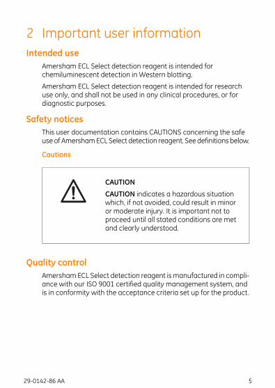
2 Important user informationIntended use
Amersham ECL Select detection reagent is intended forchemiluminescent detection in Western blotting.
Amersham ECL Select detection reagent is intended for researchuse only, and shall not be used in any clinical procedures, or fordiagnostic purposes.
Safety noticesThis user documentation contains CAUTIONS concerning the safeuse of AmershamECL Select detection reagent. See definitions below.
Cautions
CAUTION
CAUTION indicates a hazardous situationwhich, if not avoided, could result in minoror moderate injury. It is important not toproceed until all stated conditions are metand clearly understood.
Quality controlAmersham ECL Select detection reagent ismanufactured in compli-ance with our ISO 9001 certified quality management system, andis in conformity with the acceptance criteria set up for the product.
29-0142-86 AA 5

3 HandlingSafety precautions
CAUTION
Hazardous substances.When usinghazardous chemicals, take all suitableprotective measures, such as wearingprotective glasses and gloves resistant tothe substances used. Follow local and/ornational regulations for safe operation.
It is recommended to read the Safety Data Sheet (SDS) before usingAmersham ECL Select detection reagent.
StorageOn receipt, all components should be stored in a refrigerator at 2ºCto 8ºC. Amersham ECL Select detection reagent is sensitive toprolonged exposure to light. Always store the individual reagents inthe light-tight containers in which they are provided.
ExpiryThe components are stable for at least 3monthswhen stored underthe recommended conditions. See expiry date on package.
6 29-0142-86 AA

4 Required componentsKit components
The following components are included in the AmershamECL Selectdetection reagent kit.
ContentProduct
• Solution A: Luminol solution, 50 ml.• Solution B: Peroxide solution, 50 ml.
Sufficient for 1000 cm2 membrane
RPN2235
SolutionsRequired solutions are listed below.
• Phosphate buffered saline (PBS), pH 7.5
• Tris buffered saline (TBS), pH 7.6
• Dilution and wash buffer: PBS Tween™ (PBS-T) and TBS Tween(TBS-T). A Tween 20 concentration of 0.1% is suitable for mostblotting applications.
MembraneUse a suitable protocol to separate proteins by electrophoresis andtransfer them to a PVDF or nitrocellulose membrane.
Blocking reagentsBlocking reagents are typically diluted to 2% to 5% (v/v) in PBS-T orTBS-T buffer.
The following blocking reagents are recommended:
• Amersham ECL Prime Blocking Agent
• Amersham ECL Blocking Agent
• Non-fat dry milk
• Bovine Serum Albumine (BSA)
29-0142-86 AA 7
Immunodetection reagents
• Primary antibody specific to the target protein(s)
• HRP conjugated secondary antibody specific to the primary anti-body. See ECL HRP-linked secondary antibodies, on page 25.
Dilute the antibodies in PBS-T or TBS-T according to the recommen-dations in Dilution ranges, on page 19.
8 29-0142-86 AA

5 Western blotting optimizationIntroduction
To achieve an optimal Western blotting result with high signal tonoise ratio and best possible sensitivity and linearity, it is importantto optimize the method and to select compatible products.
Consider the following:
• Sample quality and loading amount – It is important that thesample is of good quality and that detectable levels of targetprotein is present.
• Membrane and blocking – Select membranes and blockingagents compatible with sample and antibodies.
• Primary and secondary antibodies – Always select specific an-tibodies of high quality and optimize the antibody dilution.
• Detection and imaging – Select detection reagent according toyour application need. A CCD imager offers high sensitivity andbroad dynamic range and provide better quantification thanX-ray film.
This chapter describes products recommended for use withAmersham ECL Select detection reagent. For more informationregarding these products, refer to www.gelifesciences.com/ecl.
Molecular weight markersMolecular weight markers are used to determine protein size. Inaddition, pre-stainedmarkers allow confirmation of protein transferand orientation (as the colored bands transfer to the membrane).
• Amersham Rainbow™ Markers are pre-stained multicoloredmarkers for monitoring progress of protein electrophoresis,confirming transfer efficiency and determination of molecularweight of blotted proteins.
29-0142-86 AA 9

• Amersham ECL DualVue™ Markers are markers optimized foruse with Amersham ECL, ECL Prime and ECL Select and containsa combinationof pre-stainedand taggedproteinsmarkers. Thesemarkers enablemonitoringof electrophoresis, confirming transferefficiency and determination of molecular weight of blotted pro-teins without staining on gel and membrane, as well as inchemiluminescence detection.
Membranes
• Amersham Hybond-P is a PVDF membrane with high proteinbinding capacity and mechanical strength, which makes it idealforWestern blotting applicationswhere stripping and re-probingare needed. The membrane is optimal for use with AmershamECL Prime and ECL Select detection reagents.
• AmershamHybond-ECL is a nitrocellulosemembranecompatiblewith all chemiluminescentWestern blotting substrates. Themainadvantage is the normally low background.
Transfer
• Wet transfer (Transfer Units TE 22 and TE 62), is the most com-monly used transfermethod. It provides efficient transfer of smallto large proteins.
• Semi-dry transfer (Transfer Units TE 70 and TE 77), is faster thanwet transfer and consumes less buffer. Semi-dry transfer workswell for most proteins but transfer may be less efficient for largeproteins. Itmight have reduced sensitivity for very lowabundanceproteins.
BlockingAfter protein transfer the membrane need to be incubated in ablocking solution to prevent non-specific binding of antibodies,whichcan cause background and non-specific protein bands on the blot.The blocking agent should be optimized for best results, no singleblocking agent is optimal for all proteins and antibodies. GEHealthcare recommend the following blocking agents compatiblewith Amersham ECL, ECL Prime and ECL Select:
10 29-0142-86 AA

• Amersham ECL Prime Blocking Agent
• Amersham ECL Blocking Agent
• BSA Blocking Agent
• Non-fat dry milk
Imagers and filmThe ImageQuant LAS systemsare flexible systems that cover awiderange of imaging applications. These CCD based imagers providean affordable option for protein detection with genuine benefits, in-cluding quantitation with high sensitivity, publication grade data,and image and data archiving.
ImageQuant LAS 4000
AmershamHyperfilm product range are trusted products for sensi-tive, qualitative chemiluminescent detection in Western blots.
Western blotting handbookMore technical help, tips, and best practices can be found in thehandbookWestern Blotting Principles and Methods from GEHealthcare (code no. 28-9998-97).
29-0142-86 AA 11

6 ProtocolProtocol overview
Below is an overview of the Western blotting detection protocol.
Gel electrophoresis
Transfer
Blocking
Antibody probing
Detection
Image analysis
12 29-0142-86 AA
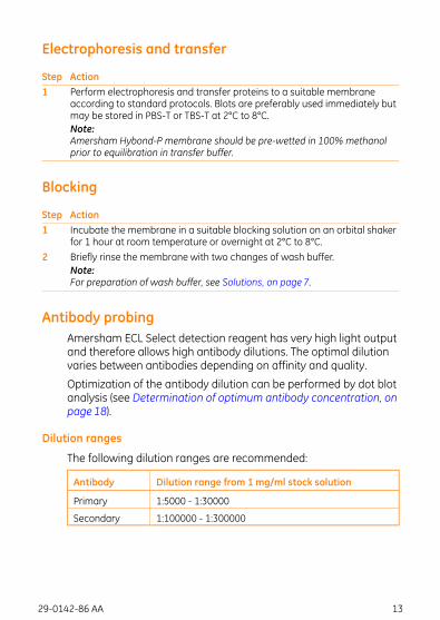
Electrophoresis and transfer
ActionStepPerform electrophoresis and transfer proteins to a suitable membraneaccording to standard protocols. Blots are preferably used immediately butmay be stored in PBS-T or TBS-T at 2°C to 8°C.
1
Note:Amersham Hybond-P membrane should be pre-wetted in 100% methanolprior to equilibration in transfer buffer.
Blocking
ActionStepIncubate the membrane in a suitable blocking solution on an orbital shakerfor 1 hour at room temperature or overnight at 2°C to 8°C.
1
Briefly rinse the membrane with two changes of wash buffer.2Note:For preparation of wash buffer, see Solutions, on page 7.
Antibody probingAmersham ECL Select detection reagent has very high light outputand therefore allows high antibody dilutions. The optimal dilutionvaries between antibodies depending on affinity and quality.
Optimization of the antibody dilution can be performed by dot blotanalysis (see Determination of optimum antibody concentration, onpage 18).
Dilution ranges
The following dilution ranges are recommended:
Dilution range from 1 mg/ml stock solutionAntibody
1:5000 - 1:30000Primary
1:100000 - 1:300000Secondary
29-0142-86 AA 13

The table below shows suggested starting dilutions for primaryantibodies with different levels of affinity.
Secondary antibodydilution
Primary antibodydilution
Type of antibody
1:1500001:10000High affinity primaryantibodies
1:1000001:5000Medium to low affinityprimary antibodies
Primary antibody incubation
ActionStepDilute the primary antibody in PBS-T or TBS-T.1Incubate themembrane in the primary antibody solution on anorbital shakerfor 1 hour at room temperature or overnight at 2°C to 8°C.
2
Briefly rinse the membrane with two changes of wash buffer.3Wash the membrane 4 to 6 times in wash buffer for 5 minutes each at roomtemperature on an orbital shaker.
4
Note:Exposure to X-ray film requires 6 wash steps, to avoid background.
Secondary antibody incubation
NotesActionStepIncrease the light output by buildinga three layer sandwich usingbiotinylated secondary antibodies andHRP conjugated streptavidin.
Dilute the secondary antibody (HRPconjugated or biotinylated antibody)in PBS-T or TBS-T.
1
Incubate the membrane in thesecondary antibody solution for 1 hourat room temperature on an orbitalshaker.
2
Briefly rinse the membrane with twochanges of wash buffer.
3
Exposure to film requires 6wash steps,to avoid background.
Wash the membrane 4 to 6 times inwash buffer for 5 minutes each atroom temperature on an orbitalshaker.
4
14 29-0142-86 AA
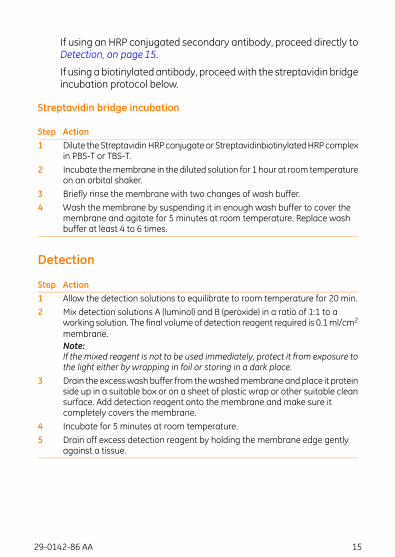
If using an HRP conjugated secondary antibody, proceed directly toDetection, on page 15.
If using a biotinylated antibody, proceedwith the streptavidin bridgeincubation protocol below.
Streptavidin bridge incubation
ActionStepDilute the StreptavidinHRP conjugate or StreptavidinbiotinylatedHRP complexin PBS-T or TBS-T.
1
Incubate themembrane in the diluted solution for 1 hour at room temperatureon an orbital shaker.
2
Briefly rinse the membrane with two changes of wash buffer.3Wash the membrane by suspending it in enough wash buffer to cover themembrane and agitate for 5 minutes at room temperature. Replace washbuffer at least 4 to 6 times.
4
Detection
ActionStepAllow the detection solutions to equilibrate to room temperature for 20 min.1Mix detection solutions A (luminol) and B (peroxide) in a ratio of 1:1 to aworking solution. The final volume of detection reagent required is 0.1ml/cm2
membrane.
2
Note:If the mixed reagent is not to be used immediately, protect it from exposure tothe light either by wrapping in foil or storing in a dark place.Drain the excesswash buffer from thewashedmembraneandplace it proteinside up in a suitable box or on a sheet of plastic wrap or other suitable cleansurface. Add detection reagent onto the membrane and make sure itcompletely covers the membrane.
3
Incubate for 5 minutes at room temperature.4Drain off excess detection reagent by holding the membrane edge gentlyagainst a tissue.
5
29-0142-86 AA 15
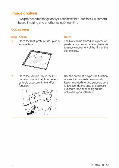
Image analysisTwoprotocols for imageanalysis are described, one for CCD camera-based imaging and another using X-ray film.
CCD camera
NotesActionStepThe blot can be placed on a piece ofplastic wrap, protein side up, to facili-tate easymovement of the film on thesample tray.
Place the blot, protein side up on asample tray.
1
Use the automatic exposure functionor select exposure time manually.Recommendedstarting exposure timeis 60 seconds. Increase or decreaseexposure time depending on theobtained signal intensity.
Place the sample tray in the CCDcamera compartment and selectsuitable exposure time and/orfunction.
2
16 29-0142-86 AA
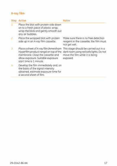
X-ray film
NotesActionStepPlace the blot with protein side downon to a fresh piece of plastic wrap,wrap the blots and gently smooth outany air bubbles.
1
Make sure there is no free detectionreagent in the cassette; the film mustnot get wet.
Place the wrapped blot with proteinside up in an X-ray film cassette.
2
This stage should be carried out in adark roomusing red safe lights. Do notmove the film while it is beingexposed.
Place a sheet of X-ray film (AmershamHyperfilmproduct range) on top of themembrane. Close the cassette andallow exposure. Suitable exposurestart time is 1 minute.
3
Develop the film immediately and, onthe basis of the signal intensityobtained, estimate exposure time fora second sheet of film.
4
29-0142-86 AA 17

7 Additional informationStripping and reprobing membranes
The complete removal of primary and secondary antibodies fromthemembrane is possible following the protocol outlined below. Themembranes may be stripped and reprobed several times.
NotesActionStepIf more stringent conditions arerequired, the incubation can beperformedat 70°C or for a longer time.
Place the membrane in strippingbuffer (100 mM 2-mercaptoethanol,2% SDS, 62.5mMTris-HCl pH 6.7 ) andincubate at 50°C for 30 minutes withoccasional agitation.
1
Membranes may be incubated withAmersham ECL Select detectionreagent and exposed to film to ensureremoval of antibodies.
Wash the membrane for 3 x 10minutes in PBS-T or TBS-T at roomtemperature using large volumes ofwash buffer. The membrane may beused immediately or stored in PBS-Tor TBS-T at 2 to 8°C.
2
Block the membrane in a suitableblocking solution for 1 hour at roomtemperature.
3
Repeat the immunodetectionprotocolin Chapter 6, Protocol from Antibodyprobing to Image analysis.
4
Determination of optimum antibody concentrationDue to the high signal intensity of the AmershamECL Select detectionreagent, optimization of antibody concentrations is recommendedto ensure the best results. In general, very highly diluted primary andsecondary antibodies are requiredwith AmershamECL Select detec-tion reagent compared to standard chemiluminescent substrates.Outlined below are protocols for determining optimal antibody con-centrations.
18 29-0142-86 AA
Dilution ranges
The following dilution ranges are recommended:
Dilution range from 1 mg/ml stock solutionAntibody
1:5000 - 1:30000Primary
1:100000 - 1:300000Secondary
Primary antibody optimization
Dot blotting is a quick and effective method of determining theoptimum dilution of a primary antibody of unknown concentration.Alternatively, a Western blot can be prepared and then cut intoseveral strips. It should be noted that some antibodies may requirealternative blocking andwashing steps to the ones suggested below.
ActionStepSpot different amounts of protein sample, preferably a dilution series, on toa PVDF or nitrocellulose membrane and allow to air dry. Alternatively, use adot blot or slot blot manifold. Make one sample series for each dilution to betested.
1
Note:PVDF membranes must be pre-wetted in methanol.Incubate in blocking solution for 1 hour at room temperature with agitation.2Rinse the membranes briefly with two changes of wash buffer.3Cut themembrane to get each sample series on a separatemembrane strip.4Prepare different primary antibody solutions within the recommended anti-body range. Incubate each membrane strip in antibody solution for 1 hourat room temperature with agitation.
5
Briefly rinse the membrane with two changes of wash buffer. Wash themembrane by suspending it in wash buffer and agitate for 5minutes in roomtemperature. Replace wash buffer at least 4 to 6 times.
6
Dilute the secondary antibody (using only one concentration) and incubatethe membranes for 1 hour at room temperature with agitation.
7
Rinse blots in two changes of wash buffer, then wash 4 to 6 times in freshchanges of wash buffer.
8
Detect using Amersham ECL Select detection reagent detailed in Detection,on page 15 of the protocol. The antibody dilution which gives the best signalwith the minimum background should be selected.
9
29-0142-86 AA 19

Secondary antibody optimization
ActionStepSpot different amounts of protein sample, preferably a dilution series, on toa PVDF or nitrocellulose membrane and allow to air dry. Alternatively, use adot blot or slot blot manifold. Make one sample series for each dilution to betested.
1
Note:PVDF membranes must be pre-wetted in methanol.Incubate in blocking solution for 1 hour at room temperature with agitation.2Incubate in diluted primary antibody (optimized concentration) for 1 hour atroom temperature with agitation.
3
Briefly rinse the membrane with two changes of wash buffer. Wash themembrane by suspending it in wash buffer and agitate for 5minutes in roomtemperature. Replace wash buffer at least 4 to 6 times.
4
Cut themembrane to get each sample series on a separatemembrane strip.5Prepare different secondary antibody solutions within the recommendedantibody range. Incubate each membrane strip in antibody solution for 1hour at room temperature with agitation.
6
Briefly rinse the membrane with two changes of wash buffer. Wash themembrane by suspending it in wash buffer and agitate for 5minutes in roomtemperature. Replace wash buffer at least 4 to 6 times.
7
Detect using Amersham ECL Select detection reagent detailed in Detection,on page 15 of the protocol. The antibody dilution which gives the best signalwith minimum background should be selected.
8
20 29-0142-86 AA

8 Troubleshooting guidePossible causes / remediesProblems
• Non-detectable amounts of target protein.• Primary antibody is not binding to target protein, which may bedue to bad quality and/or unspecific primary antibody.
• Incorrect species of secondary antibody has been used.• PVDF membrane not pre-wetted in methanol.• Check that transfer equipment is working properly and that thecorrect procedure has been followed.
• Check protein transfer by staining the membrane and/or gel.• Confirm transfer efficiency by using pre-stainedRainbowmarker.• Some proteins may be affected by the treatments required forelectrophoresis.
• Detection reagents do not function properly.
- To test the detection reagent activity, in a darkroomprepare1 to 2ml of detection reagentworking solution in a clear testtube. Add1µl of undilutedHRP-conjugatedantibody solution.The solution should immediately emit a visible blue light thatfades during the next several minutes.
• Incorrect storage of Amersham ECL Select detection reagentmay cause a loss of signal. Bacterial growth inhibit the reagent.
No signal
• Transfer efficiency may have been poor.• Insufficient protein was loaded on to the gel.• The concentration of primary and secondary antibodies couldbe too low; optimization is required.
• Bad quality and/or unspecific primary antibody.• Exposure time may have been too short.
Weaksignal
• Too much protein was loaded on to the gel.• The concentrations of primary and secondary antibodies couldbe too high; optimization is required.
Excessive,diffusesignal
• Negative bands generally occur when protein target is in excessand antibody concentrations are too high. The effect is causedby substrate depletion.
- Load less amount of protein.- Dilute both primary and secondary antibody further.
White(negative)bands onthe film
29-0142-86 AA 21
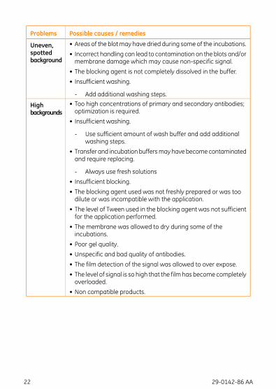
Possible causes / remediesProblems
• Areas of the blotmay have dried during some of the incubations.• Incorrect handling can lead to contamination on the blots and/ormembrane damage which may cause non-specific signal.
• The blocking agent is not completely dissolved in the buffer.• Insufficient washing.
- Add additional washing steps.
Uneven,spottedbackground
• Too high concentrations of primary and secondary antibodies;optimization is required.
• Insufficient washing.
- Use sufficient amount of wash buffer and add additionalwashing steps.
• Transfer and incubationbuffersmayhavebecomecontaminatedand require replacing.
- Always use fresh solutions• Insufficient blocking.• The blocking agent used was not freshly prepared or was toodilute or was incompatible with the application.
• The level of Tween used in the blocking agent was not sufficientfor the application performed.
• The membrane was allowed to dry during some of theincubations.
• Poor gel quality.• Unspecific and bad quality of antibodies.• The film detection of the signal was allowed to over expose.• The level of signal is so high that the filmhas becomecompletelyoverloaded.
• Non compatible products.
Highbackgrounds
22 29-0142-86 AA

9 Related productsThis chapter presents a subset of related products. For moreinformation, refer to www.gelifesciences.com/ecl.
Sample preparation
Code no.QuantityProduct
80-6484-7050 samplesSDS-PAGE Clean-up kit
28-9412-791 × 500 mlMammalian Protein Extraction Buffer
80-6483-56500 assays2-D Quant Kit
Molecular weight markers
Code no.QuantityProduct
RPN755E250 μlLow-Range Rainbow Molecular Weight Markers
RPN756E250 μlHigh-Range Rainbow Molecular Weight Markers
RPN800E250 μlFull-Range Rainbow Molecular Weight Markers
RPN8101 pack(25 loadings)
ECL DualVue Western Blotting Markers
RPN850E120 μlECL Plex™ Fluorescent Rainbow Markers
RPN851E500 μlECL Plex Fluorescent Rainbow Markers
Gel electrophoresis equipment
Code no.QuantityProduct
Gel electrophoresis
28-9906-081ECL Gel Box
28-9898-0410ECL Gel 10%, 10 wells
28-9898-0510ECL Gel 12%, 10 wells
28-9898-0610ECL Gel 4-12%, 10 wells
28-9898-0710ECL Gel 8-16%, 10 wells
28-9901-5410ECL Gel 4-20%, 10 wells
28-9901-5510ECL Gel 10%, 15 wells
28-9901-5610ECL Gel 12%, 15 wells
29-0142-86 AA 23
Code no.QuantityProduct
28-9901-5710ECL Gel 4-12%, 15 wells
28-9901-5810ECL Gel 8-16%, 15 wells
28-9901-5910ECL Gel 4-20%, 15 wells
28-9901-6010ECL Gel 10%, 2 wells
28-9901-6110ECL Gel 12%, 2 wells
28-9901-6210ECL Gel 4-12%, 2 wells
28-9901-6310ECL Gel 8-16%, 2 wells
28-9901-6410ECL Gel 4-20%, 2 wells
28-9898-082ECL Gel 10%, 10 wells
28-9898-092ECL Gel 12%, 10 wells
28-9901-512ECL Gel 4-12%, 10 wells
28-9901-522ECL Gel 8-16%, 10 wells
28-9901-532ECL Gel 4-20%, 10 wells
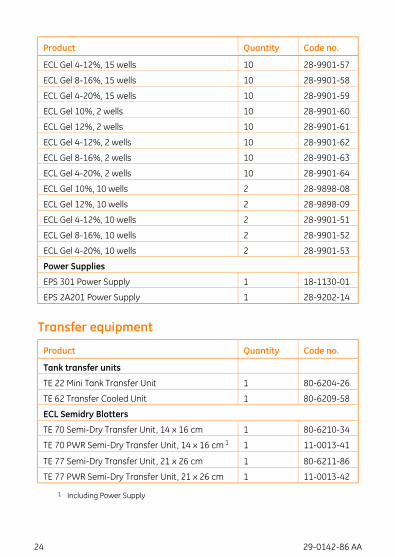
Power Supplies
18-1130-011EPS 301 Power Supply
28-9202-141EPS 2A201 Power Supply
Transfer equipment
Code no.QuantityProduct
Tank transfer units
80-6204-261TE 22 Mini Tank Transfer Unit
80-6209-581TE 62 Transfer Cooled Unit
ECL Semidry Blotters
80-6210-341TE 70 Semi-Dry Transfer Unit, 14 x 16 cm
11-0013-411TE 70 PWR Semi-Dry Transfer Unit, 14 x 16 cm 1
80-6211-861TE 77 Semi-Dry Transfer Unit, 21 x 26 cm
11-0013-421TE 77 PWR Semi-Dry Transfer Unit, 21 x 26 cm
1 Including Power Supply
24 29-0142-86 AA

Blotting equipment
Code no.QuantityProduct
Blotting paper
RPN6101M100 sheetsHybond blotting paper (20 × 20 cm)
3030-861100 sheets3MM Chr 20 × 20 cm
Membranes
RPN2020D10 sheetsHybond ECL (20 × 20 cm)
RPN7.58D10 sheetsHybond ECL (8 × 7.5 cm)
RPN2020LFP10 sheetsHybond-LFP (20 × 20 cm)
28-9909-8410 sheetsHybond-LFP (8 × 7.5 cm)
RPN2020F10 sheetsHybond-P (20 x 20 cm)
28-9909-8310 sheetsHybond-P (8 × 7.5 cm)
Blocking agents
Code no.QuantityProduct
RPN212540 gECL Blocking Agent
RPN41840 gECL Prime Blocking Agent
ECL HRP-linked secondary antibodies
Code no.QuantityProduct
NA931-1ML1 mlECLMouse IgG, HRP-LinkedWholeAb (fromsheep)
NA933-1ML1 mlECL Human IgG, HRP-Linked Whole Ab (fromsheep)
NA934-1ML1 mlECL Rabbit IgG, HRP-Linked Whole Ab (fromdonkey)
NA9310-1ML1 mlECLMouse IgG, HRP-Linked F(ab)2 fragment (fromsheep)
NA9340-1ML1 mlECL Rabbit IgG, HRP-Linked F(ab)2 fragment (fromdonkey)
28-9011-08150 μgECL Plex™ goat-α-mouse IgG-Cy2
28-9011-10150 μgECL Plex goat-α-rabbit IgG-Cy2
29-0142-86 AA 25

Code no.QuantityProduct
28-9011-06150 μgECL Plex goat-α-rabbit IgG-Cy3
PA43009150 μgECL Plex goat-α-mouse IgG-Cy3
PA45011150 μgECL Plex goat-α-rabbit IgG-Cy5
PA45010150 μgECL Plex goat-α-mouse IgG-Cy5
Detection reagents
Code no.QuantityProduct
RPN2235for 1000 cm2ECL Select Western Blotting Detection Reagent
RPN2232for 1000 cm2ECL Prime Western Blotting Detection Reagent
RPN2109for 1000 cm2ECL Western Blotting Detection Reagents
RPN2106for 4000 cm2ECL Western Blotting Detection Reagents
RPN2134for 6000 cm2ECL Western Blotting Detection Reagents
ECL Plex CyDye conjugated antibodies
Code no.QuantityProduct
RPN9981ECL PlexWestern Blotting Combination Pack (Cy3,Cy5, Hybond ECL)
RPN9991ECL PlexWestern Blotting Combination Pack (Cy3,Cy5, Hybond LFP) for two slab gels
26 29-0142-86 AA
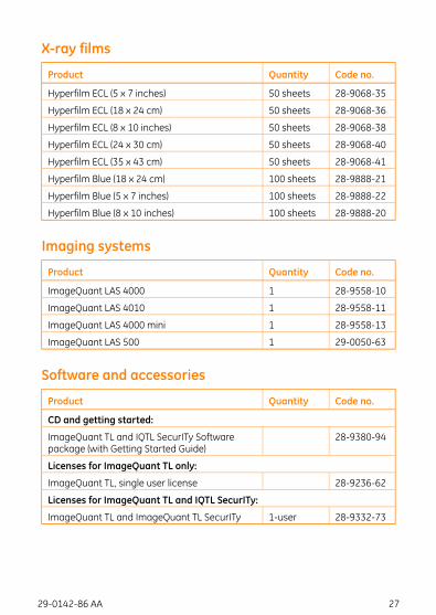
X-ray films
Code no.QuantityProduct
28-9068-3550 sheetsHyperfilm ECL (5 x 7 inches)
28-9068-3650 sheetsHyperfilm ECL (18 x 24 cm)
28-9068-3850 sheetsHyperfilm ECL (8 x 10 inches)
28-9068-4050 sheetsHyperfilm ECL (24 x 30 cm)
28-9068-4150 sheetsHyperfilm ECL (35 x 43 cm)
28-9888-21100 sheetsHyperfilm Blue (18 x 24 cm)
28-9888-22100 sheetsHyperfilm Blue (5 x 7 inches)
28-9888-20100 sheetsHyperfilm Blue (8 x 10 inches)
Imaging systems
Code no.QuantityProduct
28-9558-101ImageQuant LAS 4000
28-9558-111ImageQuant LAS 4010
28-9558-131ImageQuant LAS 4000 mini
29-0050-631ImageQuant LAS 500
Software and accessories
Code no.QuantityProduct
CD and getting started:
28-9380-94ImageQuant TL and IQTL SecurITy Softwarepackage (with Getting Started Guide)
Licenses for ImageQuant TL only:
28-9236-62ImageQuant TL, single user license
Licenses for ImageQuant TL and IQTL SecurITy:
28-9332-731-userImageQuant TL and ImageQuant TL SecurITy
29-0142-86 AA 27

For local office contact information, visitwww.gelifesciences.com/contact
GE Healthcare Bio-Sciences ABBjörkgatan 30751 84 UppsalaSweden
www.gelifesciences.com/ecl
GE, imagination at work and GE monogram are trademarks of General Electric Company.
Amersham, ECL, ECL DualVue, ECL Plex, Hybond, Hyperfilm, ImageQuant, and Rainbow aretrademarks of GE Healthcare companies.
This product or portions thereof is manufactured and sold under license from Cyanagen Srl andis subject of US patent number 7803573 and 7855287, and Italian application numberTO2010A000580, togetherwith other equivalent granted patents and patent applications in othercountries.
Tween is a trademark of ICI Americas Inc.
© 2006-2012 General Electric Company – All rights reserved.First published Feb. 2012
All goods and services are sold subject to the terms and conditions of sale of the company withinGE Healthcare which supplies them. A copy of these terms and conditions is available on request.Contact your local GE Healthcare representative for the most current information.
GE Healthcare Europe GmbHMunzinger Strasse 5, D-79111 Freiburg, Germany
GE Healthcare UK LimitedAmersham Place, Little Chalfont, Buckinghamshire, HP7 9NA, UK
GE Healthcare Bio-Sciences Corp.800 Centennial Avenue, P.O. Box 1327, Piscataway, NJ 08855-1327, USA
GE Healthcare Japan CorporationSanken Bldg. 3-25-1, Hyakunincho Shinjuku-ku, Tokyo 169-0073, Japan
29-0142-86 AA 02/2012
imagination at work


![Inhibitors of protein kinases affecting cAMP-dependent ...-P membrane (Millipore) [14]. The GATA- 6 was detected with Amersham. TM. ECL Western blotting analysis system [×2000 and](https://img.pdfslide.us/doc/110x75/5e364a58f1ebd16b5a001556/inhibitors-of-protein-kinases-affecting-camp-dependent-p-membrane-millipore.jpg)