Embed Size (px)
Citation preview
American Trypanosomiasis(Chagas Disease)
Anis Rassi Jr, MD, PhDa,*, Anis Rassi, MDa,Joffre Marcondes de Rezende, MDb
KEYWORDS
� Chagas disease � Chagas heart disease � American trypanosomiasis� Trypanosoma cruzi � Epidemiology � Treatment
KEY POINTS
� Chagas disease still represents a major public health challenge in Latin America, where 8to 10 million people are infected. Because of growing population movements, the diseasehas also spread to other continents.
� The disease is caused by the protozoan parasite T cruzi and transmitted to humans usuallyby the faeces of triatomine bugs or occasionally by nonvectorial mechanisms, such asblood transfusion and mother to fetus.
� Chagas disease has 2 phases, acute and chronic. Acute-phase disease is often asymp-tomatic. Up to 40%-50% of chronically infected patients develop progressive cardiomy-opathy and/or motility disturbances of the esophagus and colon.
� The disease, in both phases, is curable with the available antitrypanosomal drugs (benz-nidazole and nifurtimox). The sooner the treatment is initiated after infection, the greaterthe chance of cure.
� In patients with established chronic disease, several pharmacologic and nonpharmaco-logic interventions are available and may prevent or delay disease complications.
Chagas disease, or American trypanosomiasis, is caused by the parasite Trypano-soma cruzi, and was discovered in 1909 by the Brazilian physician Carlos Chagas(1879–1934).1 While still at a young age he described the etiologic agent, vectors, prin-cipal reservoirs, and mechanism of infection, as well as the acute clinical manifesta-tions of the first human case. However, Chagas disease is most likely an ancientdisease: T cruzi DNA has been recorded in tissue specimens of mummies in pre-Colombian Andean countries from as early as 9000 years ago (w7050 BC).2
T cruzi is a protozoan of the Sarcomastigophora phylum,Mastigophora subphylum,Kinetoplastida order, and Trypanosomatidae family (Fig. 1). It has a flagellum and its
The authors have nothing to disclose.a Division of Cardiology, Anis Rassi Hospital, Avenida Jose Alves 453, Setor Oeste, Goiania, GO74110-020, Brazil; b Instituto de Gastroenterologia de Goiania, Avenida B, 435, Setor Oeste,Goiania, GO 74435-010, Brazil* Corresponding author.E-mail address: [email protected]
Infect Dis Clin N Am 26 (2012) 275–291doi:10.1016/j.idc.2012.03.002 id.theclinics.com0891-5520/12/$ – see front matter � 2012 Elsevier Inc. All rights reserved.
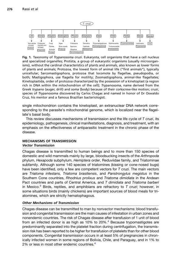
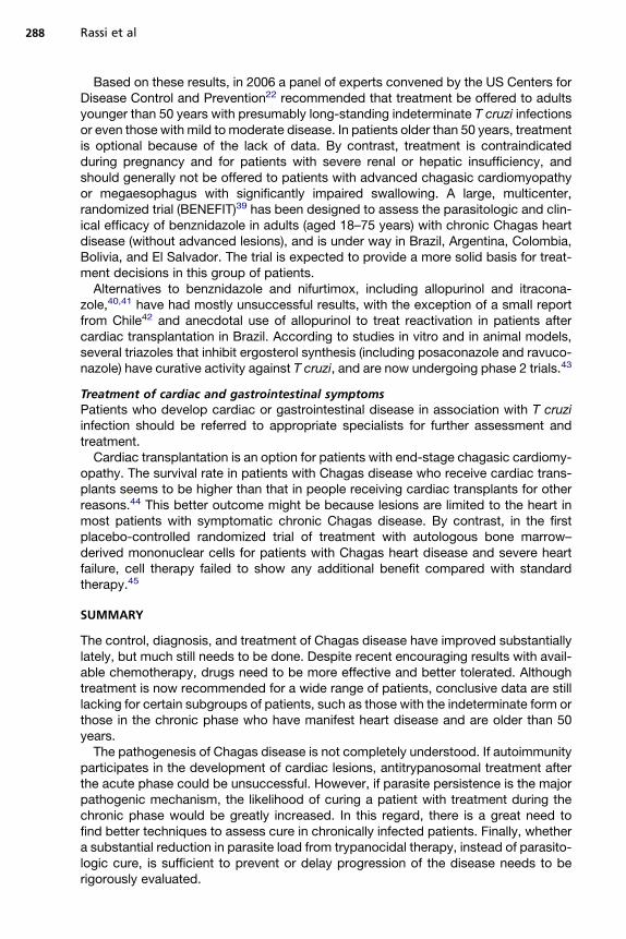
Fig. 1. Taxonomy of Trypanosoma cruzi. Eukaryota, cell organisms that have a cell nucleusand specialized organelles; Protista, a group of eukaryotic organisms (usually microorgan-isms), without the cardinal characteristics of plants and animals, also known as lower formsof plants and animals; Protozoa, the lowest form of animal life (“first animals”), typicallyunicellular; Sarcomastigophora, protozoa that locomote by flagellae, pseudopodia, orboth; Mastigophora, use flagella for motility; Zoomastigophora, animal-like flagellates;Kinetoplastida, order of protozoa characterized by the possession of a kinetoplast (a regionrich in DNA within the mitochondrion of the cell); Trypanosoma, name derived from theGreek trypano (auger, drill) and soma (body) because of their corkscrew-like motion; cruzi,species of Trypanosoma discovered by Carlos Chagas and named in honor of Dr OswaldoCruz, his mentor and a famous Brazilian bacteriologist.
Rassi et al276
single mitochondrion contains the kinetoplast, an extranuclear DNA network corre-sponding to the parasite’s mitochondrial genome, which is localized near the flagel-late’s basal body.This review discusses mechanisms of transmission and the life cycle of T cruzi, its
epidemiology, pathogenesis, clinical manifestations, diagnosis, and treatment, with anemphasis on the effectiveness of antiparasitic treatment in the chronic phase of thedisease.
MECHANISMS OF TRANSMISSIONVector Transmission
Chagas disease is transmitted to human beings and to more than 150 species ofdomestic and wild mammals mainly by large, bloodsucking insects of the Arthropodaphylum, Hexapoda subphylum, Hemiptera order, Reduviidae family, and Triatominaesubfamily. Although some 140 species of triatomines (kissing or cone-nosed bugs)have been identified, only a few are competent vectors for T cruzi. The main vectorsare Triatoma infestans, Triatoma brasiliensis, and Panstrongylus megistus in theSouthern Cone countries, Rhodnius prolixus and Triatoma dimidiata in the AndeanPact countries and parts of Central America, and T dimidiata and Triatoma barberiin Mexico.3 Birds, reptiles, and amphibians are refractory to T cruzi; however, insome situations birds (mainly chickens) are important sources of blood meals for tri-atomines, which are strictly hematophagous.
Other Mechanisms of Transmission
Chagas disease can be transmitted to man by nonvector mechanisms: blood transfu-sion and congenital transmission are the main causes of infestation in urban zones andnonendemic countries. The risk of Chagas disease after transfusion of 1 unit of bloodfrom an infected donor is as high as 10% to 20%.4 Because trypomastigotes arepredominantly separated into the platelet fraction during centrifugation, the transmis-sion risk has been reported to be higher for transfusion of platelets than for other bloodcomponents. Congenital transmission occurs in at least 5% of pregnancies in chron-ically infected women in some regions of Bolivia, Chile, and Paraguay, and in 1% to2% or less in most other endemic countries.4
American Trypanosomiasis 277
Transmission can also occur from transplantation of a solid organ or bone marrowfrom a chronically infected donor, which has been well documented in Latin America.Furthermore, Chagas disease can be orally transmitted by ingestion of food or liquidcontaminated with T cruzi. Such transmission is usually responsible for regionaloutbreaks of acute infection in areas devoid of domiciled insect vectors. More rarely,T cruzi can be transmitted through laboratory accidents to people who work with liveparasites.
LIFE CYCLE OF T CRUZI
The life cycle of T cruzi is complex, with several developmental forms in insect vectorsand mammalian hosts.4 The insects become infected by sucking blood from animalsor human beings who have circulating parasites (trypomastigote forms). In the diges-tive tract of triatomines, the trypomastigotes differentiate into epimastigotes (multipli-cative form) and then to metacyclic trypomastigotes in the final portion of the intestine.Infection of mammals occurs when they come into contact with the infective metacy-clic forms of the parasite that are eliminated with the feces of triatomines after feeding.This contact occurs through the mucosa or through injury, either preexistent or result-ing from the bite of the bug.Once in the vertebrate host, the metacyclic trypomastigotes invade the local retic-
uloendothelial and connective cells, and differentiate into amastigotes that begin repli-cating by binary fission. When the cell is swollen with amastigotes, they transformback into trypomastigotes by growing flagellae. The trypomastigotes lyse the cells,invade adjacent tissues, and spread via the lymphatics and bloodstream to distantsites, mainly muscle cells (cardiac, smooth, and skeletal) and ganglion cells, wherethey undergo further cycles of intracellular multiplication. The cycle of transmissionis completed when circulating trypomastigotes are taken up in blood meals byvectors.4
EPIDEMIOLOGY AND GEOGRAPHIC DISTRIBUTION
T cruzi is restricted to South America, Central America, and parts of North America(Mexico and southern United States). The Caribbean islands are free of Chagasdisease. In rural Latin America, poor housing conditions favor vector infestation andacute Chagas disease usually occurs in children younger than 12 years. Historicallytransmission and morbidity were concentrated in this region, but migration hasbrought chronic infected individuals to cities both in and outside of Latin America,making Chagas disease a public health problem of global concern. According to esti-mates by the Pan American Health Organization (PAHO)5 and the World Health Orga-nization (WHO),6 7.7 to 10 million people are chronically infected with T cruzi, and10,000 to 14,000 deaths per year are caused by Chagas disease.It is notable that as a result of successful programs involving vector control, blood
bank screening, and education of at-risk populations, both of these estimates aresubstantially lower than a few decades ago. A major program, begun in 1991 in theSouthern Cone nations of South America (Uruguay, Paraguay, Bolivia, Brazil, Chile,and Argentina), has provided the framework for much of this progress.7 Uruguayand Chile were certified free of transmission by the main domiciliary vector species(T infestans) in the late 1990s, and Brazil was declared transmission-free in 2006. Inaddition, blood donor screening has steadily increased, with coverage now approach-ing 100% in most endemic countries.The highest prevalences of Chagas disease have been reported from Bolivia (6.8%),
Argentina (4.1%), El Salvador (3.4%), Honduras (3.1%), and Paraguay (2.5%).
Rassi et al278
However, 2 remaining countries with prevalences of about 1% (Brazil and Mexico),together with Argentina, are home to almost 60% of all people infected with T cruziin Latin America.4
CHAGAS DISEASE IN THE UNITED STATES AND OTHER NONENDEMIC COUNTRIES
Acute Chagas disease is rare in the United States.8 Six human cases of autochtho-nous transmission and 5 instances of transmission by blood transfusion have beenreported. Furthermore, 3 infected donors transmitted T cruzi to 5 recipients of solidorgans, 2 of whom received cardiac transplants. The rarity of autochthonousvector-borne transmission is presumably due to better housing conditions and lessefficient vectors, but many infections probably go undetected. The 2 principal vectorsin the United States (Triatoma sanguisuga and Triatoma gerstaeckeri) have relativelylow infection rates (25% and 45%, respectively), display different feeding habits,and often defecate 30 minutes or more after feeding, making them likely to be some-what inefficient at stercorarian transmission to hosts.9
By contrast, the prevalence of chronic T cruzi infections in the United States hasincreased substantially in the past 20 years. An estimated 23 million immigrantsfrom endemic countries live in the United States, about 17 million of whom are Mexi-cans.10 The United States ranks seventh worldwide for the total number of peopleinfected with T cruzi: in 2009 an estimated 300,167 infected people lived in the UnitedStates.11 Screening of United States blood donations for T cruzi infection began inJanuary 2007, and now covers most of the blood supply. About 1 in 28,000 donorshas T cruzi infection, and so far more than 1400 infected donors have been identifiedand deferred permanently from donation.8
The southern states of the United States have an active sylvatic transmission cycleinvolving many wild animal reservoirs. Recent serologic and parasitologic surveyssuggest that raccoons, opossums, and woodrats are the main hosts, with prevalenceof infections of 38.7%, 28.0%, and 33.2%, respectively.9
Many individuals with Chagas disease have emigrated from Latin America to coun-tries other than the United States, such as Canada, Australia, Japan, France, Italy,Sweden, Switzerland, and England. But by far the largest population of these infectedimmigrants lives in Spain (47,000–67,000), with most originating from Ecuador,Argentina, Bolivia, and Peru.12 Nonendemic countries with large immigrant popula-tions have also begun to establish interventions to prevent transfusion-associated Tcruzi infection. European legislation prevents people with a history of Chagas diseasefrom donating blood. However, most infected people are asymptomatic and unawareof their status.
PATHOGENESIS
Chagas disease occurs in 2 phases: acute and chronic. Initial infection at the site ofparasite entry is characterized by the presence of infective trypomastigotes in leuko-cytes and cells of subcutaneous tissues, and by the development of interstitial edema,lymphocytic infiltration, and reactive hyperplasia of adjacent lymph nodes. Afterdissemination through the lymphatic system and the bloodstream, parasites concen-trate mainly in the muscles (including the myocardium) and ganglion cells. The char-acteristic pseudocysts that are present in some tissues are intracellular aggregatesof multiplying forms (amastigotes).Chagas disease is the most severe parasitic infection of the heart,13 and the heart is
the organ most often affected in individuals with chronic T cruzi infection.14 Changesinclude thinning of the ventricular walls, biventricular enlargement, apical aneurysms,
American Trypanosomiasis 279
and mural thrombi. Widespread destruction of myocardial cells, diffuse fibrosis,edema, lymphocytic infiltration of the myocardium, and scarring of the conductionsystem are often apparent, but parasites are difficult to find in myocardial tissue byconventional histologic methods. In chronic Chagas disease of the gastrointestinaltract, the esophagus and colon can be dilated to varying degrees. On microscopicexamination, focal inflammatory lesions with lymphocytic infiltration are seen, andthe number of neurons in the myenteric plexus might be markedly reduced.Evidence accumulated with highly powerful and sensitive methods, such as immu-
nohistochemistry and polymerase chain reaction (PCR), indicates that myocardialdamage in chronic T cruzi infection is due to the persistence of parasites and theaccompanying chronic inflammation, rather than autoimmune mechanisms. Cardiacdenervation (mainly parasympathetic), and abnormalities in the coronary microvascu-lature might also contribute to the pathogenesis of chronic lesions.15
CLINICAL MANIFESTATIONSAcute Chagas Disease
In most individuals, irrespective of the mechanism of transmission, acute Chagasinfection is asymptomatic, which is probably because the parasite load is fairlysmall.4,10 Symptoms that develop at around 8 to 10 days after invasion by the para-sites, or at 20 to 40 days after transfusion of T cruzi–infected blood, include prolongedfever, malaise enlargement of the liver, spleen, and lymph nodes, and subcutaneousedema (localized or generalized). In vector-borne transmission, there are signs ofportal of entry of T cruzi: entry through the skin produces the chagoma, an induratedarea of erythema and swelling, whereas entry via the ocular mucous membranesproduces Romana’s sign, the classic finding in acute Chagas disease, which consistsof unilateral painless edema of the palpebrae and periocular tissues. Severe myocar-ditis develops rarely; most deaths in acute Chagas disease are due to heart failure.Neurologic signs are not common, but meningoencephalitis occurs occasionally,especially in children younger than 2 years.4,10
An electrocardiogram (ECG) might show sinus tachycardia, first-degree atrioven-tricular block, low QRS voltage, or primary T-wave changes; and a chest radiographmight show variable degrees of cardiomegaly. Repetition of the ECG and chest radio-graph is crucial for detection of these abnormalities.16
Echocardiography was recently introduced, which explains the lack of informationabout its performance during the acute phase because most of such cases werereported before this method was available. Nevertheless, variable degrees of pericar-dial effusion, mitral or tricuspid valve regurgitation, and concentric hypertrophy of theleft ventricle have been described, with more than one abnormality often seen in thesame patient.17
Chronic Chagas Disease
The chronic phase begins 2 to 3 months after initial infection, when the clinical mani-festations (if present) of the acute disease will have resolved in virtually all infectedindividuals even if the infection has not been treated with trypanocidal drugs.4 About60% to 70% of these patients will have the indeterminate form of chronic Chagasdisease, which has no clinical symptoms. This form is characterized by positivity forantibodies against T cruzi in serum, a normal 12-lead ECG, and normal radiologicexamination of the chest, esophagus, and colon. The remaining patients (30%–40%) will develop a determinate form—cardiac, digestive (mainly megaesophagusand megacolon), or cardiodigestive—usually 10 to 30 years after initial infection.4
Rassi et al280
Reactivation of Chagas disease can also occur in chronically infected patients whobecome immunologically compromised, for example, from coinfection with humanimmunodeficiency virus (HIV) or immunosuppressive drugs. Fever, myocarditis, pan-niculitis, and skin lesions are common in recipients of solid-organ or bone marrowtransplants, whereas the most common manifestations of reactivation in patientswith AIDS are meningoencephalitis and lesions of the central nervous system thatresemble the lesions of cerebral toxoplasmosis.
Cardiac formThe cardiac form is the most serious and frequent manifestation of chronic disease.14
This form develops in 20% to 30% of individuals and typically leads to abnormalities ofthe conduction system, bradyarrhythmias and tachyarrhythmias, apical aneurysms,cardiac failure, thromboembolism, and sudden death. The most common ECG abnor-malities are right bundle branch block, left anterior fascicular block, ventricular prema-ture beats, ST-T changes, abnormal Q waves, and low QRS voltage. The combinationof right bundle branch block and left anterior fascicular block is very typical in Chagasheart disease.4,13,14 Frequent, complex ventricular premature beats, includingcouplets and runs of nonsustained ventricular tachycardia, are a common findingon Holter monitoring or stress testing.18
Sustained ventricular tachycardia is another hallmark of the disease, and seems toresult from an intramyocardial or subepicardial reentry circuit that is usually located atthe inferior-posterior-lateral wall of the left ventricle.4,14,18
Heart failure is often a late manifestation of Chagas heart disease. Such failure isusually biventricular with a predominance of right-sided failure at advanced stages,with peripheral edema, hepatomegaly, and ascites more prominent than pulmonarycongestion. Isolated left-sided failure can occur in the early stages of cardiac decom-pensation.4,14 Heart failure of chagasic etiology is associated with higher mortalitythan is heart failure from other causes.19 Systemic and pulmonary embolisms arisingfrom mural thrombi in the cardiac chambers are frequent.Sudden cardiac death accounts for nearly two-thirds of all deaths in Chagas heart
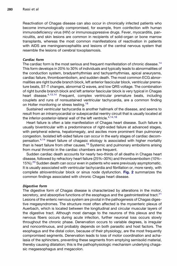
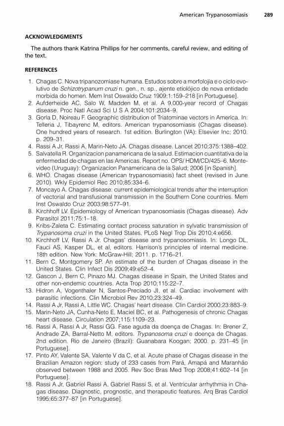
disease, followed by refractory heart failure (25%–30%) and thromboembolism (10%–15%).20 Sudden death can occur even in patients who were previously asymptomatic.It is usually associated with ventricular tachycardia and fibrillation or, more rarely, withcomplete atrioventricular block or sinus node dysfunction. Fig. 2 summarizes thecommon findings associated with chronic Chagas heart disease.
Digestive formThe digestive form of Chagas disease is characterized by alterations in the motor,secretory, and absorptive functions of the esophagus and the gastrointestinal tract.21
Lesions of the enteric nervous system are pivotal in the pathogenesis of Chagas diges-tive megasyndromes. The structure most often affected is the myoenteric plexus ofAuerbach, which is located between the longitudinal and circular muscular layers ofthe digestive tract. Although most damage to the neurons of this plexus and thenervous fibers occurs during acute infection, further neuronal loss occurs slowlythroughout the chronic phase. Denervation occurs to variable degrees, is irregularand noncontinuous, and probably depends on both parasitic and host factors. Theesophagus and the distal colon, because of their physiology, are the most frequentlycompromised segments. Denervation leads to loss of motor coordination and acha-lasia of the sphincters, preventing these segments from emptying semisolid material,thereby causing dilatation; this is the pathophysiologic mechanism underlying chaga-sic megaesophagus and megacolon.
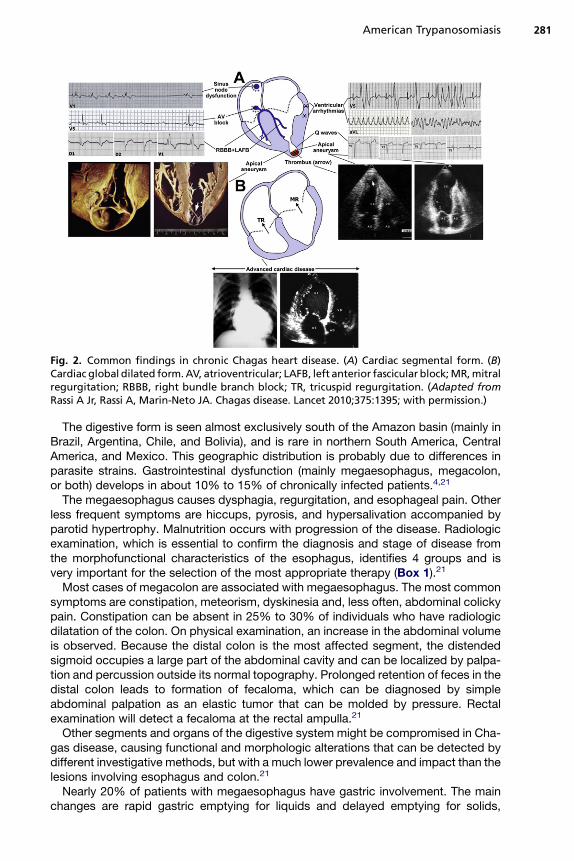
Fig. 2. Common findings in chronic Chagas heart disease. (A) Cardiac segmental form. (B)Cardiac global dilated form.AV, atrioventricular; LAFB, left anterior fascicular block;MR,mitralregurgitation; RBBB, right bundle branch block; TR, tricuspid regurgitation. (Adapted fromRassi A Jr, Rassi A, Marin-Neto JA. Chagas disease. Lancet 2010;375:1395; with permission.)
American Trypanosomiasis 281
The digestive form is seen almost exclusively south of the Amazon basin (mainly inBrazil, Argentina, Chile, and Bolivia), and is rare in northern South America, CentralAmerica, and Mexico. This geographic distribution is probably due to differences inparasite strains. Gastrointestinal dysfunction (mainly megaesophagus, megacolon,or both) develops in about 10% to 15% of chronically infected patients.4,21
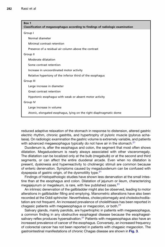
The megaesophagus causes dysphagia, regurgitation, and esophageal pain. Otherless frequent symptoms are hiccups, pyrosis, and hypersalivation accompanied byparotid hypertrophy. Malnutrition occurs with progression of the disease. Radiologicexamination, which is essential to confirm the diagnosis and stage of disease fromthe morphofunctional characteristics of the esophagus, identifies 4 groups and isvery important for the selection of the most appropriate therapy (Box 1).21
Most cases of megacolon are associated with megaesophagus. The most commonsymptoms are constipation, meteorism, dyskinesia and, less often, abdominal colickypain. Constipation can be absent in 25% to 30% of individuals who have radiologicdilatation of the colon. On physical examination, an increase in the abdominal volumeis observed. Because the distal colon is the most affected segment, the distendedsigmoid occupies a large part of the abdominal cavity and can be localized by palpa-tion and percussion outside its normal topography. Prolonged retention of feces in thedistal colon leads to formation of fecaloma, which can be diagnosed by simpleabdominal palpation as an elastic tumor that can be molded by pressure. Rectalexamination will detect a fecaloma at the rectal ampulla.21
Other segments and organs of the digestive system might be compromised in Cha-gas disease, causing functional and morphologic alterations that can be detected bydifferent investigative methods, but with a much lower prevalence and impact than thelesions involving esophagus and colon.21
Nearly 20% of patients with megaesophagus have gastric involvement. The mainchanges are rapid gastric emptying for liquids and delayed emptying for solids,
Box 1
Classification of megaesophagus according to findings of radiologic examination
Group I
Normal diameter
Minimal contrast retention
Presence of a residual air column above the contrast
Group II
Moderate dilatation
Some contrast retention
Increase in uncoordinated motor activity
Relative hypertony of the inferior third of the esophagus
Group III
Large increase in diameter
Great contrast retention
Hypotonic esophagus with weak or absent motor activity
Group IV
Large increase in volume
Atonic, elongated esophagus, lying on the right diaphragmatic dome
Rassi et al282
reduced adaptive relaxation of the stomach in response to distension, altered gastricelectric rhythm, chronic gastritis, and hypertrophy of pyloric muscle (pylorus acha-lasia). On radiologic examination the gastric volume is extremely variable, and patientswith advanced megaesophagus typically do not have air in the stomach.21
Duodenum is, after the esophagus and colon, the segment that most often showsdilatation. Megaduodenum is nearly always associated with other visceromegaly.The dilatation can be localized only at the bulb (megabulb) or at the second and thirdsegments, or can affect the entire duodenal arcade. Even when no dilatation ispresent, dyskinesia and hyperreactivity to cholinergic stimuli are common becauseof enteric denervation. Symptoms caused by megaduodenum can be confused withdyspepsia of gastric origin, of the dysmotility type.21
Findings of histopathologic studies have shown less denervation at the small intes-tine than at the esophagus and colon. Dilatation of jejunum or ileum, characterizingmegajejunum or megaileum, is rare, with few published cases.21
An intrinsic denervation of the gallbladder might also be observed, leading to motoralterations in gallbladder filling and emptying. Manometric alterations have also beenrecorded at the Oddi sphincter. Nevertheless, cholecystomegaly and choledochodila-tation are not frequent. An increased prevalence of cholelithiasis has been reported inchagasic patients with megaesophagus or megacolon, or both.21
Salivary glands, mainly parotids, are hypertrophic in patients with megaesophagus,a common finding in any obstructive esophageal disease because the esophageal-salivary reflex produces hypersalivation.21 Patients with megaesophagus also have anincreased prevalence of cancer of the esophagus. Conversely, an increased frequencyof colorectal cancer has not been reported in patients with chagasic megacolon. Thegastrointestinal manifestations of chronic Chagas disease are shown in Fig. 3.
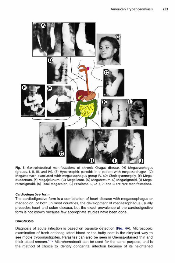
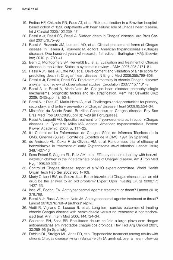
Fig. 3. Gastrointestinal manifestations of chronic Chagas disease. (A) Megaesophagus(groups, I, II, III, and IV). (B) Hypertrophic parotids in a patient with megaesophagus. (C)Megastomach associated with megaesophagus group IV. (D) Cholecystomegaly. (E) Mega-duodenum. (F) Megajejunum. (G) Megaileum. (H) Megarectum. (I) Megasigmoid. (J) Mega-rectosigmoid. (K) Total megacolon. (L) Fecaloma. C, D, E, F, and G are rare manifestations.
American Trypanosomiasis 283
Cardiodigestive formThe cardiodigestive form is a combination of heart disease with megaesophagus ormegacolon, or both. In most countries, the development of megaesophagus usuallyprecedes heart and colon disease, but the exact prevalence of the cardiodigestiveform is not known because few appropriate studies have been done.
DIAGNOSIS
Diagnosis of acute infection is based on parasite detection (Fig. 4A). Microscopicexamination of fresh anticoagulated blood or the buffy coat is the simplest way tosee motile trypomastigotes. Parasites can also be seen in Giemsa-stained thin andthick blood smears.4,10 Microhematocrit can be used for the same purpose, and isthe method of choice to identify congenital infection because of its heightened
Chronic Chagas infection
Incubation
period
1-2 weeks
infection
In
ten
sity o
f re
sp
on
se
Acute Chagas infection
Clinical disease
Chronic Chagas infection
Incubation
period
1-2 weeks
infection
Anti T. cruzi IgM
In
te
ns
ity o
f re
sp
on
se
8-10 weeks
Time
Acute Chagas infection
Clinical diseaseClinical disease
Etiological treatment
after 3 to 5 years8-10 weeks
Time
after 1 year
Congenitaltransmission
Vectorialtransmission
parasitemia
Anti T. cruzi IgG
parasitemia
Anti T. cruzi IgG
B
A
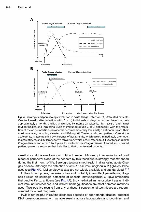
Fig. 4. Serologic and parasitologic evolution in acute Chagas infection. (A) Untreated patients.One to 2 weeks after infection with T cruzi, individuals undergo an acute phase that lastsapproximately 2 months, and is characterized by intense parasitemia, high levels of anti T cruziIgM antibodies, and increasing levels of immunoglobulin G (IgG) antibodies; with the resolu-tion of the acute infection, parasitemia becomes extremely low and IgG antibodies reach theirmaximum level, persisting elevated and lifelong. (B) Treated and cured patients. Cure at theacute phase is accompanied by clearance of parasitemia, which occurs immediately after etio-logic treatment, and by seronegative conversion,which occurs after about 1 year for congenitalChagas disease and after 3 to 5 years for vector-borne Chagas disease. Treated and uncuredpatients present a response that is similar to that of untreated patients.
Rassi et al284
sensitivity and the small amount of blood needed. Microscopic examination of cordblood or peripheral blood of the neonate by this technique is strongly recommendedduring the first month of life. Serologic testing is not helpful in diagnosing acute Cha-gas disease. Although the detection of anti–T cruzi immunoglobulin M (IgM) could beused (see Fig. 4A), IgM serology assays are not widely available and standardized.4,10
In the chronic phase, because of low and probably intermittent parasitemia, diag-nosis relies on serologic detection of specific immunoglobulin G (IgG) antibodiesthat bind to T cruzi antigens (see Fig. 4A). Enzyme-linked immunosorbent assay, indi-rect immunofluorescence, and indirect hemagglutination are most common methodsused. Two positive results from any of these 3 conventional techniques are recom-mended for a final diagnosis.PCR is not helpful in routine diagnosis because of poor standardization, potential
DNA cross-contamination, variable results across laboratories and countries, and
American Trypanosomiasis 285
the need for specific laboratory facilities. However, PCR has higher sensitivity than doother parasitologic methods, and therefore could be useful to confirm diagnosis incases of inconclusive serology and as an auxiliary method to monitor treatment.PCR can identify treatment failure from positive detection of T cruziDNA, but not treat-ment success, because even repeated negative PCR results do not necessarily indi-cate parasitologic cure. At best, such results indicate the absence of parasite DNA atthe time of the test.4,10
Low parasitemia during the chronic phase means that hemoculture and xenodiag-nosis have low sensitivity for parasite detection. However, these methods could beused to confirm diagnosis in rare cases of serologically doubtful results or to identifytreatment failures at specialized centers when PCR is not available.4,10
Initial assessment of a patient newly diagnosed with chronic T cruzi infectionincludes a complete medical history and physical examination, and a resting 12-lead ECG.4,22 Asymptomatic patients with a normal ECG have a favorable prognosisand should be followed up annually or biannually. Patients with ECG changes consis-tent with Chagas cardiomyopathy should undergo a routine cardiac assessment,including ambulatory 24-hour Holter monitoring (together with an exercise test when-ever possible) to detect arrhythmias and assess functional capacity, chest radiog-raphy and 2-dimensional echocardiography to assess ventricular function, andother cardiologic tests as indicated. The results of these tests should be used tostratify individual patients by risk and implement appropriate therapy.23–25 Bariumswallow and enema are indicated for patients with symptoms of the digestive form.
TREATMENT
The aim of treatment is to cure infection in acute Chagas disease, to prevent organdamage in chronic asymptomatic infection, and to limit incapacity and preventmorbidity and mortality once the disease is already clinically manifested.26 In patientswith chronic long-standing T cruzi infection, research has not elucidated whether theparasite has to be eliminated from the body, or if only a reduction in the parasiteburden is sufficient to prevent or delay disease progression.
Antitrypanosomal Treatment
Only 2 drugs, benznidazole and nifurtimox, are recommended for the treatment ofChagas disease. Benznidazole (a nitroimidazole derivative) has been more extensivelyinvestigated in clinical studies and has the better safety and efficacy profile, and there-fore is usually used for first-line treatment. Children should be given 5 to 10 mg/kgbenznidazole in 2 or 3 divided doses per day for 60 days, or 15 mg/kg nifurtimox in3 divided doses per day for 60 to 90 days; both drugs should preferably be given aftermeals. For adults the recommended doses are 5 mg/kg benznidazole per day or 8 to10 mg/kg nifurtimox per day, for the same duration as for children.27
The most common adverse effect of benznidazole is a generalized or, sometimes,localized allergic dermatitis, which affects about 20% to 30% of patients and consistsof pruritic and nonbullous polymorphous erythematous rashes, often followed bydesquamation. This dermatitis is autolimited, usually of mild to moderate intensity,and begins 8 to 10 days after treatment starts (occasionally later); the dose does notneed to be reduced or interrupted in most patients. Another adverse effect, whichoccurs in about 5% to 10% of patients usually late in the treatment course, isa dose-dependent peripheral sensitive neuropathy, affecting mainly the distal partsof the lower limbs; in such cases, treatment should be stopped. Polyneuropathy isnearly always reversible but can take months to resolve. It is not relieved by the
Rassi et al286
administration of B-complex vitamins, but might respond to systemic corticosteroids.Rare serious adverse events include leukopenia with granulocytopenia or agranulocy-tosis (sometimes followed by fever and tonsillitis), and thrombocytopenic purpura.Bone marrow suppression usually occurs by the third week of therapy, or eventuallylater, and should trigger immediate treatment interruption. Leukopenia usually resolvesa few days after discontinuation of benznidazole, and tonsillitis should be treated withantibiotics. Additional reported side effects include nausea, vomiting, anorexia, weightloss, insomnia, loss of taste, and onycholysis.26,28
Nifurtimox is associated with various adverse effects that usually resolve whentreatment is stopped. Gastrointestinal symptoms are the most common side effectsreported in clinical studies, occurring in about 50% of patients, and include anorexialeading to weight loss, nausea, vomiting, abdominal discomfort, and occasionallydiarrhea. Other common side effects include symptoms of central nervous systemtoxicity, such as insomnia, irritability, and disorientation. Polyneuropathy, paresthe-sias, and peripheral neuritis are more serious but less common adverse effects. Addi-tional side effects include headache, myalgia, arthralgia, dizziness or vertigo, andmood changes.26,28 In general, children treated with benznidazole or nifurtimoxhave fewer adverse effects than adults.In acute Chagas disease, antitrypanosomal treatment clears the parasitemia, as
shown by conversion to negative serologic and parasitologic tests, reduces the severityand duration of symptoms, and decreases mortality (Fig. 4B). Cure rates of up to 81%have been reported,16 and treatment is mandatory for all patients with acute infection(vector borne, oral, or accidental), congenital infection, or reactivated infection fromimmunosuppressive treatment (eg, after organ transplantation) or coinfection with HIV.29
In children aged 6 to 12 years, findings of 2 randomized placebo-controlled trialsshowed that benznidazole cured about 60% of asymptomatic infections, as measuredby conversion of IgG serologic tests to negative at 3 to 4 years after treatment comple-tion.30,31 Together with growing favorable anecdotal experiences of individual clini-cians across Latin America, these studies prompted the WHO to recommend earlydiagnosis and antitrypanosomal treatment for all children with chronic T cruzi infec-tion32; this recommendation was extended to children aged up to 18 years in theUnited States guidelines.22
Whether adults with the indeterminate or chronic symptomatic form of Chagasdisease should be treated with benznidazole or nifurtimox has been debated for years.Some researchers argue that both drugs have frequent and unpleasant side effects,might be carcinogenic, need to be taken for a long period, and lack efficacy, as shownby low seroconversion rates.33,34 However, none of these arguments is solid orcompelling.35 To improve benznidazole tolerability, the authors have adopted 3 strat-egies. First, the daily dose should not exceed 300 mg; in patients weighing more than60 kg, a fixed daily dose of 300 mg should be given for a total number of days equal tothe patient’s weight in kilograms, resulting in a total dose that is equivalent to 5 mg/kgper day for 60 days.26 Second, patients with mild to moderate allergic dermatitisshould be treated immediately with low-dose systemic corticosteroids (eg, 20 mg/dprednisolone orally for 10 days, followed by 10 mg/d for 10 days), without the needfor benznidazole interruption.26,28 Third, in patients with severe dermatitis, the authorshave achieved a tolerance rate of 72% by retreating 25 patients with benznidazole andprednisolone (20 mg/d during the first 14 days followed by 10 mg/d for the remainingdays of treatment) at the same time (Rassi A, unpublished data, 2010).Seroconversion might not be the most appropriate criterion to monitor drug efficacy
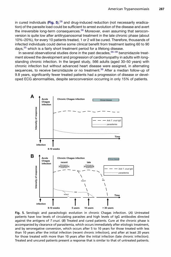
after chemotherapy at a late stage of chronic infection. Contrary to parasitologictesting, serologic test results can take decades to convert from positive to negative
American Trypanosomiasis 287
in cured individuals (Fig. 5),28 and drug-induced reduction (not necessarily eradica-tion) of the parasite load could be sufficient to arrest evolution of the disease and avertthe irreversible long-term consequences.36 Moreover, even assuming that serocon-version is quite low after antitrypanosomal treatment in the late chronic phase (about10%–20%), for every 10 patients treated, 1 or 2 will be cured. Therefore, thousands ofinfected individuals could derive some clinical benefit from treatment lasting 60 to 90days,35 which is a fairly short treatment period for a lifelong disease.In several observational studies done in the past decades,36–38 benznidazole treat-
ment slowed the development and progression of cardiomyopathy in adults with long-standing chronic infection. In the largest study, 566 adults (aged 30–50 years) withchronic infection but without advanced heart disease were assigned, in alternatingsequences, to receive benznidazole or no treatment.36 After a median follow-up of9.8 years, significantly fewer treated patients had a progression of disease or devel-oped ECG abnormalities, despite seroconversion occurring in only 15% of patients.
Fig. 5. Serologic and parasitologic evolution in chronic Chagas infection. (A) Untreatedpatients have low levels of circulating parasites and high levels of IgG antibodies directedagainst the antigens of T cruzi. (B) Treated and cured patients. Cure at the chronic phase isaccompanied by clearance of parasitemia, which occurs immediately after etiologic treatment,and by seronegative conversion, which occurs after 5 to 10 years for those treated with lessthan 10 years after the initial infection (recent chronic infection), and after at least 20 yearsfor those treated with more than 10 years after the initial infection (late chronic infection).Treated and uncured patients present a response that is similar to that of untreated patients.
Rassi et al288
Based on these results, in 2006 a panel of experts convened by the US Centers forDisease Control and Prevention22 recommended that treatment be offered to adultsyounger than 50 years with presumably long-standing indeterminate T cruzi infectionsor even those with mild to moderate disease. In patients older than 50 years, treatmentis optional because of the lack of data. By contrast, treatment is contraindicatedduring pregnancy and for patients with severe renal or hepatic insufficiency, andshould generally not be offered to patients with advanced chagasic cardiomyopathyor megaesophagus with significantly impaired swallowing. A large, multicenter,randomized trial (BENEFIT)39 has been designed to assess the parasitologic and clin-ical efficacy of benznidazole in adults (aged 18–75 years) with chronic Chagas heartdisease (without advanced lesions), and is under way in Brazil, Argentina, Colombia,Bolivia, and El Salvador. The trial is expected to provide a more solid basis for treat-ment decisions in this group of patients.Alternatives to benznidazole and nifurtimox, including allopurinol and itracona-
zole,40,41 have had mostly unsuccessful results, with the exception of a small reportfrom Chile42 and anecdotal use of allopurinol to treat reactivation in patients aftercardiac transplantation in Brazil. According to studies in vitro and in animal models,several triazoles that inhibit ergosterol synthesis (including posaconazole and ravuco-nazole) have curative activity against T cruzi, and are now undergoing phase 2 trials.43
Treatment of cardiac and gastrointestinal symptomsPatients who develop cardiac or gastrointestinal disease in association with T cruziinfection should be referred to appropriate specialists for further assessment andtreatment.Cardiac transplantation is an option for patients with end-stage chagasic cardiomy-
opathy. The survival rate in patients with Chagas disease who receive cardiac trans-plants seems to be higher than that in people receiving cardiac transplants for otherreasons.44 This better outcome might be because lesions are limited to the heart inmost patients with symptomatic chronic Chagas disease. By contrast, in the firstplacebo-controlled randomized trial of treatment with autologous bone marrow–derived mononuclear cells for patients with Chagas heart disease and severe heartfailure, cell therapy failed to show any additional benefit compared with standardtherapy.45
SUMMARY
The control, diagnosis, and treatment of Chagas disease have improved substantiallylately, but much still needs to be done. Despite recent encouraging results with avail-able chemotherapy, drugs need to be more effective and better tolerated. Althoughtreatment is now recommended for a wide range of patients, conclusive data are stilllacking for certain subgroups of patients, such as those with the indeterminate form orthose in the chronic phase who have manifest heart disease and are older than 50years.The pathogenesis of Chagas disease is not completely understood. If autoimmunity
participates in the development of cardiac lesions, antitrypanosomal treatment afterthe acute phase could be unsuccessful. However, if parasite persistence is the majorpathogenic mechanism, the likelihood of curing a patient with treatment during thechronic phase would be greatly increased. In this regard, there is a great need tofind better techniques to assess cure in chronically infected patients. Finally, whethera substantial reduction in parasite load from trypanocidal therapy, instead of parasito-logic cure, is sufficient to prevent or delay progression of the disease needs to berigorously evaluated.
American Trypanosomiasis 289
ACKNOWLEDGMENTS
The authors thank Katrina Phillips for her comments, careful review, and editing ofthe text.
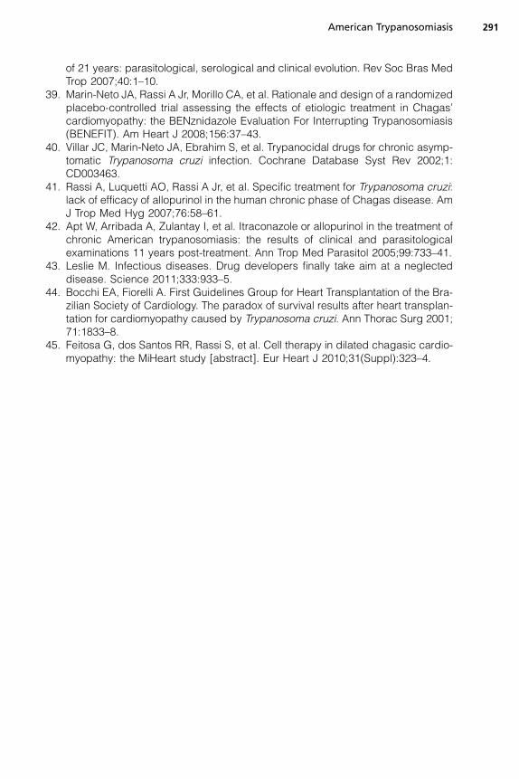
REFERENCES
1. ChagasC. Nova tripanozomiase humana. Estudos sobre amorfolojıa e o ciclo evo-lutivo de Schizotrypanum cruzi n. gen., n. sp., ajente etiolojico de nova entidademorbida do homen. Mem Inst Oswaldo Cruz 1909;1:159–218 [in Portuguese].
2. Aufderheide AC, Salo W, Madden M, et al. A 9,000-year record of Chagasdisease. Proc Natl Acad Sci U S A 2004;101:2034–9.
3. Gorla D, Noireau F. Geographic distribution of Triatominae vectors in America. In:Telleria J, Tibayrenc M, editors. American trypanosomiasis (Chagas disease).One hundred years of research. 1st edition. Burlington (VA): Elsevier Inc; 2010.p. 209–31.
4. Rassi A Jr, Rassi A, Marin-Neto JA. Chagas disease. Lancet 2010;375:1388–402.5. Salvatella R.Organizacion panamericana de la salud. Estimacion cuantitativa de la
enfermedad de chagas en las Americas. Report no. OPS/ HDM/CD/425–6. Monte-video (Uruguay): Organizacion Panamericana de la Salud; 2006 [in Spanish].
6. WHO. Chagas disease (American trypanosomiasis) fact sheet (revised in June2010). Wkly Epidemiol Rec 2010;85:334–6.
7. Moncayo A. Chagas disease: current epidemiological trends after the interruptionof vectorial and transfusional transmission in the Southern Cone countries. MemInst Oswaldo Cruz 2003;98:577–91.
8. Kirchhoff LV. Epidemiology of American trypanosomiasis (Chagas disease). AdvParasitol 2011;75:1–18.
9. Kribs-Zaleta C. Estimating contact process saturation in sylvatic transmission ofTrypanosoma cruzi in the United States. PLoS Negl Trop Dis 2010;4:e656.
10. Kirchhoff LV, Rassi A Jr. Chagas’ disease and trypanosomiasis. In: Longo DL,Fauci AS, Kasper DL, et al, editors. Harrison’s principles of internal medicine.18th edition. New York: McGraw-Hill; 2011. p. 1716–21.
11. Bern C, Montgomery SP. An estimate of the burden of Chagas disease in theUnited States. Clin Infect Dis 2009;49:e52–4.
12. Gascon J, Bern C, Pinazo MJ. Chagas disease in Spain, the United States andother non-endemic countries. Acta Trop 2010;115:22–7.
13. Hidron A, Vogenthaler N, Santos-Preciado JI, et al. Cardiac involvement withparasitic infections. Clin Microbiol Rev 2010;23:324–49.
14. Rassi A Jr, Rassi A, Little WC. Chagas’ heart disease. Clin Cardiol 2000;23:883–9.15. Marin-Neto JA, Cunha-Neto E, Maciel BC, et al. Pathogenesis of chronic Chagas
heart disease. Circulation 2007;115:1109–23.16. Rassi A, Rassi A Jr, Rassi GG. Fase aguda da doenca de Chagas. In: Brener Z,
Andrade ZA, Barral-Netto M, editors. Trypanosoma cruzi e doenca de Chagas.2nd edition. Rio de Janeiro (Brazil): Guanabara Koogan; 2000. p. 231–45 [inPortuguese].
17. Pinto AY, Valente SA, Valente V da C, et al. Acute phase of Chagas disease in theBrazilian Amazon region: study of 233 cases from Para, Amapa and Maranhaoobserved between 1988 and 2005. Rev Soc Bras Med Trop 2008;41:602–14 [inPortuguese].
18. Rassi A Jr, Gabriel Rassi A, Gabriel Rassi S, et al. Ventricular arrhythmia in Cha-gas disease. Diagnostic, prognostic, and therapeutic features. Arq Bras Cardiol1995;65:377–87 [in Portuguese].
Rassi et al290
19. Freitas HF, Chizzola PR, Paes AT, et al. Risk stratification in a Brazilian hospital-based cohort of 1220 outpatients with heart failure: role of Chagas heart disease.Int J Cardiol 2005;102:239–47.
20. Rassi A Jr, Rassi SG, Rassi A. Sudden death in Chagas’ disease. Arq Bras Car-diol 2001;76:75–96.
21. Rassi A, Rezende JM, Luquetti AO, et al. Clinical phases and forms of Chagasdisease. In: Telleria J, Tibayrenc M, editors. American trypanosomiasis (Chagasdisease). One hundred years of research. 1st edition. Burlington (MA): ElsevierInc; 2010. p. 709–41.
22. Bern C, Montgomery SP, Herwaldt BL, et al. Evaluation and treatment of Chagasdisease in the United States: a systematic review. JAMA 2007;298:2171–81.
23. Rassi A Jr, Rassi A, Little WC, et al. Development and validation of a risk score forpredicting death in Chagas’ heart disease. N Engl J Med 2006;355:799–808.
24. Rassi A Jr, Rassi A, Rassi SG. Predictors of mortality in chronic Chagas disease:a systematic review of observational studies. Circulation 2007;115:1101–8.
25. Rassi A Jr, Rassi A, Marin-Neto JA. Chagas heart disease: pathophysiologicmechanisms, prognostic factors and risk stratification. Mem Inst Oswaldo Cruz2009;104(Suppl 1):152–8.
26. Rassi A Jr, Dias JC, Marin-Neto JA, et al. Challenges and opportunities for primary,secondary, and tertiary prevention of Chagas’ disease. Heart 2009;95:524–34.
27. Ministerio da Saude Brasil. Brazilian Consensus on Chagas disease. Rev SocBras Med Trop 2005;38(Suppl 3):7–29 [in Portuguese].
28. Rassi A, Luquetti AO. Specific treatment for Trypanosoma cruzi infection (Chagasdisease). In: Tyler KM, Miles MA, editors. American trypanosomiasis. Boston:Kluwer Academic; 2003. p. 117–25.
29. 811Control de La Enfermedad de Chagas. Serie de Informes Tecnicos de laOMS. Ginebra (Suiza): Comite de Expertos de la OMS; 1991 [in Spanish].
30. de Andrade AL, Zicker F, de Oliveira RM, et al. Randomised trial of efficacy ofbenznidazole in treatment of early Trypanosoma cruzi infection. Lancet 1996;348:1407–13.
31. Sosa Estani S, Segura EL, Ruiz AM, et al. Efficacy of chemotherapy with benzni-dazole in children in the indeterminate phase of Chagas’ disease. Am J Trop MedHyg 1998;59:526–9.
32. Control of Chagas disease: report of a WHO expert committee. World HealthOrgan Tech Rep Ser 2002;905:1–109.
33. Mady C, Ianni BM, de Souza JL Jr. Benznidazole and Chagas disease: can an olddrug be the answer to an old problem? Expert Opin Investig Drugs 2008;17:1427–33.
34. Issa VS, Bocchi EA. Antitrypanosomal agents: treatment or threat? Lancet 2010;376:768.
35. Rassi A Jr, Rassi A, Marin-Neto JA. Antitrypanosomal agents: treatment or threat?Lancet 2010;376:768–9 [authors‘ reply].
36. Viotti R, Vigliano C, Lococo B, et al. Long-term cardiac outcomes of treatingchronic Chagas disease with benznidazole versus no treatment: a nonrandom-ized trial. Ann Intern Med 2006;144:724–34.
37. Gallerano RH, Sosa RR. Resultados de um estudio a largo plazo com drogasantiparasitarias em infectados chagasicos cronicos. Rev Fed Arg Cardiol 2001;30:289–96 [in Spanish].
38. Fabbro DL, Streiger ML, Arias ED, et al. Trypanocide treatment among adults withchronic Chagas disease living in Santa Fe city (Argentina), over a mean follow-up
American Trypanosomiasis 291
of 21 years: parasitological, serological and clinical evolution. Rev Soc Bras MedTrop 2007;40:1–10.
39. Marin-Neto JA, Rassi A Jr, Morillo CA, et al. Rationale and design of a randomizedplacebo-controlled trial assessing the effects of etiologic treatment in Chagas’cardiomyopathy: the BENznidazole Evaluation For Interrupting Trypanosomiasis(BENEFIT). Am Heart J 2008;156:37–43.
40. Villar JC, Marin-Neto JA, Ebrahim S, et al. Trypanocidal drugs for chronic asymp-tomatic Trypanosoma cruzi infection. Cochrane Database Syst Rev 2002;1:CD003463.
41. Rassi A, Luquetti AO, Rassi A Jr, et al. Specific treatment for Trypanosoma cruzi:lack of efficacy of allopurinol in the human chronic phase of Chagas disease. AmJ Trop Med Hyg 2007;76:58–61.
42. Apt W, Arribada A, Zulantay I, et al. Itraconazole or allopurinol in the treatment ofchronic American trypanosomiasis: the results of clinical and parasitologicalexaminations 11 years post-treatment. Ann Trop Med Parasitol 2005;99:733–41.
43. Leslie M. Infectious diseases. Drug developers finally take aim at a neglecteddisease. Science 2011;333:933–5.
44. Bocchi EA, Fiorelli A. First Guidelines Group for Heart Transplantation of the Bra-zilian Society of Cardiology. The paradox of survival results after heart transplan-tation for cardiomyopathy caused by Trypanosoma cruzi. Ann Thorac Surg 2001;71:1833–8.
45. Feitosa G, dos Santos RR, Rassi S, et al. Cell therapy in dilated chagasic cardio-myopathy: the MiHeart study [abstract]. Eur Heart J 2010;31(Suppl):323–4.