Embed Size (px)
Citation preview
Medical Parasitology/ 2nd Stage/ College of Pharmacy Dr. Muhannad
1
(Haemoflagelates)
Trypanosomiasis:-
Introduction:
Trypanosomes are hemoflagellates and three species of the genus Trypanosoma are
responsible for disease in humans such as sleeping sickness. Trypanosomes occur in the
blood of the majority of vertebrate animals. The life cycle involves intermediate host,
which usually is an insect. Many species of trypanosomes can live in harmony with their
hosts producing no pathogenic effect, but the best known species are those that are
pathogenic to their definitive hosts. The disease is caused by the pathogenic types is called
trypanosomiasis.
Salivarian Trypanosomes:
Trypanosoma brucei rhodesiense and Trypanosoma brucei gambiense - The metacyclic
trypanosomes are found in the proboscis of the insect vector - infection is therefore
inoculative. The above are the causative agents of African trypanosomiasis. It is a zoonotic
species in that it multiplies in the blood of a range of many mammals including man.
Trypanosoma brucei rhodesiense causes acute sleeping sickness in East Africa, while T. b.
gambiense causes chronic sleeping sickness in West Africa.
These are known as salivarian trypanosomes as they complete their development in the
salivary system (anterior portion of the vector). Transmission takes place by inoculation of
the metacyclic stage.
Stercorarian Trypanosomes:
Trypanosoma cruzi - The metacyclic trypanosomes occupy a posterior position in the gut of
the insect vector and are passed out in the feces - infection is therefore contaminative. This
is the causative agent of American Trypanosomiasis.
These trypanosomes are known as stercocarian as they complete their development in the
posterior region of the vector, so that the infective forms appear in the insect’s feces. Hosts
are infected by the contaminative route.
Medical Parasitology/ 2nd Stage/ College of Pharmacy Dr. Muhannad
2
Etiologic agents:
Trypanosoma brucei complex – African trypanosomiasis (sleeping sickness)
Trypanosoma cruzi – American trypanosomiasis (Chagas’ disease)
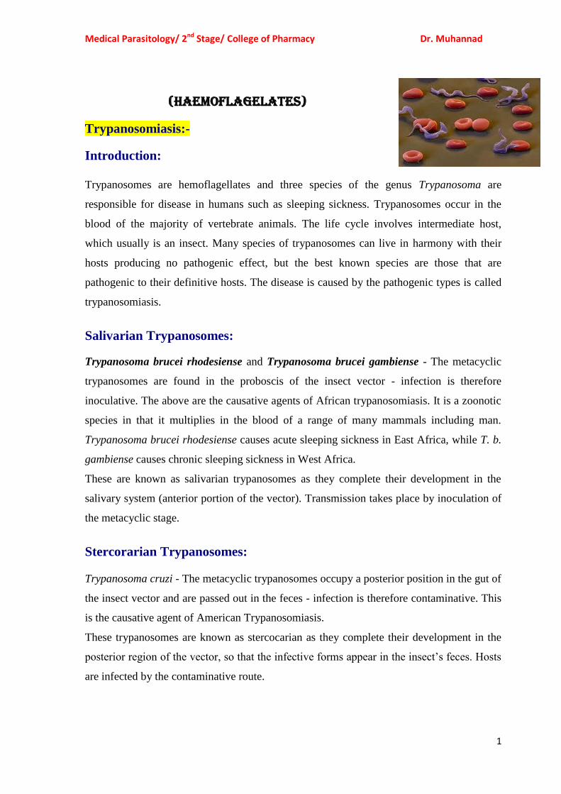
Important features:
These species may have amastigote, promastigote, epimastigote, and trypomastigote
stages in their life cycle. In human trypanosomes of the African form, however, the
amastigote and promastigote stages of development are absent. Typical trypanosome
structure is an elongated spindle-shaped body that more or less tapers at both ends, a
centrally situated nucleus, a kinetoplast posterior to nucleus, an undulating membrane
arising from the kinetoplast and proceeding forward along the margin of the cell membrane
and a single free flagellum at the anterior end.
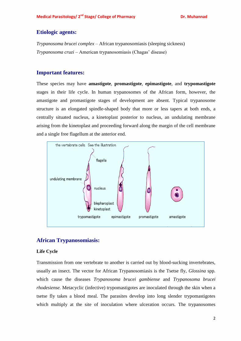
African Trypanosomiasis:
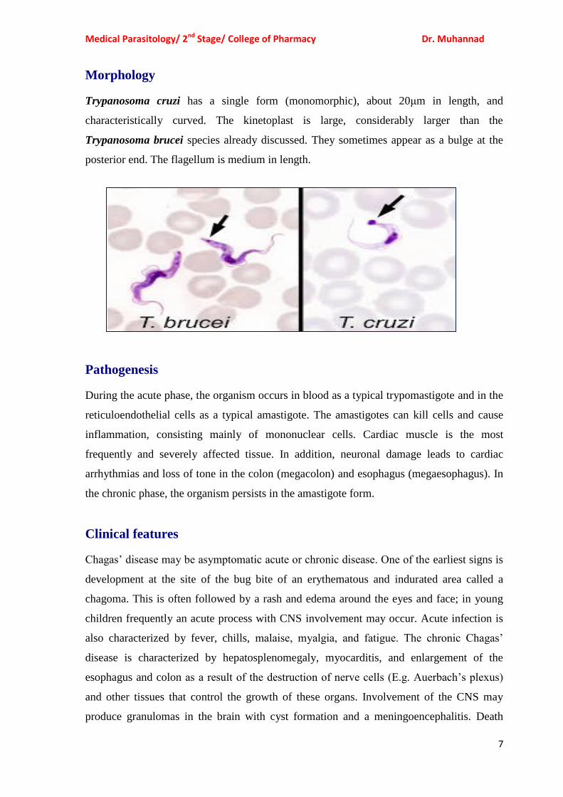
Life Cycle
Transmission from one vertebrate to another is carried out by blood-sucking invertebrates,
usually an insect. The vector for African Trypanosomiasis is the Tsetse fly, Glossina spp.
which cause the diseases Trypanosoma brucei gambiense and Trypanosoma brucei
rhodesiense. Metacyclic (infective) trypomastigotes are inoculated through the skin when a
tsetse fly takes a blood meal. The parasites develop into long slender trypomastigotes
which multiply at the site of inoculation where ulceration occurs. The trypanosomes
Medical Parasitology/ 2nd Stage/ College of Pharmacy Dr. Muhannad
3
continue to develop and then may invade the lymphatic tissues, the heart, various organs
and in later stages, the central nervous system. Trypomastigotes are taken up by the tsetse
fly (male and female) during a blood meal. The parasites develop in the midgut of the fly
where they multiply. 2-3 weeks later the trypomastigotes move to the salivary glands
transforming from epimastigotes into metacyclic (infective) trypomastigotes. The tsetse fly
remains infective for life i.e. about three months.
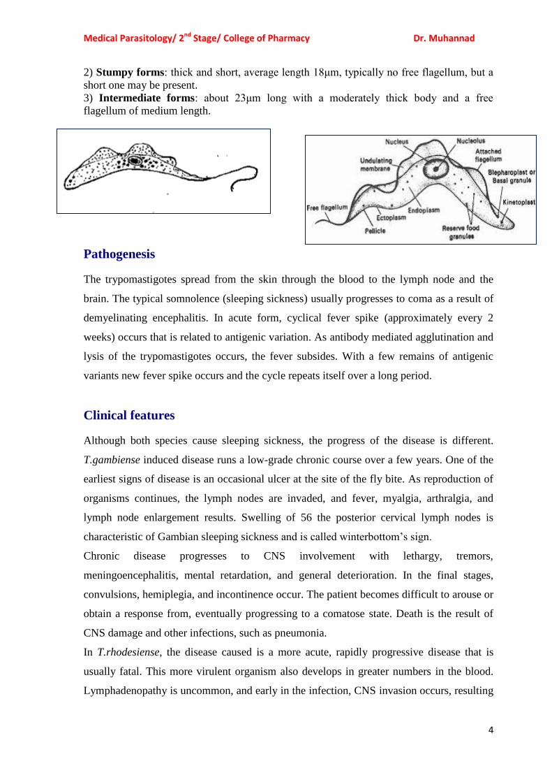
Morphology
The parasite is an elongated cell with single nucleus which usually lies near the centre of
the cell. Each cell bears a single flagellum which appears to arise from a small granule - the
kinetoplast. The kinetoplast is a specialized part of the mitochondria and contains DNA.
The length and position of the trypanosome’s flagellum is variable. In trypanosomes from
the blood of a host the flagellum originates near the posterior end of the cell and passes
forward over the cell surface, its sheath is expanded and forms a wavy flange called an
undulating membrane. Development is characterized by the occurrence of three types of
blood forms (polymorphic), these are:
1) Slender forms: long and thin, about 29μm long, free flagellum.
Medical Parasitology/ 2nd Stage/ College of Pharmacy Dr. Muhannad
4
2) Stumpy forms: thick and short, average length 18μm, typically no free flagellum, but a
short one may be present.
3) Intermediate forms: about 23μm long with a moderately thick body and a free
flagellum of medium length.
Pathogenesis
The trypomastigotes spread from the skin through the blood to the lymph node and the
brain. The typical somnolence (sleeping sickness) usually progresses to coma as a result of
demyelinating encephalitis. In acute form, cyclical fever spike (approximately every 2
weeks) occurs that is related to antigenic variation. As antibody mediated agglutination and
lysis of the trypomastigotes occurs, the fever subsides. With a few remains of antigenic
variants new fever spike occurs and the cycle repeats itself over a long period.
Clinical features
Although both species cause sleeping sickness, the progress of the disease is different.
T.gambiense induced disease runs a low-grade chronic course over a few years. One of the
earliest signs of disease is an occasional ulcer at the site of the fly bite. As reproduction of
organisms continues, the lymph nodes are invaded, and fever, myalgia, arthralgia, and
lymph node enlargement results. Swelling of 56 the posterior cervical lymph nodes is
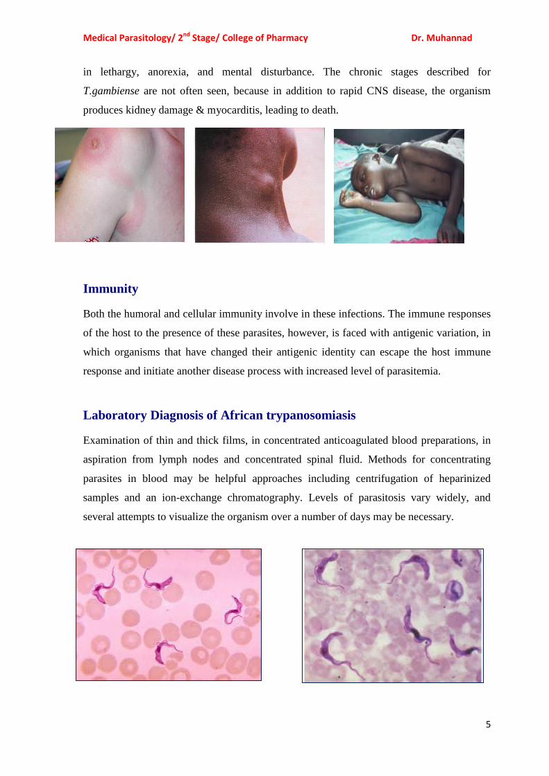
characteristic of Gambian sleeping sickness and is called winterbottom’s sign.
Chronic disease progresses to CNS involvement with lethargy, tremors,
meningoencephalitis, mental retardation, and general deterioration. In the final stages,
convulsions, hemiplegia, and incontinence occur. The patient becomes difficult to arouse or
obtain a response from, eventually progressing to a comatose state. Death is the result of
CNS damage and other infections, such as pneumonia.
In T.rhodesiense, the disease caused is a more acute, rapidly progressive disease that is
usually fatal. This more virulent organism also develops in greater numbers in the blood.
Lymphadenopathy is uncommon, and early in the infection, CNS invasion occurs, resulting
Medical Parasitology/ 2nd Stage/ College of Pharmacy Dr. Muhannad
5
in lethargy, anorexia, and mental disturbance. The chronic stages described for
T.gambiense are not often seen, because in addition to rapid CNS disease, the organism
produces kidney damage & myocarditis, leading to death.
Immunity
Both the humoral and cellular immunity involve in these infections. The immune responses
of the host to the presence of these parasites, however, is faced with antigenic variation, in
which organisms that have changed their antigenic identity can escape the host immune
response and initiate another disease process with increased level of parasitemia.
Laboratory Diagnosis of African trypanosomiasis
Examination of thin and thick films, in concentrated anticoagulated blood preparations, in
aspiration from lymph nodes and concentrated spinal fluid. Methods for concentrating
parasites in blood may be helpful approaches including centrifugation of heparinized
samples and an ion-exchange chromatography. Levels of parasitosis vary widely, and
several attempts to visualize the organism over a number of days may be necessary.
Medical Parasitology/ 2nd Stage/ College of Pharmacy Dr. Muhannad
6
Treatment:
The same treatment protocol is applied for these parasites. For the acute stages of the
disease the drug of choice is suramin with pentamidine as an alternative. In chronic
disease with CNS involvement, the drug of choice is melarsoprol. Alternatives include
trypars amide combined with suramin.
Prevention:
• Control of breeding sites of tsetse flies and use of insecticides.
• Treatment of human cases to reduce transmission to flies.
• Avoiding insect bite by wearing protective clothing & use of screen, bed netting and
insect repellants.
American trypanosomiasis
Trypanosoma cruzi is a pleomorphic trypanosome that includes an additional form of
amastigote in its life cycle. The vector for transmission is reduviid bugs.
Medical Parasitology/ 2nd Stage/ College of Pharmacy Dr. Muhannad
7
Morphology
Trypanosoma cruzi has a single form (monomorphic), about 20μm in length, and
characteristically curved. The kinetoplast is large, considerably larger than the
Trypanosoma brucei species already discussed. They sometimes appear as a bulge at the
posterior end. The flagellum is medium in length.
Pathogenesis
During the acute phase, the organism occurs in blood as a typical trypomastigote and in the
reticuloendothelial cells as a typical amastigote. The amastigotes can kill cells and cause
inflammation, consisting mainly of mononuclear cells. Cardiac muscle is the most
frequently and severely affected tissue. In addition, neuronal damage leads to cardiac
arrhythmias and loss of tone in the colon (megacolon) and esophagus (megaesophagus). In
the chronic phase, the organism persists in the amastigote form.
Clinical features
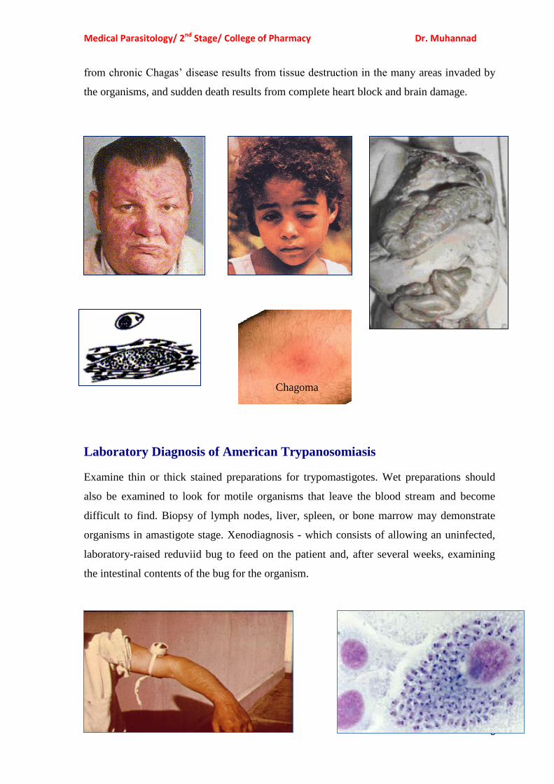
Chagas’ disease may be asymptomatic acute or chronic disease. One of the earliest signs is
development at the site of the bug bite of an erythematous and indurated area called a
chagoma. This is often followed by a rash and edema around the eyes and face; in young
children frequently an acute process with CNS involvement may occur. Acute infection is
also characterized by fever, chills, malaise, myalgia, and fatigue. The chronic Chagas’
disease is characterized by hepatosplenomegaly, myocarditis, and enlargement of the
esophagus and colon as a result of the destruction of nerve cells (E.g. Auerbach’s plexus)
and other tissues that control the growth of these organs. Involvement of the CNS may
produce granulomas in the brain with cyst formation and a meningoencephalitis. Death
Medical Parasitology/ 2nd Stage/ College of Pharmacy Dr. Muhannad
8
from chronic Chagas’ disease results from tissue destruction in the many areas invaded by
the organisms, and sudden death results from complete heart block and brain damage.
Laboratory Diagnosis of American Trypanosomiasis
Examine thin or thick stained preparations for trypomastigotes. Wet preparations should
also be examined to look for motile organisms that leave the blood stream and become
difficult to find. Biopsy of lymph nodes, liver, spleen, or bone marrow may demonstrate
organisms in amastigote stage. Xenodiagnosis - which consists of allowing an uninfected,
laboratory-raised reduviid bug to feed on the patient and, after several weeks, examining
the intestinal contents of the bug for the organism.
Chagoma
Medical Parasitology/ 2nd Stage/ College of Pharmacy Dr. Muhannad
9
Immunity
Unlike African trypanosomiasis, the antigenic variation is less common in T.cruzi
infection. Therefore, the humoral and cellular immune responses function in the immune
system.
Treatment
The drug of choice is nifurtimox. Alternative agents include allopurinol &
benzimidazole.
Prevention
• Bug control, eradication of nests
• Treating infected person & exclusion of donors by screening blood.
• Development of vaccine.
--------------------------------------------------------------------------------------------------------------
Leishmanaiasis:-
Leishmania Species:
Clinical disease
- Veseral leishmaniasis
- Cutaneous leishmaniasis
- Mucocutaneous leishmaniasis
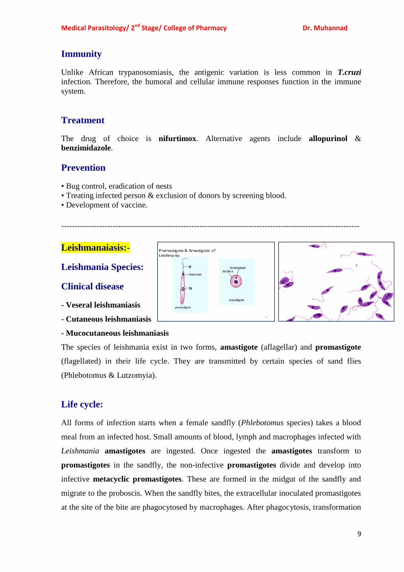
The species of leishmania exist in two forms, amastigote (aflagellar) and promastigote
(flagellated) in their life cycle. They are transmitted by certain species of sand flies
(Phlebotomus & Lutzomyia).
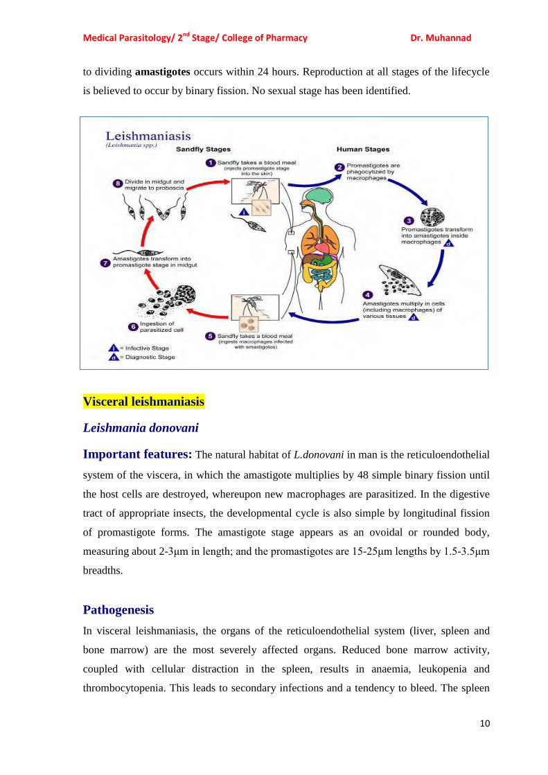
Life cycle:
All forms of infection starts when a female sandfly (Phlebotomus species) takes a blood
meal from an infected host. Small amounts of blood, lymph and macrophages infected with
Leishmania amastigotes are ingested. Once ingested the amastigotes transform to
promastigotes in the sandfly, the non-infective promastigotes divide and develop into
infective metacyclic promastigotes. These are formed in the midgut of the sandfly and
migrate to the proboscis. When the sandfly bites, the extracellular inoculated promastigotes
at the site of the bite are phagocytosed by macrophages. After phagocytosis, transformation
Medical Parasitology/ 2nd Stage/ College of Pharmacy Dr. Muhannad
11
to dividing amastigotes occurs within 24 hours. Reproduction at all stages of the lifecycle
is believed to occur by binary fission. No sexual stage has been identified.
Visceral leishmaniasis
Leishmania donovani
Important features: The natural habitat of L.donovani in man is the reticuloendothelial
system of the viscera, in which the amastigote multiplies by 48 simple binary fission until
the host cells are destroyed, whereupon new macrophages are parasitized. In the digestive
tract of appropriate insects, the developmental cycle is also simple by longitudinal fission
of promastigote forms. The amastigote stage appears as an ovoidal or rounded body,
measuring about 2-3μm in length; and the promastigotes are 15-25μm lengths by 1.5-3.5μm
breadths.
Pathogenesis
In visceral leishmaniasis, the organs of the reticuloendothelial system (liver, spleen and
bone marrow) are the most severely affected organs. Reduced bone marrow activity,
coupled with cellular distraction in the spleen, results in anaemia, leukopenia and
thrombocytopenia. This leads to secondary infections and a tendency to bleed. The spleen
Medical Parasitology/ 2nd Stage/ College of Pharmacy Dr. Muhannad
11
and liver become markedly enlarged, and hypersplenism contributes to the development of
anaemia and lymphadenopathy also occurs. Increased production of globulin results in
hyperglobulinemia, and reversal of the albumin-to-globulin ratio.
Clinical features
Symptoms begin with intermittent fever, weakness, and diarrhea; chills and sweating that
may resemble malaria symptoms are also common early in the infection. As organisms
proliferate & invade cells of the liver and spleen, marked enlargement of the organs, weight
loss, anemia, and emaciation occurs. With persistence of the disease, deeply pigmented,
granulomatous lesion of skin, referred to as post-kala-azar dermal leishmaniasis occurs.
Untreated visceral leishmaniasis is nearly always fatal as a result of secondary infection.
Immunity
Host cellular and humoral defence mechanisms are stimulated.
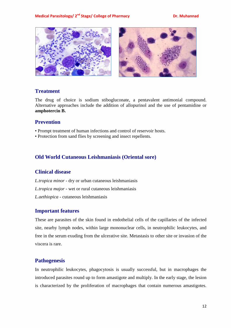
Laboratory diagnosis
• Examination of tissue biopsy, spleen aspiration, bone marrow aspiration or lymph node
aspiration in properly stained smear (e.g. Giemsa stain).
• The amastigotes appear as intracellular & extra cellular L. donovan (LD) bodies.
• Culture of blood, bone marrow, and other tissue often demonstrates the
promastigote stage of the organisms.
• Serologic testing is also available.
Medical Parasitology/ 2nd Stage/ College of Pharmacy Dr. Muhannad
12
Treatment
The drug of choice is sodium stibogluconate, a pentavalent antimonial compound.
Alternative approaches include the addition of allopurinol and the use of pentamidine or
amphotercin B.
Prevention
• Prompt treatment of human infections and control of reservoir hosts.
• Protection from sand flies by screening and insect repellents.
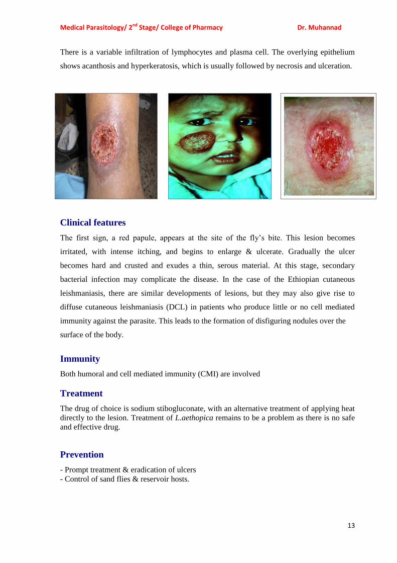
Old World Cutaneous Leishmaniasis (Oriental sore)
Clinical disease
L.tropica minor - dry or urban cutaneous leishmaniasis
L.tropica major - wet or rural cutaneous leishmaniasis
L.aethiopica - cutaneous leishmaniasis
Important features
These are parasites of the skin found in endothelial cells of the capillaries of the infected
site, nearby lymph nodes, within large mononuclear cells, in neutrophilic leukocytes, and
free in the serum exuding from the ulcerative site. Metastasis to other site or invasion of the
viscera is rare.
Pathogenesis
In neutrophilic leukocytes, phagocytosis is usually successful, but in macrophages the
introduced parasites round up to form amastigote and multiply. In the early stage, the lesion
is characterized by the proliferation of macrophages that contain numerous amastigotes.
Medical Parasitology/ 2nd Stage/ College of Pharmacy Dr. Muhannad
13
There is a variable infiltration of lymphocytes and plasma cell. The overlying epithelium
shows acanthosis and hyperkeratosis, which is usually followed by necrosis and ulceration.
Clinical features
The first sign, a red papule, appears at the site of the fly’s bite. This lesion becomes
irritated, with intense itching, and begins to enlarge & ulcerate. Gradually the ulcer
becomes hard and crusted and exudes a thin, serous material. At this stage, secondary
bacterial infection may complicate the disease. In the case of the Ethiopian cutaneous
leishmaniasis, there are similar developments of lesions, but they may also give rise to
diffuse cutaneous leishmaniasis (DCL) in patients who produce little or no cell mediated
immunity against the parasite. This leads to the formation of disfiguring nodules over the
surface of the body.
Immunity
Both humoral and cell mediated immunity (CMI) are involved
Treatment
The drug of choice is sodium stibogluconate, with an alternative treatment of applying heat
directly to the lesion. Treatment of L.aethopica remains to be a problem as there is no safe
and effective drug.
Prevention
- Prompt treatment & eradication of ulcers
- Control of sand flies & reservoir hosts.
Medical Parasitology/ 2nd Stage/ College of Pharmacy Dr. Muhannad
14
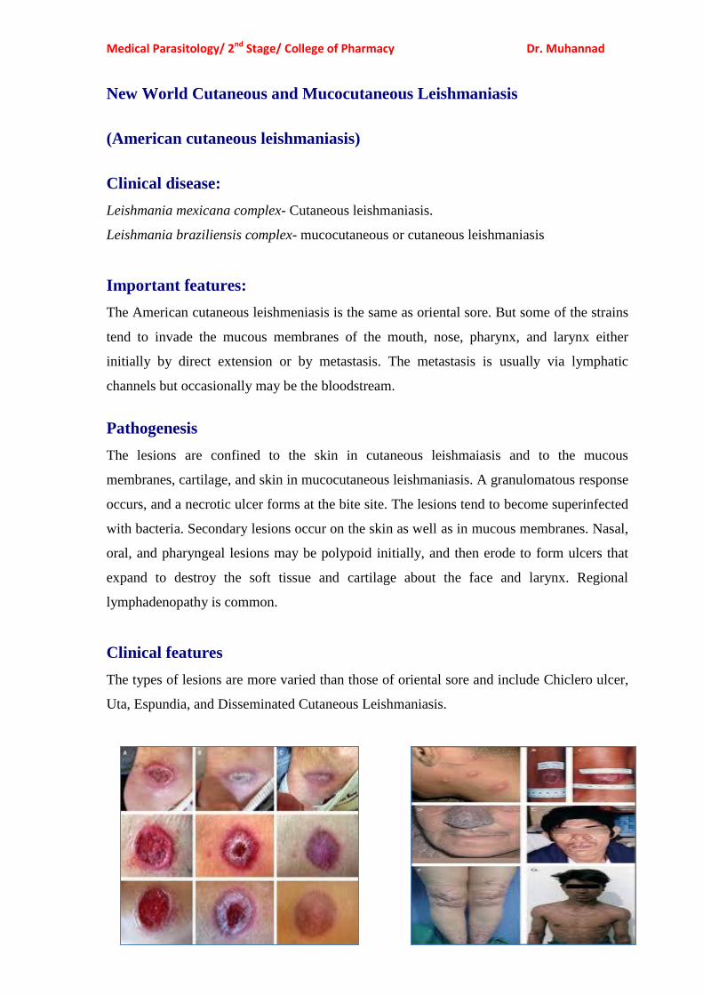
New World Cutaneous and Mucocutaneous Leishmaniasis
(American cutaneous leishmaniasis)
Clinical disease:
Leishmania mexicana complex- Cutaneous leishmaniasis.
Leishmania braziliensis complex- mucocutaneous or cutaneous leishmaniasis
Important features:
The American cutaneous leishmeniasis is the same as oriental sore. But some of the strains
tend to invade the mucous membranes of the mouth, nose, pharynx, and larynx either
initially by direct extension or by metastasis. The metastasis is usually via lymphatic
channels but occasionally may be the bloodstream.
Pathogenesis
The lesions are confined to the skin in cutaneous leishmaiasis and to the mucous
membranes, cartilage, and skin in mucocutaneous leishmaniasis. A granulomatous response
occurs, and a necrotic ulcer forms at the bite site. The lesions tend to become superinfected
with bacteria. Secondary lesions occur on the skin as well as in mucous membranes. Nasal,
oral, and pharyngeal lesions may be polypoid initially, and then erode to form ulcers that
expand to destroy the soft tissue and cartilage about the face and larynx. Regional
lymphadenopathy is common.
Clinical features
The types of lesions are more varied than those of oriental sore and include Chiclero ulcer,
Uta, Espundia, and Disseminated Cutaneous Leishmaniasis.
Medical Parasitology/ 2nd Stage/ College of Pharmacy Dr. Muhannad
15
Laboratory diagnosis
• Demonstration of the amastigotes in properly stained smears from touch preparations of
ulcer biopsy specimen.
• Serological tests based on fluorescent antibody tests.
• Leishman skin test in some species.
Immunity
The humoral and cellular immune systems are involved
Treatment
The drug of choice is sodium stibogluconate.
Prevention
• Avoiding endemic areas especially during times when local vectors are most active.
• Prompt treatment of infected individuals.
Dr. Muhannad Shweash
PhD, Post-doctorate in Clinical Pathological Analyses
United Kingdom (UK)
Medical Parasitology/ Second Year