Embed Size (px)
Citation preview
American Thoracic Society Documents
Diagnosis and Initial Management of NonmalignantDiseases Related to AsbestosThis official statement of the American Thoracic Society was adopted by the ATS Board of Directorson December 12, 2003
CONTENTS
Diagnostic Criteria and Guidelines for Documenting ThemAsbestos as a HazardAsbestos in Lung Tissue
Clinical Evaluation and IndicatorsSymptomsOccupational and Environmental HistoryPhysical ExaminationConventional ImagingComputed TomographyBronchoalveolar LavagePulmonary Function Tests
Nonmalignant Disease OutcomesAsbestosisNonmalignant Pleural Abnormalities Associated
with AsbestosChronic Airway Obstruction
Implications of Diagnosis for Patient ManagementActions Required before Disease Is ApparentActions Required after Diagnosis
Conclusions
Asbestos is a general term for a heterogeneous group of hydratedmagnesium silicate minerals that have in common a tendencyto separate into fibers (1). These fibers, inhaled and displacedby various means to lung tissue, can cause a spectrum of diseasesincluding cancer and disorders related to inflammation and fi-brosis. Asbestos has been the largest single cause of occupationalcancer in the United States and a significant cause of diseaseand disability from nonmalignant disease. To this demonstrableburden of asbestos-related disease is added the burden of publicconcern and fear regarding risk after minimal exposure.
This statement presents guidance for the diagnosis of nonma-lignant asbestos-related disease. Nonmalignant asbestos-relateddisease refers to the following conditions: asbestosis, pleuralthickening or asbestos-related pleural fibrosis (plaques or diffusefibrosis), “benign” (nonmalignant) pleural effusion, and airflowobstruction. This document is intended to assist the clinician inmaking a diagnosis that will be the basis for individual manage-ment of the patient. It therefore provides overarching criteriafor the diagnosis, specific guidelines for satisfying these criteria,and descriptions of the clinical implications of the diagnosis,including the basic management plan that should be triggeredby the diagnosis. It is understood that disease may be present
Members of the Ad Hoc Statement Committee have disclosed any direct commer-cial associations (financial relationships or legal obligations) related to the prepara-tion of this statement. This information is kept on file at the ATS headquarters.
Am J Respir Crit Care Med Vol 170. pp 691–715, 2004DOI: 10.1164/rccm.200310-1436STInternet address: www.atsjournals.org
at a subclinical level and may not be sufficiently advanced to beapparent on histology, imaging, or functional studies.
One of the most important implications of the diagnosis ofnonmalignant asbestos-related disease is that there is a closecorrelation between the presence of nonmalignant disease andthe risk of malignancy, which may arise from exposure levelsrequired to produce nonmalignant disease or mechanisms sharedwith premalignant processes that lead to cancer. The major ma-lignancies associated with asbestos are cancer of the lung (witha complex relationship to cigarette smoking) and mesothelioma(pleural or peritoneal), with excess risk also reported for othersites. There is a strong statistical association between asbestos-related disease and malignancy, but the majority of patients withnonmalignant asbestos-related disease do not develop cancer.On the other hand, the risk of cancer may be elevated in aperson exposed to asbestos without obvious signs of nonmalig-nant asbestos-related disease. However, a diagnosis of nonmalig-nant asbestos-related disease does imply a lifelong elevated riskfor asbestos-related cancer.
DIAGNOSTIC CRITERIA AND GUIDELINES FORDOCUMENTING THEM
People with past exposure to asbestos consult physicians formany relevant reasons: to be screened for asbestos-related dis-ease, for evaluation of specific symptoms that may relate to pastasbestos exposure (known or unsuspected), for treatment andadvice, and for evaluation of impairment. In 1986, the AmericanThoracic Society convened a group of experts to review theliterature and to present an authoritative consensus view of thecurrent state of knowledge with respect to diagnosis of nonmalig-nant disease related to asbestos (2). In 2001, a new group wasconvened to review and to update the 1986 criteria. This state-ment constitutes that committee’s report, completed in 2004.
The criteria formulated in this statement are intended for thediagnosis of nonmalignant asbestos-related disease in an individ-ual in a clinical setting for the purpose of managing that person’scurrent condition and future health. These general criteria areslightly modified from those presented in 1986 (Table 1) (2):
• Evidence of structural pathology consistent with asbestos-related disease as documented by imaging or histology
• Evidence of causation by asbestos as documented by theoccupational and environmental history, markers of expo-sure (usually pleural plaques), recovery of asbestos bodies,or other means
• Exclusion of alternative plausible causes for the findings
The rest of this statement is largely devoted to presentingclinical guidelines required to document that each of these crite-ria is met. Demonstration of functional impairment is not re-quired for the diagnosis of a nonmalignant asbestos-related dis-ease, but where present should be documented as part of thecomplete evaluation. Evaluation of impairment has been exten-
692 AMERICAN JOURNAL OF RESPIRATORY AND CRITICAL CARE MEDICINE VOL 170 2004
TABLE 1. CRITERIA FOR DIAGNOSIS OF NONMALIGNANT LUNG DISEASE RELATED TO ASBESTOS
1986 Guidelines 2004 Guidelines Comparison and Notes
Evidence of structural change, as Demonstrates the existence of a structural lesion consistentdemonstrated by one or more of the with the effects of asbestos. The criteria outlined in the 1986following: guidelines were most explicit for asbestosis
Chest film (irregular opacities) • Imaging methods Chest film, HRCT, and possibly future methods based onimaging. The 1986 guidelines specified ILO classification 1/1
Pathology (College of American • Histology (College of American Criteria for identifying asbestosis on microscopic examinationPathologists) Pathologists) of tissue are unchanged
Consistent time interval Evidence of plausible causation, as Evidence of plausible causation implies that the temporaldemonstrated by one or more of the relationship, including latency, is plausiblefollowing:
Occupational and environmental history • Occupational and environmental history ofexposure (with plausible latency)
• Markers of exposure (e.g., pleural plaques)Asbestos bodies or fibers in lung tissue • Recovery of asbestos bodies The 2004 guidelines are not limited to lung tissue, consider
the role of BAL to be established, and deemphasize fibersbecause they are difficult to detect and a systematic analysisfor asbestos fibers is not generally available
Rule out other causes of interstitial fibrosis Exclusion of alternative diagnoses The 1986 guidelines primarily addressed asbestosis butor obstructive disease mentioned smoking as a cause of obstructive disease.
Implicit in the article, however, is that nonmalignantdiseases presenting similarly to asbestos-related diseaseshould also be ruled out
“Evidence of abnormal test” Evidence of functional impairment, as Functional assessment is not required for diagnosis but is partdemonstrated by one or more of the of a complete evaluation. It contributes to diagnosis infollowing: defining the activity of disease and the resulting impairment
Crackles, bilateral, not cleared by cough • Signs and symptoms (including crackles) Signs and symptoms are not specific for diagnosis but arevaluable in assessing impairment
Restrictive disease • Change in ventilatory function (restrictive, The 1986 criteria admitted the possibility of obstructiveobstructive patterns in context or disease disease; the 2004 criteria address this specificallyhistory)
Reduced diffusing capacity • Impaired gas exchange (e.g., reduceddiffusing capacity)
• Inflammation (e.g., by bronchoalveolar The 1986 guidelines noted possible utility of bronchoalveolarlavage) lavage and gallium scanning but considered them to be
experimental techniques. The 2004 guidelines excludegallium scanning, suggest that additional indicators ofactive inflammation may become useful in future
• Exercise testing
Definition of abbreviations: BAL � bronchoalveolar lavage; HRCT � high-resolution computed tomography; ILO � International Labour Organization.From References 64 and 65.
sively reviewed elsewhere and is not repeated here (3). Func-tional impairment may be demonstrated by evidence of symp-toms or signs, ventilatory dysfunction, impaired gas exchange,and inflammation. Pulmonary function testing should be con-ducted in conformity with standards already published by theAmerican Thoracic Society (4, 5), including multiple trials to con-firm reproducibility and documentation of all trials attempted.
These guidelines are designed for clinical application, notfor research, epidemiologic surveillance, screening, litigation, oradjudication. They balance the need to be as accurate as possiblewith protection of the patient’s safety and the yield, cost, andaccessibility of the diagnostic procedures available. These guide-lines, if they err, err on the side of specificity rather than sensitiv-ity. This is because nonmalignant asbestos-related disorders aredifficult to detect in their earliest stages and because there is noearly intervention that has been proven to alter the subsequentevolution of the disease. On the other hand, the documentationof causation by asbestos carries important implications for thepatient and can be established with reasonable certainty, oncethe disease is identified.
Asbestos as a Hazard
The generic term “asbestos” is used to describe a group ofminerals that, when crushed, break into fibers. As defined by
the National Research Council (1), the term “asbestos” is a“commercial-industrial term rather than a mineralogical term.It refers to well-developed and hair-like long-fibered varietiesof certain minerals that satisfy particular industrial needs.” Theyare chemically heterogeneous hydrated silicates and each haschemical analogs with different structures that do not form fibers.Fibers have parallel sides with length three or more times greaterthan width. Asbestos fibers have great tensile strength, heatresistance, and acid resistance; varieties are also flexible. Thesix minerals that are traditionally defined as asbestos includechrysotile asbestos (the asbestiform variety of serpentine); theamphiboles, which include crocidolite (the asbestiform varietyof riebeckite) and amosite (the asbestiform variety of cumming-tonite-grunerite); and the asbestiform varieties of the amphi-boles, which include anthophyllite (anthophyllite asbestos), ac-tinolite (actinolite asbestos), and tremolite (tremolite asbestos)(6). Just as all forms of asbestos, by the definition and classifica-tion above, appear to cause malignancy, all may cause the non-malignant diseases described. Issues of relative potency amongthe forms of asbestos, and particularly between chrysotile andthe amphiboles, are primarily of concern with respect to the riskof malignancy and are not discussed in this document.
Commercial-grade asbestos is made up of fiber bundles.These bundles, in turn, are composed of extremely long and thinfibers, often with splayed ends, that can easily be separated from

American Thoracic Society Documents 693
one another. Commercial asbestos has high tensile strength,flexibility, resistance to chemical and thermal degradation, andhigh electrical resistance, and can often be woven. On the basisof these characteristics, asbestos was broadly used in the pastin insulation, brake linings, flooring, cement, paint, textiles, andmany other products; however, commercial use has declinedsubstantially in more recent years.
Asbestos and asbestiform minerals may occur as a naturalaccessory mineral in other industrial mineral deposits or rocks.These asbestiform amphiboles and some other fibrous mineralsmay not completely fit the commercial definition of asbestos butmay have similar effects, such as the tremolite-like asbestiformmineral found in association with vermiculite in Libby, Montana(7). Although the general criteria still apply, the specific diagnos-tic guidelines provided in this statement may or may not applyin such situations, depending on the mineral and exposure cir-cumstances. Documentation of health effects in the scientificliterature for these minerals is not as extensive as for chrysotileand the common amphiboles.
World production and use of asbestos climbed steadily sinceits commercial introduction in the late nineteenth century andfell rapidly after documentation of its hazards in the 1970s and1980s. In Western industrialized countries, the widespread useof asbestos in industry and in the built environment in the firstseven decades of the twentieth century has resulted in an epi-demic of asbestos-related illness that now continues into thetwenty-first century, despite decline in global production anduse. Its use has now been banned in many Western countries.Asbestos is still mined in Russia and China, mainly for localuse, and in Canada, where most of the product is exported toAsia and Africa.
Today, with stringent regulation of asbestos use and the disap-pearance of almost all asbestos-containing products from themarket, nonmalignant asbestos-related disease is primarily aconcern in four settings in the developed world: (1) the historicallegacy of asbestos exposure affecting older workers; (2) thecurrent risk experienced by the workforce engaged in certainoccupations managing the remaining hazard, such as buildingand facility maintenance; (3) asbestos abatement operations,removing insulation and other asbestos-containing products; and(4) renovation and demolition of structures containing asbestos.In the developing world, workers and their families continue tobe exposed. In some countries, including industrialized countriesformerly belonging to the Eastern bloc and rapidly industrializ-ing countries in Asia, the use of asbestos continues and mayeven be increasing.
Asbestos is still a hazard for an estimated 1.3 million workersin the construction industry in the United States and for workersinvolved in maintenance of buildings and equipment (8). Mostasbestos in the United States today exists in building and machin-ery insulation and old products, such as appliances, that may beavailable for resale. New products that may contain asbestostoday in the United States include friction surfaces (brake pads),roofing materials, vinyl tile, and imported cement pipe and sheet-ing. Significant asbestos content may be present as a contaminantin vermiculite insulation often found in homes (7).
Historically, occupations at greatest risk for nonmalignantasbestos-related disease have tended to be those engaged in theproduction and end use of products made from asbestos. Thesehave included a wide assortment of items, including friction pads,brake linings, gas masks, cement water pipe, insulation, andtextiles. Occupations engaged in the mining and extraction ofasbestos have usually shown lower frequencies of nonmalignantasbestos-related disease. Passive exposure, including workerscarrying home asbestos on their clothing, was historically associ-ated with elevated cancer risk, particularly mesothelioma, and
risk of nonmalignant asbestos-related disease. Workers in build-ing and equipment maintenance may still encounter asbestosinsulation even though asbestos is no longer widely used incommerce. Asbestos abatement activities, including removal andreplacement of insulation, provide opportunities for exposureamong contemporary workers (8).
Asbestos in Lung Tissue
Asbestos fibers carried to the deep lung induce an alveolitis thatresults in fibrosis. Inhaled asbestos fibers can also result in pleuralinflammation. Asbestos fibers are transported to the pleural sur-face along lymphatic channels by macrophages and/or by directpenetration. The degree of fibrosis in asbestosis is dose depen-dent (9–12).
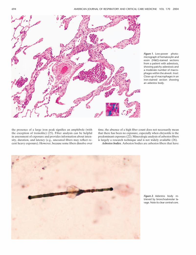
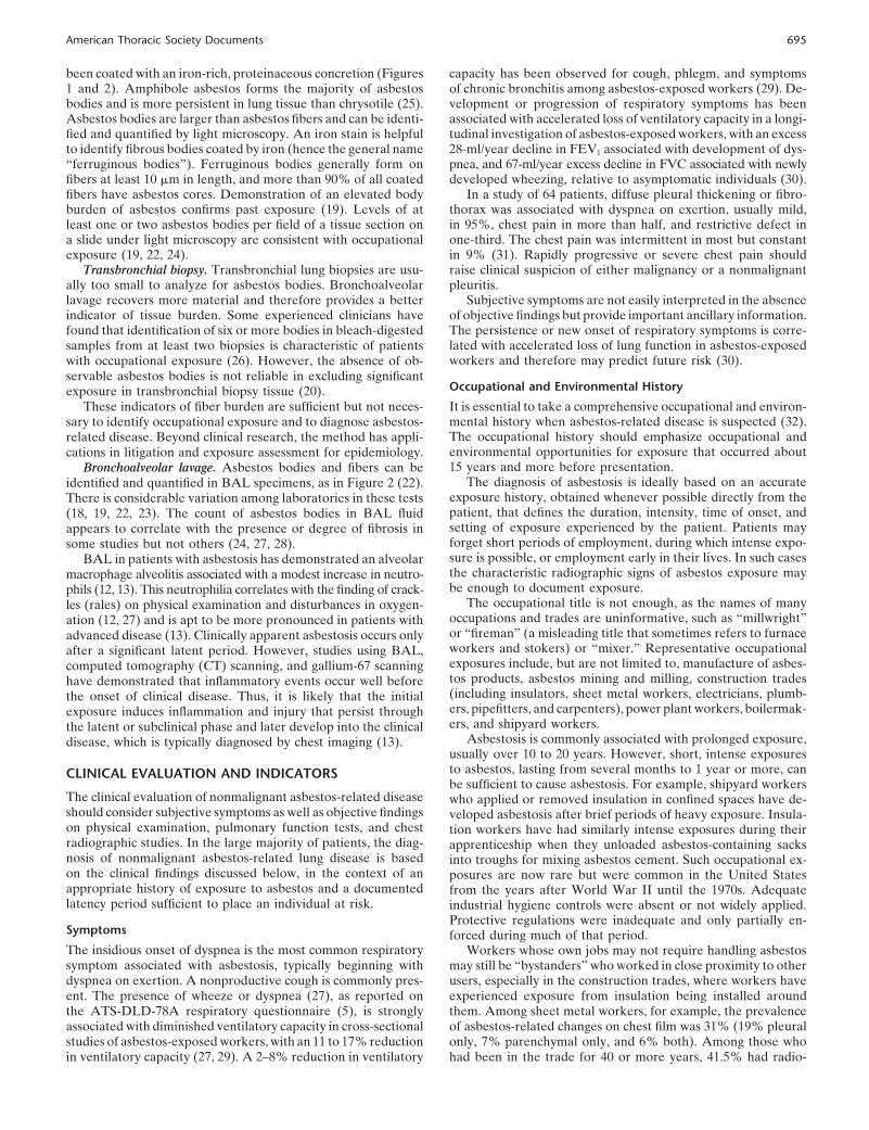
Asbestos fibers are deposited at airway bifurcations and inrespiratory bronchioles and alveoli primarily by impaction andinterception. Fibers migrate into the interstitium, in part via anuptake process involving Type I alveolar epithelial cells. Thiscauses an alveolar macrophage–dominated alveolitis, as demon-strated in Figure 1 (12, 13). Thereafter, many of the fibers arecleared.
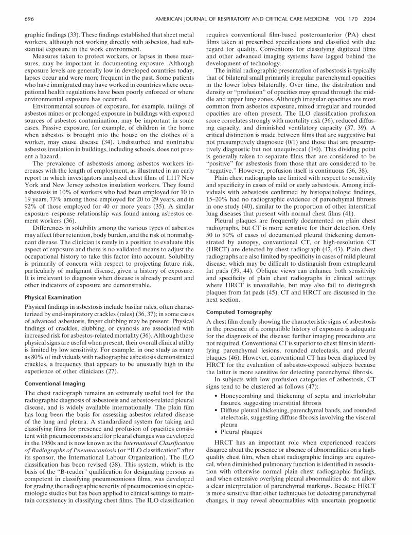
Activated macrophages are stimulated to engulf and removeasbestos fibers. This process is not uniformly successful, however,and many fibers are retained (9, 10). The long fibers cannot becompletely engulfed by the macrophage, as demonstrated inFigure 2.
Chrysotile fibers also split longitudinally, creating additionalfibrils. These are cleared more efficiently than amphibole asbes-tos fibers, which may be retained indefinitely (12). The fibersinduce apoptosis, a form of controlled cell death, in the macro-phage and stimulate inflammation. This effect is reduced oncethe fiber is coated to create an asbestos body, but the greatmajority of fibers in the lung remain uncoated. For these reasons,asbestos has a prolonged residence in the lung, penetrates theinterstitium of the distal lung, and shows extensive mobility bothin the lung and around the body (9).
Asbestos fibers, in particular, stimulate macrophages to pro-duce a variety of mediators. Oxygen radicals contribute to tissueinjury. Granulocytes are recruited to sites of disease activity andthey in turn release mediators that contribute to tissue fibrosisby stimulating fibroblast proliferation and chemotaxis and ulti-mately promoting collagen synthesis (11–15).
The inflammatory processes induced by asbestos include alve-olitis, inflammation in the surrounding interstitium, and inflam-mation followed by fibrotic change in the respiratory bronchiolesthat extends into adjacent alveolar tissue (11, 14, 16). Studiesof the lung tissue of asbestos-exposed workers, including non-smokers, have demonstrated a form of peribronchiolitis involv-ing the walls of membranous and respiratory bronchioles, thatshows characteristics of a more intense fibrotic response thanthe small airway lesions caused by nonspecific mineral dusts thatthe lesions otherwise resemble (17, 18).
Asbestos fibers and their derivatives, asbestos bodies, canbe identified and quantified in lung tissue and bronchoalveolarlavage (BAL) specimens, as demonstrated in Figure 2 (19).Transbronchial lung biopsy is less reliable than BAL or openlung biopsy in recovering sufficient tissue to demonstrate ele-vated asbestos body or fiber counts when they do occur (20).
Asbestos fibers, unlike asbestos bodies, are rarely seen bylight microscopy and must be analyzed by scanning/transmissionelectron microscopy (19, 21, 22). There is considerable variationamong laboratories in procedures to quantify asbestos fibers intissue (18, 23, 24), which has led to efforts to standardize proce-dures (19). Asbestos mineralogical types can be identified byenergy-dispersive X-ray analysis, in which detection of magne-sium and silicon is characteristic of most forms of asbestos and
694 AMERICAN JOURNAL OF RESPIRATORY AND CRITICAL CARE MEDICINE VOL 170 2004
Figure 1. Low-power photo-micrograph of hematoxylin andeosin (H&E)-stained sectionsfrom a patient with asbestosis,showing patchy asbestosis anda moderate number of macro-phages within the alveoli. Inset:Close-up of macrophages in aniron-stained section showingan asbestos body.
the presence of a large iron peak signifies an amphibole (withthe exception of tremolite) (25). Fiber analysis can be helpfulin assessment of exposure and provides information about inten-sity, duration, and latency (e.g., uncoated fibers may reflect re-cent heavy exposure). However, because some fibers dissolve over
Figure 2. Asbestos body re-trieved by bronchoalveolar la-vage. Note its clear central core.
time, the absence of a high fiber count does not necessarily meanthat there has been no exposure, especially when chrysotile is thepredominant exposure (22). Mineralogic analysis of asbestos fibersis largely a research technique and is not widely available (26).
Asbestos bodies. Asbestos bodies are asbestos fibers that have
American Thoracic Society Documents 695
been coated with an iron-rich, proteinaceous concretion (Figures1 and 2). Amphibole asbestos forms the majority of asbestosbodies and is more persistent in lung tissue than chrysotile (25).Asbestos bodies are larger than asbestos fibers and can be identi-fied and quantified by light microscopy. An iron stain is helpfulto identify fibrous bodies coated by iron (hence the general name“ferruginous bodies”). Ferruginous bodies generally form onfibers at least 10 �m in length, and more than 90% of all coatedfibers have asbestos cores. Demonstration of an elevated bodyburden of asbestos confirms past exposure (19). Levels of atleast one or two asbestos bodies per field of a tissue section ona slide under light microscopy are consistent with occupationalexposure (19, 22, 24).
Transbronchial biopsy. Transbronchial lung biopsies are usu-ally too small to analyze for asbestos bodies. Bronchoalveolarlavage recovers more material and therefore provides a betterindicator of tissue burden. Some experienced clinicians havefound that identification of six or more bodies in bleach-digestedsamples from at least two biopsies is characteristic of patientswith occupational exposure (26). However, the absence of ob-servable asbestos bodies is not reliable in excluding significantexposure in transbronchial biopsy tissue (20).
These indicators of fiber burden are sufficient but not neces-sary to identify occupational exposure and to diagnose asbestos-related disease. Beyond clinical research, the method has appli-cations in litigation and exposure assessment for epidemiology.
Bronchoalveolar lavage. Asbestos bodies and fibers can beidentified and quantified in BAL specimens, as in Figure 2 (22).There is considerable variation among laboratories in these tests(18, 19, 22, 23). The count of asbestos bodies in BAL fluidappears to correlate with the presence or degree of fibrosis insome studies but not others (24, 27, 28).
BAL in patients with asbestosis has demonstrated an alveolarmacrophage alveolitis associated with a modest increase in neutro-phils (12, 13). This neutrophilia correlates with the finding of crack-les (rales) on physical examination and disturbances in oxygen-ation (12, 27) and is apt to be more pronounced in patients withadvanced disease (13). Clinically apparent asbestosis occurs onlyafter a significant latent period. However, studies using BAL,computed tomography (CT) scanning, and gallium-67 scanninghave demonstrated that inflammatory events occur well beforethe onset of clinical disease. Thus, it is likely that the initialexposure induces inflammation and injury that persist throughthe latent or subclinical phase and later develop into the clinicaldisease, which is typically diagnosed by chest imaging (13).
CLINICAL EVALUATION AND INDICATORS
The clinical evaluation of nonmalignant asbestos-related diseaseshould consider subjective symptoms as well as objective findingson physical examination, pulmonary function tests, and chestradiographic studies. In the large majority of patients, the diag-nosis of nonmalignant asbestos-related lung disease is basedon the clinical findings discussed below, in the context of anappropriate history of exposure to asbestos and a documentedlatency period sufficient to place an individual at risk.
Symptoms
The insidious onset of dyspnea is the most common respiratorysymptom associated with asbestosis, typically beginning withdyspnea on exertion. A nonproductive cough is commonly pres-ent. The presence of wheeze or dyspnea (27), as reported onthe ATS-DLD-78A respiratory questionnaire (5), is stronglyassociated with diminished ventilatory capacity in cross-sectionalstudies of asbestos-exposed workers, with an 11 to 17% reductionin ventilatory capacity (27, 29). A 2–8% reduction in ventilatory
capacity has been observed for cough, phlegm, and symptomsof chronic bronchitis among asbestos-exposed workers (29). De-velopment or progression of respiratory symptoms has beenassociated with accelerated loss of ventilatory capacity in a longi-tudinal investigation of asbestos-exposed workers, with an excess28-ml/year decline in FEV1 associated with development of dys-pnea, and 67-ml/year excess decline in FVC associated with newlydeveloped wheezing, relative to asymptomatic individuals (30).
In a study of 64 patients, diffuse pleural thickening or fibro-thorax was associated with dyspnea on exertion, usually mild,in 95%, chest pain in more than half, and restrictive defect inone-third. The chest pain was intermittent in most but constantin 9% (31). Rapidly progressive or severe chest pain shouldraise clinical suspicion of either malignancy or a nonmalignantpleuritis.
Subjective symptoms are not easily interpreted in the absenceof objective findings but provide important ancillary information.The persistence or new onset of respiratory symptoms is corre-lated with accelerated loss of lung function in asbestos-exposedworkers and therefore may predict future risk (30).
Occupational and Environmental History
It is essential to take a comprehensive occupational and environ-mental history when asbestos-related disease is suspected (32).The occupational history should emphasize occupational andenvironmental opportunities for exposure that occurred about15 years and more before presentation.
The diagnosis of asbestosis is ideally based on an accurateexposure history, obtained whenever possible directly from thepatient, that defines the duration, intensity, time of onset, andsetting of exposure experienced by the patient. Patients mayforget short periods of employment, during which intense expo-sure is possible, or employment early in their lives. In such casesthe characteristic radiographic signs of asbestos exposure maybe enough to document exposure.
The occupational title is not enough, as the names of manyoccupations and trades are uninformative, such as “millwright”or “fireman” (a misleading title that sometimes refers to furnaceworkers and stokers) or “mixer.” Representative occupationalexposures include, but are not limited to, manufacture of asbes-tos products, asbestos mining and milling, construction trades(including insulators, sheet metal workers, electricians, plumb-ers, pipefitters, and carpenters), power plant workers, boilermak-ers, and shipyard workers.
Asbestosis is commonly associated with prolonged exposure,usually over 10 to 20 years. However, short, intense exposuresto asbestos, lasting from several months to 1 year or more, canbe sufficient to cause asbestosis. For example, shipyard workerswho applied or removed insulation in confined spaces have de-veloped asbestosis after brief periods of heavy exposure. Insula-tion workers have had similarly intense exposures during theirapprenticeship when they unloaded asbestos-containing sacksinto troughs for mixing asbestos cement. Such occupational ex-posures are now rare but were common in the United Statesfrom the years after World War II until the 1970s. Adequateindustrial hygiene controls were absent or not widely applied.Protective regulations were inadequate and only partially en-forced during much of that period.
Workers whose own jobs may not require handling asbestosmay still be “bystanders” who worked in close proximity to otherusers, especially in the construction trades, where workers haveexperienced exposure from insulation being installed aroundthem. Among sheet metal workers, for example, the prevalenceof asbestos-related changes on chest film was 31% (19% pleuralonly, 7% parenchymal only, and 6% both). Among those whohad been in the trade for 40 or more years, 41.5% had radio-
696 AMERICAN JOURNAL OF RESPIRATORY AND CRITICAL CARE MEDICINE VOL 170 2004
graphic findings (33). These findings established that sheet metalworkers, although not working directly with asbestos, had sub-stantial exposure in the work environment.
Measures taken to protect workers, or lapses in these mea-sures, may be important in documenting exposure. Althoughexposure levels are generally low in developed countries today,lapses occur and were more frequent in the past. Some patientswho have immigrated may have worked in countries where occu-pational health regulations have been poorly enforced or whereenvironmental exposure has occurred.
Environmental sources of exposure, for example, tailings ofasbestos mines or prolonged exposure in buildings with exposedsources of asbestos contamination, may be important in somecases. Passive exposure, for example, of children in the homewhen asbestos is brought into the house on the clothes of aworker, may cause disease (34). Undisturbed and nonfriableasbestos insulation in buildings, including schools, does not pres-ent a hazard.
The prevalence of asbestosis among asbestos workers in-creases with the length of employment, as illustrated in an earlyreport in which investigators analyzed chest films of 1,117 NewYork and New Jersey asbestos insulation workers. They foundasbestosis in 10% of workers who had been employed for 10 to19 years, 73% among those employed for 20 to 29 years, and in92% of those employed for 40 or more years (35). A similarexposure–response relationship was found among asbestos ce-ment workers (36).
Differences in solubility among the various types of asbestosmay affect fiber retention, body burden, and the risk of nonmalig-nant disease. The clinician is rarely in a position to evaluate thisaspect of exposure and there is no validated means to adjust theoccupational history to take this factor into account. Solubilityis primarily of concern with respect to projecting future risk,particularly of malignant disease, given a history of exposure.It is irrelevant to diagnosis when disease is already present andother indicators of exposure are demonstrable.
Physical Examination
Physical findings in asbestosis include basilar rales, often charac-terized by end-inspiratory crackles (rales) (36, 37); in some casesof advanced asbestosis, finger clubbing may be present. Physicalfindings of crackles, clubbing, or cyanosis are associated withincreased risk for asbestos-related mortality (36). Although thesephysical signs are useful when present, their overall clinical utilityis limited by low sensitivity. For example, in one study as manyas 80% of individuals with radiographic asbestosis demonstratedcrackles, a frequency that appears to be unusually high in theexperience of other clinicians (27).
Conventional Imaging
The chest radiograph remains an extremely useful tool for theradiographic diagnosis of asbestosis and asbestos-related pleuraldisease, and is widely available internationally. The plain filmhas long been the basis for assessing asbestos-related diseaseof the lung and pleura. A standardized system for taking andclassifying films for presence and profusion of opacities consis-tent with pneumoconiosis and for pleural changes was developedin the 1950s and is now known as the International Classificationof Radiographs of Pneumoconiosis (or “ILO classification” afterits sponsor, the International Labour Organization). The ILOclassification has been revised (38). This system, which is thebasis of the “B-reader” qualification for designating persons ascompetent in classifying pneumoconiosis films, was developedfor grading the radiographic severity of pneumoconiosis in epide-miologic studies but has been applied to clinical settings to main-tain consistency in classifying chest films. The ILO classification
requires conventional film-based posteroanterior (PA) chestfilms taken at prescribed specifications and classified with dueregard for quality. Conventions for classifying digitized filmsand other advanced imaging systems have lagged behind thedevelopment of technology.
The initial radiographic presentation of asbestosis is typicallythat of bilateral small primarily irregular parenchymal opacitiesin the lower lobes bilaterally. Over time, the distribution anddensity or “profusion” of opacities may spread through the mid-dle and upper lung zones. Although irregular opacities are mostcommon from asbestos exposure, mixed irregular and roundedopacities are often present. The ILO classification profusionscore correlates strongly with mortality risk (36), reduced diffus-ing capacity, and diminished ventilatory capacity (37, 39). Acritical distinction is made between films that are suggestive butnot presumptively diagnostic (0/1) and those that are presump-tively diagnostic but not unequivocal (1/0). This dividing pointis generally taken to separate films that are considered to be“positive” for asbestosis from those that are considered to be“negative.” However, profusion itself is continuous (36, 38).
Plain chest radiographs are limited with respect to sensitivityand specificity in cases of mild or early asbestosis. Among indi-viduals with asbestosis confirmed by histopathologic findings,15–20% had no radiographic evidence of parenchymal fibrosisin one study (40), similar to the proportion of other interstitiallung diseases that present with normal chest films (41).
Pleural plaques are frequently documented on plain chestradiographs, but CT is more sensitive for their detection. Only50 to 80% of cases of documented pleural thickening demon-strated by autopsy, conventional CT, or high-resolution CT(HRCT) are detected by chest radiograph (42, 43). Plain chestradiographs are also limited by specificity in cases of mild pleuraldisease, which may be difficult to distinguish from extrapleuralfat pads (39, 44). Oblique views can enhance both sensitivityand specificity of plain chest radiographs in clinical settingswhere HRCT is unavailable, but may also fail to distinguishplaques from fat pads (45). CT and HRCT are discussed in thenext section.
Computed Tomography
A chest film clearly showing the characteristic signs of asbestosisin the presence of a compatible history of exposure is adequatefor the diagnosis of the disease: further imaging procedures arenot required. Conventional CT is superior to chest films in identi-fying parenchymal lesions, rounded atelectasis, and pleuralplaques (46). However, conventional CT has been displaced byHRCT for the evaluation of asbestos-exposed subjects becausethe latter is more sensitive for detecting parenchymal fibrosis.
In subjects with low profusion categories of asbestosis, CTsigns tend to be clustered as follows (47):
• Honeycombing and thickening of septa and interlobularfissures, suggesting interstitial fibrosis
• Diffuse pleural thickening, parenchymal bands, and roundedatelectasis, suggesting diffuse fibrosis involving the visceralpleura
• Pleural plaques
HRCT has an important role when experienced readersdisagree about the presence or absence of abnormalities on a high-quality chest film, when chest radiographic findings are equivo-cal, when diminished pulmonary function is identified in associa-tion with otherwise normal plain chest radiographic findings,and when extensive overlying pleural abnormalities do not allowa clear interpretation of parenchymal markings. Because HRCTis more sensitive than other techniques for detecting parenchymalchanges, it may reveal abnormalities with uncertain prognostic
American Thoracic Society Documents 697
significance. HRCT is more specific than plain chest radiographs,excluding conditions such as emphysema, vessel prominence,overlying pleural disease, and bronchiectasis, which may confoundradiographic interpretation.
HRCT is much more sensitive in the detection of asbestosisthan plain chest radiographs (46, 48), although even a normalHRCT cannot completely exclude asbestosis (49). Among asbes-tos-exposed individuals with unremarkable chest radiographicfindings (ILO score 0/0 or 0/1), 34% were identified by HRCTas having findings suggestive of asbestosis. HRCT findings alsocorrelated with decrements in pulmonary function tests in thesecases, with a significantly diminished vital capacity and diffusingcapacity (50).
HRCT can detect early pleural thickening (i.e., 1–2 mm inthickness) much more sensitively than plain chest radiographs.Pleural thickening is frequently discontinuous and interspersedwith normal regions. It is usually bilateral but may be unilateralin a third of cases (48). HRCT also offers an advantage overplain chest radiographs in specificity, being able to distinguishpleural disease from extrapleural fat (51).
HRCT should be obtained at 2-cm intervals, to allow a moreaccurate assessment of pleural abnormalities, as well as otherabnormal findings such as pulmonary masses (52). Prone viewsshould always be obtained, as it is essential to distinguish be-tween dependent atelectasis and parenchymal fibrosis in theposterior lung fields. HRCT findings in asbestosis are typicallybilateral, and include evidence of fibrosis (e.g., intralobular inter-stitial thickening and interlobular septal thickening), subpleural“dotlike” opacities, subpleural lines, parenchymal bands, occa-sionally ground-glass opacity, and honeycombing in advanceddisease (47, 52, 53). A proposal has been put forward for aclassification system analogous to that of the ILO system forplain chest radiographs (54), but none has been widely adopted.
The extent of plaque formation does not correlate with cumu-lative asbestos exposure and thus cannot be used to estimateexposure (55).
Bronchoalveolar Lavage
Sputum analyses for asbestos bodies miss almost half of occupa-tionally exposed individuals in whom asbestos bodies are foundon BAL (56). Thus, on the rare occasions in which the diagnosisof asbestosis hinges on demonstration of asbestos bodies andfibers to document exposure, BAL should be performed if spu-tum analysis is negative (19). Subjects with long-term exposurehave higher concentrations of fibers than those with more recentexposure, probably because of higher workplace exposures inthe past (19).
Asbestos bodies (ABs) in BAL fluid correlate with occupa-tional exposure and asbestosis (10, 19, 56, 57) and with asbestosbodies in the lung (57). Patients with asbestosis consistently have2 to 5 orders of magnitude more ABs per milliliter than dopleural plaque subjects. Recovery of more than 1 AB/ml indi-cates a high probability of substantial occupational exposure toasbestos (19, 58). In one large series, patients with asbestosishad a log mean of 120 AB/ml, those with pleural plaques had5 AB/ml, those exposed to asbestos who had a normal chestX-ray had 4 AB/ml, and those with malignant mesothelioma orlung cancer had 8 AB/ml. Of those with more than 100 AB/ml,60% had asbestosis; others had pleural plaques, mesothelioma,or lung cancer, and only 6% were exposed but had no evidenceof pathology (59).
BAL cells can also be digested with bleach and the residueanalyzed by electron microscopy, with fibers expressed per 106
alveolar macrophages (58). In U.S. asbestos insulation workers,electron microscopy identified 1 chrysotile fiber in every 35 alve-olar macrophages and 1 amosite fiber per 215 macrophages, with
no crocidolite detected. BAL performed on asbestos-exposedsubjects has recovered 28 �103 fibers compared with 1 �103 inunexposed subjects (60). For every 100 fibers, there is typically1 asbestos body (61). Clinically, the appearance of fibers orbeaded fibers on a single centrifuged BAL sample mountedon a Diff-Quik slide represents an indicator of parenchymalasbestosis (28).
Amphibole fiber recovery on BAL correlates well with am-phibole fiber burden in the lung, but the relationship does nothold for chrysotile because of translocation, clearance, and disso-lution (57, 61–63).
Pulmonary Function Tests
Evaluation of subjects with suspected asbestos-related diseaseshould include spirometry (with a hard copy of the flow–volumeloop for the permanent medical record), all lung volumes, andthe carbon monoxide diffusing capacity. Care should be taken todiscriminate among effects due to asbestosis, chronic obstructivepulmonary disease, and restrictive changes due to obesity.
As with other interstitial lung diseases, the classic findingin asbestosis is a restrictive impairment. Mixed restrictive andobstructive impairment is frequently seen; isolated obstructiveimpairment is unusual. Restrictive impairment may also be ob-served with pleural disease (see section on pleural abnormalitiesbelow).
In addition to diminished lung volumes, the carbon monoxidediffusing capacity is commonly reduced due to diminished alveo-lar–capillary gas diffusion, as well as ventilation–perfusion mis-matching. Although a low diffusing capacity for carbon monox-ide is often reported as the most sensitive indicator of earlyasbestosis, it is also a relatively nonspecific finding.
Exercise testing is generally not required for diagnostic pur-poses, but may be useful in assessing aerobic work capacity inselected cases, or when the degree of dyspnea correlates poorlywith objective pulmonary function measurements.
NONMALIGNANT DISEASE OUTCOMES
Asbestosis
Asbestosis is the interstitial pneumonitis and fibrosis caused byinhalation of asbestos fibers. After asbestos exposure, asbestosisbecomes evident only after an appreciable latent period. Theduration and intensity of exposure influence the prevalence ofradiographically evident parenchymal pulmonary fibrosis. Inwork sites around the world that meet recommended controllevels, high exposure to asbestos is now uncommon and clinicalasbestosis is becoming a less severe disease that manifests itselfafter a longer latent interval.
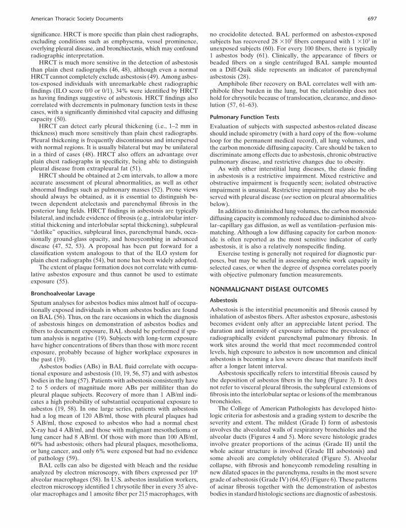
Asbestosis specifically refers to interstitial fibrosis caused bythe deposition of asbestos fibers in the lung (Figure 3). It doesnot refer to visceral pleural fibrosis, the subpleural extensions offibrosis into the interlobular septae or lesions of the membranousbronchioles.
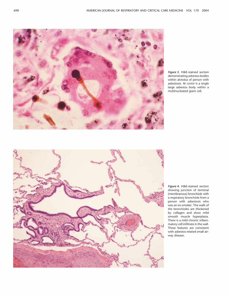
The College of American Pathologists has developed histo-logic criteria for asbestosis and a grading system to describe theseverity and extent. The mildest (Grade I) form of asbestosisinvolves the alveolated walls of respiratory bronchioles and thealveolar ducts (Figures 4 and 5). More severe histologic gradesinvolve greater proportions of the acinus (Grade II) until thewhole acinar structure is involved (Grade III asbestosis) andsome alveoli are completely obliterated (Figure 5). Alveolarcollapse, with fibrosis and honeycomb remodeling resulting innew dilated spaces in the parenchyma, results in the most severegrade of asbestosis (Grade IV) (64, 65) (Figure 6). These patternsof acinar fibrosis together with the demonstration of asbestosbodies in standard histologic sections are diagnostic of asbestosis.
698 AMERICAN JOURNAL OF RESPIRATORY AND CRITICAL CARE MEDICINE VOL 170 2004
Figure 3. H&E-stained sectiondemonstrating asbestos bodieswithin alveolus of person withasbestosis. At center is a singlelarge asbestos body within amultinucleated giant cell.
Figure 4. H&E-stained sectionshowing junction of terminal(membranous) bronchiole witha respiratory bronchiole from aperson with asbestosis whowas an ex-smoker. The walls ofthe bronchioles are thickenedby collagen and show mildsmooth muscle hyperplasia.There is a mild chronic inflam-matory cell infiltrate in the wall.These features are consistentwith asbestos-related small air-way disease.
American Thoracic Society Documents 699
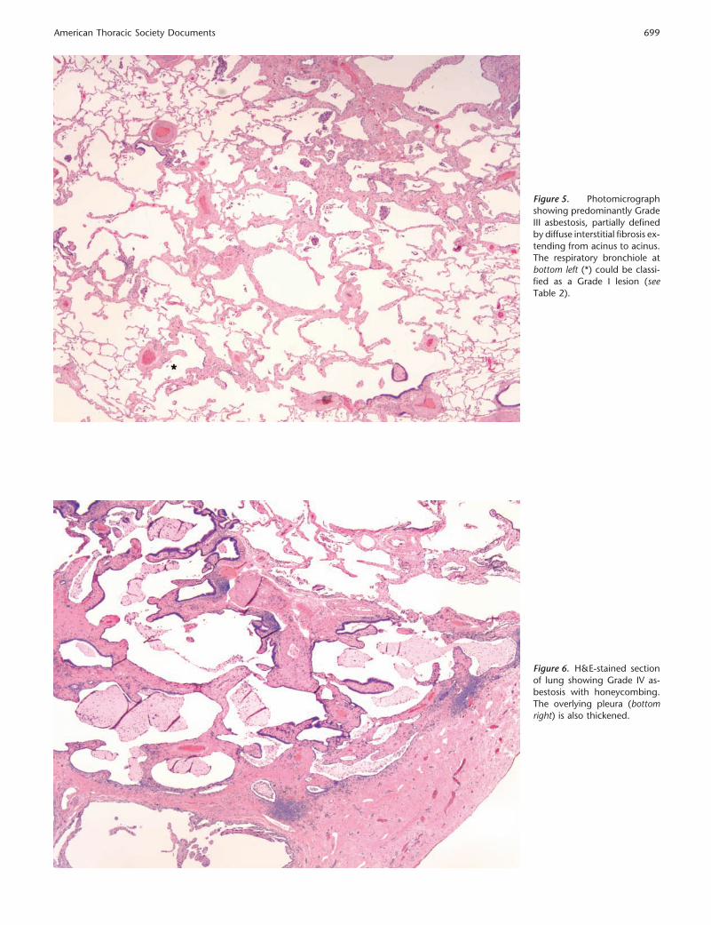
Figure 5. Photomicrographshowing predominantly GradeIII asbestosis, partially definedby diffuse interstitial fibrosis ex-tending from acinus to acinus.The respiratory bronchiole atbottom left (*) could be classi-fied as a Grade I lesion (seeTable 2).
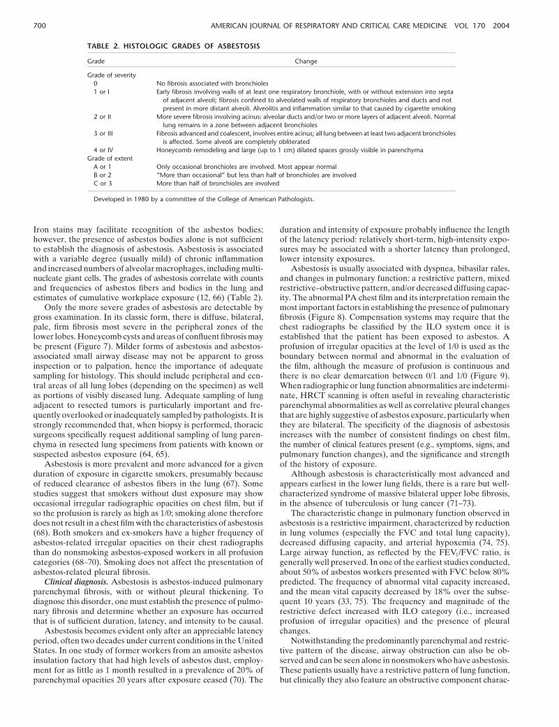
Figure 6. H&E-stained sectionof lung showing Grade IV as-bestosis with honeycombing.The overlying pleura (bottomright) is also thickened.
700 AMERICAN JOURNAL OF RESPIRATORY AND CRITICAL CARE MEDICINE VOL 170 2004
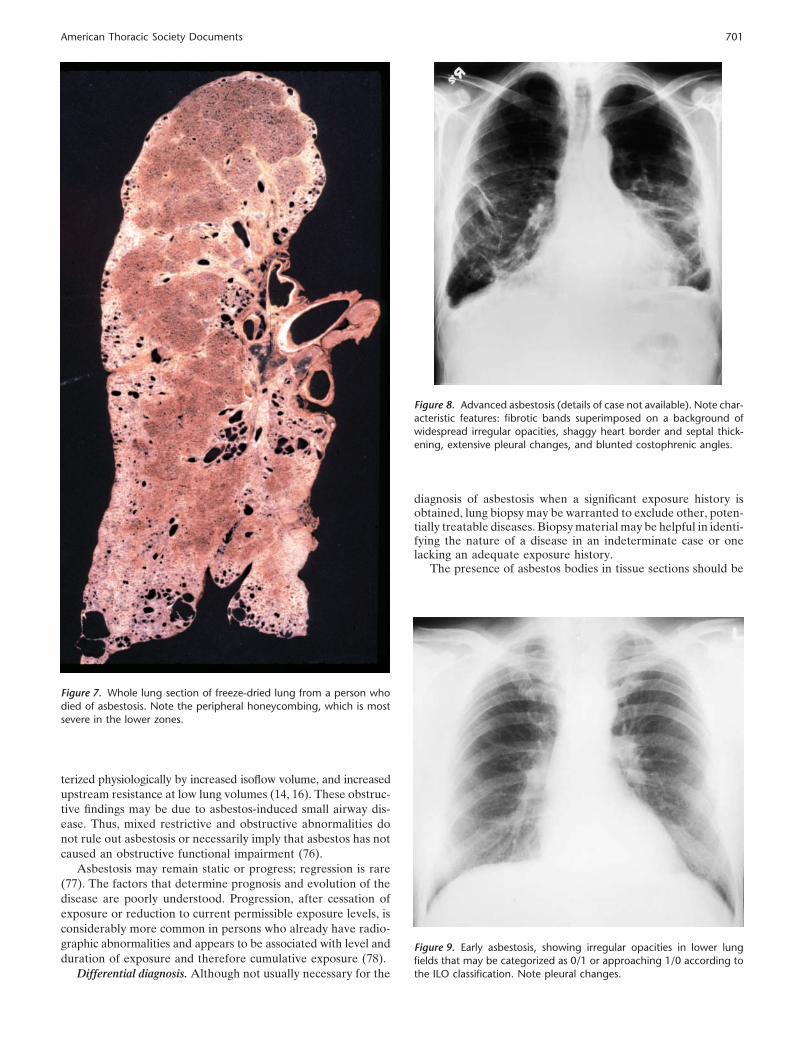
TABLE 2. HISTOLOGIC GRADES OF ASBESTOSIS
Grade Change
Grade of severity0 No fibrosis associated with bronchioles1 or I Early fibrosis involving walls of at least one respiratory bronchiole, with or without extension into septa
of adjacent alveoli; fibrosis confined to alveolated walls of respiratory bronchioles and ducts and notpresent in more distant alveoli. Alveolitis and inflammation similar to that caused by cigarette smoking
2 or II More severe fibrosis involving acinus: alveolar ducts and/or two or more layers of adjacent alveoli. Normallung remains in a zone between adjacent bronchioles
3 or III Fibrosis advanced and coalescent, involves entire acinus; all lung between at least two adjacent bronchiolesis affected. Some alveoli are completely obliterated
4 or IV Honeycomb remodeling and large (up to 1 cm) dilated spaces grossly visible in parenchymaGrade of extent
A or 1 Only occasional bronchioles are involved. Most appear normalB or 2 “More than occasional” but less than half of bronchioles are involvedC or 3 More than half of bronchioles are involved
Developed in 1980 by a committee of the College of American Pathologists.
Iron stains may facilitate recognition of the asbestos bodies;however, the presence of asbestos bodies alone is not sufficientto establish the diagnosis of asbestosis. Asbestosis is associatedwith a variable degree (usually mild) of chronic inflammationand increased numbers of alveolar macrophages, including multi-nucleate giant cells. The grades of asbestosis correlate with countsand frequencies of asbestos fibers and bodies in the lung andestimates of cumulative workplace exposure (12, 66) (Table 2).
Only the more severe grades of asbestosis are detectable bygross examination. In its classic form, there is diffuse, bilateral,pale, firm fibrosis most severe in the peripheral zones of thelower lobes. Honeycomb cysts and areas of confluent fibrosis maybe present (Figure 7). Milder forms of asbestosis and asbestos-associated small airway disease may not be apparent to grossinspection or to palpation, hence the importance of adequatesampling for histology. This should include peripheral and cen-tral areas of all lung lobes (depending on the specimen) as wellas portions of visibly diseased lung. Adequate sampling of lungadjacent to resected tumors is particularly important and fre-quently overlooked or inadequately sampled by pathologists. It isstrongly recommended that, when biopsy is performed, thoracicsurgeons specifically request additional sampling of lung paren-chyma in resected lung specimens from patients with known orsuspected asbestos exposure (64, 65).
Asbestosis is more prevalent and more advanced for a givenduration of exposure in cigarette smokers, presumably becauseof reduced clearance of asbestos fibers in the lung (67). Somestudies suggest that smokers without dust exposure may showoccasional irregular radiographic opacities on chest film, but ifso the profusion is rarely as high as 1/0; smoking alone thereforedoes not result in a chest film with the characteristics of asbestosis(68). Both smokers and ex-smokers have a higher frequency ofasbestos-related irregular opacities on their chest radiographsthan do nonsmoking asbestos-exposed workers in all profusioncategories (68–70). Smoking does not affect the presentation ofasbestos-related pleural fibrosis.
Clinical diagnosis. Asbestosis is asbestos-induced pulmonaryparenchymal fibrosis, with or without pleural thickening. Todiagnose this disorder, one must establish the presence of pulmo-nary fibrosis and determine whether an exposure has occurredthat is of sufficient duration, latency, and intensity to be causal.
Asbestosis becomes evident only after an appreciable latencyperiod, often two decades under current conditions in the UnitedStates. In one study of former workers from an amosite asbestosinsulation factory that had high levels of asbestos dust, employ-ment for as little as 1 month resulted in a prevalence of 20% ofparenchymal opacities 20 years after exposure ceased (70). The
duration and intensity of exposure probably influence the lengthof the latency period: relatively short-term, high-intensity expo-sures may be associated with a shorter latency than prolonged,lower intensity exposures.
Asbestosis is usually associated with dyspnea, bibasilar rales,and changes in pulmonary function: a restrictive pattern, mixedrestrictive–obstructive pattern, and/or decreased diffusing capac-ity. The abnormal PA chest film and its interpretation remain themost important factors in establishing the presence of pulmonaryfibrosis (Figure 8). Compensation systems may require that thechest radiographs be classified by the ILO system once it isestablished that the patient has been exposed to asbestos. Aprofusion of irregular opacities at the level of 1/0 is used as theboundary between normal and abnormal in the evaluation ofthe film, although the measure of profusion is continuous andthere is no clear demarcation between 0/1 and 1/0 (Figure 9).When radiographic or lung function abnormalities are indetermi-nate, HRCT scanning is often useful in revealing characteristicparenchymal abnormalities as well as correlative pleural changesthat are highly suggestive of asbestos exposure, particularly whenthey are bilateral. The specificity of the diagnosis of asbestosisincreases with the number of consistent findings on chest film,the number of clinical features present (e.g., symptoms, signs, andpulmonary function changes), and the significance and strengthof the history of exposure.
Although asbestosis is characteristically most advanced andappears earliest in the lower lung fields, there is a rare but well-characterized syndrome of massive bilateral upper lobe fibrosis,in the absence of tuberculosis or lung cancer (71–73).
The characteristic change in pulmonary function observed inasbestosis is a restrictive impairment, characterized by reductionin lung volumes (especially the FVC and total lung capacity),decreased diffusing capacity, and arterial hypoxemia (74, 75).Large airway function, as reflected by the FEV1/FVC ratio, isgenerally well preserved. In one of the earliest studies conducted,about 50% of asbestos workers presented with FVC below 80%predicted. The frequency of abnormal vital capacity increased,and the mean vital capacity decreased by 18% over the subse-quent 10 years (33, 75). The frequency and magnitude of therestrictive defect increased with ILO category (i.e., increasedprofusion of irregular opacities) and the presence of pleuralchanges.
Notwithstanding the predominantly parenchymal and restric-tive pattern of the disease, airway obstruction can also be ob-served and can be seen alone in nonsmokers who have asbestosis.These patients usually have a restrictive pattern of lung function,but clinically they also feature an obstructive component charac-
American Thoracic Society Documents 701
Figure 7. Whole lung section of freeze-dried lung from a person whodied of asbestosis. Note the peripheral honeycombing, which is mostsevere in the lower zones.
terized physiologically by increased isoflow volume, and increasedupstream resistance at low lung volumes (14, 16). These obstruc-tive findings may be due to asbestos-induced small airway dis-ease. Thus, mixed restrictive and obstructive abnormalities donot rule out asbestosis or necessarily imply that asbestos has notcaused an obstructive functional impairment (76).
Asbestosis may remain static or progress; regression is rare(77). The factors that determine prognosis and evolution of thedisease are poorly understood. Progression, after cessation ofexposure or reduction to current permissible exposure levels, isconsiderably more common in persons who already have radio-graphic abnormalities and appears to be associated with level andduration of exposure and therefore cumulative exposure (78).
Differential diagnosis. Although not usually necessary for the
Figure 8. Advanced asbestosis (details of case not available). Note char-acteristic features: fibrotic bands superimposed on a background ofwidespread irregular opacities, shaggy heart border and septal thick-ening, extensive pleural changes, and blunted costophrenic angles.
diagnosis of asbestosis when a significant exposure history isobtained, lung biopsy may be warranted to exclude other, poten-tially treatable diseases. Biopsy material may be helpful in identi-fying the nature of a disease in an indeterminate case or onelacking an adequate exposure history.
The presence of asbestos bodies in tissue sections should be
Figure 9. Early asbestosis, showing irregular opacities in lower lungfields that may be categorized as 0/1 or approaching 1/0 according tothe ILO classification. Note pleural changes.
702 AMERICAN JOURNAL OF RESPIRATORY AND CRITICAL CARE MEDICINE VOL 170 2004
sufficient to differentiate asbestosis from other forms of intersti-tial fibrosis. The chance of finding one asbestos body from back-ground exposure alone has been shown to be about 1 per 1,000(79). Conversely, the presence of interstitial fibrosis in the ab-sence of asbestos bodies is most likely not asbestosis, althoughrare cases of pulmonary fibrosis with large numbers of uncoatedasbestos fibers have been described (80–82). Idiopathic pulmo-nary fibrosis (IPF in clinical terms or usual interstitial pneumoni-tis in terms of pathology) has an acinar pattern of fibrosis differ-ent from that of asbestosis and is not associated with asbestosbodies in tissue sections. On occasion, asbestosis is seen in con-junction with an unrelated interstitial lung disease (such as sar-coidosis) or in association with another pneumoconiosis, forexample, silicosis. In the absence of fibrosis, asbestos bodies arean indication of exposure, not disease.
Asbestosis resembles a variety of other diffuse interstitialinflammatory and fibrotic processes in the lung and must bedistinguished from other pneumoconioses, IPF, hypersensitivitypneumonitis, sarcoidosis, and other diseases of this class. Theclinical features of asbestosis, although characteristic, are notindividually unique or pathognomonic, but the characteristicsigns of the disease are highly suggestive when they occur to-gether. The presence of pleural plaques provides useful corollaryevidence that the parenchymal process is asbestos related.
Diagnostic uncertainty is most likely in certain groups ofpatients. Patients may have a heavy cigarette-smoking historyand concurrent emphysema (which also reduces the diffusingcapacity). In such cases, one expects a history of asbestos expo-sure commensurate with the degree of disease. On occasion, apatient with another interstitial lung disease, such as IPF, willhave a history of asbestos exposure. Rapid progression, with avisible, year-to-year increase in symptoms, progression of radio-graphic findings, and loss of pulmonary function in the absenceof intense asbestos exposure, suggests the diagnosis of IPF ratherthan asbestosis.
Patients may be exposed at various times in their workinglife to more than one dust, such as silica and asbestos, or tomixed exposures, such as dusts in combination with fumes andvapors in welding (83). These patients may have combined dis-ease or the effects of one dust or other exposure may dominate.For example, predominantly upper lobe rounded opacities, hilarnode enlargement, and progressive massive fibrosis are not fea-tures of asbestosis and if present suggest other causes for thelung disease than asbestos, such as silicosis.
On occasion, isolated fibrotic lesions associated with asbestosresemble solitary pulmonary nodules. These are sometimescalled “asbestomas” and usually occur against a background ofirregular opacities; they rarely appear in isolation. They normallyrequire biopsy because they are not distinguishable from lungmalignancies otherwise (84).
Nonmalignant Pleural Abnormalities Associatedwith Asbestos
Pleural abnormalities associated with asbestos exposure are theresult of collagen deposition resulting in subpleural thickening,which may subsequently calcify, and which in the visceral pleuramay be associated with parenchymal fibrosis in adjacent subpleu-ral alveoli (Figures 10 and 11). Pleural thickening, as a markerof asbestos exposure, has continued to be a prominent featureof exposure to asbestos while other outcomes, such as asbestosis,have become less frequent due to declining exposure levels. Themajor determinant of pleural thickening is duration from firstexposure (70).
It is unclear whether the relative frequency of diffuse andcircumscribed pleural thickening has changed. The InternationalClassification of Radiographs of Pneumoconioses (38) provides
a basis for recording and classifying both types of pleural thick-ening, allowing correlation with indices of exposure and mea-surements of lung function. Manifestations of disease of the lungand of the pleura have become less evident and less characteristicon plain films as exposures have decreased. However, CT scan(including high-resolution images) detects pleural thickening notevident on the plain film, and sometimes fails to confirm apparentpleural thickening read on the plain film. Schemes to quantifyextent of pleural thickening on CT scan have been published(55, 85). Rarely, interlobar pleural thickening may mimic lungnodules on CT scan (86).
Pleuritis: acute pleural effusion, chronic pleuritic pain. Asbes-tos may cause an acute pleural effusion, often lasting severalmonths, that is exudative and often hemorrhagic, with variablenumbers of erythrocytes, neutrophils, lymphocytes, mesothelialcells, and often eosinophils (87–89). It may occur early (within10 years, unlike other asbestos-related diseases) or late after theonset of asbestos exposure (90). It may be superimposed onlong-standing pleural plaques (91). Although it is usually asymp-tomatic, the acute pleural effusion due to asbestos may also beexuberant, with fever and severe pleuritic pain. It is sometimesdetected only incidentally on a radiograph taken for anotherpurpose (87, 88). The effusion may persist for months, presentbilaterally, or recur on the same or the opposite side (87). Afriction rub may be present (92, 93). The traces of pleural effusionmay be observed years later as a blunted costophrenic angle or asdiffuse pleural thickening. Acute pleuritis is thought to underliemany cases of diffuse pleural thickening. Of 20 insulators with apast history of definite pleural effusion, diffuse pleural thickeningwas detected on radiograph in 16 (90). Dose–response relation-ships or characteristic features of exposure associated with effu-sion have not been described.
Chronic severe pleuritic pain is rare in patients with asbestos-related pleural disease (92, 93). Vague discomfort appears to bemore frequent. Studies examining the frequency of atypical chestpain in asbestos-exposed patients have not been performed. Inthe few cases described, it was present for many years, disabling,and often bilateral. Radiographic evidence of pleural diseaseranged from plaques to extensive diffuse and circumscribed pleu-ral thickening; several cases followed pleural effusions. The diag-nosis of acute asbestos-related pleural effusion is by exclusionof other causes of acute pleuritis, and most often is not arrivedat until the pleural space is fully explored and biopsied, generallyby thoracoscopy. Differentiation from Dressler’s syndrome isdifficult in asbestos-exposed patients who have undergone recentcardiac surgery. Differentiation from mesothelioma or pleuralextension of a pulmonary malignancy is critical, and may bedifficult on clinical grounds (including positive gallium and posi-tron emission scan). Pleural fluid cytology is useful for distin-guishing benign from malignant effusions. It is not unusual fornonspecific effusions to precede mesothelioma by several years.If a malignancy has not manifested itself within 3 years, theeffusion is generally considered benign.
The diagnosis of chronic pleuritis manifested by pleuritic painis reached by excluding malignancies, because most other causesof acute pleuritis do not result in chronic pain. Malignancy isunlikely when pain persists for years with little or no clinical orradiographic change.
Plaques: circumscribed pleural thickening. Pleural plaques areindicators of exposure to asbestos. They are clearly the mostcommon manifestation of the inhalation, retention, and biologiceffect of asbestos. Their prevalence is most directly related toduration from first exposure; they are rare within less than 20years. Pleural plaques consistent with asbestos exposure appearin chest films of 2.3% of U.S. males, a percentage that has been
American Thoracic Society Documents 703
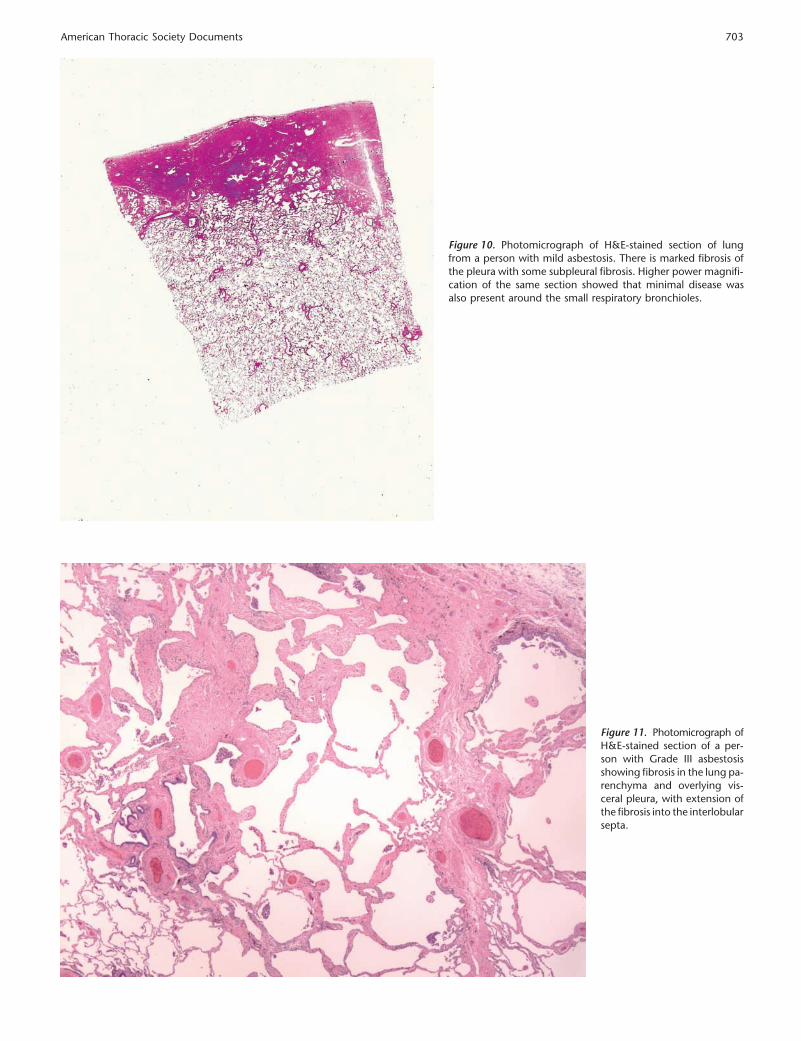
Figure 10. Photomicrograph of H&E-stained section of lungfrom a person with mild asbestosis. There is marked fibrosis ofthe pleura with some subpleural fibrosis. Higher power magnifi-cation of the same section showed that minimal disease wasalso present around the small respiratory bronchioles.
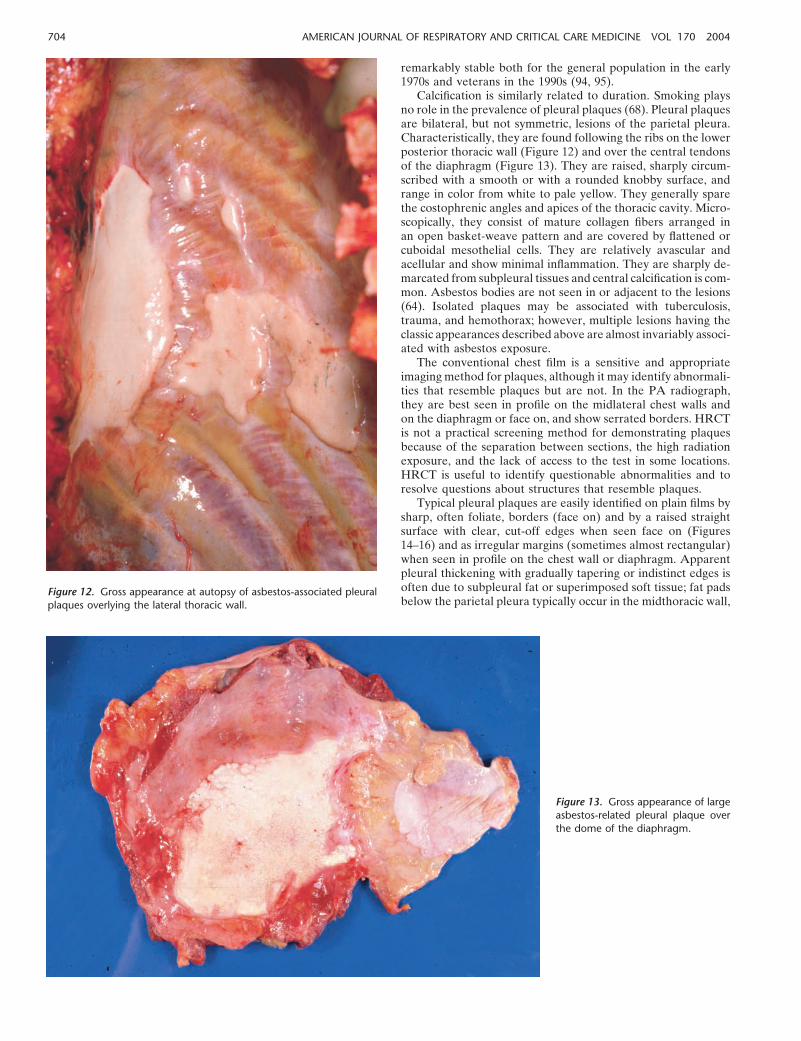
Figure 11. Photomicrograph ofH&E-stained section of a per-son with Grade III asbestosisshowing fibrosis in the lung pa-renchyma and overlying vis-ceral pleura, with extension ofthe fibrosis into the interlobularsepta.
704 AMERICAN JOURNAL OF RESPIRATORY AND CRITICAL CARE MEDICINE VOL 170 2004
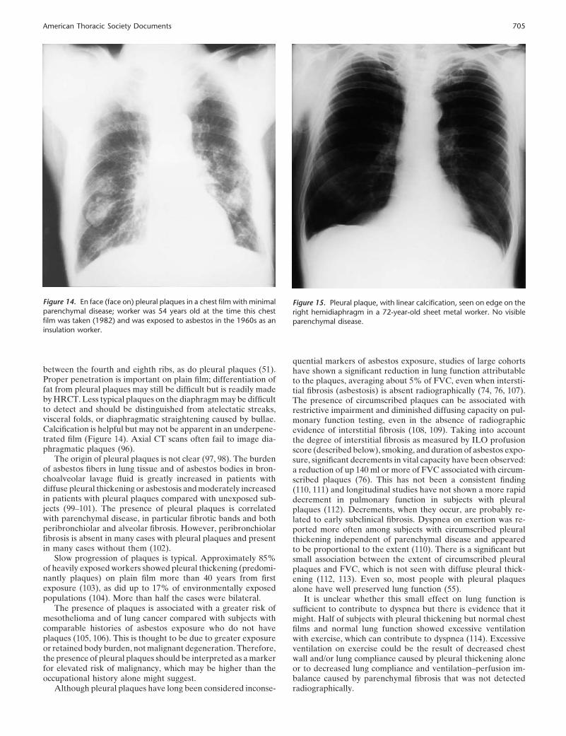
Figure 12. Gross appearance at autopsy of asbestos-associated pleuralplaques overlying the lateral thoracic wall.
Figure 13. Gross appearance of largeasbestos-related pleural plaque overthe dome of the diaphragm.
remarkably stable both for the general population in the early1970s and veterans in the 1990s (94, 95).
Calcification is similarly related to duration. Smoking playsno role in the prevalence of pleural plaques (68). Pleural plaquesare bilateral, but not symmetric, lesions of the parietal pleura.Characteristically, they are found following the ribs on the lowerposterior thoracic wall (Figure 12) and over the central tendonsof the diaphragm (Figure 13). They are raised, sharply circum-scribed with a smooth or with a rounded knobby surface, andrange in color from white to pale yellow. They generally sparethe costophrenic angles and apices of the thoracic cavity. Micro-scopically, they consist of mature collagen fibers arranged inan open basket-weave pattern and are covered by flattened orcuboidal mesothelial cells. They are relatively avascular andacellular and show minimal inflammation. They are sharply de-marcated from subpleural tissues and central calcification is com-mon. Asbestos bodies are not seen in or adjacent to the lesions(64). Isolated plaques may be associated with tuberculosis,trauma, and hemothorax; however, multiple lesions having theclassic appearances described above are almost invariably associ-ated with asbestos exposure.
The conventional chest film is a sensitive and appropriateimaging method for plaques, although it may identify abnormali-ties that resemble plaques but are not. In the PA radiograph,they are best seen in profile on the midlateral chest walls andon the diaphragm or face on, and show serrated borders. HRCTis not a practical screening method for demonstrating plaquesbecause of the separation between sections, the high radiationexposure, and the lack of access to the test in some locations.HRCT is useful to identify questionable abnormalities and toresolve questions about structures that resemble plaques.
Typical pleural plaques are easily identified on plain films bysharp, often foliate, borders (face on) and by a raised straightsurface with clear, cut-off edges when seen face on (Figures14–16) and as irregular margins (sometimes almost rectangular)when seen in profile on the chest wall or diaphragm. Apparentpleural thickening with gradually tapering or indistinct edges isoften due to subpleural fat or superimposed soft tissue; fat padsbelow the parietal pleura typically occur in the midthoracic wall,
American Thoracic Society Documents 705
Figure 14. En face (face on) pleural plaques in a chest film with minimalparenchymal disease; worker was 54 years old at the time this chestfilm was taken (1982) and was exposed to asbestos in the 1960s as aninsulation worker.
between the fourth and eighth ribs, as do pleural plaques (51).Proper penetration is important on plain film; differentiation offat from pleural plaques may still be difficult but is readily madeby HRCT. Less typical plaques on the diaphragm may be difficultto detect and should be distinguished from atelectatic streaks,visceral folds, or diaphragmatic straightening caused by bullae.Calcification is helpful but may not be apparent in an underpene-trated film (Figure 14). Axial CT scans often fail to image dia-phragmatic plaques (96).
The origin of pleural plaques is not clear (97, 98). The burdenof asbestos fibers in lung tissue and of asbestos bodies in bron-choalveolar lavage fluid is greatly increased in patients withdiffuse pleural thickening or asbestosis and moderately increasedin patients with pleural plaques compared with unexposed sub-jects (99–101). The presence of pleural plaques is correlatedwith parenchymal disease, in particular fibrotic bands and bothperibronchiolar and alveolar fibrosis. However, peribronchiolarfibrosis is absent in many cases with pleural plaques and presentin many cases without them (102).
Slow progression of plaques is typical. Approximately 85%of heavily exposed workers showed pleural thickening (predomi-nantly plaques) on plain film more than 40 years from firstexposure (103), as did up to 17% of environmentally exposedpopulations (104). More than half the cases were bilateral.
The presence of plaques is associated with a greater risk ofmesothelioma and of lung cancer compared with subjects withcomparable histories of asbestos exposure who do not haveplaques (105, 106). This is thought to be due to greater exposureor retained body burden, not malignant degeneration. Therefore,the presence of pleural plaques should be interpreted as a markerfor elevated risk of malignancy, which may be higher than theoccupational history alone might suggest.
Although pleural plaques have long been considered inconse-
Figure 15. Pleural plaque, with linear calcification, seen on edge on theright hemidiaphragm in a 72-year-old sheet metal worker. No visibleparenchymal disease.
quential markers of asbestos exposure, studies of large cohortshave shown a significant reduction in lung function attributableto the plaques, averaging about 5% of FVC, even when intersti-tial fibrosis (asbestosis) is absent radiographically (74, 76, 107).The presence of circumscribed plaques can be associated withrestrictive impairment and diminished diffusing capacity on pul-monary function testing, even in the absence of radiographicevidence of interstitial fibrosis (108, 109). Taking into accountthe degree of interstitial fibrosis as measured by ILO profusionscore (described below), smoking, and duration of asbestos expo-sure, significant decrements in vital capacity have been observed:a reduction of up 140 ml or more of FVC associated with circum-scribed plaques (76). This has not been a consistent finding(110, 111) and longitudinal studies have not shown a more rapiddecrement in pulmonary function in subjects with pleuralplaques (112). Decrements, when they occur, are probably re-lated to early subclinical fibrosis. Dyspnea on exertion was re-ported more often among subjects with circumscribed pleuralthickening independent of parenchymal disease and appearedto be proportional to the extent (110). There is a significant butsmall association between the extent of circumscribed pleuralplaques and FVC, which is not seen with diffuse pleural thick-ening (112, 113). Even so, most people with pleural plaquesalone have well preserved lung function (55).
It is unclear whether this small effect on lung function issufficient to contribute to dyspnea but there is evidence that itmight. Half of subjects with pleural thickening but normal chestfilms and normal lung function showed excessive ventilationwith exercise, which can contribute to dyspnea (114). Excessiveventilation on exercise could be the result of decreased chestwall and/or lung compliance caused by pleural thickening aloneor to decreased lung compliance and ventilation–perfusion im-balance caused by parenchymal fibrosis that was not detectedradiographically.
American Thoracic Society Documents 707
Plaques are indicators of increased risk for the future develop-ment of asbestosis (94). This may reflect greater exposure orretained body burden. An autopsy study has demonstrated morefrequent peribronchiolar fibrosis when plaques are present (90).This finding, as well as derangements in gas exchange (114) andevidence from HRCT, indicate that subradiographic asbestosismay be present in some patients with only pleural plaques. Thepresence of plaques is therefore an indication to monitor thepatient over time for interstitial fibrosis (115).
Diffuse pleural thickening. Diffuse thickening of the visceralpleura is not sharply demarcated and is often associated withfibrous strands (“crow’s feet”) extending into the parenchyma.In large surveys of asbestos-exposed workers, diffuse pleuralthickening has ranged from 9 to 22% of those with pleuraldisease. Both circumscribed and diffuse pleural thickening maybe present in the same hemithorax. Diffuse pleural thickeningsuperimposed on circumscribed plaques has been observed, of-ten after pleural effusion (91).
The frequency of diffuse pleural thickening increases withtime from first exposure and is thought to be dose related (104).Diffuse pleural thickening has been observed after acute pleuritis(90). It may also be caused by extension of interstitial fibrosisto the visceral pleura, consistent with the pleural migration ofasbestos fibers. The extent of diffuse pleural thickening seemsto be more or less uniformly distributed, the different degreesbeing fairly equally often seen, however, in contradistinction tocircumscribed pleural thickening, in which the lowest categoriesare more frequent (113). Lung burdens of asbestos in thesecases are intermediate between asbestosis and pleural plaques(116–118).
This condition affects the visceral pleural surface and is quitedifferent in appearance from the parietal pleural plaque. It con-sists of pale gray diffuse thickening that blends at the edges withthe more normal pleura. It may be extensive and cover a wholelobe or whole lung and obliterate lobar fissures. It ranges inthickness from less than 1 mm up to 1 cm or more. Adhesions tothe parietal pleura are common, particularly opposite to pleuralplaques. The lesion may show a gradient with immature granula-tion tissue and fibrin at the surface, progressing to mature colla-gen adjacent to the lung. The fibrosis may extend for a fewmillimeters into the lung parenchyma and into the lobular septae.The latter features do not constitute asbestosis.
Diffuse pleural thickening may have a significantly greaterimpact on pulmonary function than circumscribed plaques. Areduction of 270 ml of FVC has been associated with diffusepleural thickening (76, 119). Workers with diffuse pleural thick-ening have a significantly greater decrement in FVC (by a factorof two or more) than those with circumscribed pleural thickening(76, 113). This effect is unrelated to the radiographic extent ofpleural thickening; a similar reduction in FVC was seen withlittle more than costophrenic angle blunting as with extensiveinvolvement (113). Decrements associated with diffuse pleuralthickening reflect pulmonary restriction as a result of adhesionsof the parietal with the visceral pleura. Restrictive impairmentis characteristic, with relative preservation of diffusing capacity(pattern of entrapped lung).
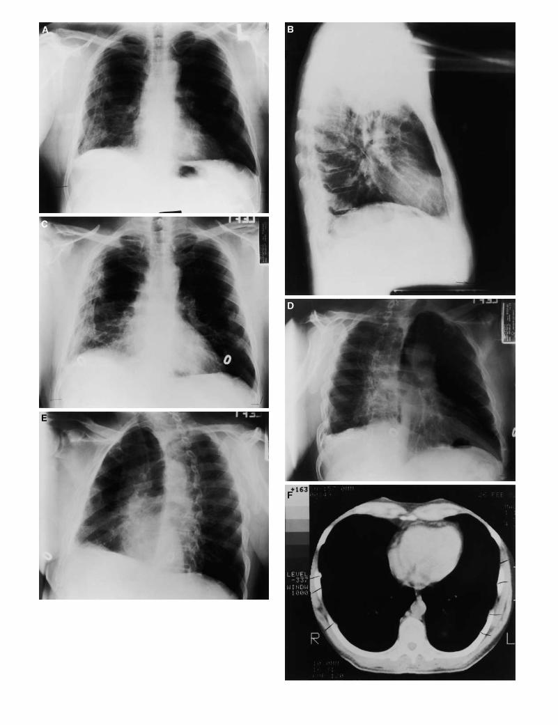
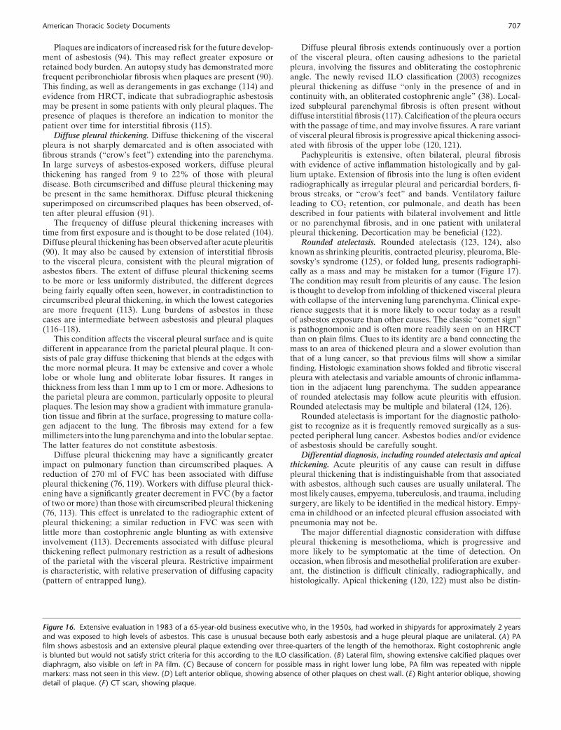
Figure 16. Extensive evaluation in 1983 of a 65-year-old business executive who, in the 1950s, had worked in shipyards for approximately 2 yearsand was exposed to high levels of asbestos. This case is unusual because both early asbestosis and a huge pleural plaque are unilateral. (A ) PAfilm shows asbestosis and an extensive pleural plaque extending over three-quarters of the length of the hemothorax. Right costophrenic angleis blunted but would not satisfy strict criteria for this according to the ILO classification. (B ) Lateral film, showing extensive calcified plaques overdiaphragm, also visible on left in PA film. (C ) Because of concern for possible mass in right lower lung lobe, PA film was repeated with nipplemarkers: mass not seen in this view. (D ) Left anterior oblique, showing absence of other plaques on chest wall. (E ) Right anterior oblique, showingdetail of plaque. (F ) CT scan, showing plaque.
Diffuse pleural fibrosis extends continuously over a portionof the visceral pleura, often causing adhesions to the parietalpleura, involving the fissures and obliterating the costophrenicangle. The newly revised ILO classification (2003) recognizespleural thickening as diffuse “only in the presence of and incontinuity with, an obliterated costophrenic angle” (38). Local-ized subpleural parenchymal fibrosis is often present withoutdiffuse interstitial fibrosis (117). Calcification of the pleura occurswith the passage of time, and may involve fissures. A rare variantof visceral pleural fibrosis is progressive apical thickening associ-ated with fibrosis of the upper lobe (120, 121).
Pachypleuritis is extensive, often bilateral, pleural fibrosiswith evidence of active inflammation histologically and by gal-lium uptake. Extension of fibrosis into the lung is often evidentradiographically as irregular pleural and pericardial borders, fi-brous streaks, or “crow’s feet” and bands. Ventilatory failureleading to CO2 retention, cor pulmonale, and death has beendescribed in four patients with bilateral involvement and littleor no parenchymal fibrosis, and in one patient with unilateralpleural thickening. Decortication may be beneficial (122).
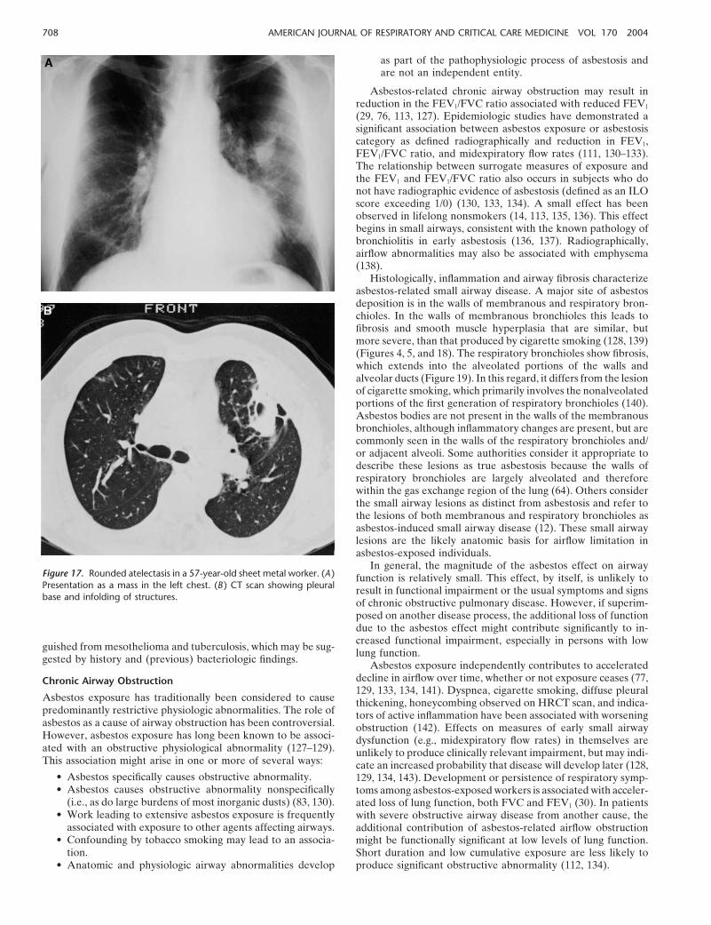
Rounded atelectasis. Rounded atelectasis (123, 124), alsoknown as shrinking pleuritis, contracted pleurisy, pleuroma, Ble-sovsky’s syndrome (125), or folded lung, presents radiographi-cally as a mass and may be mistaken for a tumor (Figure 17).The condition may result from pleuritis of any cause. The lesionis thought to develop from infolding of thickened visceral pleurawith collapse of the intervening lung parenchyma. Clinical expe-rience suggests that it is more likely to occur today as a resultof asbestos exposure than other causes. The classic “comet sign”is pathognomonic and is often more readily seen on an HRCTthan on plain films. Clues to its identity are a band connecting themass to an area of thickened pleura and a slower evolution thanthat of a lung cancer, so that previous films will show a similarfinding. Histologic examination shows folded and fibrotic visceralpleura with atelectasis and variable amounts of chronic inflamma-tion in the adjacent lung parenchyma. The sudden appearanceof rounded atelectasis may follow acute pleuritis with effusion.Rounded atelectasis may be multiple and bilateral (124, 126).
Rounded atelectasis is important for the diagnostic patholo-gist to recognize as it is frequently removed surgically as a sus-pected peripheral lung cancer. Asbestos bodies and/or evidenceof asbestosis should be carefully sought.
Differential diagnosis, including rounded atelectasis and apicalthickening. Acute pleuritis of any cause can result in diffusepleural thickening that is indistinguishable from that associatedwith asbestos, although such causes are usually unilateral. Themost likely causes, empyema, tuberculosis, and trauma, includingsurgery, are likely to be identified in the medical history. Empy-ema in childhood or an infected pleural effusion associated withpneumonia may not be.
The major differential diagnostic consideration with diffusepleural thickening is mesothelioma, which is progressive andmore likely to be symptomatic at the time of detection. Onoccasion, when fibrosis and mesothelial proliferation are exuber-ant, the distinction is difficult clinically, radiographically, andhistologically. Apical thickening (120, 122) must also be distin-
708 AMERICAN JOURNAL OF RESPIRATORY AND CRITICAL CARE MEDICINE VOL 170 2004
Figure 17. Rounded atelectasis in a 57-year-old sheet metal worker. (A )Presentation as a mass in the left chest. (B ) CT scan showing pleuralbase and infolding of structures.
guished from mesothelioma and tuberculosis, which may be sug-gested by history and (previous) bacteriologic findings.
Chronic Airway Obstruction
Asbestos exposure has traditionally been considered to causepredominantly restrictive physiologic abnormalities. The role ofasbestos as a cause of airway obstruction has been controversial.However, asbestos exposure has long been known to be associ-ated with an obstructive physiological abnormality (127–129).This association might arise in one or more of several ways:
• Asbestos specifically causes obstructive abnormality.• Asbestos causes obstructive abnormality nonspecifically
(i.e., as do large burdens of most inorganic dusts) (83, 130).• Work leading to extensive asbestos exposure is frequently
associated with exposure to other agents affecting airways.• Confounding by tobacco smoking may lead to an associa-
tion.• Anatomic and physiologic airway abnormalities develop
as part of the pathophysiologic process of asbestosis andare not an independent entity.
Asbestos-related chronic airway obstruction may result inreduction in the FEV1/FVC ratio associated with reduced FEV1
(29, 76, 113, 127). Epidemiologic studies have demonstrated asignificant association between asbestos exposure or asbestosiscategory as defined radiographically and reduction in FEV1,FEV1/FVC ratio, and midexpiratory flow rates (111, 130–133).The relationship between surrogate measures of exposure andthe FEV1 and FEV1/FVC ratio also occurs in subjects who donot have radiographic evidence of asbestosis (defined as an ILOscore exceeding 1/0) (130, 133, 134). A small effect has beenobserved in lifelong nonsmokers (14, 113, 135, 136). This effectbegins in small airways, consistent with the known pathology ofbronchiolitis in early asbestosis (136, 137). Radiographically,airflow abnormalities may also be associated with emphysema(138).
Histologically, inflammation and airway fibrosis characterizeasbestos-related small airway disease. A major site of asbestosdeposition is in the walls of membranous and respiratory bron-chioles. In the walls of membranous bronchioles this leads tofibrosis and smooth muscle hyperplasia that are similar, butmore severe, than that produced by cigarette smoking (128, 139)(Figures 4, 5, and 18). The respiratory bronchioles show fibrosis,which extends into the alveolated portions of the walls andalveolar ducts (Figure 19). In this regard, it differs from the lesionof cigarette smoking, which primarily involves the nonalveolatedportions of the first generation of respiratory bronchioles (140).Asbestos bodies are not present in the walls of the membranousbronchioles, although inflammatory changes are present, but arecommonly seen in the walls of the respiratory bronchioles and/or adjacent alveoli. Some authorities consider it appropriate todescribe these lesions as true asbestosis because the walls ofrespiratory bronchioles are largely alveolated and thereforewithin the gas exchange region of the lung (64). Others considerthe small airway lesions as distinct from asbestosis and refer tothe lesions of both membranous and respiratory bronchioles asasbestos-induced small airway disease (12). These small airwaylesions are the likely anatomic basis for airflow limitation inasbestos-exposed individuals.
In general, the magnitude of the asbestos effect on airwayfunction is relatively small. This effect, by itself, is unlikely toresult in functional impairment or the usual symptoms and signsof chronic obstructive pulmonary disease. However, if superim-posed on another disease process, the additional loss of functiondue to the asbestos effect might contribute significantly to in-creased functional impairment, especially in persons with lowlung function.
Asbestos exposure independently contributes to accelerateddecline in airflow over time, whether or not exposure ceases (77,129, 133, 134, 141). Dyspnea, cigarette smoking, diffuse pleuralthickening, honeycombing observed on HRCT scan, and indica-tors of active inflammation have been associated with worseningobstruction (142). Effects on measures of early small airwaydysfunction (e.g., midexpiratory flow rates) in themselves areunlikely to produce clinically relevant impairment, but may indi-cate an increased probability that disease will develop later (128,129, 134, 143). Development or persistence of respiratory symp-toms among asbestos-exposed workers is associated with acceler-ated loss of lung function, both FVC and FEV1 (30). In patientswith severe obstructive airway disease from another cause, theadditional contribution of asbestos-related airflow obstructionmight be functionally significant at low levels of lung function.Short duration and low cumulative exposure are less likely toproduce significant obstructive abnormality (112, 134).
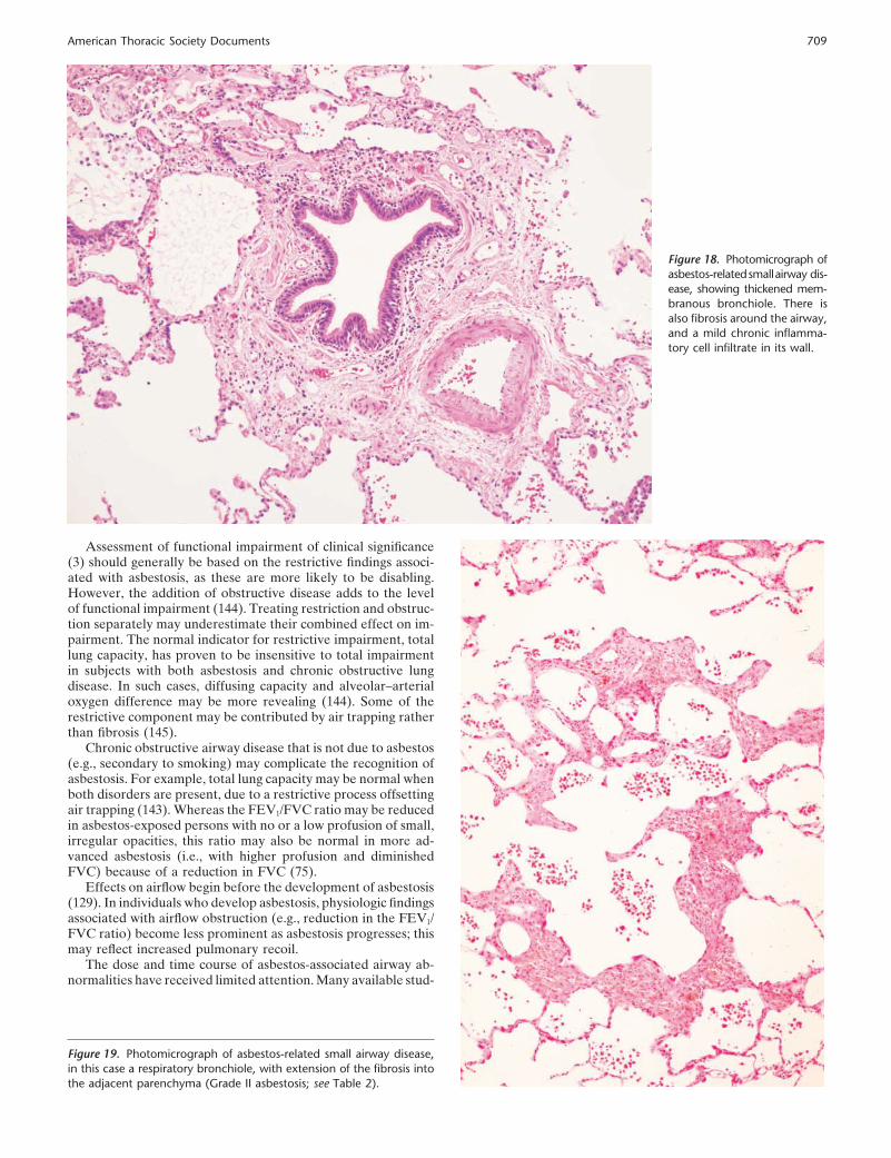
American Thoracic Society Documents 709
Figure 18. Photomicrograph ofasbestos-relatedsmallairway dis-ease, showing thickened mem-branous bronchiole. There isalso fibrosis around the airway,and a mild chronic inflamma-tory cell infiltrate in its wall.
Assessment of functional impairment of clinical significance(3) should generally be based on the restrictive findings associ-ated with asbestosis, as these are more likely to be disabling.However, the addition of obstructive disease adds to the levelof functional impairment (144). Treating restriction and obstruc-tion separately may underestimate their combined effect on im-pairment. The normal indicator for restrictive impairment, totallung capacity, has proven to be insensitive to total impairmentin subjects with both asbestosis and chronic obstructive lungdisease. In such cases, diffusing capacity and alveolar–arterialoxygen difference may be more revealing (144). Some of therestrictive component may be contributed by air trapping ratherthan fibrosis (145).
Chronic obstructive airway disease that is not due to asbestos(e.g., secondary to smoking) may complicate the recognition ofasbestosis. For example, total lung capacity may be normal whenboth disorders are present, due to a restrictive process offsettingair trapping (143). Whereas the FEV1/FVC ratio may be reducedin asbestos-exposed persons with no or a low profusion of small,irregular opacities, this ratio may also be normal in more ad-vanced asbestosis (i.e., with higher profusion and diminishedFVC) because of a reduction in FVC (75).
Effects on airflow begin before the development of asbestosis(129). In individuals who develop asbestosis, physiologic findingsassociated with airflow obstruction (e.g., reduction in the FEV1/FVC ratio) become less prominent as asbestosis progresses; thismay reflect increased pulmonary recoil.
The dose and time course of asbestos-associated airway ab-normalities have received limited attention. Many available stud-
Figure 19. Photomicrograph of asbestos-related small airway disease,in this case a respiratory bronchiole, with extension of the fibrosis intothe adjacent parenchyma (Grade II asbestosis; see Table 2).
710 AMERICAN JOURNAL OF RESPIRATORY AND CRITICAL CARE MEDICINE VOL 170 2004
TABLE 3. RECOMMENDATIONS FOR MANAGEMENT AFTER DIAGNOSIS OF ASBESTOSIS
1. Patient notification1.1. Inform patient of work-related illness1.2. Report to appropriate authority as occupational disease, as required by law1.3. Inform patient that there are options for compensation
2. Impairment assessment2.1. Conduct an assessment of functional impairment2.2. Rate impairment in accordance with ATS criteria,* which are incorporated into the AMA Guides†
3. Tertiary prevention3.1. Smoking cessation (primary prevention for smoking-related disorders)3.2. Withdrawal from further excessive exposure‡
3.3. Immunization (pneumococcal pneumonia, influenza)3.4. Management of concurrent respiratory and other diseases
4. Monitoring4.1. Chest film and pulmonary function testing§ should be conducted every 3 to 5 years4.2. Active monitoring (periodic screening) for colon cancer4.3. Observation and elevated index of suspicion but not screening for lung cancer, mesothelioma, gastrointestinal cancers
(other than colon)5. Development of a patient-specific management plan for symptomatic disease
Definition of abbreviations: AMA � American Medical Association; ATS � American Thoracic Society.* See Reference 3.† See Reference 157.‡ See text.§ See References 4 and 5.
ies reflect relatively high historical levels of exposure. Amongnonsmoking Chinese asbestos workers, association of cumulativeexposure with functional effects was seen only among those withlong-term exposure (133).
Tobacco smoking is the predominant cause of chronic airwayobstruction in asbestos-exposed workers who smoke, althoughoccupational exposures can be significant. The association be-tween airway obstruction and exposure to asbestos has been welldemonstrated in nonsmokers, and in some studies the associationbetween exposure and airway obstruction is seen only amongnonsmokers (131); among smoking asbestos-exposed workers,smoking accounts for most of the small airway abnormality (111,127, 135, 141, 142). In addition to smoking, other occupationalexposures might contribute to chronic obstructive airway dis-ease; effects of asbestos in producing airflow obstruction arelikely to be additive to these. There may be an interaction be-tween smoking and asbestos in the development of airway ob-struction, as has been demonstrated in animal models (146), butthis has not yet been demonstrated for human subjects.
IMPLICATIONS OF DIAGNOSIS FORPATIENT MANAGEMENT
A history of significant asbestos exposure obligates the responsi-ble physician to provide a management plan for the patient thattakes into consideration current disease and impairment as wellas future risk (147). A recommended management plan is sum-marized in Table 3.
Workers referred for evaluation of asbestos-related diseasetoday differ from those referred in past years. Exposure to asbes-tos among these workers is likely to be more remote in timeand to have been less intense. Exposed workers may live longerand progress later to more advanced stages of disease. They aremore likely to survive to develop additional outcomes associatedwith asbestos, such as malignancy, and to present more compli-cated management challenges (148).
Actions Required before Disease Is Apparent
A recent or short-term history of exposure to asbestos, particu-larly in the absence of detail on duration and intensity, requiresthe clinician at a minimum to educate the patient with respect
to latency, the exposure–response relationship characteristic ofasbestos-related diseases, and the future risk of malignant dis-ease. Reassurance should be offered where appropriate and therisk placed into the context of the exposure history. This is oftenan excellent opportunity at the same time to review the patient’shistory, work hygiene practices, behavior and attitudes towardcigarette smoking, as well as exposure to other occupational andenvironmental carcinogens (149).
For all patients presenting with a history of significant orpossibly significant exposure, at a minimum a baseline, high-quality chest film should be obtained, together with spirometryand a single-breath diffusing capacity that conform to AmericanThoracic Society guidelines. Complete pulmonary function test-ing should be obtained if clinically indicated. Workers who havehad exposure to asbestos have also often worked in other dustyoccupations. They and their families may have lived in communi-ties where they experienced environmental exposures.
The sensitivity of the plain chest film for identifying asbestosisat a profusion level of 1/0 (in the ILO classification system) hasbeen estimated at or slightly below 90%. The correspondingspecificity has been estimated at 93%. Applied to populationswith varying prevalence of disease, the positive predictive valueof the minimally abnormal chest film alone in making the diagno-sis of asbestosis may fall below 30% when exposure to asbestoshas been infrequent and exceed 50% when it has been prevalent.This suggests that screening programs based on the chest filmalone may vary considerably in their yield of true cases de-pending on the characteristics of the population being screened.In the general population and for occupational groups with lowlevels of exposure they may be unreliable in identifying asbesto-sis. The application of multiple criteria, as outlined in this state-ment, is a preferable approach (150). However, combinationsof tests for a specific criterion, such as a hypothetical requirementthat multiple tests for pulmonary function be abnormal, wouldreduce the sensitivity without enhancing specificity for asbestos-related disease; in general, the most sensitive test for a particularcriterion is preferable (2).
Persons identified as having asbestos-related disease or asignificant exposure history should be informed of the risk ofprogression of disease, the risk of malignancy, and especially
American Thoracic Society Documents 711
the interaction between smoking and asbestos exposure in en-hancing the risk of lung cancer. Such persons who smoke maybe more motivated to consider cessation when the connectionbetween asbestos and the risk of respiratory impairment and ofmalignancy is brought up at this time (151). The risk conferredby other occupational and environmental carcinogens shouldalso be emphasized at this time.
The question of monitoring for asbestos-related disease iscomplicated by requirements for occupational surveillance, espe-cially for those with minimal exposure. The Occupational Safetyand Health Administration asbestos standard requires employ-ers to monitor their asbestos-exposed workers during employ-ment but makes no provision beyond the period of employment,despite the latency, and private insurance may or may not allowthe expense thereafter (8).
Persons with a history of exposure to asbestos but no manifestdisease, and for whom the time since initial exposure is 10 yearsor more, may reasonably be monitored with chest films andpulmonary function studies every 3 to 5 years to identify theonset of asbestos-related disease.
Persons with a history of exposure to asbestos are also at riskfor asbestos-related malignancies. Periodic health surveillancefor lung cancer or mesothelioma is not recommended. Screeningfor lung cancer using periodic (annual) chest films, low-dosecomputed tomography, or sputum cytology has not been shownto be effective in preventing mortality or improving quality of lifein populations of smokers without known adverse occupationalexposures (152, 153). New technologies (e.g., low-dose spiral CTscanning) are being evaluated for use in high-risk groups (153).The risk of extrathoracic malignancies may also be increased inasbestos-exposed workers. Studies suggest that there may be anelevation in the risk of colon cancer (149, 150), although thisremains controversial (154). Because colon cancer is often treat-able and screening for colorectal cancer is recommended by theAmerican Cancer Society for persons more than 50 years of age(155), it is reasonable on the basis of current evidence to screenfor this condition. The risk of cancer of the larynx (156) andpossibly gastrointestinal cancers other than colon, including pan-creas, stomach, and esophagus (154), may also be increased withasbestos exposure, but the presence and magnitude of an associa-tion with asbestos remain controversial for extrathoracic cancers(154). Routine screening for these cancers is in any case not practicalat present.
No prophylactic medication or treatment is currently avail-able to prevent the development or progression of asbestosis orother asbestos-related diseases, once exposure has occurred.
Actions Required after Diagnosis
The diagnosis of asbestosis, in particular, imposes a duty toinform the patient that he or she has a disease that is work-related, to report the disease, and to inform the patient that heor she may have legal or adjudication options for compensation.The role of the physician in this compensation process includesperforming an objective evaluation of impairment consistentwith the rules of the specific compensation system. Guidelinesdeveloped by the American Thoracic Society (3) may be of useand are incorporated into the AMA Guides to the Evaluationof Permanent Impairment (157). As in the management of anylung disorder, the physician should also manage the clinical mani-festations of the disease and counsel the patient to protect re-maining lung function.
The patient with evidence of asbestosis should be consideredto be at risk of progressive lung disease, whatever the level ofimpairment on first encounter. It seems logical that removalfrom further exposure to asbestos or other significant occupa-tional and environmental exposures may avoid more rapid pro-
gression of lung disease, although specific evidence for this islacking. However, if such exposures are minimal and are wellwithin occupational guidelines, care must be taken not to deprivethe patient of a livelihood for no clinical benefit.
Immunization against pneumococcal pneumonia and annualinfluenza vaccine should be administered unless contraindicatedfor other reasons. Effective management of concurrent chronicobstructive pulmonary disease or asthma, if present, may reducemorbidity from mixed disease.
Severe asbestosis is rare in the United States and other coun-tries with generally effective occupational health regulation. Corpulmonale, secondary polycythemia, and respiratory insufficiencyand failure are all treated in the conventional manner in patientswith asbestosis.
In the spring of 2000, the Association of Occupational andEnvironmental Clinics adopted a resolution recommending nec-essary standards for screening programs (158). This action wastaken in response to the proliferation of screening programsundertaken to identify cases for possible legal actions in whichcounseling and education may be lacking (159), but the recom-mendations also apply to those conducted for patient care andprotection. Their recommendations were consistent with thosegiven above and also emphasized timely physician disclosure ofresults to the patient, appropriate medical follow-up, and patienteducation. The National Institute of Occupational Safety andHealth has outlined elements of an adequate screening program,with special reference to screening for asbestos-related disordersin currently employed mineworkers, in a white paper producedin 2002 that has received little attention (160). The NationalInstitute for Occupational Safety and Health recommended thatsuch programs should be under the direction of a “qualifiedphysician or other qualified health care provider” knowledgeablein the field and competent to administer it, and documentedwith written reports to workers and employers (the latter provi-sion that would not necessarily be applicable to workers whohad separated from the employer). However, the National Insti-tute for Occupational Safety and Health did not address theissue of counseling in that document or clinical interventions toreduce future risk.
CONCLUSIONS
The diagnosis of nonmalignant asbestos-related disease rests, asit did in 1986, on the essential criteria described: a compatiblestructural lesion, evidence of exposure, and exclusion of otherplausible conditions, with an additional requirement for impair-ment assessment if the other three criteria suggest asbestos-related disease (2). Each criterion may be satisfied by one of anumber of findings or tests. The 2004 criteria are open to futuretesting modalities if and when they are validated. For example,HRCT has greatly increased the sensitivity of detection and hasbecome a standard method of imaging. Evidence for exposurestill rests on the occupational history, the demonstration of asbes-tos fibers or bodies, or pleural plaques. Impairment evaluationis largely unchanged from 1986 and remains an essential part ofthe clinical assessment. Potentially confounding conditions, suchas idiopathic pulmonary fibrosis, are better understood and many,such as tuberculosis, are less common than in the past so thatthe clinical picture is less often confusing.
These criteria and the guidelines that support them are com-patible with the Helsinki criteria, developed by an expert groupin 1997, which represents substantial consensus worldwide (147).The guidelines supporting these criteria will undoubtedly changeagain in future, but the present guidelines should provide areliable basis for clinical diagnosis for some years to come.
712 AMERICAN JOURNAL OF RESPIRATORY AND CRITICAL CARE MEDICINE VOL 170 2004
This statement was developed by an ad hoc subcommittee of theScientific Assembly on Environmental and Occupational Health ofthe American Thoracic Society. Members of the committee are asfollows:
TEE L. GUIDOTTI, M.D., M.P.H., ChairGeorge Washington University Medical Center, Washington, DC
ALBERT MILLER, M.D.St. Vincent Catholic Medical Center of New York, Brooklyn-QueensService Region, Jamaica, NY
DAVID CHRISTIANI, M.D., M.P.H.Harvard School of Public Health, Boston, MA
GREGORY WAGNER, M.D.National Institute for Occupational Safety and Health,Morgantown, WV
JOHN BALMES, M.D.University of California San Francisco, San Francisco, CA
PHILIP HARBER, M.D., M.P.H.University of California Los Angeles, Los Angeles, CA
CARL ANDREW BRODKIN, M.D., M.P.H.University of Washington, Seattle, WA
WILLIAM ROM, M.D., M.P.H.New York University School of Medicine, New York, NY
GUNNAR HILLERDAL, M.D.Karolinska Institute, Stockholm, Sweden
MICHAEL HARBUT, M.D., M.P.H.Center for Occupational and Environmental Medicine,Royal Oak, MI
FRANCIS H. Y. GREEN, M.D.University of Calgary, Calgary, AB, Canada
Acknowledgment : The statement subcommittee thanks the following for contri-butions, assistance, and suggestions: William Beckett, M.D., M.P.H. (Universityof Rochester, Rochester, NY) and Jerrold L. Abraham, M.D. (State Universityof New York, Syracuse, NY). Gross histology and photomicrographs were suppliedby Francis H. Y. Green, M.D. Radiographs were supplied by Tee L. Guidotti,M.D., M.P.H.
References
1. Committee on Nonoccupational Health Risks of Asbestiform Fibers,Board of Toxicology and Environmental Health Hazards, NationalResearch Council. Asbestiform fibers: nonoccupational health risks.Washington DC: National Academies Press; 1984. p. 24–29.
2. American Thoracic Society. The diagnosis of nonmalignant diseasesrelated to asbestos. Am Rev Respir Dis 1986;134:363–368.
3. American Thoracic Society Ad Hoc Committee on Impairment/Disabil-ity Criteria. Evaluation of impairment/disability secondary to respira-tory disorders. Am Rev Respir Dis 1986;134:1205–1209.
4. American Thoracic Society. Lung function testing: selection of referencevalues and interpretive strategies. Am Rev Respir Dis 1991;144:1202–1218.
5. Ferris B. Epidemiology standardization project. Am Rev Respir Dis1978;118:1–113.
6. Virta RL. Some facts about asbestos. Fact sheet FS-012–01. Reston,VA: U.S. Geologic Survey, U.S. Department of Interior. Availableonline at http://pubs.usgs.gov/factsheet/fs012-01/. Accessed July 29,2004.
7. U.S. Environmental Protection Agency. Region 8 Superfund, Libby.www.epa.gov/region8/superfund/libby/. Accessed July 29, 2004.
8. U.S. Department of Labor, Occupational Safety and Health Administra-tion. Safety and health topics: asbestos. Available online at http://www.osha.gov/SLTC/asbestos. Accessed July 29, 2004.
9. Churg A, Wright JL, Depaoli L, Wiggs B. Mineralogic correlates offibrosis in chrysotile miners and millers. Am Rev Respir Dis 1989;139:891–896.
10. Churg A, Wright J, Wiggs B, DePaoli L. Mineralogic parameters relatedto amosite asbestos-induced fibrosis in humans. Am Rev Respir Dis1990;142:1331–1336.
11. Rom WV. Asbestos-related diseases. In: Environmental and occupa-tional medicine, 3rd ed. Philadelphia, PA: Lippincott-Raven; 1998.p. 349–375.
12. Rom WN, Bitterman PB, Rennard SI, Cantin AC, Crystal RG. Charac-terization of the lower respiratory tract inflammation of non-smokingindividuals with interstitual lung disease associated with the chronicinhalation of inorganic dusts. Am Rev Respir Dis 1987;137:1429–1434.
13. Rom WN, Travis WD, Brody AR. Cellular and molecular basis ofthe asbestos-related diseases [State of the Art]. Am Rev Respir Dis1991;143:408–422.
14. Wright JL, Cagle P, Churg A, Colby TV, Myers J. Diseases of the smallairways. Am Rev Respir Dis 1992;146:240–262.
15. Jaurand MC, Gaudichet A, Atassi K, Sebastien P, Bignon J. Relation-ship between the number of asbestos fibres and the cellular andenzymatic content of bronchoalveolar fluid in asbestos exposed sub-jects. Bull Eur Physiopathol Respir 1980;16:595–606.
16. Begin R, Cantin A, Berthiaume Y, Boileau R, Peloquin S, Masse S.Airway function in lifetime-nonsmoking older asbestos workers. AmJ Med 1983;75:631–638.
17. Churg A, Wright JL. Small-airway lesions in patients exposed to non-asbestos mineral dusts. Hum Pathol 1983;14:688–693.
18. Dai J, Gilks B, Price K, Churg A. Mineral dusts directly induce epithelialand interstitial fibrogenic mediators and matrix components in theairway wall. Am J Respir Crit Care Med 1998;158:1907–1913.
19. De Vuyst P, Karjalainen A, Dumortier P, Pairon J-C, Monso E, Bro-chard P, Teschler H, Tossavainen A, Gibbs A. Guidelines for mineralfibre analyses in biological samples: report of the ERS WorkingGroup. Eur Respir J 1998;11:1416–1426.
20. Churg A. Fiber counting and analysis in the diagnosis of asbestos-relateddisease. Hum Pathol 1982;13:381–392.
21. Roggli VL, Coin PG, MacIntyre NR, Bell DY. Asbestos content ofbronchoalveolar lavage fluid: a comparison of light and scanningelectron microscopic analysis. Acta Cytol 1994;38:502–510.
22. Johnson NF, Haslam PL, Dewar A, Newman-Taylor AJ, Turner-War-wick M. Identification of inorganic dust particles in bronchoalveolarlavage macrophages by energy dispersive x-ray microanalysis. ArchEnviron Health 1986;41:133–144.
23. Morgan A, Holmes A. Concentrations and characteristics of amphibolefibres in the lungs of workers exposed to crocidolite in the Britishgas-mask factories, and elsewhere, during the Second World War. BrJ Ind Med 1982;39:62–69.
24. Schwartz DA, Galvin JR, Burmeister LF, Merchant RK, Dayton CS,Merchant JA, Hunninghake GW. The clinical utility and reliabilityof asbestos bodies in bronchoalveolar fluid. Am Rev Respir Dis 1991;144:684–688.
25. Churg A, Warnock ML. Analysis of the cores of asbestos bodies frommembers of the general population: patients with probable low-degree exposure to asbestos. Am Rev Respir Dis 1979;120:781–786.
26. Dodson RF, Williams MG, Corn CJ, Brollo A, Bianchi C. Asbestoscontent of lung tissue, lymph nodes, and pleural plaques from formershipyard workers. Am Rev Respir Dis 1990;142:843–847.
27. Xaubet A, Rodriguez-Roisin R, Bombi JA, Marin A, Roca J, Agusti-Vidal A. Correlation of bronchoalveolar lavage and clinical and func-tional findings in asbestosis. Am Rev Respir Dis 1986;133:848–854.
28. Vathesatogkit P, Harkin TJ, Addrizzo-Harris DJ, Bodkin M, Crane M,Rom WN. Clinical correlation of asbestos bodies in bronchoalveolarlavage fluid [abstract]. Am J Respir Crit Care Med 2003;167:A684.
29. Brodkin CA, Barnhart S, Anderson G, Checkoway H, Omenn GS,Rosenstock L. Correlation between respiratory symptoms and pulmo-nary function in asbestos-exposed workers. Am Rev Respir Dis 1993;148:32–37.
30. Brodkin CA, Barnhart S, Checkoway H, Balmes J, Omenn GS, Rosens-tock L. Longitudinal pattern of reported symptoms and acceleratedventilatory loss in asbestos-exposed workers. Chest 1996;109:120–126.
31. Yates DH, Browne K, Stidolph PN, Neville E. Asbestos-related bilateraldiffuse pleural thickening: natural history of radiographic and lungfunction abnormalities. Am J Respir Crit Care Med 1996;153:301–306.
32. Committee on Occupational and Environmental Health of the Ameri-can Lung Association of San Diego and Imperial Counties. Takingthe occupational history. Ann Intern Med l983;99:641–651.
33. Welch LS, Michaels D, Zoloth SR. The national sheet metal workerasbestos disease screening program: radiologic findings. Am J IndMed 1994;25:635–648.
34. Epler GR, Gerald MX, Gaensler EA, Carrington CB. Asbestos-relateddisease from household exposure. Respiration (Herrlisheim) 1980;39:229–240.
35. Weill H, Waggenspack C, Bailey W, Ziskin M, Rossiter C. Radiographic
American Thoracic Society Documents 713
and physiologic patterns among workers engaged in manufacture ofasbestos cement products. J Occup Med 1973;15:248–252.
36. Markowitz SB, Marabia A, Lilis R, Miller A, Nicholson WJ, Levin S.Clinical predictors of mortality from asbestosis in the North AmericanInsulator Cohort, 1981–1991. Am J Respir Crit Care Med 1997;156:101–108.
37. Murphy RL Jr, Gaensler EA, Holford SK, Del Bono EA, Epler G.Crackles in the early detection of asbestosis. Am Rev Respir Dis 1984;129:375–379.
38. International Labour Office. International classification of radiographsof pneumoconioses. Geneva, Switzerland: International Labour Or-ganization; 2003.
39. Harkin TJ, McGuinness G, Goldring R, Parker JE, Crane M, NaidichDP. Differentiation of the ILO boundary chest roentgenograph (0/1to 1/0) in asbestosis by high-resolution computed tomography scan,alveolitis, and respiratory impairment. J Occup Environ Med 1996;38:46–52.
40. Kipen HM, Lilis R, Suzuki Y, Valciukas JA, Selikoff IJ. Pulmonaryfibrosis in asbestos insulation workers with lung cancer: a radiologicaland histopathological evaluation. Br J Ind Med 1987;44:96–100.
41. Epler GR, McLoud TC, Gaensler EA, Mikus JP, Carrington CB. Nor-mal chest roentgenograms in chronic diffuse infiltrative lung disease.N Engl J Med 1978;298:934–939.
42. Aberle DR, Gamsu G, Ray CS. High-resolution CT of benign asbestos-related diseases: clinical and radiographic correlation. Am J Roent-genol 1988;151:883–891.
43. Schwartz DA, Galvin JR, Yagla SJ, Speakman SB, Merchant JA, Hun-ninghake GW. Restrictive lung function and asbestos-induced pleuralfibrosis: a quantitative approach. J Clin Invest 1993;91:2685–2692.
44. Lee YC, Runnion CK, Pang SC, de Klerk NH, Musk AW. Increasedbody mass index is related to apparent circumscribed pleural thick-ening on plain chest radiographs. Am J Ind Med 2001;39:112–116.
45. Ameille J, Brochard, Brechot JM, Pascano T, Cherin A, Raix A, FredyM, Bignon J. Pleural thickening: a comparison of oblique chest radio-graphs and high-resolution computed tomography in subjects exposedto low levels of asbestos pollution. Int Arch Occup Environ Health1993;64:545–548.
46. Genevois PA, De Vuyst P, Dedeire S, Cosaert J, Vande Weyer R,Sturyven J. Conventional and high-resolution CT in asymptomaticasbestos-exposed workers. Acta Radiol 1994;35:226–229.
47. Genevois PA, de Maertaeler V, Madani A, Winant C, Sergent G, DeVuyst P. Asbestosis, pleural plaques and diffuse pleural thickening:three distinct benign responses to asbestos exposure. Eur Respir J 1998;11:1021–1027.
48. Neri S, Borashi P, Antonelli A, Falaschi F, Baschieri L. Pulmonaryfunction, smoking habits, and high resolution computed tomography(HRCT) early abnormality of lung and pleural fibrosis in shipyardworkers exposed to asbestos. Am J Ind Med 1996;30:588–595.
49. Gamsu G, Salmon CJ, Warnock ML, Blanc PD. CT quantification ofinterstitial fibrosis in patients who have asbestosis: a comparison oftwo methods. AJR Am J Roentgenol 1995;164:63–68.
50. Staples CA, Gamsu G, Ray CS, Webb WR. High resolution computedtomography and lung function in asbestos-exposed workers with nor-mal chest radiographs. Am Rev Respir Dis 1989;139:1502–1508.
51. Sargent EN, Boswell WD Jr, Ralls PW, Markovitz A. Subpleural fatpads in patients exposed to asbestos: distinction from non-calcifiedpleural plaques. Radiology 1984;152:273–277.
52. Staples CA. Computed tomography in the evaluation of benign asbestos-related disorders. Radiol Clin North Am 1992;30:1191–1207.
53. Webb WR, Muller NL, Naidich DP. High-resolution CT of the lung,3rd ed. Philadelphia: Lippincott Williams & Wilkins; 2001. Chapter4, Diseases characterized primarily by linear and reticular opacities.p. 236–251.
54. Kraus T, Raithel HJ, Lehnert G. Computer-assisted classification systemfor chest X-ray and computed tomography findings in occupationallung disease. Int Arch Occup Environ Health 1997;69:482–486.
55. Van Cleemput J, De Raeve H, Verschakelen JA, Rombouts J, LacquetLM, Nemery B. Surface of localized pleural plaques quantitated bycomputed tomography scanning: no relation with cumulative asbestosexposure and no effect on lung function. Am J Respir Crit Care Med2001;163:705–710.
56. De Vuyst P, Jedwab J, Dumortier P, Vandermoten G, Vande WeyerR, Yernault JC. Asbestos bodies in bronchoalveolar lavage. Am RevRespir Dis 1982;126:972–976.
57. De Vuyst P, Dumortier P, Moulin E, Yourassowsky N, Roomans P, deFrancquen P, Yernault JC. Asbestos bodies in bronchoalveolar lavagereflect lung asbestos body concentration. Eur Respir J 1988;1:362–367.
58. Karjalainen A, Antilla S, Mantyla T, Taskinen E, Kyyronen P, TukiainenP. Asbestos bodies in bronchoalveolar lavage fluid in relation tooccupational history. Am J Ind Med 1994;26:645–654.
59. De Vuyst P, Dumortier P, Moulin E, Yourassowsky N, Yernault JC.Diagnostic value of asbestos bodies in bronchoalveolar lavage fluid.Am Rev Respir Dis 1987;136:1219–1224.
60. Rom WN, Churg A, Leapman R, Fiori C, Swyt C. Evaluation of alveolarmacrophage particle burden in individuals occupationally exposed toinorganic dusts. J Aerosol Med 1990;3:S43–S56.
61. Sartorelli P, Scancarello G, Romeo R, Marciano G, Rottoli P, ArcangeliG, Palmi S. Asbestos exposure assessment by mineralogical analysis ofbronchoalveolar lavage fluid. J Occup Environ Med 2001;43:872–881.
62. Sebastien P, Armstrong B, Monchaux G, Bignon J. Asbestos bodies inbronchoalveolar lavage fluid and in lung parenchyma. Am Rev RespirDis 1988;137:75–78.
63. Teschler H, Heinz-Friedrichs K, Hoheisel GR, Wick G, Soltner U,Thompson AB, Konietzko N, Costabel U. Asbestos fibers in bron-choalveolar lavage and lung tissue of former asbestos workers. AmJ Respir Crit Care Med 1994;149:641–645.
64. Craighead JE, Abraham JL, Churg A, Green FHY, Kleinerman J, PrattPC, Seemayer TA, Vallyathan V, Weill H. The pathology of asbestos-associated diseases of the lungs and pleural cavities: diagnostic criteriaand proposed grading schema. Report of the Pneumoconiosis Com-mittee of the College of American Pathologists and the NationalInstitute for Occupational Safety and Health. Arch Pathol Lab Med1982;106:543–596.
65. Green FH, Attfield M. Pathology standards for asbestosis. Scand J WorkEnviron Health 1983;9:162–168.
66. Green FHY, Harley R, Vallyathan V, Althouse R, Fick G, Dement J,Mitha R, Pooley F. Exposure and mineralogic correlates of pulmonaryfibrosis in chrysotile asbestos workers. Occup Environ Med 1997;54:549–559.
67. Barnhart S, Thornquist M, Omenn GS, Goodman G, Feigl P, Rosen-stock L. The degree of roentgenographic parenchymal opacities attrib-utable to smoking among asbestos-exposed subjects. Am Rev RespirMed 1990;141:1102–1106.
68. Zitting AJ, Karjalainen A, Impivaara O, Kuusela T, Maki J, TossavainenA, Jarvisalo J. Radiographic small lung opacities and pleural abnor-malities in relation to smoking, urbanization status and occupationalasbestos exposure in Finland. J Occup Environ Med 1996;38:602–609.
69. Lilis R, Selikoff IJ, Lerman Y, Seidman H, Gelb SK. Asbestosis: intersti-tial pulmonary fibrosis and pleural fibrosis in a cohort of asbestosinsulation workers: influence of cigarette smoking. Am J Ind Med 1986;10:459–470.
70. Ehrlich R, Lilis R, Chan E, Nicholson WJ, Selikoff IJ. Long-term radio-logical effects of short-term exposure to amosite among factory work-ers. Br J Ind Med 1992;49:268–275.
71. Green RA, Dimcheff DG. Massive bilateral upper lobe fibrosis second-ary to asbestos exposure. Chest 1974;65:52–55.
72. Hillerdal G. Pleural and parenchymal fibrosis mainly affecting the upperlung in persons exposed to asbestos. Respir Med 1990;84:129–134.
73. Morgan A, Holmes A. Distribution and characteristics of amphiboleasbestos fibres, measured with the light microscope, in the left lungof an insulation worker. Br J Ind Med 1983;40:45–50.
74. Miller A, Lilis R, Godbold J, Chan E, Selikoff IJ. Relationship ofpulmonary function to radiographic interstitial fibrosis in 2,611 long-term asbestos insulators: an assessment of the International LabourOffice profusion score. Am Rev Respir Dis 1992;145:263–270.
75. Selikoff IJ, Churg J, Hammond EC. Asbestos exposure and neoplasia.JAMA 1964;188:22–26.
76. Schwartz DA, Fuortes LJ, Galvin JR, Burmeister LF, Schmidt LE,Leistikow BN, LaMarte FP, Merchant JA. Asbestos-induced pleuralfibrosis and impaired lung function. Am Rev Respir Dis 1990;141:321–326.
77. Rom WN. Accelerated loss of lung function and alveolitis in a longitudi-nal study of non-smoking individuals with occupational exposure toasbestos. Am J Ind Med 1992;21:835–844.
78. Becklake M, Case B. Fiber burden and asbestos-related lung disease:determinants of dose–response relationships. Am J Respir Crit CareMed 1994;150:1488–1492.
79. Roggli VL, Pratt PC. Numbers of asbestos bodies on iron-stained sec-tions in relation to asbestos body counts in lung tissue digests. HumPathol 1983;14:355–361.
80. Dodson RF, Williams MG Jr, O’Sullivan MF, Corn CJ, GreenbergSD, Hurst GA. A comparison of the ferruginous and uncoated fibercontent in the lungs of former asbestos workers. Am Rev Respir Dis1985;132:143–147.
714 AMERICAN JOURNAL OF RESPIRATORY AND CRITICAL CARE MEDICINE VOL 170 2004
81. Seal RME. Current views on pathological aspects of asbestosis: theunresolved questions and problems. In: Wagner JC, editor. Biologicaleffects of mineral fibers. Lyon, France: International Agency forResearch on Cancer; 1980. p. 217–235.
82. Miller A, Langer AM, Tierstein AS, Selikoff IJ. “Nonspecific” intersti-tial pulmonary fibrosis: association with asbestos fibers detected byelectron microscopy. N Engl J Med 1975;292:91–94.
83. Hnizdo E, Sullivan P, Bang KM, Wagner GR. Association betweenchronic obstructive pulmonary disease and employment by industryand occupational in the US population: a study of data from theThird National Health and Nutrition Examination Survey. Am JEpidemiol 2002;156:738–746.
84. Hammar SP, Hallman KO. Localized inflammatory pulmonary diseasein subjects occupationally exposed to asbestos. Chest 1993;103:1792–1799.
85. Al-Jarad N, Wilkinson P, Pearson MC, Rudd RM. A new high resolutioncomputed tomography scoring system for pulmonary fibrosis, pleuraldisease, and emphysema in patients who have asbestos related dis-ease. Br J Ind Med 1992;49:73–84.
86. Webb WR, Cooper C, Gamsu G. Interlobar pleural plaque mimickinga lung nodule in a patient with asbestos exposure. J Comput AssistTomogr 1983;7:135–136.
87. Hillerdal G, Ozesmi M. Benign asbestos pleural effusion: 73 exudatesin 60 patients. Eur J Respir Dis 1987;71:113–121.
88. Epler GR, Gaensler EA. Prevalence of asbestos pleural effusion in aworking population. JAMA 1982;247:617–622.
89. Robinson BWS, Musk AW. Benign asbestos pleural effusion: diagnosisand course. Thorax 1981;36:896–900.
90. Lilis R, Lerman Y, Selikoff IJ. Symptomatic benign pleural effusionsamong asbestos insulation workers: residual radiographic abnormali-ties. Br J Ind Med 1988;45:443–449.
91. Miller A, Miller JA. Diffuse pleural thickening superimposed on circum-scribed pleural thickening related to asbestos exposure. Am J IndMed 1993;23:859–871.
92. Miller A. Chronic pleuritic pain in four patients with asbestos inducedpleural fibrosis. Br J Ind Med 1990;47:147–153.
93. Fielding DI, McKeon JL, Oliver WA, Matar K, Brown IG. Pleurectomyfor persistent pain in benign asbestos-related disease. Thorax 1995;50:181–183.
94. Rogan WJ, Gladen BC, Ragan NB, Anderson HA. US prevalence ofoccupational pleural thickening: a look at x-rays from the First Na-tional Health and Nutrition Examination Survey. Am J Epidemiol1987;126:893–900.
95. Miller JA, Zurlo JV. Asbestos plaques in a typical veteran’s hospitalpopulation. Am J Ind Med 1996;30:726–729.
96. Begin R, Boctor M, Bergeron D, Cantin A, Berthiaume Y, PeloquinS, Bisson G, Lamoureux G. Radiographic assessment of pleuropulmo-nary disease in asbestos workers: posteroanterior, four view films, andcomputed tomograms of the thorax. Br J Ind Med 1984;41:373–383.
97. Churg A. The pathogenesis of pleural plaques. Indoor Built Environ1997;6:73–78.
98. Taskinen E, Ahlmon K, Wiikeri M. A current hypothesis of the lym-phatic transport of inspired dust to the parietal pleura. Chest 1973;64:193–196.
99. Churg A, Vedal S. Fiber burden and patterns of asbestos-related diseasein workers with heavy mixed amosite and chrysotile exposure. Am JRespir Crit Care Med 1994;150:663–669.
100. Churg A, Wright JL, Vedal S. Fiber burden and patterns of asbestos-related disease chrysotile miners and millers. Am Rev Respir Dis1993;148:25–31.
101. Kishimoto T, Ono T, Okada K, Ito H. Relationship between numberof asbestos bodies in autopsy lung and pleural plaques on chest x-rayfilm. Chest 1989;95:549–552.
102. Sison RF, Hruban RH, Moore GW, Kuhlman JE, Wheeler PS, HutchinsGM. Pulmonary disease associated with pleural “asbestos” plaques.Chest 1989;95:831–835.
103. Lilis R, Miller A, Godbold J, Chan E, Selikoff IJ. Radiographic abnor-malities in asbestos insulators: effects of duration from onset of expo-sure and smoking; relationships of dyspnea with parenchymal andpleural fibrosis. Am J Ind Med 1991;20:1–15.
104. Schwartz DA. New developments in asbestos-induced pleural disease.Chest 1991;99:191–198.
105. Hillerdal G, Henderson DW. Asbestos, asbestosis, pleural plaques andlung cancer. Scand J Work Environ Health 1997;23:93–103.
106. Hillerdal G. Pleural plaques and the risk for bronchial carcinoma andmesothelioma. Chest 1994;105:144–150.
107. Jarvolm B, Sanden A. Pleural plaques and respiratory function. Am JInd Med 1986;10:419–426.
108. Hjortsberg U, Ørbaek P, Aborelius M Jr, Ranstam J, Welinder H.Railroad workers with pleural plaques. I. Spirometric and nitrogenwashout investigation on smoking and nonsmoking asbestos-exposedworkers. Am J Ind Med 1988;14:635–641.
109. Oliver LC, Eisen EA, Greene R, Sprince NL. Asbestos-related pleuralplaques and lung function. Am J Ind Med 1988;14:649–656.
110. Bourbeau J, Ernst P, Chrome J, Armstrong B, Becklake MR. Therelationship between respiratory impairment and asbestos-relatedpleural abnormality in an active work force. Am Rev Respir Dis1990;142:837–842.
111. Ohlson G, Rydman T, Sundell L, Bodin L, Hogstedt C. Decreasedlung function in long-term asbestos cement workers: a cross-sectionalstudy. Am J Ind Med 1984;5:359–366.
112. Lilis R, Miller A, Godbold J, Chan E, Selikoff IJ. Pulmonary functionand pleural fibrosis: quantitative relationships with an integrativeindex of pleural abnormalities. Am J Ind Med 1991;29:145–161.
113. Ohlson C-G, Bodin L, Rydman T, Hogstedt C. Ventilatory decrementsin former asbestos cement workers: a four year follow up. Br J IndMed 1985;42:612–616.
114. Miller A, Bhuptani A, Sloane MF, Brown LK, Teirstein AS. Cardiore-spiratory responses to incremental exercise in patients with asbestos-related pleural thickening and normal or slightly abnormal lung func-tion. Chest 1993;103:1045–1050.
115. Hillerdal G. Non-malignant asbestos pleural disease. Thorax 1981;36:669–675.
116. Finkelstein MM. A study of dose–response relationships for asbestosassociated disease. Br J Ind Med 1985;42:319–325.
117. Gibbs AR, Stephens M, Griffiths DM, Blight BJN, Pooley FD. Fibredistribution in the lungs and pleura of subjects with asbestos relateddiffuse pleural fibrosis. Br J Ind Med 1991;48:762–770.
118. Stephens M, Gibbs AR, Pooley FD, Wagner JC. Asbestos induceddiffuse pleural fibrosis: pathology and mineralogy. Thorax 1987;42:583–588.
119. Kee ST, Gamsu G, Blanc P. Causes of pulmonary impairment in asbes-tos-exposed individuals with diffuse pleural thickening. Am J RespirCrit Care Med 1996;154:789–793.
120. Green RA, Dimcheff DG. Massive bilateral upper lobe fibrosis second-ary to asbestos exposure. Chest 1974;65:52–55.
121. Hillerdal G. Pleural and parenchymal fibrosis mainly affecting the upperlobes in persons exposed to asbestos. Respir Med 1990;84:129–134.
122. Miller A, Teirstein AS, Selikoff IJ. Ventilatory failure due to asbestospleurisy. Am J Med 1983;75:911–919.
123. Hanke R. Rundatelektasen (Kugel- und Walzenatelektasen): ein Bei-trag zur differential diagnose intrapulmonaler Rundherde. FortschrRoentgenstr 1971;114:164–183.
124. Hillerdal G. Rounded atelectasis: clinical experience with 74 patients.Chest 1989;95:836–841.
125. Blesovsky A. The folded lung. Br J Dis Chest 1996;60:19–22.126. Gamsu G, Aberle DR, Lynch D. Computed tomography in the diagnosis
of asbestos-related thoracic disease. J Thorac Imaging 1989;4:61–67.127. Kennedy SM, Vedal S, Muller N, Kassam A, Chan-Yeung M. Lung
function and chest radiograph abnormalities among construction insu-lators. Am J Ind Med 1991;20:673–684.
128. Jodoin G, Gibbs GW, Macklem PT, McDonald JC, Becklake MR. Earlyeffects of asbestos exposure on lung function. Am Rev Respir Dis1971;104:525–535.
129. Mohsenifar Z, Jasper AJ, Mahrer T, Koerner SK. Asbestos and airflowlimitation. J Occup Med 1985;28:817–820.
130. Wright JL, Churg A. Morphology of small airways disease induced byasbestos exposure. Hum Pathol 1984;15:68–74.
131. Fournier-Massey G, Becklake MR. Pulmonary function profiles in Que-bec asbestos workers. Bull Physiopathol Respir (Nancy) 1975;11:429–445.
132. Rodriguez-Roisin R, Merchant JE, Cochrane GM, Hickey BP, Turner-Warwick M, Clark TJ. Maximal expiratory flow volume curves inworkers exposed to asbestos. Respiration (Herrlisheim) 1980;39:58–65.
133. Wang XR, Yano E, Nonaka K, Wang M, Wang Z. Pulmonary functionof nonsmoking female asbestos workers without radiographic signsof asbestosis. Arch Environ Health 1998;53:292–298.
134. Nkadate T. Decline in annual lung function in workers exposed toasbestos with and without pre-existing fibrotic changes on chest radi-ography. Occup Environ Med 1995;52:368–373.
135. Wang XR, Christiani DC. Respiratory symptoms and functional statusin workers exposed to silica, asbestos, and coal mine dusts. J OccupEnviron Med 2000;42:1076–1084.
American Thoracic Society Documents 715
136. Kilburn K, Warshaw RH, Einstein K, Bernstein J. Airway disease innon-smoking asbestos workers. Arch Environ Health 1985;40:293–295.
137. Griffith DE, Garcia JGN, Dodson RF, Levin JL, Kronenberg RS. Air-flow obstruction in nonsmoking, asbestos- and mixed dust-exposedworkers. Lung 1993;171:213–224.
138. Begin R, Filion R, Ostiguy G. Emphysema in silica- and asbestos-exposed workers seeking compensation: CT scan study. Chest 1995;108:647–655.
139. Wright JL, Churg A. Severe diffuse small airways disease in long-termchrysotile miners. Br J Ind Med 1985;42:556–559.
140. Pinkerton KE, Green FHY, Saiki C, Vallyathan V, Plopper CG, GopalV, Hung D, Bahne EB, Lin SS, Menache MG, et al. Distribution ofparticulate matter and tissue remodeling in the human lung. EnvironHealth Perspect 2000;108:1063–1069.
141. Siracusa A, Forcina A, Volpi R, Mollichella E, Cicioni C, Fiordi T. An11-year longitudinal study of the occupational dust exposure and lungfunction of polyvinyl chloride, cement and asbestos cement factoryworkers. Scand J Work Environ Health 1988;14:181–188.
142. Schwartz DA, Davis CS, Merchant JA, Bunn WB, Galvin JR, vanFossen DS, Dayton CS, Hunninghake GW. Longitudinal changes inlung function among asbestos-exposed workers. Am J Respir CritCare Med 1994;150:1243–1249.
143. Dujic Z, Tocilj J, Saric M. Early detection of interstitial lung diseasein asbestos exposed non-smoking workers by mid-expiratory flowrate and high resolution computed tomography. Br J Ind Med 1991;48:663–664.
144. Barnhart S, Hudson LD, Mason SE, Pierson DJ, Rosenstock L. Totallung capacity: an insensitive measure of impairment in patients withasbestosis and chronic obstructive pulmonary disease? Chest 1988;93:299–302.
145. Kilburn K, Miller A, Warshaw RH. Measuring lung volumes in advancedasbestosis: comparability of plethysmographic and radiographic ver-sus helium rebreathing and single breath methods. Respir Med 1993;87:115–120.
146. Wright JL, Tron V, Wiggs B, Churg A. Cigarette smoke potentiatesasbestos-induced airflow abnormalities. Exp Lung Res 1988;14:537–548.
147. Anonymous. Asbestos, asbestosis, and cancer: the Helsinki criteria fordiagnosis and attribution. Scand J Work Environ Health 1997;23:311–316.
148. Ohar J, Sterling DA, Bleecker E, Donohue J. Changing patterns inasbestos-induced lung disease. Chest 2004;125:744–753.
149. Osinubi Y, Afilaka AA, Doucette J, Golden A, Soriano T, Rovner E,Anselm E. Study of smoking behavior in asbestos workers. Am J IndMed 2002;41:62–69.
150. Ross RM. The clinical diagnosis of asbestosis in this century requiresmore than a chest radiograph. Chest 2003;124:1120–1128.
151. Homa DM, Garabrant DH, Gillespie BW. A meta-analysis of colorectalcancer and asbestos exposure. Am J Epidemiol 1994;139:1210–1222.
152. U.S. Preventive Services Task Force. Lung cancer screening: recommen-dation statement. Ann Intern Med 2004;140:738–739.
153. Humphrey LL, Teutsch S, Johnson M. Lung cancer screening withsputum cytologic examination, chest radiography, and computed to-mography: an update for the US Preventive Services Task Force.Ann Intern Med 2004;140:740–753.
154. Goodman M, Morgan RW, Ray R, Malloy CD, Zhao K. Cancer inasbestos-exposed occupational cohorts: a meta-analysis. Cancer CausesControl 1999;10:453–465.
155. Smith RA, Mettlin CJ, Davis KJ, Eyre H. American Cancer Societyguidelines for the early detection of cancer. CA Cancer J Clin 2000;50:34–49.
156. Seidman H, Selikoff IJ, Gelb SK. Mortality experience of amosite asbes-tos factory workers: dose–response relationships 5 to 40 years afteronset of short-term work exposure. Am J Ind Med 1986;10:479–514.
157. Cocchiarella L, Abdersson GBJ, editors. Guides to the evaluation ofpermanent impairment. Chicago, IL: AMA Press; 2001. p. 87–115.
158. Association of Occupational and Environmental Clinics. Asbestosscreening (resolution adopted Spring 2000). Available online atwww.aoec.org/asbestos-screen.htm. Accessed July 29, 2004.
159. Egilman D, Bohme SR. Attorney-directed screenings can be dangerous.Am J Ind Med 2004;45:305–307.
160. National Institute for Occupational Safety and Health. Comments toDOL: Comments of the National Institute for Occupational Safetyand Health on the Mine Safety and Health Administration AdvancedNotice of Proposed Rulemaking on Measuring and Controlling As-bestos Exposure, 30 CFR Parts 58 and 72, 2002.