Embed Size (px)
Citation preview

Alucinógenos e Club Drugs
As Novas Drogas e Seu MecanismoDe Ação
Leonardo Ludwig Paim
Médico Psiquiatra
Mestre em Psiquiatria – UFRGS

“O termo designer drug refere-se a um grupo de substâncias sintéticas com efeitos similares a
drogas ilícitas”
NIDA
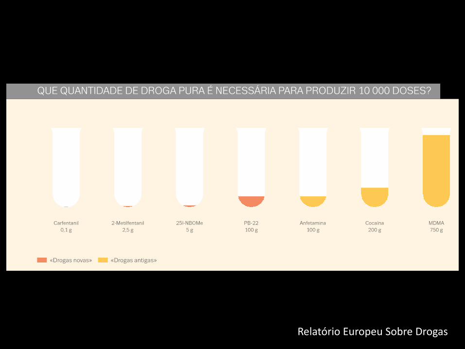
Relatório Europeu Sobre Drogas

Identificação de Novas Substâncias Psicoativas(NPS)
UNODC, 2014

Apreensões de Novas SubstânciasPsicoativas
UNODC, 2014


UNODC, 2014

Visitas a emergência por intoxicação
Maxwell JC, Drug Alcohol Dep, 2014

• 5% dos jovens europeus já fizeram uso de alguma nova droga sintética
EMMCDA, 2014

Apreensões Ecstasy America Latina
UNODC, 2014

Uso de “substâncias de pesquisa” nos últimos 12 mesesentre os participantes do GDS2015


Ecstasy x Cocaína
LENAD II, resultados parciais

Composição dos Comprimidos de Ecstasy no Brasil
Togni LR et al, J Forensic Sci, 2014

Plano de apresentação
• MDMA
• N-BOME
• Quetamina


“Quantos de vocês viram a Molly hoje?”
Madonna Ultra Music Festival, Miami, 2012


MDMA3-4 metilenodioximetanfetamina
EMCDA, 2013

MDMA3-4 metilenodioximetanfetamina
1912 – Síntese pelo laboratório Merck
1950 – Primeiros estudos em animais
1978 – Primeira descrição de seus efeitos
1980 – Inicio do uso recreativo
1985 – Proibida nos Estados Unidos
Meyer JS, Subst Abuse Reab 2013

Nomenclatura
• Ecstasy
• Bala
• XTC
• A
• E
• MD
• ADAM


Mecanismo de Ação
• Ligação ao transportador de monoaminas
– Serotonina
– Dopamina
– Noradrenalina

Principais Vias
• Estriado
• Nucleo da Rafe
• Neocortex
• Amidala
• Hipocampo
NIDA

Ocupação do Transportador
dancesafe.org

Liberação Mediada por Transportador
dancesafe.org

Inundação de 5-HT, DA, NE na FendaSináptica
dancesafe.org

Dopamina
• Euforia
• Excitabilidade
• Reforço ao uso

Noradrenalina
• Taquicardia
• Aumento da pressão arterial
• Hipertermia
Hysek CM et al, Br J Pharmacol, 2012

Serotonina
• Alterações de percepção – 5HT2a
• Prazer no movimento e toque
• Efeito pró-social – 5HT1a
– Empatia
– Proximidade com o outro
– Conexão com o mundo ou com sentimentos
Meyer JS, Subst Abuse Reab 2013

Ocitocina e MDMA
• 5-HT1a estimula liberação de ocitocina nos núcleos supraóptico e paraventricular
• Ativação de circuitos de “affiliative behavior”
• Hiponatremia, desidratação e hipertermia aumentam ainda mais a secreção de ocitocina
McGregor IS, Hormon Behav 2012

Efeitos Pós-Uso Agudo
• Midweekend blues
• Alterações cognitivas
Depleção de serotonina
– Bloqueio do transportador de monoaminas
– Bloqueio da triptofano-hidroxilase

Efeitos Crônicos
• Trajetória de uso de cerca de 1 ano
– 1 a 2 comprimidos por semana
• Alteração de Funções cognitivas superiores
• Maior presença de sintomas psiquiátricos
Smirnov et al, Addict Behav, 2013; Parrot ES, Neurosci Biobehav Rev, 2013
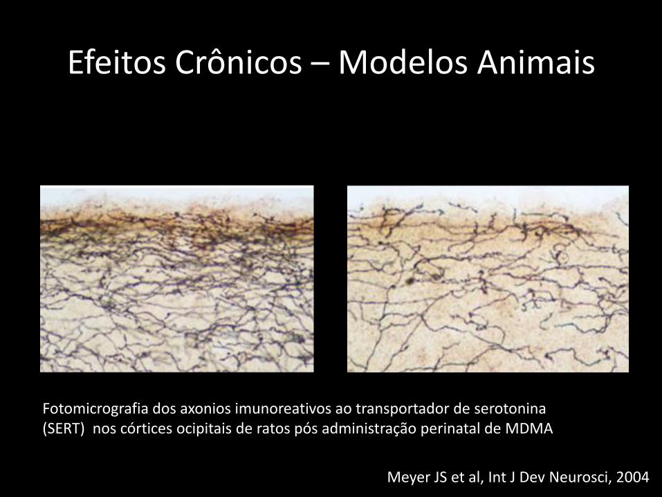
Efeitos Crônicos – Modelos Animais
Substance Abuse and Rehabilitation 2013:4submit your manuscript | www.dovepress.com
Dovepress
Dovepress
90
Meyer
Fisk and Sharp68 proposed that working memory consists
of a central executive function along with four subcompo-
nents which they termed “updating,” “attention shifting,”
“inhibition,” and “access to long-term memory.” A recent
meta-analysis comparing ecstasy users to polydrug-using
controls on these four subcomponents found significant
ecstasy-associated deficits in updating, attention shifting, and
access to long-term memory.69 Effect sizes were generally
moderate for the three significant subcomponents, whereas
the inhibition subcomponent was not significantly affected.
Meta-analyses that examined aspects of cognitive function
other than memory found significant impairment in attention
and concentration, verbal comprehension, processing speed,
and motor/psychomotor speed.70,71 Together with the reviews
of memory performance, these findings suggest that regular
ecstasy users suffer from widespread problems across a wide
range of cognitive domains.
Despite the positive findings from a number of different
analyses, the literature on cognitive deficits in ecstasy users
suffers from some of the same interpretive problems as the lit-
erature on mood changes. For example, Gouzoulis-Mayfrank
and Daumann19 discuss the issue that, even if ecstasy users
are compared to drug users who do not consume ecstasy, the
latter often have a more moderate pattern of drug consumption
than the ecstasy-using group. Cannabis is particularly prob-
lematic in studies of cognitive function, given the evidence
for significant cognitive deficits in heavy cannabis users.72
Comorbidity of psychopathology with ecstasy use may
additionally contribute to the cognitive impairment observed
in some studies.73 Finally, the typical use of cross-sectional
studies again makes it impossible, in such cases, to ascertain
whether cognitive differences between the ecstasy-using and
control group(s) preceded or followed the onset of ecstasy
use. Two prospective studies have addressed this issue by
recruiting new ecstasy users, assessing their cognitive function
at baseline, and then retesting the subjects from 1 to 3 years
later.74,75 Interestingly, in both cases, the users did not differ
from controls at baseline but did exhibit significant deficits in
immediate and delayed verbal recall memory. These findings
point up the limitations inherent in cross-sectional studies in
which baseline mood or cognitive function prior to the onset
of ecstasy use is not known. Importantly, the results suggest
that, at least for some ecstasy users, repeated exposure to the
drug leads to later impairment in certain cognitive domains.
Longitudinal studies of up to 2 years in length have also
asked whether cognitive function keeps declining with con-
tinued ecstasy use, and whether cessation of use leads to a
recovery of cognitive function. In general, such studies have
found neither further deterioration in users nor recovery of
function in subjects who discontinued ecstasy use76–78 (see
Zakzanis and Campbell79 for an exception to these findings).
Because in these studies the ecstasy users already differed from
controls at the time of initial testing, the results are difficult to
interpret. There may have been preexisting cognitive differences
that were unchanged by ecstasy use, or the typical use patterns
of the subjects in the first three studies may have caused an
asymptotic decline in cognitive performance that neither got
worse over time nor recovered upon 2 years of abstinence.
NeurotoxicityThe abovementioned evidence for mood changes and neurop-
sychological deficits in ecstasy users raises the question of
how MDMA might be acting within the brain to cause these
effects. The answer given most often centers around numer-
ous findings obtained from experimental animals, mainly
rats and nonhuman primates (eg, squirrel monkeys), for a
toxic effect of MDMA on the serotonergic system. The first
evidence for MDMA neurotoxicity was published in the mid-
1980s, and these and numerous follow-up studies showed that
high doses of MDMA (or MDA) lead to a long-lasting deple-
tion of 5-HT, reduced 5-HT uptake and SERT binding sites,
and decreased activity of tryptophan hydroxylase in forebrain
areas such as the neocortex, hippocampus, and striatum (see
Green et al33). Time-course studies demonstrated a biphasic
action of MDMA consisting of the initial 5-HT depletion
discussed earlier, a short-term recovery of 5-HT levels seen
at 1–2 days following dosing, and then a secondary, more
long-lasting depletion measured at 1–2 weeks and beyond.33
Histological examination of brain tissues from treated rats
and monkeys consistently showed a persistent reduction in
the density of axons immunoreactive for 5-HT or for SERT
(Figure 1). Moreover, staining of brain tissues obtained at
Figure 1 Photomicrograph of serotonin transporter-immunoreactive axons
in the upper layers of occipital cortex of a control rat (A) compared to a 3,4-
methylenedioxymethamphetamine (MDMA)-treated rat ( B).
Notes: The animals received twice-daily injections of either saline or MDMA
(10 mg/kg peq injection) fqom the fiqs t to the fouqth day of life and then weqe
killed at 9 months of age (see Meyer et al80 for experimental details). These results
illustqate the long-lasting natuqe of MDMA-induced seqotoneqgic defici ts when the
drug is administered early in development.
Fotomicrografia dos axonios imunoreativos ao transportador de serotonina (SERT) nos córtices ocipitais de ratos pós administração perinatal de MDMA
Meyer JS et al, Int J Dev Neurosci, 2004

Efeitos Crônicos - Humanos
Urban NB et al, neuropsychophamacology, 2012

Efeitos Crônicos - Humanos
Iorio CR et al, Arch Gen Psych, 2012

Mecanismos de Neurotoxidade
• Pouco elucidado
• Envolve Espécies Ativas de Oxigênio (ROS)
• Hipotese do metabólito neurotóxico
• Hipotese do influxo de dopamina nos neurônios serotonérgicos
Meyer JS, Subst Abuse Reab 2013

Morbi-Mortalidade
• Série de casos de uma rave em San Francisco:
– 12 pacientes admitidos em CTI
– 10 deles com temperaturas de 40-43oC
– 8 necessitaram entubação endotraqueal
– 2 óbitos
• Análise de 106 casos de óbitos por MDMA
– 43% envolviam antidepressivos ou fármacos serotonérgicos
Pilgrim JL et al 2011; Armenian P et al, 2013


“Seu Doce era LSD ou N-BOME?”
Anônimo
Web Forum

N-BOME


N-BOME
• Família de compostos derivados da feniletilamina
• Notoriedade recente devido a múltiplos óbitos relacionados com overdose
• Ingerido inadvertidamente com LSD
• Vendido livremente em webpages
Ninneman A et al, 2013; Bersani FS et al, 2013, Zuba D, 2013
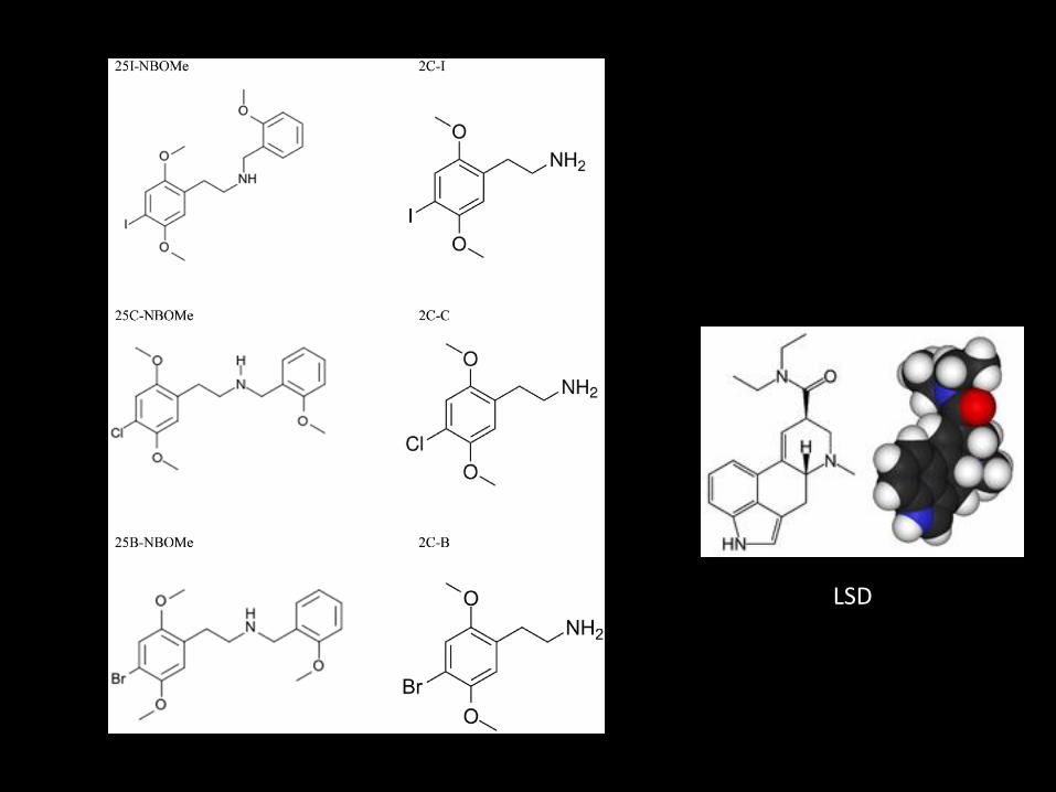
LSD

Nomenclatura
• N-Bomb
• Pandora
• Boom
• Holland Film
• Dime

Mecanismo de Ação
• Potente agonista parcial 5HT2A
• Efeitos lembram os alucinógenos clássicos como LSD com algumas características de anfetaminas
Bersani FS et al, 2013

Efeitos
• Adormecimento da língua
– Diferencia da intoxicação por LSD
• Body high: formigamento em todo corpo
• Euforia
• Agitação psicomotora intensa
Bersani FS et al, 2013
Efeitos Psicodélicos
• Instrospeção
• Despersonalização
• Desrealização
• Alucinações visuais e auditivas
– Reconhecimento de padrões
– Borramento visual
– Tracing
Bersani FS et al, 2013

Litgens RP et al 2014

Litgens RP et al 2014

Litgens RP et al 2014

Modulação de Impulsos Visuais porNeurônios Serotonérgicos
Litgens RP et al 2014

Toxicidade
• Confusão
• Agitação psicomotora
• Hipertensão
• Taquicardia
• Hipertermia
• Convulsões
• Acidose metabólica
• Falência de múltiplos orgãos
SíndromeSerotonérgica

Toxicidade
• 29 óbitos por toxicidade aguda
• 25 casos adicionais relatados no EMMCDA
• Muito mais tóxico que o ácido lisérgico
Bersani FS et al, 2013 EMMCDA, 2014


Quetamina
“A peeping tom at the keyhole of eternity”
John Lilly, médico, final dos anos 70


Nomenclatura
• Special K
• Ket
• K
• The Horse anesthesic
• Calvin Klein ( cocaína e quetamina)

História
• Introduzido como anestésico em 1964
• Visava substituir fenciclidina
• Atualmente quase restrita ao veterinário
• Usada em modelos animais de esquizofrenia
• Ensaios como antidepressivo
Morgan CJ et al, 2012

Uso Não Médico
• 1960: primeiros relatos de abuso
• 1990: adulterante em comprimidos de ecstasy
• 2001: 25% de frequentadores de raves já haviam utilizado no reino unido
• 2009: 68% frequentadores de raves no reino unido
• Não há dados quanto a prevalência de dependência
Morgan CJ et al, 2012, McCambridge, 2007, MixMag 2010

Mecanismo de Ação
• Atuação em vias glutamatérgias
• Antagonista não competitivo de receptores NMDA
• Menor efeito em GABA, dopamina e receptores muscarínicos
Morgan CJ et al, 2012

Efeitos Psicoativos
• Distorção do tempo e espaço
• Alucinações
• Efeitos dissociativos
• Sensação de “derreter no ambiente”
• Experiências extracorpóreas
• K-HOLE
– Estado intenso de dissociação da realidade
Stewart C, 2001

Riscos Agudos
Green SM et al 1999, Schifino F, 2008

Riscos Agudos
• Série de casos com doses acima de 100x a margem de segurança sem relato de efeitos letais
• Risco elevado de acidentes
• Risco cardiovascular quando associado a estimulantes
Green SM et al 1999, Morgan CJ et al, 2012

Riscos Crônicos
• Cistite ulcerativa
• Hidronefrose
• Alterações de função hepática
• Dilatação de vias biliares
Morgan CJ et al, 2012

Riscos Crônicos - SNC
• Sintomas depressivos em usuários diários
• Graves deficits cognitivos
• Lesões em substância branca cerebral• Diminuição da integração de certas areas corticais
Liang HJ, 2013; Roberts RE, 2014

Considerações Finais
• Aumento Global do consumo de drogas sintéticas
• Parecem ser mais potentes que as drogas originais
• Poucas informações sobre seu uso crônico
• Dificuldades legais para controlar o uso das novas substâncias psicoativas
• Desafio para governos, sociedade e profissionais de saúde