Embed Size (px)
Citation preview
376
CHAPTER
21Alterations in
Respiratory Function:Disorders of Gas Exchange
Disorders of Lung InflationDisorders of the Pleura
Pleuritis and Pleural PainPleural EffusionPneumothorax
AtelectasisObstructive Airway Disorders
Physiology of Airway DiseaseBronchial Asthma
PathogenesisClinical FeaturesStatus Asthmaticus and Fatal AsthmaBronchial Asthma in Children
Chronic Obstructive Pulmonary DiseaseEmphysemaChronic BronchitisClinical Features
BronchiectasisCystic Fibrosis
Interstitial Lung DiseasesPulmonary Vascular Disorders
Pulmonary EmbolismPulmonary Hypertension
Secondary Pulmonary HypertensionPrimary Pulmonary HypertensionCor Pulmonale
Acute Respiratory Distress Syndrome
Respiratory FailureAlterations in Blood Gases
Mechanisms of Altered Gas ExchangeHypoxemiaHypercapnia
Treatment of Respiratory Failure
T he major function of the lungs is to oxygenate and removecarbon dioxide from the blood as a means of supportingthe metabolic functions of body cells. The gas exchange
function of the lungs depends on a system of open airways, ex-pansion of the lungs, an adequate area for gas diffusion, andblood flow that carries the gases to the rest of the body. Thischapter focuses on diseases that disrupt ventilation and gas ex-change and on respiratory failure and hyperventilation.
DISORDERS OF LUNG INFLATION
Air entering through the airways inflates the lung, and the neg-ative pressure in the pleural cavity keeps the lung from col-lapsing. Disorders of lung inflation are caused by conditionsthat produce lung compression or lung collapse. There can becompression of the lung by an accumulation of fluid in the in-trapleural space, complete collapse of an entire lung as in pneu-mothorax, or collapse of a segment of the lung as in atelectasis.
Disorders of the PleuraThe pleura is a thin, double-layered membrane that encases thelungs. The two layers of the pleurae are separated by a thin layerof serous fluid (Fig. 21-1). The right and left pleural cavities areseparated by the mediastinum, which contains the heart andother thoracic structures. Both the chest wall and the lungs haveelastic properties. The pressure in the pleural cavity, which isnegative in relation to atmospheric pressure, holds the lungs

against the chest wall and keeps them from collapsing (seeChapter 19). Disorders of the pleura include pleural pain,pleural effusion, and pneumothorax.
Pleuritis and Pleural PainPain is a common symptom of pleuritis, or inflammation ofthe pleura. Pleuritis is common in infectious processes such asviral respiratory infections or pneumonia that extend to in-volve the pleura. Most commonly the pain is abrupt in onset,such that the person experiencing it can cite almost to theminute when the pain started. It usually is unilateral and tendsto be localized to the lower and lateral part of the chest. Whenthe central part of the diaphragm is irritated, the pain may bereferred to the shoulder. The pain is usually made worse bychest movements, such as deep breathing and coughing, thatexaggerate pressure changes in the pleural cavity and increasemovement of the inflamed or injured pleural surfaces. Becausedeep breathing is painful, tidal volumes usually are kept small,and breathing becomes more rapid. Reflex splinting of thechest muscles may occur, causing a lesser respiratory excursionon the affected side.
It is important to differentiate pleural pain from pain pro-duced by other conditions, such as musculoskeletal strain ofchest muscles, bronchial irritation, and myocardial disease.Musculoskeletal pain may occur as the result of frequent, force-ful coughing. This type of pain usually is bilateral and locatedin the inferior portions of the rib cage, where the abdominalmuscles insert into the anterior rib cage. It is made worse bymovements associated with contraction of the abdominal mus-cles. The pain associated with irritation of the bronchi usuallyis substernal and dull, rather than sharp, in character. It is madeworse with coughing but is not affected by deep breathing.Myocardial pain, which is discussed in Chapter 17, usually islocated in the substernal area and is not affected by respiratorymovements.
Pleural EffusionPleural effusion refers to an abnormal collection of fluid in the pleural cavity (see Fig. 21-1). The fluid may be a transu-date, exudate, purulent drainage (empyema), chyle, or blood.
Normally, only a thin layer (<10 to 20 mL) of serous fluidseparates the visceral and parietal layers of the pleural cavity.Like fluid developing in other transcellular spaces in thebody, pleural effusion occurs when the rate of fluid formationexceeds the rate of its removal (see Chapter 6). Five mecha-nisms have been linked to the abnormal collection of fluid inthe pleural cavity: (1) increased capillary pressure, as in con-gestive heart failure; (2) increased capillary permeability,which occurs with inflammatory conditions; (3) decreasedcolloidal osmotic pressure, such as the hypoalbuminemia oc-curring with liver disease and nephrosis; (4) increased nega-tive intrapleural pressure, which develops with atelectasis;and (5) impaired lymphatic drainage of the pleural space,which results from obstructive processes such as mediastinalcarcinoma.
The accumulation of a serous transudate (clear fluid) in thepleural cavity often is referred to as hydrothorax. The conditionmay be unilateral or bilateral. The most common cause ofhydrothorax is congestive heart failure.1 Other causes are renalfailure, nephrosis, liver failure, and malignancy. An exudate is apleural fluid that has a specific gravity greater than 1.020 and,often, inflammatory cells. Conditions that produce exudativepleural effusions are infections, pulmonary infarction, malig-nancies, rheumatoid arthritis, and lupus erythematosus.
Empyema refers to pus in the pleural cavity. It is caused bydirect infection of the pleural space from an adjacent bacterialpneumonia, rupture of a lung abscess into the pleural space, in-vasion from a subdiaphragmatic infection, or infection associ-ated with trauma.
Chylothorax is the effusion of lymph in the thoracic cavity.2
Chyle, a milky fluid containing chylomicrons, is found in thelymph fluid originating in the gastrointestinal tract. The tho-racic duct transports chyle to the central circulation. Chylo-thorax also results from trauma, inflammation, or malignantinfiltration obstructing chyle transport from the thoracicduct into the central circulation. It also can occur as a com-plication of intrathoracic surgical procedures and use of thegreat veins for total parenteral nutrition and hemodynamicmonitoring.
Hemothorax is the presence of blood in the pleural cavity.Bleeding may arise from chest injury, a complication of chestsurgery, malignancies, or rupture of a great vessel such as anaortic aneurysm. It is usually diagnosed by the presence ofblood in the pleural fluid. Hemorrhagic pleural fluid is a mix-ture of blood and pleural fluid. Hemothorax usually requiresdrainage, and if the bleeding continues, surgery to control thebleeding may be required.
The manifestations of pleural effusion vary with the cause.Hemothorax may be accompanied by signs of blood loss andempyema by fever and other signs of inflammation. Fluid inthe pleural cavity acts as a space-occupying mass; it causes a de-crease in lung expansion on the affected side that is propor-tional to the amount of fluid that is present. The effusion maycause a shift in the mediastinal structures toward the oppositeside of the chest with a decrease in lung volume on that side aswell as the side with the pneumothorax. Characteristic signs ofpleural effusion are dullness or flatness to percussion and di-minished breath sounds. Dyspnea, the most common symp-tom, occurs when fluid compresses the lung, resulting in de-creased ventilation. Pleuritic pain usually occurs only wheninflammation is present, although constant discomfort may be
377Chapter 21: Alterations in Respiratory Function: Disorders of Gas Exchange
■ FIGURE 21-1 ■ The parietal and visceral pleura and site of fluidaccumulation in pleural effusions.
Visceralpleura
Pleuralcavity
Parietalpleura

felt with large effusions. Mild hypoxemia may occur and usu-ally is corrected with supplemental oxygen.
Diagnosis of pleural effusion is based on chest radiographs,chest ultrasound, and computed tomography (CT). Thora-centesis is the aspiration of fluid from the pleural space. It canbe used to obtain a sample of pleural fluid for diagnosis, or itcan be used for therapeutic purposes. The treatment of pleuraleffusion is directed at the cause of the disorder. With large ef-fusions, thoracentesis may be used to remove fluid from theintrapleural space and allow for re-expansion of the lung. A pal-liative method used for treatment of pleural effusions causedby a malignancy is the injection of a sclerosing agent into thepleural cavity. This method of treatment causes obliteration ofthe pleural space and prevents the reaccumulation of fluid.Open surgical drainage may be necessary in cases of continuedeffusion.
PneumothoraxNormally, the pleural cavity is free of air and contains only athin layer of fluid. When air enters the pleural cavity, it iscalled pneumothorax. Pneumothorax causes partial or completecollapse of the affected lung. Pneumothorax can occur with-out an obvious cause or injury (i.e., spontaneous pneumo-thorax) or as a result of direct injury to the chest or major air-ways (i.e., traumatic pneumothorax). Tension pneumothoraxdescribes a life-threatening condition of excessive pressure inthe pleural cavity.
Spontaneous Pneumothorax. Spontaneous pneumothoraxoccurs when an air-filled bleb, or blister, on the lung surfaceruptures. Rupture of these blebs allows atmospheric air fromthe airways to enter the pleural cavity (Fig. 21-2). Because alve-olar pressure normally is greater than pleural pressure, air flowsfrom the alveoli into the pleural space, causing the involvedportion of the lung to collapse as a result of its own recoil. Aircontinues to flow into the pleural space until a pressure gradi-ent no longer exists or until the decline in lung size causes theleak to seal. Spontaneous pneumothoraces can be divided intoprimary and secondary pneumothoraces.3 Primary sponta-
neous pneumothorax occurs in otherwise healthy persons.Secondary spontaneous pneumothorax occurs in persons withunderlying lung disease.
In primary spontaneous pneumothorax, the air-filled blebthat ruptures is usually on the top of the lung. The condition isseen most often in tall boys and young men between 10 and 30 years of age.3 It has been suggested that the difference inpleural pressure from the top to the bottom of the lung is greaterin tall persons and that this difference in pressure may contrib-ute to the development of blebs. Another factor that has beenassociated with primary spontaneous pneumothorax is smok-ing. Disease of the small airways related to smoking probablycontributes to the condition.
Secondary spontaneous pneumothoraces usually are moreserious because they occur in persons with lung disease. Theyare associated with many different types of lung conditionsthat cause trapping of gases and destruction of lung tissue,including asthma, tuberculosis, cystic fibrosis, sarcoidosis,bronchogenic carcinoma, and metastatic pleural diseases. Themost common cause of secondary spontaneous pneumothoraxis emphysema.
Traumatic Pneumothorax. Traumatic pneumothorax may becaused by penetrating or nonpenetrating chest injuries. Frac-tured or dislocated ribs that penetrate the pleura are the mostcommon cause of pneumothorax from nonpenetrating chest in-juries. Hemothorax often accompanies these injuries. Pneumo-thorax also may accompany fracture of the trachea or majorbronchus or rupture of the esophagus. Persons with pneumo-thorax caused by chest trauma frequently have other compli-cations and may require chest surgery. Medical procedures suchas transthoracic needle aspirations, intubation, and positive-pressure ventilation occasionally may cause pneumothorax.Traumatic pneumothorax also can occur as a complication ofcardiopulmonary resuscitation.
Tension Pneumothorax. Tension pneumothorax occurs whenthe intrapleural pressure exceeds atmospheric pressure. It is alife-threatening condition and occurs when injury to the chestor respiratory structures permits air to enter but not leave thepleural space (Fig. 21-3). This results in a rapid increase inpressure in the chest with a compression atelectasis of the un-affected lung, a shift in the mediastinum to the opposite sideof the chest, and compression of the vena cava with impair-ment of venous return to the heart.4 Although tension pneumo-thorax can develop in persons with spontaneous pneumo-thoraces, it is seen most often in persons with traumaticpneumothoraces.
With tension pneumothorax, the structures in the mediasti-nal space shift toward the opposite side of the chest (see Fig.21-3). When this occurs, the position of the trachea, normallylocated in the midline of the neck, deviates with the medi-astinum. There may be distention of the neck veins and subcu-taneous emphysema (i.e., air bubbles in the subcutaneoustissues of the chest and neck) and clinical signs of shock.
Clinical Features. The manifestations of pneumothorax de-pend on its size and the integrity of the underlying lung. Inspontaneous pneumothorax, manifestations of the disorder in-clude development of ipsilateral (same side) chest pain in anotherwise healthy person. There is an almost immediate in-
378 Unit Five: Alterations in the Respiratory System
■ FIGURE 21-2 ■ Mechanism for development of spontaneouspneumothorax.
Rupturedbleb
Pleuralspace

crease in respiratory rate, often accompanied by dyspnea thatoccurs as a result of the activation of receptors that monitorlung volume. Heart rate is increased. Asymmetry of chestmovement may occur because of the air trapped in the pleuralcavity on the affected side. Percussion of the chest produces amore hyperresonant sound, and breath sounds are decreasedor absent over the area of the pneumothorax.
Hypoxemia usually develops immediately after a largepneumothorax, followed by vasoconstriction of the blood ves-sels in the affected lung, causing the blood flow to shift to theunaffected lung. In persons with primary spontaneous pneu-mothorax, this mechanism usually returns oxygen saturationto normal within 24 hours. Hypoxemia usually is more seriousin persons with underlying lung disease in whom secondaryspontaneous pneumothorax develops. In these persons, thehypoxemia caused by the partial or total loss of lung functioncan be life threatening.
Diagnosis of pneumothorax can be confirmed by chest ra-diograph or CT scan. Blood gas analysis may be done to de-termine the effect of the condition on blood oxygen levels.Treatment varies with the cause and extent of the disorder.Even without treatment, air in the pleural space usually reab-sorbs after the pleural leak seals. In small spontaneous pneu-mothoraces, the air usually reabsorbs, and observation and
follow-up chest radiographs are all that is required. Sup-plemental oxygen may be used to increase the rate at which theair is reabsorbed. In larger pneumothoraces, the air is removedby needle aspiration or a closed drainage system used with orwithout an aspiration pump. This type of drainage system usesa one-way valve or a tube submerged in water to allow air to exit the pleural space and prevent it from re-entering thechest. In secondary pneumothorax, surgical closure of thechest wall defect, ruptured airway, or perforated esophagusmay be required.
Emergency treatment of tension pneumothorax involves theprompt insertion of a large-bore needle or chest tube into theaffected side of the chest along with one-way valve drainage orcontinuous chest suction to aid in lung expansion. Suckingchest wounds, which allow air to pass in and out of the chestcavity, should be treated by promptly covering the area with anairtight covering. Chest tubes are inserted as soon as possible.
AtelectasisAtelectasis refers to the incomplete expansion of a lung or por-tion of a lung. It can be caused by airway obstruction, lungcompression such as occurs in pneumothorax or pleural effu-sion, or the increased recoil of the lung caused by inadequatepulmonary surfactant (see Chapter 19).
Atelectasis is caused most commonly by airway obstruction(Fig. 21-4). Obstruction can be caused by a mucus plug in theairway or by external compression by fluid, tumor mass, exu-date, or other matter in the area surrounding the airway. Asmall segment of lung or an entire lung lobe may be involvedin obstructive atelectasis. Complete obstruction of an airway isfollowed by the absorption of air from the dependent alveoliand collapse of that portion of the lung. The danger of ob-structive atelectasis increases after surgery. Anesthesia, pain,administration of narcotics, and immobility tend to promoteretention of viscid bronchial secretions and thus airwayobstruction.
Another cause of atelectasis is compression of lung tissue.It occurs when the pleural cavity is partially or completelyfilled with fluid, exudate, blood, a tumor mass, or air. It is ob-served most commonly in persons with pleural effusion from
379Chapter 21: Alterations in Respiratory Function: Disorders of Gas Exchange
Tension Pneumothorax
Open Pneumothorax
Inspiration Expiration
Inspiration Expiration
■ FIGURE 21-3 ■ Open or communicating pneumothorax (top)and tension pneumothorax (bottom). In an open pneumothorax,air enters the chest during inspiration and exits during expiration.There may be slight inflation of the affected lung due to a decreasein pressure as air moves out of the chest. In tension pneumotho-rax, air can enter but not leave the chest. As the pressure in thechest increases, the heart and great vessels are compressed andthe mediastinal structures are shifted toward the opposite side ofthe chest. The trachea is pushed from its normal midline positiontoward the opposite side of the chest, and the unaffected lung iscompressed.
Absorption Compression
■ FIGURE 21-4 ■ Atelectasis caused by airway obstruction andabsorption of air from the involved lung area on the left and bycompression of lung tissue on the right.

congestive heart failure or cancer. In compression atelectasis,the mediastinum shifts away from the affected lung.
The clinical manifestations of atelectasis include tachyp-nea, tachycardia, dyspnea, cyanosis, signs of hypoxemia, di-minished chest expansion, absence of breath sounds, andintercostal retractions. Fever and other signs of infection maydevelop. Both chest expansion and breath sounds are de-creased on the affected side. There may be intercostal retrac-tion (pulling in of the intercostal spaces) over the involvedarea during inspiration. If the collapsed area is large, themediastinum and trachea shift to the affected side. Signs ofrespiratory distress are proportional to the extent of lungcollapse.
The diagnosis of atelectasis is based on signs and symptoms.Chest radiographs are used to confirm the diagnosis. CT scansmay be used to show the exact location of the obstruction.Treatment depends on the cause and extent of lung involve-ment. It is directed at reducing the airway obstruction or lungcompression and at reinflating the collapsed area of the lung.Ambulation and body positions that favor increased lung ex-pansion are used when appropriate. Administration of oxygenmay be needed to treat the hypoxemia. Bronchoscopy may beused as a diagnostic and treatment method.
Physiology of Airway DiseaseAir moves through the upper airways (i.e., trachea and majorbronchi) into the lower or pulmonary airways (i.e., bronchiand alveoli), which are located in the lung. In the pulmonaryairways, the cartilaginous layer that provides support for thetrachea and major bronchi gradually disappears and is replacedwith crisscrossing strips of smooth muscle (see Chapter 19).The contraction and relaxation of the smooth muscle layer,which is innervated by the autonomic nervous system (ANS),controls the diameter of the airways and consequent resistanceto airflow. Parasympathetic stimulation, through the vagusnerve and cholinergic receptors, produces bronchoconstriction,and sympathetic stimulation, through β2-adrenergic receptors,increases bronchodilation. Normally, a slight vagal-mediatedbronchoconstrictor tone predominates. When there is need forincreased airflow, as during exercise, the vagal-mediated bron-choconstrictor tone is inhibited, and the bronchodilator effectsof the sympathetic nervous system are increased.
Bronchial smooth muscle also responds to inflammatorymediators, such as histamine, that act directly on smooth mus-cle cells to produce bronchoconstriction. During an antigen-antibody response, inflammatory mediators are released by aspecial type of cell, called the mast cell, which is present in theairways. The binding of immunoglobulin E (IgE) antibodies toreceptors on mast cells prepares them for an allergic responsewhen antigen appears (see Chapter 10).
Bronchial AsthmaBronchial asthma is a chronic inflammatory airway disease.According to 1998 data, an estimated 26 million Americanshave received diagnoses of asthma, and 10.6 million have had
380 Unit Five: Alterations in the Respiratory System
In summary, lung inflation depends on a negative intrapleural pressure and unobstructed intrapulmonary airways. Disorders of the pleura include pleuritis and pain,pleural effusion, and pneumothorax. Pain is commonly asso-ciated with conditions that produce inflammation of thepleura. Characteristically, it is unilateral, abrupt in onset, andexaggerated by respiratory movements. Pleural effusionrefers to the abnormal accumulation of fluid in the pleuralcavity. The fluid may be a transudate (i.e., hydrothorax),exudate (i.e., empyema), blood (i.e., hemothorax), or chyle(i.e., chylothorax). Pneumothorax refers to an accumulationof air in the pleural cavity with the partial or complete col-lapse of the lung. It can result from rupture of an air-filledbleb on the lung surface or from penetrating or nonpenetrat-ing injuries. A tension pneumothorax is a life-threateningevent in which air progressively accumulates in the thorax,collapsing the lung on the injured side and progressivelyshifting the mediastinum to the opposite side of the thorax,producing severe cardiorespiratory impairment.
Atelectasis refers to an incomplete expansion of the lung.In adults, atelectasis usually results from airway obstructioncaused by mucus plug or because of external compression byfluid, tumor mass, exudate, or other matter in the area surrounding the airway.
OBSTRUCTIVE AIRWAY DISORDERS
Obstructive airway diseases are caused by disorders that limitexpiratory airflow. Bronchial asthma represents a reversibleform of airway disease caused by narrowing of airways due tobronchospasm, inflammation, and increased airway secretions.Chronic obstructive airway disease can be caused by a varietyof airway diseases, including chronic bronchitis, emphysema,bronchiectasis, and cystic fibrosis.
KEY CONCEPTS
AIRWAY DISORDERS
■ Airway disorders involve the movement of gases intoand out of the lung. They involve bronchial smoothmuscle tone, mucosal injury, and obstruction due tosecretions.
■ The tone of the bronchial smooth muscles surround-ing the airways determines airway radius, and thepresence or absence of airway secretions influencesairway patency.
■ Bronchial smooth muscle is innervated by the auto-nomic nervous system—the parasympathetic ner-vous system, via the vagus nerve, produces bron-choconstriction and the sympathetic nervous system produces bronchodilation.
■ Inflammatory mediators that are released in responseto environmental irritants, immune responses, andinfectious agents increase airway responsiveness byproducing bronchospasm, increasing mucus secre-tion, and producing injury to the mucosal lining ofthe airways.

an asthma episode during the past 12 months.5 Of the 26 mil-lion Americans with diagnoses of asthma, 8.6 million areyounger than 18 years. In the general population, asthmaprevalence rates increased 102% between 1980 and 1994.5
There also has been a reported increase in incidence and mor-tality associated with asthma during the past several decades.
The National Heart, Lung, and Blood Institute’s SecondExpert Panel on the Management of Asthma defined bronchialasthma as “a chronic inflammatory disorder of the airways inwhich many cells and cellular elements play a role, in partic-ular, mast cells, eosinophils, T lymphocytes, and epithelialcells.”6 This inflammatory process produces recurrent episodesof airway obstruction, characterized by wheezing, breathless-ness, chest tightness, and a cough that often is worse at nightand in the early morning. These episodes, which usually are re-versible either spontaneously or with treatment, also cause anassociated increase in bronchial responsiveness to a variety ofstimuli.6
PathogenesisIn susceptible persons, an asthma attack can be triggered by avariety of stimuli that do not normally cause symptoms. Basedon their mechanism of response, these triggers can be dividedinto two categories—bronchospastic or inflammatory. Broncho-spastic triggers depend on the existing level of airway respon-siveness. They do not normally increase airway responsiveness
but produce symptoms in persons who already are predisposedto bronchospasm. Bronchospastic triggers include cold air, exer-cise, emotional upset, and exposure to bronchial irritantssuch as cigarette smoke. Inflammatory triggers exert their effectsthrough the inflammatory response. They cause inflammationand prime the sensitive airways so they are hyperresponsive tononallergic stimuli. The mechanisms whereby these two typesof triggers produce an asthmatic attack can be further describedas the early or acute response versus the late phase response.7The acute or early response results in immediate broncho-constriction on exposure to an inhaled antigen or irritant (Fig. 21-5). The symptoms of the acute response, which usuallydevelop within 10 to 20 minutes, are caused by the release ofchemical mediators from IgE-sensitized mast cells. In the caseof airborne antigens, the reaction occurs when antigen binds tosensitized mast cells on the mucosal surface of the airways.Mediator release results in the infiltration of inflammatory cellsand opening of the mucosal intercellular junctions and en-hancement of antigen movement to the more prevalent sub-mucosal mast cells (Fig. 21-5). In addition, there is broncho-constriction caused by direct stimulation of parasympatheticreceptors, mucosal edema caused by increased vascular perme-ability, and increased mucus secretions.
The late phase response develops 4 to 8 hours after exposureto an asthmatic trigger.7,8 The late phase response involves in-flammation and increased airway responsiveness that prolong
381Chapter 21: Alterations in Respiratory Function: Disorders of Gas Exchange
■ FIGURE 21-5 ■ Mechanisms ofearly and late phase Ig-E mediatedbronchospasm.
Allergen
Mast cells
Release histamine, leukotrienes,interleukins, and prostaglandins
Infiltration of inflammatory cells
Release cytokines, interleukins,and other inflammatory mediators
Edema Epithelial injury
Impaired mucociliary function
Airflowlimitation
Bronchospasm
Increased airwayresponsivenessAirway inflammation
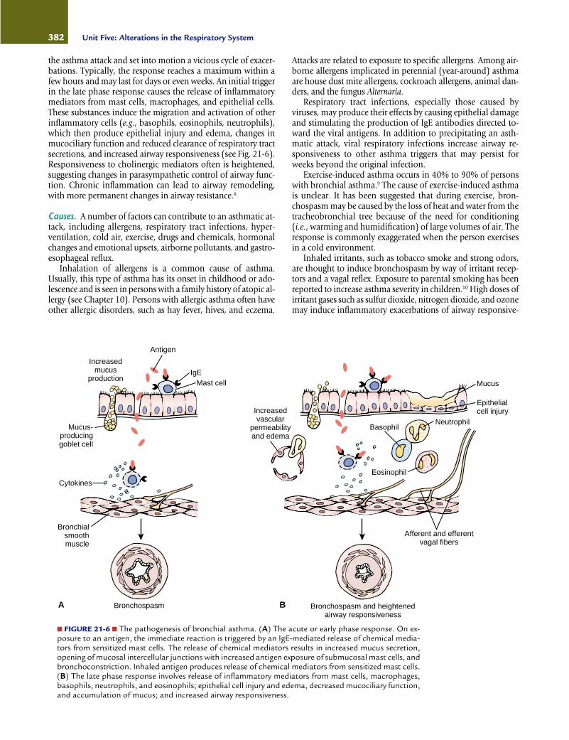
the asthma attack and set into motion a vicious cycle of exacer-bations. Typically, the response reaches a maximum within afew hours and may last for days or even weeks. An initial triggerin the late phase response causes the release of inflammatorymediators from mast cells, macrophages, and epithelial cells.These substances induce the migration and activation of otherinflammatory cells (e.g., basophils, eosinophils, neutrophils),which then produce epithelial injury and edema, changes inmucociliary function and reduced clearance of respiratory tractsecretions, and increased airway responsiveness (see Fig. 21-6).Responsiveness to cholinergic mediators often is heightened,suggesting changes in parasympathetic control of airway func-tion. Chronic inflammation can lead to airway remodeling,with more permanent changes in airway resistance.6
Causes. A number of factors can contribute to an asthmatic at-tack, including allergens, respiratory tract infections, hyper-ventilation, cold air, exercise, drugs and chemicals, hormonalchanges and emotional upsets, airborne pollutants, and gastro-esophageal reflux.
Inhalation of allergens is a common cause of asthma.Usually, this type of asthma has its onset in childhood or ado-lescence and is seen in persons with a family history of atopic al-lergy (see Chapter 10). Persons with allergic asthma often haveother allergic disorders, such as hay fever, hives, and eczema.
Attacks are related to exposure to specific allergens. Among air-borne allergens implicated in perennial (year-around) asthmaare house dust mite allergens, cockroach allergens, animal dan-ders, and the fungus Alternaria.
Respiratory tract infections, especially those caused byviruses, may produce their effects by causing epithelial damageand stimulating the production of IgE antibodies directed to-ward the viral antigens. In addition to precipitating an asth-matic attack, viral respiratory infections increase airway re-sponsiveness to other asthma triggers that may persist forweeks beyond the original infection.
Exercise-induced asthma occurs in 40% to 90% of personswith bronchial asthma.9 The cause of exercise-induced asthmais unclear. It has been suggested that during exercise, bron-chospasm may be caused by the loss of heat and water from thetracheobronchial tree because of the need for conditioning(i.e., warming and humidification) of large volumes of air. Theresponse is commonly exaggerated when the person exercisesin a cold environment.
Inhaled irritants, such as tobacco smoke and strong odors,are thought to induce bronchospasm by way of irritant recep-tors and a vagal reflex. Exposure to parental smoking has beenreported to increase asthma severity in children.10 High doses ofirritant gases such as sulfur dioxide, nitrogen dioxide, and ozonemay induce inflammatory exacerbations of airway responsive-
382 Unit Five: Alterations in the Respiratory System
■ FIGURE 21-6 ■ The pathogenesis of bronchial asthma. (A) The acute or early phase response. On ex-posure to an antigen, the immediate reaction is triggered by an IgE-mediated release of chemical media-tors from sensitized mast cells. The release of chemical mediators results in increased mucus secretion,opening of mucosal intercellular junctions with increased antigen exposure of submucosal mast cells, andbronchoconstriction. Inhaled antigen produces release of chemical mediators from sensitized mast cells.(B) The late phase response involves release of inflammatory mediators from mast cells, macrophages,basophils, neutrophils, and eosinophils; epithelial cell injury and edema, decreased mucociliary function,and accumulation of mucus; and increased airway responsiveness.
Antigen
IgEMast cell
Increasedmucus
production
Mucus-producinggoblet cell
Increasedvascular
permeabilityand edema
Cytokines
Bronchialsmoothmuscle
Bronchospasm
Epithelialcell injury
Mucus
NeutrophilBasophil
Eosinophil
Afferent and efferentvagal fibers
Bronchospasm and heightenedairway responsiveness
A B
ness (e.g., smog-related asthma). Occupational asthma is stim-ulated by fumes and gases (e.g., epoxy resins, plastics, toluene),organic and chemical dusts (i.e., wood, cotton, platinum), andother chemicals (e.g., formaldehyde) in the workplace.11
There is a small group of persons with asthma in whom as-pirin and nonsteroidal anti-inflammatory drugs (NSAIDs) areassociated with asthmatic attacks, the presence of nasal polyps,and recurrent episodes of rhinitis.12 An addition to the list ofchemicals that can provoke an asthmatic attack are the sulfitesused in food processing and as a preservative added to beer,wine, and fresh vegetables.
Both emotional factors and changes in hormone levels arethought to contribute to an increase in asthma symptoms.Emotional factors produce bronchospasm by way of vagalpathways. They can act as a bronchospastic trigger, or they canincrease airway responsiveness to other triggers through non-inflammatory mechanisms. The role of sex hormones inasthma is unclear, although there is much circumstantial evi-dence to suggest they may be important. As many as 40% ofwomen with asthma report a premenstrual increase in asthmasymptoms.13 Female sex hormones have a regulatory role on β2-adrenergic function, and it has been suggested that abnor-mal regulation may be a possible mechanism for premen-strual asthma.13
Symptoms of gastroesophageal reflux are common in bothadults and children with asthma, suggesting that reflux of gas-tric secretions may act as a bronchospastic trigger. Reflux dur-ing sleep can contribute to nocturnal asthma.6
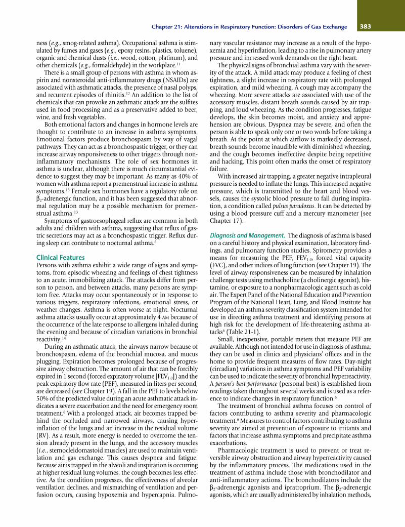
Clinical FeaturesPersons with asthma exhibit a wide range of signs and symp-toms, from episodic wheezing and feelings of chest tightnessto an acute, immobilizing attack. The attacks differ from per-son to person, and between attacks, many persons are symp-tom free. Attacks may occur spontaneously or in response tovarious triggers, respiratory infections, emotional stress, orweather changes. Asthma is often worse at night. Nocturnalasthma attacks usually occur at approximately 4 AM because ofthe occurrence of the late response to allergens inhaled duringthe evening and because of circadian variations in bronchialreactivity.14
During an asthmatic attack, the airways narrow because ofbronchospasm, edema of the bronchial mucosa, and mucusplugging. Expiration becomes prolonged because of progres-sive airway obstruction. The amount of air that can be forciblyexpired in 1 second (forced expiratory volume [FEV1.0]) and thepeak expiratory flow rate (PEF), measured in liters per second,are decreased (see Chapter 19). A fall in the PEF to levels below50% of the predicted value during an acute asthmatic attack in-dicates a severe exacerbation and the need for emergency roomtreatment.6 With a prolonged attack, air becomes trapped be-hind the occluded and narrowed airways, causing hyper-inflation of the lungs and an increase in the residual volume(RV). As a result, more energy is needed to overcome the ten-sion already present in the lungs, and the accessory muscles(i.e., sternocleidomastoid muscles) are used to maintain venti-lation and gas exchange. This causes dyspnea and fatigue.Because air is trapped in the alveoli and inspiration is occurringat higher residual lung volumes, the cough becomes less effec-tive. As the condition progresses, the effectiveness of alveolarventilation declines, and mismatching of ventilation and per-fusion occurs, causing hypoxemia and hypercapnia. Pulmo-
nary vascular resistance may increase as a result of the hypo-xemia and hyperinflation, leading to a rise in pulmonary arterypressure and increased work demands on the right heart.
The physical signs of bronchial asthma vary with the sever-ity of the attack. A mild attack may produce a feeling of chesttightness, a slight increase in respiratory rate with prolongedexpiration, and mild wheezing. A cough may accompany thewheezing. More severe attacks are associated with use of theaccessory muscles, distant breath sounds caused by air trap-ping, and loud wheezing. As the condition progresses, fatiguedevelops, the skin becomes moist, and anxiety and appre-hension are obvious. Dyspnea may be severe, and often theperson is able to speak only one or two words before taking abreath. At the point at which airflow is markedly decreased,breath sounds become inaudible with diminished wheezing,and the cough becomes ineffective despite being repetitiveand hacking. This point often marks the onset of respiratoryfailure.
With increased air trapping, a greater negative intrapleuralpressure is needed to inflate the lungs. This increased negativepressure, which is transmitted to the heart and blood ves-sels, causes the systolic blood pressure to fall during inspira-tion, a condition called pulsus paradoxus. It can be detected byusing a blood pressure cuff and a mercury manometer (seeChapter 17).
Diagnosis and Management. The diagnosis of asthma is basedon a careful history and physical examination, laboratory find-ings, and pulmonary function studies. Spirometry provides ameans for measuring the PEF, FEV1.0, forced vital capacity(FVC), and other indices of lung function (see Chapter 19). Thelevel of airway responsiveness can be measured by inhalationchallenge tests using methacholine (a cholinergic agonist), his-tamine, or exposure to a nonpharmacologic agent such as coldair. The Expert Panel of the National Education and PreventionProgram of the National Heart, Lung, and Blood Institute hasdeveloped an asthma severity classification system intended foruse in directing asthma treatment and identifying persons athigh risk for the development of life-threatening asthma at-tacks6 (Table 21-1).
Small, inexpensive, portable meters that measure PEF areavailable. Although not intended for use in diagnosis of asthma,they can be used in clinics and physicians’ offices and in thehome to provide frequent measures of flow rates. Day-night(circadian) variations in asthma symptoms and PEF variabilitycan be used to indicate the severity of bronchial hyperreactivity.A person’s best performance (personal best) is established fromreadings taken throughout several weeks and is used as a refer-ence to indicate changes in respiratory function.6
The treatment of bronchial asthma focuses on control offactors contributing to asthma severity and pharmacologictreatment.6 Measures to control factors contributing to asthmaseverity are aimed at prevention of exposure to irritants andfactors that increase asthma symptoms and precipitate asthmaexacerbations.
Pharmacologic treatment is used to prevent or treat re-versible airway obstruction and airway hyperreactivity causedby the inflammatory process. The medications used in thetreatment of asthma include those with bronchodilator andanti-inflammatory actions. The bronchodilators include theβ2-adrenergic agonists and ipratroprium. The β2-adrenergicagonists, which are usually administered by inhalation methods,
383Chapter 21: Alterations in Respiratory Function: Disorders of Gas Exchange

relax bronchial smooth muscle. Ipratropium is an inhaled an-ticholinergic drug that blocks the postganglionic efferentvagal pathways that cause bronchoconstriction. The anti-inflammatory drugs include the corticosteroids, mast cell sta-bilizers, and leukotriene modifiers. The corticosteroids, whichare often administered by inhalation methods, are consideredthe most effective anti-inflammatory agents for use in thelong-term treatment of asthma. The anti-inflammatory agentssodium cromolyn and nedocromil are used to prevent an asth-matic attack. These agents, which are used prophylactically,act by stabilizing mast cells, thereby preventing release of theinflammatory mediators that cause an asthmatic attack. Anewer group of drugs called the leukotriene modifiers are avail-able for use in the treatment of asthma. The leukotrienes arepotent biochemical mediators released from mast cells thatcause bronchoconstriction, increased mucus secretion, andattraction and activation of inflammatory cells in the airwaysof people with asthma.
Status Asthmaticus and Fatal AsthmaStatus asthmaticus is severe, prolonged asthma that is refrac-tory to conventional methods of therapy. Most asthma deathshave occurred outside the hospital. Persons at highest risk arethose with previous exacerbations resulting in respiratory fail-ure, respiratory acidosis, and the need for intubation. Risk fac-tors for fatal asthma are described in Chart 21-1.6 Although thecause of death during an acute asthmatic attack is largely un-known, both cardiac dysrhythmias and asphyxia caused by se-vere airway obstruction have been implicated. It has been sug-gested that an underestimation of the severity of the attack maybe a contributing factor. Deterioration often occurs rapidly dur-ing an acute attack, and underestimation of its severity maylead to a life-threatening delay in seeking medical attention.Frequent and repetitive use of β2-agonist inhalers (more thantwice in a month) far in excess of the recommended doses maytemporarily blunt symptoms and mask the severity of the con-dition. Lack of access to medical care is another risk factor as-sociated with asthma-related death. Distance, as in rural areas,
or lack of financial resources, as in the uninsured or under-insured, may limit access to emergency care.
Bronchial Asthma in ChildrenAsthma is a leading cause of chronic illness in children and isresponsible for a significant number of lost school days. It is themost frequently occurring admitting diagnosis in children’shospitals. As many as 10% to 15% of boys and 7% to 10% ofgirls have asthma at some time during childhood.15 Asthma
384 Unit Five: Alterations in the Respiratory System
Classification of Asthma SeverityTABLE 21-1
Mild intermittent
Mild persistent
Moderate persistent
Severe persistent
Symptoms ≤2 times a weekAsymptomatic and normal PEF between
exacerbationsExacerbations brief (from a few hours to a
few days); intensity may varySymptoms >2 times a week but <1 time a dayExacerbations may affect activityDaily symptomsDaily use of inhaled short-acting β2-agonistExacerbations affect activityExacerbations ≥2 times a week; may last daysContinual symptomsLimited physical activityFrequent exacerbations
Symptoms Nighttime Symptoms Lung Function
FEV1.0, forced expiratory volume in 1 second; PEF, peak expiratory flow rate.(Adapted from National Education and Prevention Program. [1997]. Expert Panel report 2: Guidelines for the diagnosis and man-
agement of asthma. National Institutes of Health publication no. 97-4051. Bethesda, MD: National Institutes of Health.)
≤2 times a month
>2 times a month
>1 time a week
Frequent
FEV1.0 or PEF ≥80% predictedPEF variability <20%
FEV1.0 or PEF ≥80% predictedPEF variability 20%–30%FEV1.0 or PEF >60%–<80% predictedPEF variability >30%
FEV1.0 or PEF ≤60% predictedPEF variability >30%
CHART 21-1 Risk Factors for Death From Asthma
■ Past history of sudden severe exacerbations■ Prior intubation for asthma■ Two or more hospitalizations for asthma in the past year■ Three or more emergency care visits for asthma in the
past year■ Hospitalization or an emergency care visit for asthma
within the past month■ Use of more than two canisters per month of inhaled
short-acting β2-agonist■ Current use of systemic corticosteroids or recent with-
drawal from systemic corticosteroids■ Difficulty perceiving airflow obstruction or its severity■ Comorbidity, as from cardiovascular diseases or chronic
obstructive pulmonary disease■ Serious psychiatric disease or psychosocial problems■ Low socioeconomic status and urban residence■ Illicit drug use■ Sensitivity to Alternaria
(From National Education and Prevention Program. [1997]. Expert Panelreport 2: Guidelines for the diagnosis and management of asthma.National Institutes of Health publication no. 97-4051. Bethesda, MD: National Institutes of Health.)
may have its onset at any age; 30% of children are symptomaticby 1 year of age, and 80% to 90% are symptomatic by 4 to 5 years of age.15
As with adults, asthma in children commonly is associatedwith an IgE-related reaction. It has been suggested that IgE di-rected against respiratory viruses in particular may be impor-tant in the pathogenesis of the wheezing illnesses in infants(i.e., bronchiolitis), which often precede the onset of asthma.The respiratory syncytial virus and parainfluenza viruses are themost commonly involved.15 Other contributing factors includeexposure to environmental allergens such as pet danders, dustmite antigens, and cockroach allergens. Exposure to environ-mental tobacco smoke also may contribute to asthma in chil-dren. Of particular concern is the effect of in utero exposure tomaternal smoking on lung function in infants and children.16
The signs and symptoms of asthma in infants and smallchildren vary with the stage and severity of an attack. Becauseairway patency decreases at night, many children have acutesigns of asthma at this time. Often, previously well infants andchildren experience what may seem to be a cold with rhinor-rhea, rapidly followed by irritability, a tight and nonproduc-tive cough, wheezing, tachypnea, dyspnea with prolongedexpiration, and use of accessory muscles of respiration. Cya-nosis, hyperinflation of the chest, and tachycardia indicate in-creasing severity of the attack. Wheezing may be absent in chil-dren with extreme respiratory distress. The symptoms mayprogress rapidly and require a trip to the emergency room orhospitalization.
The Expert Panel of the National Heart, Lung, and BloodInstitute’s National Asthma Education Program has developedguidelines for the management of asthma in infants and chil-dren younger than 5 years and for adults and children olderthan 5 years.6 The Panel recommends that adolescents (andyounger children when appropriate) be directly involved in de-veloping their asthma management plans. Active participationin physical activities, exercise, and sports should be encour-aged. A written asthma management plan should be preparedfor the student’s school, including plans to ensure reliable,prompt access to medications.6
Chronic Obstructive Pulmonary DiseaseChronic obstructive pulmonary disease (COPD) denotes agroup of respiratory disorders characterized by chronic and re-current obstruction of airflow in the pulmonary airways. Air-flow obstruction usually is progressive, may be accompaniedby airway hyperreactivity, and may be partially reversible.17 Themost common cause of COPD is smoking.17,18 Thus, the diseaseis largely preventable. Unfortunately, clinical findings are al-most always absent during the early stages of COPD, and by thetime symptoms appear, the disease usually is far advanced. Forsmokers with early signs of airway disease, there is hope thatearly recognition, combined with appropriate treatment andsmoking cessation, may prevent or delay the usually relentlessprogression of the disease.
The term chronic obstructive pulmonary disease encompassestwo types of obstructive airway disease: emphysema, with en-largement of air spaces and destruction of lung tissue, andchronic obstructive bronchitis, with obstruction of small airways.The mechanisms involved in the pathogenesis of COPD usu-ally are multiple and include inflammation and fibrosis of the
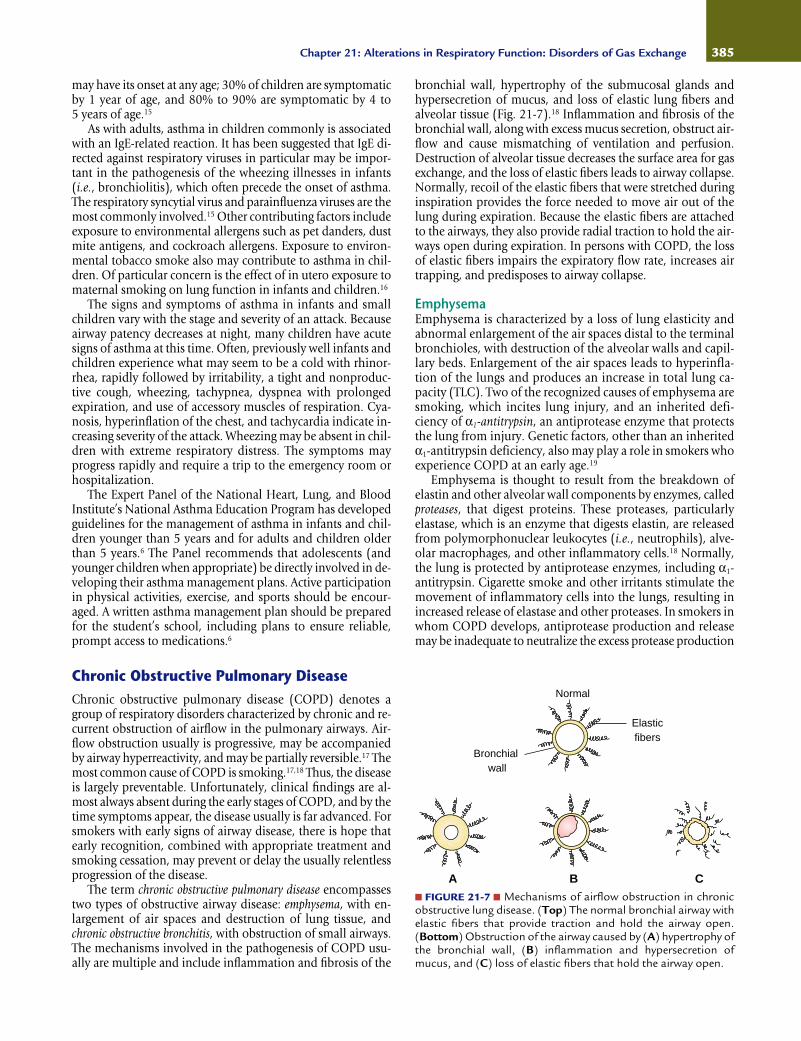
bronchial wall, hypertrophy of the submucosal glands andhypersecretion of mucus, and loss of elastic lung fibers andalveolar tissue (Fig. 21-7).18 Inflammation and fibrosis of thebronchial wall, along with excess mucus secretion, obstruct air-flow and cause mismatching of ventilation and perfusion.Destruction of alveolar tissue decreases the surface area for gasexchange, and the loss of elastic fibers leads to airway collapse.Normally, recoil of the elastic fibers that were stretched duringinspiration provides the force needed to move air out of thelung during expiration. Because the elastic fibers are attachedto the airways, they also provide radial traction to hold the air-ways open during expiration. In persons with COPD, the lossof elastic fibers impairs the expiratory flow rate, increases airtrapping, and predisposes to airway collapse.
EmphysemaEmphysema is characterized by a loss of lung elasticity andabnormal enlargement of the air spaces distal to the terminalbronchioles, with destruction of the alveolar walls and capil-lary beds. Enlargement of the air spaces leads to hyperinfla-tion of the lungs and produces an increase in total lung ca-pacity (TLC). Two of the recognized causes of emphysema aresmoking, which incites lung injury, and an inherited defi-ciency of α1-antitrypsin, an antiprotease enzyme that protectsthe lung from injury. Genetic factors, other than an inheritedα1-antitrypsin deficiency, also may play a role in smokers whoexperience COPD at an early age.19
Emphysema is thought to result from the breakdown ofelastin and other alveolar wall components by enzymes, calledproteases, that digest proteins. These proteases, particularlyelastase, which is an enzyme that digests elastin, are releasedfrom polymorphonuclear leukocytes (i.e., neutrophils), alve-olar macrophages, and other inflammatory cells.18 Normally,the lung is protected by antiprotease enzymes, including α1-antitrypsin. Cigarette smoke and other irritants stimulate themovement of inflammatory cells into the lungs, resulting inincreased release of elastase and other proteases. In smokers inwhom COPD develops, antiprotease production and releasemay be inadequate to neutralize the excess protease production
385Chapter 21: Alterations in Respiratory Function: Disorders of Gas Exchange
■ FIGURE 21-7 ■ Mechanisms of airflow obstruction in chronicobstructive lung disease. (Top) The normal bronchial airway withelastic fibers that provide traction and hold the airway open.(Bottom) Obstruction of the airway caused by (A) hypertrophy ofthe bronchial wall, (B) inflammation and hypersecretion ofmucus, and (C) loss of elastic fibers that hold the airway open.
Bronchialwall
Elasticfibers
Normal
A B C

such that the process of elastic tissue destruction goes un-checked (Fig. 21-8).
A hereditary deficiency in α1-antitrypsin accounts for ap-proximately 1% of all cases of COPD and is more common inyoung persons with emphysema.18 An α1-antitrypsin deficiencyis inherited as an autosomal recessive disorder. Homozygoteswho carry two defective genes have only about 15% to 20% ofthe normal plasma concentration of α1-antitrypsin. It is mostcommon in persons of Scandinavian descent and is rare inJews, blacks, and the Japanese.20 Smoking and repeated respi-ratory tract infections, which also decrease α1-antitrypsin lev-els, contribute to the risk of emphysema in persons with an α1-antitrypsin deficiency. Laboratory methods are available formeasuring α1-antitrypsin levels. Human α1-antitrypsin is avail-able for replacement therapy in persons with a hereditary defi-ciency of the enzyme.
There are two commonly recognized types of emphysema:centriacinar and panacinar (Fig. 21-9). The centriacinar typeaffects the bronchioles in the central part of the respiratory lob-ule, with initial preservation of the alveolar ducts and sacs20
(Fig. 21-10). It is the most common type of emphysema and isseen predominantly in male smokers. The panacinar type pro-duces initial involvement of the peripheral alveoli and later ex-tends to involve the more central bronchioles. This type of em-physema is more common in persons with α1-antitrypsindeficiency. It also is found in smokers in association with cen-trilobular emphysema. In such cases, panacinar changes areseen in the lower parts of the lung and the centriacinar changesin the upper parts of the lung.
Chronic BronchitisIn chronic bronchitis, airway obstruction is caused by inflam-mation of the major and small airways. There is edema and hy-perplasia of submucosal glands and excess mucus excretion
into the bronchial tree. A history of a chronic productive coughof more than 3 months’ duration for more than 2 consecutiveyears is necessary for the diagnosis of chronic bronchitis.17,18
Typically, the cough has been present for many years, with agradual increase in acute exacerbations that produce franklypurulent sputum. Chronic bronchitis without airflow obstruc-tion often is referred to as simple bronchitis, and chronic bron-chitis with airflow obstruction as chronic obstructive bronchitis.The outlook for persons with simple bronchitis is good, com-pared with the premature morbidity and mortality associatedwith chronic obstructive bronchitis.
Chronic bronchitis is seen most commonly in middle-aged men and is associated with chronic irritation from smok-
386 Unit Five: Alterations in the Respiratory System
■ FIGURE 21-8 ■ Protease (elastase)-antiprotease (antitrypsin)mechanisms of emphysema. The effects of smoking and an inher-ited α1-antitrypsin deficiency on the destruction of elastic fibers inthe lung and development of emphysema.
Smoking Attraction ofinflammatory cells
Release of elastase
Action inhibitedby α -antitrypsin
Macrophagesand neutrophils
Decreasedα -antitrypsin
activity
Inherited α -antitrypsin
deficiency
Destruction of elastic fibers in lung
Emphysema
11
1
Normal Centriacinar Panacinar
TB TB TBRB RB RB
AA A
■ FIGURE 21-9 ■ Centriacinar and panacinar emphysema. In cen-triacinar emphysema, the destruction is confined to the terminal(TB) and respiratory bronchioles (RB). In panacinar emphysema,the peripheral alveoli (A) are also involved. (West J.B. [1997].Pulmonary pathophysiology [5th ed., p. 53]. Philadelphia: Lippincott-Raven)
■ FIGURE 21-10 ■ Centrilobular emphysema. A whole mount ofthe left lung of a smoker with mild emphysema shows enlarged airspaces scattered throughout both lobes, which represent destruc-tion of terminal bronchioles in the central part of the pulmonarylobule. These abnormal spaces are surrounded by intact pul-monary parenchyma. (Rubin E., Farber J.L. [1999]. Pathology[3rd ed., p. 628]. Philadelphia: Lippincott Williams & Wilkins)

ing and recurrent infections. In the United States, smoking isthe most important cause of chronic bronchitis. Viral and bac-terial infections are common in persons with chronic bron-chitis and are thought to be a result, rather than a cause, of theproblem.
Clinical FeaturesThe mnemonics “pink puffer” and “blue bloater” have beenused to differentiate the clinical manifestations of emphysemaand chronic obstructive bronchitis. The important features ofthese two forms of COPD are described in Table 21-2. In prac-tice, differentiation between the two types is not as vivid as pre-sented here. This is because persons with COPD often havesome degree of both emphysema and chronic bronchitis.
A major difference between the pink puffers and the bluebloaters is the respiratory responsiveness to the hypoxic stim-uli. With pulmonary emphysema, there is a proportionate lossof ventilation and perfusion area in the lung. These persons arepink puffers, or fighters, who are able to overventilate and thusmaintain relatively normal blood gas levels until late in the dis-ease. Chronic obstructive bronchitis is characterized by exces-sive bronchial secretions and airway obstruction that causesmismatching of ventilation and perfusion. Thus, persons withchronic bronchitis are unable to compensate by increasingtheir ventilation; instead, hypoxemia and cyanosis develop.These are the blue bloaters, or nonfighters.
Persons with emphysema have marked dyspnea and strug-gle to maintain normal blood gas levels with increased breath-ing effort, including prominent use of the accessory muscles.The seated position, which stabilizes chest structures and al-
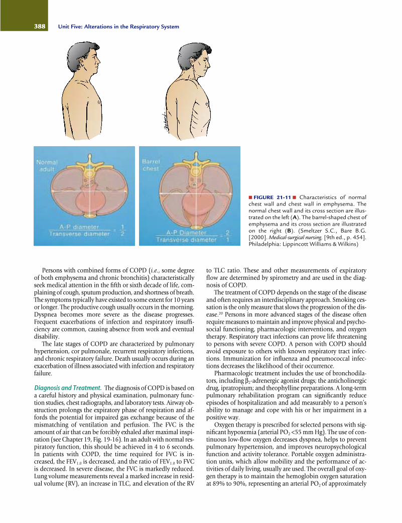
lows for maximum chest expansion and use of accessory mus-cles, is preferred. With loss of lung elasticity and hyperinflationof the lungs, the airways often collapse during expiration be-cause pressure in surrounding lung tissues exceeds airway pres-sure. Air becomes trapped in lungs, producing an increase inthe anteroposterior dimensions of the chest, the so-called bar-rel chest that is typical of persons with emphysema (Fig. 21-11).Expiration often is accomplished through pursed lips. Pursed-lip breathing, which increases the resistance to the outflow ofair, helps to prevent airway collapse by increasing airway pres-sure. The work of breathing is greatly increased in persons withemphysema, and eating often is difficult. As a result, there oftenis considerable weight loss.
Chronic obstructive bronchitis is characterized by shortnessof breath with a progressive decrease in exercise tolerance. Asthe disease progresses, breathing becomes increasingly more la-bored, even at rest. The expiratory phase of respiration is pro-longed, and expiratory wheezes and crackles can be heard onauscultation. In contrast to persons with emphysema, thosewith chronic obstructive bronchitis are unable to maintainnormal blood gases by increasing their breathing effort.Hypoxemia, hypercapnia, and cyanosis develop, reflecting animbalance between ventilation and perfusion. Hypoxemia, inwhich arterial PO2 levels fall below 55 mm Hg, causes reflexvasoconstriction of the pulmonary vessels and further impair-ment of gas exchange in the lung. Hypoxemia also stimulatesred blood cell production, causing polycythemia. As a result,persons with chronic obstructive bronchitis develop pulmo-nary hypertension and, eventually, right-sided heart failure withperipheral edema (i.e., cor pulmonale).
387Chapter 21: Alterations in Respiratory Function: Disorders of Gas Exchange
Characteristics of Chronic Bronchitis and Emphysematous Types ofChronic Obstructive Lung Disease
TABLE 21-2
Smoking historyAge of onsetClinical features
Barrel chest (hyperinflation of the lungs)
Weight lossShortness of breath
Decreased breath soundsWheezingRhonchiSputum
Cyanosis
Blood gases
Cor pulmonale
PolycythemiaPrognosis
Type A Pulmonary Emphysema Type B Chronic Bronchitis Characteristic (“Pink Puffers”) (“Blue Bloaters”)
Usual40 to 50 years of age
Often dramatic
May be severe in advanced diseaseMay be absent early in disease
CharacteristicUsually absentUsually absent or minimalMay be absent or may develop late in the course
Often absent, even late in the disease whenthere is low PO2
Relatively normal until late in the disease process
Only in advanced cases
Only in advanced casesSlowly debilitating disease
Usual30 to 40 years of age; disability in middle age
May be present
InfrequentPredominant early symptom, insidious in
onset, exertionalVariableVariableOften prominentFrequent early manifestation, frequent infec-
tions, abundant purulent sputumOften dramatic
Hypercapnia may be presentHypoxemia may be presentFrequentPeripheral edemaFrequentNumerous life-threatening episodes due to
acute exacerbations
Persons with combined forms of COPD (i.e., some degreeof both emphysema and chronic bronchitis) characteristicallyseek medical attention in the fifth or sixth decade of life, com-plaining of cough, sputum production, and shortness of breath.The symptoms typically have existed to some extent for 10 yearsor longer. The productive cough usually occurs in the morning.Dyspnea becomes more severe as the disease progresses.Frequent exacerbations of infection and respiratory insuffi-ciency are common, causing absence from work and eventualdisability.
The late stages of COPD are characterized by pulmonaryhypertension, cor pulmonale, recurrent respiratory infections,and chronic respiratory failure. Death usually occurs during anexacerbation of illness associated with infection and respiratoryfailure.
Diagnosis and Treatment. The diagnosis of COPD is based ona careful history and physical examination, pulmonary func-tion studies, chest radiographs, and laboratory tests. Airway ob-struction prolongs the expiratory phase of respiration and af-fords the potential for impaired gas exchange because of themismatching of ventilation and perfusion. The FVC is theamount of air that can be forcibly exhaled after maximal inspi-ration (see Chapter 19, Fig. 19-16). In an adult with normal res-piratory function, this should be achieved in 4 to 6 seconds.In patients with COPD, the time required for FVC is in-creased, the FEV1.0 is decreased, and the ratio of FEV1.0 to FVCis decreased. In severe disease, the FVC is markedly reduced.Lung volume measurements reveal a marked increase in resid-ual volume (RV), an increase in TLC, and elevation of the RV
to TLC ratio. These and other measurements of expiratoryflow are determined by spirometry and are used in the diag-nosis of COPD.
The treatment of COPD depends on the stage of the diseaseand often requires an interdisciplinary approach. Smoking ces-sation is the only measure that slows the progression of the dis-ease.20 Persons in more advanced stages of the disease oftenrequire measures to maintain and improve physical and psycho-social functioning, pharmacologic interventions, and oxygentherapy. Respiratory tract infections can prove life threateningto persons with severe COPD. A person with COPD shouldavoid exposure to others with known respiratory tract infec-tions. Immunization for influenza and pneumococcal infec-tions decreases the likelihood of their occurrence.
Pharmacologic treatment includes the use of bronchodila-tors, including β2-adrenergic agonist drugs; the anticholinergicdrug, ipratropium; and theophylline preparations. A long-termpulmonary rehabilitation program can significantly reduceepisodes of hospitalization and add measurably to a person’sability to manage and cope with his or her impairment in apositive way.
Oxygen therapy is prescribed for selected persons with sig-nificant hypoxemia (arterial PO2 <55 mm Hg). The use of con-tinuous low-flow oxygen decreases dyspnea, helps to preventpulmonary hypertension, and improves neuropsychologicalfunction and activity tolerance. Portable oxygen administra-tion units, which allow mobility and the performance of ac-tivities of daily living, usually are used. The overall goal of oxy-gen therapy is to maintain the hemoglobin oxygen saturationat 89% to 90%, representing an arterial PO2 of approximately
388 Unit Five: Alterations in the Respiratory System
■ FIGURE 21-11 ■ Characteristics of normalchest wall and chest wall in emphysema. Thenormal chest wall and its cross section are illus-trated on the left (A). The barrel-shaped chest ofemphysema and its cross section are illustratedon the right (B). (Smeltzer S.C., Bare B.G.[2000]. Medical-surgical nursing. [9th ed., p. 454].Philadelphia: Lippincott Williams & Wilkins)
60 mm Hg (see Chapter 19, Fig. 19-18). Because the ventila-tory drive associated with hypoxic stimulation of the periph-eral chemoreceptors does not occur until the arterial PO2 hasbeen reduced to about 60 mm Hg or less, the oxygen flow rateusually is titrated to provide an arterial PO2 of 55 to 65 mmHg. Increasing the arterial PO2 above that level tends to de-press ventilation, leading to hypoventilation and carbon diox-ide retention.
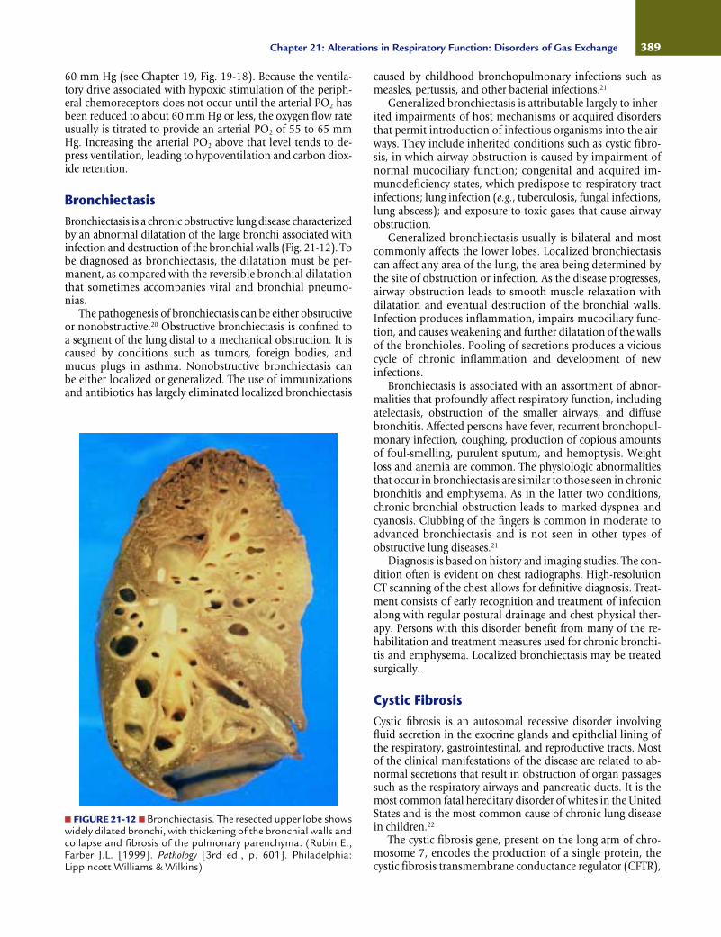
BronchiectasisBronchiectasis is a chronic obstructive lung disease characterizedby an abnormal dilatation of the large bronchi associated withinfection and destruction of the bronchial walls (Fig. 21-12). Tobe diagnosed as bronchiectasis, the dilatation must be per-manent, as compared with the reversible bronchial dilatationthat sometimes accompanies viral and bronchial pneumo-nias.
The pathogenesis of bronchiectasis can be either obstructiveor nonobstructive.20 Obstructive bronchiectasis is confined toa segment of the lung distal to a mechanical obstruction. It iscaused by conditions such as tumors, foreign bodies, andmucus plugs in asthma. Nonobstructive bronchiectasis can be either localized or generalized. The use of immunizationsand antibiotics has largely eliminated localized bronchiectasis
caused by childhood bronchopulmonary infections such asmeasles, pertussis, and other bacterial infections.21
Generalized bronchiectasis is attributable largely to inher-ited impairments of host mechanisms or acquired disordersthat permit introduction of infectious organisms into the air-ways. They include inherited conditions such as cystic fibro-sis, in which airway obstruction is caused by impairment ofnormal mucociliary function; congenital and acquired im-munodeficiency states, which predispose to respiratory tractinfections; lung infection (e.g., tuberculosis, fungal infections,lung abscess); and exposure to toxic gases that cause airwayobstruction.
Generalized bronchiectasis usually is bilateral and mostcommonly affects the lower lobes. Localized bronchiectasiscan affect any area of the lung, the area being determined bythe site of obstruction or infection. As the disease progresses,airway obstruction leads to smooth muscle relaxation with dilatation and eventual destruction of the bronchial walls.Infection produces inflammation, impairs mucociliary func-tion, and causes weakening and further dilatation of the wallsof the bronchioles. Pooling of secretions produces a viciouscycle of chronic inflammation and development of new infections.
Bronchiectasis is associated with an assortment of abnor-malities that profoundly affect respiratory function, includingatelectasis, obstruction of the smaller airways, and diffusebronchitis. Affected persons have fever, recurrent bronchopul-monary infection, coughing, production of copious amountsof foul-smelling, purulent sputum, and hemoptysis. Weightloss and anemia are common. The physiologic abnormalitiesthat occur in bronchiectasis are similar to those seen in chronicbronchitis and emphysema. As in the latter two conditions,chronic bronchial obstruction leads to marked dyspnea andcyanosis. Clubbing of the fingers is common in moderate toadvanced bronchiectasis and is not seen in other types ofobstructive lung diseases.21
Diagnosis is based on history and imaging studies. The con-dition often is evident on chest radiographs. High-resolutionCT scanning of the chest allows for definitive diagnosis. Treat-ment consists of early recognition and treatment of infectionalong with regular postural drainage and chest physical ther-apy. Persons with this disorder benefit from many of the re-habilitation and treatment measures used for chronic bronchi-tis and emphysema. Localized bronchiectasis may be treatedsurgically.
Cystic FibrosisCystic fibrosis is an autosomal recessive disorder involvingfluid secretion in the exocrine glands and epithelial lining ofthe respiratory, gastrointestinal, and reproductive tracts. Mostof the clinical manifestations of the disease are related to ab-normal secretions that result in obstruction of organ passagessuch as the respiratory airways and pancreatic ducts. It is themost common fatal hereditary disorder of whites in the UnitedStates and is the most common cause of chronic lung diseasein children.22
The cystic fibrosis gene, present on the long arm of chro-mosome 7, encodes the production of a single protein, thecystic fibrosis transmembrane conductance regulator (CFTR),
389Chapter 21: Alterations in Respiratory Function: Disorders of Gas Exchange
■ FIGURE 21-12 ■ Bronchiectasis. The resected upper lobe showswidely dilated bronchi, with thickening of the bronchial walls andcollapse and fibrosis of the pulmonary parenchyma. (Rubin E.,Farber J.L. [1999]. Pathology [3rd ed., p. 601]. Philadelphia:Lippincott Williams & Wilkins)
which functions in chloride transport across cell membranes.23,24
Because of defective chloride transport, there is a threefoldincrease in sodium reabsorption. Water moves out of theextracellular fluid with the sodium, causing exocrine (e.g., mu-cus) secretions to become exceedingly viscid. The cystic fibro-sis gene is rare in African blacks and Asians. Homozygotes (i.e., persons with two defective genes) have all or substan-tially all of the clinical symptoms of the disease, comparedwith heterozygotes, who are carriers of the disease but haveno recognizable symptoms.
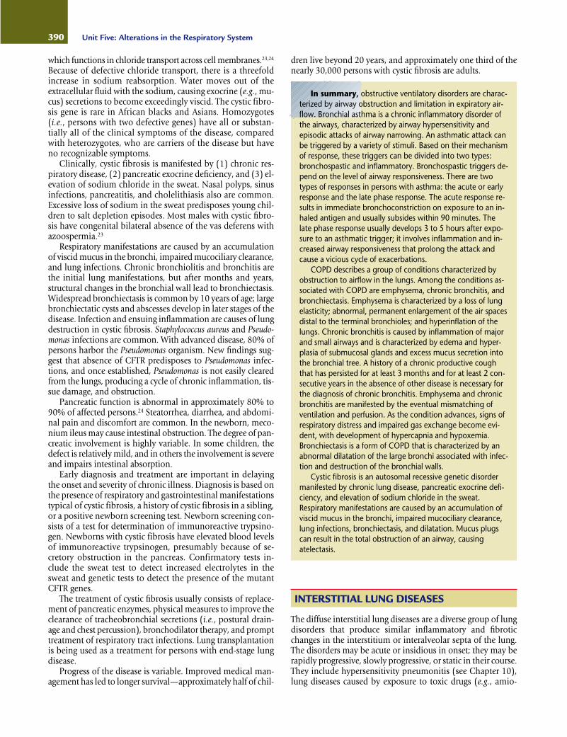
Clinically, cystic fibrosis is manifested by (1) chronic res-piratory disease, (2) pancreatic exocrine deficiency, and (3) el-evation of sodium chloride in the sweat. Nasal polyps, sinusinfections, pancreatitis, and cholelithiasis also are common.Excessive loss of sodium in the sweat predisposes young chil-dren to salt depletion episodes. Most males with cystic fibro-sis have congenital bilateral absence of the vas deferens withazoospermia.23
Respiratory manifestations are caused by an accumulationof viscid mucus in the bronchi, impaired mucociliary clearance,and lung infections. Chronic bronchiolitis and bronchitis arethe initial lung manifestations, but after months and years,structural changes in the bronchial wall lead to bronchiectasis.Widespread bronchiectasis is common by 10 years of age; largebronchiectatic cysts and abscesses develop in later stages of thedisease. Infection and ensuing inflammation are causes of lungdestruction in cystic fibrosis. Staphylococcus aureus and Pseudo-monas infections are common. With advanced disease, 80% ofpersons harbor the Pseudomonas organism. New findings sug-gest that absence of CFTR predisposes to Pseudomonas infec-tions, and once established, Pseudomonas is not easily clearedfrom the lungs, producing a cycle of chronic inflammation, tis-sue damage, and obstruction.
Pancreatic function is abnormal in approximately 80% to90% of affected persons.24 Steatorrhea, diarrhea, and abdomi-nal pain and discomfort are common. In the newborn, meco-nium ileus may cause intestinal obstruction. The degree of pan-creatic involvement is highly variable. In some children, thedefect is relatively mild, and in others the involvement is severeand impairs intestinal absorption.
Early diagnosis and treatment are important in delayingthe onset and severity of chronic illness. Diagnosis is based onthe presence of respiratory and gastrointestinal manifestationstypical of cystic fibrosis, a history of cystic fibrosis in a sibling,or a positive newborn screening test. Newborn screening con-sists of a test for determination of immunoreactive trypsino-gen. Newborns with cystic fibrosis have elevated blood levelsof immunoreactive trypsinogen, presumably because of se-cretory obstruction in the pancreas. Confirmatory tests in-clude the sweat test to detect increased electrolytes in thesweat and genetic tests to detect the presence of the mutantCFTR genes.
The treatment of cystic fibrosis usually consists of replace-ment of pancreatic enzymes, physical measures to improve theclearance of tracheobronchial secretions (i.e., postural drain-age and chest percussion), bronchodilator therapy, and prompttreatment of respiratory tract infections. Lung transplantationis being used as a treatment for persons with end-stage lungdisease.
Progress of the disease is variable. Improved medical man-agement has led to longer survival—approximately half of chil-
dren live beyond 20 years, and approximately one third of thenearly 30,000 persons with cystic fibrosis are adults.
390 Unit Five: Alterations in the Respiratory System
In summary, obstructive ventilatory disorders are charac-terized by airway obstruction and limitation in expiratory air-flow. Bronchial asthma is a chronic inflammatory disorder ofthe airways, characterized by airway hypersensitivity andepisodic attacks of airway narrowing. An asthmatic attack canbe triggered by a variety of stimuli. Based on their mechanismof response, these triggers can be divided into two types:bronchospastic and inflammatory. Bronchospastic triggers de-pend on the level of airway responsiveness. There are twotypes of responses in persons with asthma: the acute or earlyresponse and the late phase response. The acute response re-sults in immediate bronchoconstriction on exposure to an in-haled antigen and usually subsides within 90 minutes. Thelate phase response usually develops 3 to 5 hours after expo-sure to an asthmatic trigger; it involves inflammation and in-creased airway responsiveness that prolong the attack andcause a vicious cycle of exacerbations.
COPD describes a group of conditions characterized byobstruction to airflow in the lungs. Among the conditions as-sociated with COPD are emphysema, chronic bronchitis, andbronchiectasis. Emphysema is characterized by a loss of lungelasticity; abnormal, permanent enlargement of the air spacesdistal to the terminal bronchioles; and hyperinflation of thelungs. Chronic bronchitis is caused by inflammation of majorand small airways and is characterized by edema and hyper-plasia of submucosal glands and excess mucus secretion intothe bronchial tree. A history of a chronic productive coughthat has persisted for at least 3 months and for at least 2 con-secutive years in the absence of other disease is necessary forthe diagnosis of chronic bronchitis. Emphysema and chronicbronchitis are manifested by the eventual mismatching ofventilation and perfusion. As the condition advances, signs ofrespiratory distress and impaired gas exchange become evi-dent, with development of hypercapnia and hypoxemia.Bronchiectasis is a form of COPD that is characterized by anabnormal dilatation of the large bronchi associated with infec-tion and destruction of the bronchial walls.
Cystic fibrosis is an autosomal recessive genetic disordermanifested by chronic lung disease, pancreatic exocrine defi-ciency, and elevation of sodium chloride in the sweat.Respiratory manifestations are caused by an accumulation ofviscid mucus in the bronchi, impaired mucociliary clearance,lung infections, bronchiectasis, and dilatation. Mucus plugscan result in the total obstruction of an airway, causingatelectasis.
INTERSTITIAL LUNG DISEASES
The diffuse interstitial lung diseases are a diverse group of lungdisorders that produce similar inflammatory and fibroticchanges in the interstitium or interalveolar septa of the lung.The disorders may be acute or insidious in onset; they may berapidly progressive, slowly progressive, or static in their course.They include hypersensitivity pneumonitis (see Chapter 10),lung diseases caused by exposure to toxic drugs (e.g., amio-

darone) and radiation, and occupational lung diseases includ-ing the pneumoconioses that are caused by the inhalation ofinorganic dusts such as silica, coal dust, and asbestos. Some ofthe most common interstitial lung diseases are caused by ex-posure to the inhaled dust and particles. In many cases, no spe-cific cause can be found.25,26 Examples of interstitial lung dis-eases and their causes are listed in Chart 21-2.
Current theory suggests that most interstitial lung diseases,regardless of the causes, have a common pathogenesis. It isthought that these disorders are initiated by some type of in-jury to the alveolar epithelium, followed by an inflammatoryprocess that involves the alveoli and interstitium of the lung.An accumulation of inflammatory and immune cells causescontinued damage of lung tissue and the replacement of nor-mal, functioning lung tissue with fibrous scar tissue.
Because the interstitial lung diseases result in a stiff andnoncompliant lung, they are commonly classified as fibroticor restrictive lung disorders. In contrast to obstructive lungdiseases, the lungs are stiff and difficult to expand, despite nor-mal functioning airways. Persons with interstitial lung diseasesexperience dyspnea, tachypnea, and eventual cyanosis, with-out evidence of wheezing or signs of airway obstruction.Usually there is an insidious onset of breathlessness that ini-tially occurs during exercise and may progress to the point thatthe person is totally incapacitated. A nonproductive coughmay develop, particularly with continued exposure to the in-
haled irritant. Typically, a person with a restrictive lung diseasebreathes with a pattern of rapid, shallow respirations. Thistachypneic pattern of breathing, in which the respiratory rateis increased and the tidal volume is decreased, reduces thework of breathing because it takes less work to move airthrough the airways at an increased rate than it does to stretcha stiff lung to accommodate a larger tidal volume.
Although resting arterial blood gases usually are normalearly in the course of the disease, arterial PO2 levels may fallduring exercise, and in cases of advanced disease, hypoxemiaoften is present, even at rest. In the late stages of the disease,hypercapnia and respiratory acidosis develop. Clubbing of thefingers and toes may develop because of chronic hypoxemia.
The diagnosis of interstitial lung disease requires a carefulpersonal and family history, with particular emphasis on ex-posure to environmental, occupational, and other injuriousagents. Chest radiographs may be used as an initial diagnosticmethod, and serial chest films often are used to follow theprogress of the disease. A biopsy specimen for histologic studyand culture may be obtained by surgical incision or bron-choscopy using a fiberoptic bronchoscope. Gallium lung scansoften are used to detect and quantify the chronic alveolitis thatoccurs in interstitial lung disease. Gallium does not localize innormal lung tissue, but uptake of the radionuclide is increasedin interstitial lung disease and other diffuse lung diseases.
The treatment goals for persons with interstitial lung diseasefocus on identifying and removing the injurious agent, sup-pressing the inflammatory response, preventing progression ofthe disease, and providing supportive therapy for persons withadvanced disease. In general, the treatment measures vary withthe type of lung disease. Corticosteroid drugs frequently areused to suppress the inflammatory response. Many of the sup-portive treatment measures used in the late stages of the dis-ease, such as oxygen therapy and measures to prevent infection,are similar to those discussed for persons with COPD.
391Chapter 21: Alterations in Respiratory Function: Disorders of Gas Exchange
CHART 21-2 Causes of Interstitial Lung Diseases*Occupational and Environmental Inhalants
Inorganic dustsAsbestosisSilicosisCoal miner’s pneumoconiosis
Organic dustsHypersensitivity pneumonitis
Gases and fumesAmmonia, phosgene, sulfur dioxide
Drugs and Therapeutic Agents
Cancer chemotherapeutic agentsBusulfanBleomycinMethotrexate
Ionizing radiation
Immunologic Lung Disease
SarcoidosisCollagen vascular diseases
Systemic lupus erythematosusRheumatoid arthritisSclerodermaDermatomyositis-polymyositis
Miscellaneous
Postacute respiratory distress syndromeIdiopathic pulmonary fibrosis
*This list is not intended to be inclusive.
In summary, the interstitial lung diseases are character-ized by fibrosis and decreased compliance of the lung. Theyinclude the occupational lung diseases, lung diseases causedby toxic drugs and radiation, and lung diseases of unknownorigin, such as sarcoidosis. These disorders are thought to re-sult from an inflammatory process that begins in the alveoliand extends to involve the interstitial tissues of the lung.
KEY CONCEPTS
INTERSTITIAL OR RESTRICTIVE LUNG DISEASES
■ Interstitial lung diseases result from inflammatoryconditions that affect the interalveolar structures ofthe lung and produce lung fibrosis and a stiff lung.
■ A stiff and noncompliant lung is difficult to inflate,increasing the work of breathing and causing de-creased exercise tolerance due to hypoxemia.
■ Because of the increased effort needed for lung ex-pansion, persons with interstitial lung disease tendto take small but more frequent breaths.

PULMONARY VASCULAR DISORDERS
As blood moves through the lung, blood oxygen levels areraised, and carbon dioxide is removed. These processes dependon the matching of ventilation (i.e., gas exchange) and per-fusion (i.e., blood flow). This section discusses three majordisorders of the pulmonary circulation: pulmonary embolism,pulmonary hypertension, and acute respiratory distress syn-drome. Pulmonary edema, another major problem of the pul-monary circulation, is discussed in Chapter 18.
Pulmonary EmbolismPulmonary embolism develops when a blood-borne substancelodges in a branch of the pulmonary artery and obstructs theflow. The embolism may consist of a thrombus (Fig. 21-13), airthat has accidentally been injected during intravenous infu-sion, fat that has been mobilized from the bone marrow aftera fracture or from a traumatized fat depot (see Chapter 42), or
amniotic fluid that has entered the maternal circulation afterrupture of the membranes at the time of delivery.
Almost all pulmonary emboli arise from deep vein throm-bosis (DVT) in the lower extremities (see Chapter 15). The pres-ence of thrombosis in the deep veins of the legs or pelvis oftenis unsuspected until embolism occurs. The effects of emboli onthe pulmonary circulation are related to mechanical obstruc-tion of the pulmonary circulation and neurohumoral reflexescausing vasoconstriction. Obstruction of pulmonary bloodflow causes reflex bronchoconstriction in the affected area ofthe lung, wasted ventilation and impaired gas exchange, andloss of alveolar surfactant. Pulmonary hypertension and rightheart failure may develop when there is massive vasoconstric-tion because of a large embolus. Although small areas of in-farction may occur, frank pulmonary infarction is uncommon.
Persons at risk for DVT also are at risk for thromboemboli.Among the physiologic factors that contribute to venousthrombosis are venous stasis, venous endothelial injury, andhypercoagulability states. Venous stasis and venous endothe-lial injury can result from prolonged bed rest, trauma, surgery,childbirth, fractures of the hip and femur, myocardial infarc-tion and congestive heart failure, and spinal cord injury. Per-sons undergoing orthopedic surgery and gynecologic cancersurgery are at particular risk, as are bedridden patients in anintensive care unit. Cancer cells can produce thrombin and syn-thesize procoagulation factors, increasing the risk of thrombo-embolism. Use of oral contraceptive, pregnancy, and hormonereplacement therapy are thought to increase the resistance to endogenous anticoagulants. The risk of pulmonary em-bolism among users of oral contraceptives is approximatelythree times the risk of nonusers.27 Women who smoke are atparticular risk.
The manifestations of pulmonary embolism depend on thesize and location of the obstruction. Chest pain, dyspnea, andincreased respiratory rate are the most frequent signs and symp-toms of pulmonary embolism. Pulmonary infarction oftencauses pleuritic pain that changes with respiration; it is moresevere on inspiration and less severe on expiration. Moderatehypoxemia without carbon dioxide retention occurs as a resultof impaired gas exchange. Small emboli that become lodged inthe peripheral branches of the pulmonary artery may exert lit-tle effect and go unrecognized. However, repeated small em-boli often result in a gradual the size of the pulmonary capil-lary bed, resulting in pulmonary hypertension. Moderate-sizeemboli often present with breathlessness accompanied bypleuritic pain, apprehension, slight fever, rapid and shallowbreathing, and cough productive of blood-streaked sputum.Persons with massive emboli usually present with sudden col-lapse, crushing substernal chest pain, shock, and sometimesloss of consciousness. The pulse is rapid and weak, the bloodpressure is low, the neck veins are distended, and the skin iscyanotic and diaphoretic. Massive pulmonary emboli often arefatal.
The diagnosis of pulmonary embolism is based on clinicalsigns and symptoms, blood gas determinations, venous throm-bosis studies, lung scans, CT scans, and in selected cases,pulmonary angiography. Laboratory studies and radiologicfilms are useful in ruling out other conditions that might giverise to similar symptoms. The electrocardiogram (ECG) maybe used to detect signs of right heart strain resulting in an in-crease in pulmonary vascular resistance. Because almost all
392 Unit Five: Alterations in the Respiratory System
■ FIGURE 21-13 ■ Pulmonary embolism. The main pulmonaryartery and its bifurcation have opened to reveal a large saddle em-bolus. (Rubin E., Farber J.L. [1999]. Pathology [3rd ed., p. 289].Philadelphia: Lippincott Williams & Wilkins)
Unlike COPD, which affects the airways, interstitial lung dis-eases affect the supporting collagen and elastic tissues that liebetween the airways and blood vessels. These lung diseasesdecrease lung volumes, reduce the diffusing capacity of thelung, and cause various degrees of hypoxia. Because lungcompliance is reduced, persons with this form of lung diseasehave a rapid, shallow breathing pattern.

pulmonary emboli originate from DVT, venous studies such aslower limb compression ultrasonography, impedance plethysmogra-phy, and contrast venography often are used as initial diagnosticprocedures.
The treatment goals for pulmonary emboli focus on the pre-vention DVT and the development of thromboemboli, pro-tecting the lungs from exposure to thromboemboli when theyoccur, and in the case of large and life-threatening pulmonaryemboli, sustaining life and restoring pulmonary blood flow.Thrombolytic therapy may be indicated in persons with multi-ple or large emboli. Restoration of blood flow in personswith life-threatening pulmonary emboli can often be accom-plished through the surgical removal of the embolus or emboli.
Prevention focuses on identification of persons at risk,avoidance of venous stasis and hypercoagulability states, andearly detection of venous thrombosis. For persons at risk,graded compression elastic stockings and intermittent pneu-matic compression (IPC) boots can be used to prevent venousstasis. Pharmacologic prophylaxis involves the use of anticoag-ulant drugs.
Pulmonary HypertensionThe term pulmonary hypertension describes the elevation of pres-sure in the pulmonary arterial system. The pulmonary circula-tion is a low-pressure system designed to accommodate vary-ing amounts of blood delivered from the right heart and tofacilitate gas exchange. The normal mean pulmonary arterypressure is approximately 15 mm Hg (e.g., 28 systolic/8 dia-stolic). The main pulmonary artery and major branches are rel-atively thin-walled, compliant vessels. The distal pulmonary ar-terioles also are thin walled and have the capacity to dilate,collapse, or constrict, depending on the presence of vasoactivesubstances released from the endothelial cells of the vessel,neurohumoral influences, flow velocity, oxygen tension, andalveolar ventilation.
Pulmonary hypertension can be caused by an elevation inleft atrial pressure, increased pulmonary blood flow, or in-creased pulmonary vascular resistance. Because of the increasedpressure in the pulmonary circulation, pulmonary hyperten-sion increases the workload of the right heart. Although pul-monary hypertension can develop as a primary disorder, mostcases develop secondary to some other condition.
Secondary Pulmonary HypertensionSecondary pulmonary hypertension refers to an increase in pul-monary pressures associated with other disease conditions,usually cardiac or pulmonary. Secondary causes, or mecha-nisms, of pulmonary hypertension can be divided into fourmajor categories: (1) elevation of pulmonary venous pressure,(2) increased pulmonary blood flow, (3) pulmonary vascularobstruction, and (4) hypoxemia.28 Often more than one factor,such as COPD, heart failure, and sleep apnea, contributes tothe elevation in pulmonary pressures.
Elevation of pulmonary venous pressure is common in condi-tions such as mitral valve stenosis and left ventricular heartfailure, in which an elevated left atrial pressure is transmittedto the pulmonary circulation. Continued increases in leftatrial pressure can lead to medial hypertrophy and intimalthickening of the small pulmonary arteries, causing sustainedhypertension.
Increased pulmonary blood flow results from increased flowthrough left-to-right shunts in congenital heart diseases suchas atrial or ventricular septal defects and patent ductus arte-riosus. If the high-flow state is allowed to continue, morpho-logic changes occur in the pulmonary vessels, leading to sus-tained pulmonary hypertension. The pulmonary vascularchanges that occur with congenital heart disorders are dis-cussed in Chapter 17.
Obstruction of pulmonary blood vessels is most commonly theresult of pulmonary emboli. Once initiated, the pulmonaryhypertension that develops is self-perpetuating because ofhypertrophy and proliferation of vascular smooth muscle.
Hypoxemia is another common cause of pulmonary hyper-tension. Unlike the vessels in the systemic circulation, most ofwhich dilate in response to hypoxemia and hypercapnia, thepulmonary vessels constrict. The stimulus for constriction isthought to originate in the air spaces near the smaller branchesof the pulmonary arteries. In situations in which certain re-gions of the lung are hypoventilated, the response is adaptivein that it diverts blood flow away from the poorly ventilatedareas to more adequately ventilated portions of the lung.However, this effect becomes less beneficial as more and moreareas of the lung become poorly ventilated. Pulmonary hyper-tension is a common problem in persons with advancedCOPD. It also may develop at high altitudes in persons withnormal lungs. Persons who experience marked hypoxemiaduring sleep (i.e., those with sleep apnea) may also experiencemarked elevations in pulmonary arterial pressure.
The signs and symptoms of secondary pulmonary hyper-tension reflect not only the underlying cause, but the effectthat the elevated pressures have on right heart function andoxygen transport. Dyspnea and fatigue are common. Periph-eral edema, ascites, and signs of right heart failure (cor pul-monale, to be discussed) develop as the condition progresses.
Diagnosis is based on radiographic findings, echocardiog-raphy, and Doppler ultrasonography. Precise measurement ofpulmonary pressures can be obtained only through right heartcardiac catheterization. Treatment measures are directed to-ward the underlying disorder. Vasodilator therapy may beindicated for some persons.
Primary Pulmonary HypertensionPrimary pulmonary hypertension is a relatively rare and ra-pidly progressive form of pulmonary hypertension that leadsto right ventricular failure and death within a few years. Esti-mates of incidence range from 1 to 2 cases per million peoplein the general population.29 The disease can occur at any age,and familial occurrences have been reported. Persons with thedisorder usually have a steadily progressive downhill course,with death occurring in 3 to 4 years. Overall, the 5-yearsurvival rate of untreated primary pulmonary hypertension isapproximately 20%.30
Primary pulmonary hypertension is thought to be associ-ated with a number of factors, including an autosomal domi-nant genetic predisposition along with an exogenous trigger.30
Triggers include low oxygen levels that occur at high altitudes,exposure to certain drugs, human immunodeficiency virusinfection, and autoimmune disorders.
The disorder is characterized by endothelial damage, co-agulation abnormalities, and marked intimal fibrosis leadingto obliteration or obstruction of the pulmonary arteries and
393Chapter 21: Alterations in Respiratory Function: Disorders of Gas Exchange

arterioles. Most of the manifestations of the disorder are at-tributable to increased work demands on the right heart and adecrease in cardiac output. Symptoms are the same as those forsecondary hypertension. The most obvious are dyspnea andfatigue that is out of proportion to other signs of a person’swell being.
The diagnosis of primary pulmonary hypertension is basedon an absence of disorders that cause secondary hypertensionand mean pulmonary artery pressures greater than 25 mm Hgat rest or 30 mm Hg with exercise.
Treatment consists of measures to improve right heart func-tion to reduce fatigue and peripheral edema. Supplementaloxygen may be used to increase exercise tolerance. The mostwidely used drugs for long-term therapy are the calcium chan-nel blockers. Anticoagulant (warfarin) therapy may be used todecrease the risk of thrombosis caused by sluggish pulmonaryblood flow. Epoprostenol, a short-acting (half-life, 3 to 5 min-utes) analog of the naturally occurring vasodilator prostacyclin(prostaglandin I2), is increasingly used in the long-term care ofpersons with the disorder. Because of its short half-life, thedrug must be administered by continuous infusion, usuallythrough an indwelling venous catheter with an automatic am-bulatory pump. Lung transplantation may be an alternative forpersons who do not experience response to other forms oftreatment.
Cor PulmonaleThe term cor pulmonale refers to right heart failure resultingfrom primary lung disease and longstanding primary or sec-ondary pulmonary hypertension. It involves hypertrophy andthe eventual failure of the right ventricle. The manifestations ofcor pulmonale include the signs and symptoms of the primarylung disease and the signs of right-sided heart failure (seeChapter 18). Signs of right-sided heart failure include venouscongestion, peripheral edema, shortness of breath and a pro-ductive cough, which becomes worse during periods of wors-ening failure. Plethora (i.e., redness) and cyanosis and warm,moist skin may result from the compensatory polycythemiaand desaturation of arterial blood that accompany chroniclung disease. Drowsiness and altered consciousness may occuras the result of carbon dioxide retention. Management of corpulmonale focuses on the treatment of the lung disease and theheart failure. Low-flow oxygen therapy may be used to reducethe pulmonary hypertension and polycythemia associated withsevere hypoxemia caused by chronic lung disease.
Acute Respiratory Distress SyndromeAcute respiratory distress syndrome (ARDS), first described in1967, is a devastating syndrome of acute lung injury. Initiallycalled the adult respiratory distress syndrome, it is now called theacute respiratory distress syndrome because it also affects children.ARDS affects approximately 150,000 to 200,000 persons eachyear; at least 50% to 60% of these persons die, despite the mostsophisticated intensive care.31
ARDS is the final common pathway through which manyserious localized and systemic disorders produce diffuse injuryto the alveolar-capillary membrane. It may result from a num-ber of conditions, including aspiration of gastric contents,major trauma (with or without fat emboli), sepsis secondary topulmonary or nonpulmonary infections, acute pancreatitis,
hematologic disorders, metabolic events, and reactions todrugs and toxins31,32 (Chart 21-3).
Although a number of conditions may lead to ARDS, theyall produce similar pathologic lung changes that include dif-fuse endothelial and epithelial cell injury with increased per-meability of the alveolar-capillary membrane (Fig. 21-14).The increased permeability permits fluid, protein, cellular de-bris and blood cells to move out of the vascular compartmentinto the interstitium and alveoli of the lung. Alveolar celldamage leads to accumulation of edema fluid, surfactant inac-tivation, and formation of a hyaline membrane that is imper-vious to gas exchange. The lungs become very stiff and difficultto inflate. There is increased intrapulmonary shunting of blood,impaired gas exchange, and profound hypoxia. Gas exchangeis further compromised by alveolar collapse resulting from abnormalities in surfactant production. When injury to thealveolar epithelium is severe, disorganized epithelial repairmay lead to fibrosis.
The pathogenesis of ARDS is unclear. Neutrophils accumu-late early in the course of the disorder and are thought to playa role in the pathogenesis of ARDS. Activated neutrophils re-lease a variety of products, including oxidants, proteases,platelet activating factor, and leukotrienes that damage thealveolar capillary endothelium and alveolar cells.
Clinically, the syndrome consists of progressive respiratorydistress, an increase in respiratory rate, and signs of respiratoryfailure. Radiologic findings usually show extensive bilateralconsolidation of the lung tissue. Severe hypoxia persists despiteincreased inspired oxygen levels.
The treatment goals in ARDS are to supply oxygen to vitalorgans and provide supportive care until the condition causingthe pathologic process has been reversed and the lungs havehad a chance to heal. Assisted ventilation using high concen-trations of oxygen may be required to overcome the hypoxia.
394 Unit Five: Alterations in the Respiratory System
CHART 21-3 Conditions in Which ARDS Can Develop*Aspiration
Near drowningAspiration gastric contents
Drugs, Toxins, Therapeutic Agents
HeroinInhaled gases (e.g., smoke, ammonia)OxygenRadiation
Infections
Gram-negative septicemiaOther bacterial infectionsViral infections
Trauma and Shock
BurnsFat embolismChest trauma
*This list is not intended to be inclusive.
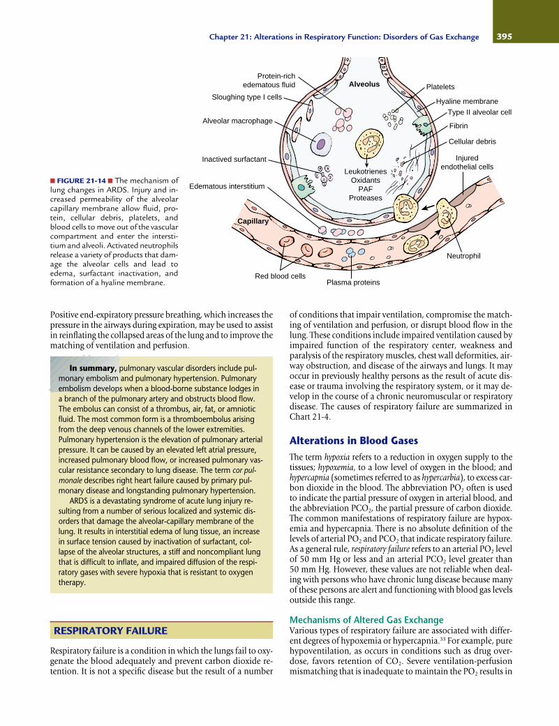
395Chapter 21: Alterations in Respiratory Function: Disorders of Gas Exchange
■ FIGURE 21-14 ■ The mechanism oflung changes in ARDS. Injury and in-creased permeability of the alveolarcapillary membrane allow fluid, pro-tein, cellular debris, platelets, andblood cells to move out of the vascularcompartment and enter the intersti-tium and alveoli. Activated neutrophilsrelease a variety of products that dam-age the alveolar cells and lead toedema, surfactant inactivation, andformation of a hyaline membrane.
Platelets
Cellular debris
Hyaline membrane
Injuredendothelial cells
Neutrophil
Plasma proteins
Capillary
Red blood cells
Edematous interstitium
Alveolar macrophageType II alveolar cell
Sloughing type I cells
Protein-richedematous fluid
Inactived surfactant
Fibrin
LeukotrienesOxidants
PAFProteases
Alveolus
In summary, pulmonary vascular disorders include pul-monary embolism and pulmonary hypertension. Pulmonaryembolism develops when a blood-borne substance lodges ina branch of the pulmonary artery and obstructs blood flow.The embolus can consist of a thrombus, air, fat, or amnioticfluid. The most common form is a thromboembolus arisingfrom the deep venous channels of the lower extremities.Pulmonary hypertension is the elevation of pulmonary arterialpressure. It can be caused by an elevated left atrial pressure,increased pulmonary blood flow, or increased pulmonary vas-cular resistance secondary to lung disease. The term cor pul-monale describes right heart failure caused by primary pul-monary disease and longstanding pulmonary hypertension.
ARDS is a devastating syndrome of acute lung injury re-sulting from a number of serious localized and systemic dis-orders that damage the alveolar-capillary membrane of thelung. It results in interstitial edema of lung tissue, an increasein surface tension caused by inactivation of surfactant, col-lapse of the alveolar structures, a stiff and noncompliant lungthat is difficult to inflate, and impaired diffusion of the respi-ratory gases with severe hypoxia that is resistant to oxygentherapy.
Positive end-expiratory pressure breathing, which increases thepressure in the airways during expiration, may be used to assistin reinflating the collapsed areas of the lung and to improve thematching of ventilation and perfusion.
of conditions that impair ventilation, compromise the match-ing of ventilation and perfusion, or disrupt blood flow in thelung. These conditions include impaired ventilation caused byimpaired function of the respiratory center, weakness andparalysis of the respiratory muscles, chest wall deformities, air-way obstruction, and disease of the airways and lungs. It mayoccur in previously healthy persons as the result of acute dis-ease or trauma involving the respiratory system, or it may de-velop in the course of a chronic neuromuscular or respiratorydisease. The causes of respiratory failure are summarized inChart 21-4.
Alterations in Blood GasesThe term hypoxia refers to a reduction in oxygen supply to thetissues; hypoxemia, to a low level of oxygen in the blood; andhypercapnia (sometimes referred to as hypercarbia), to excess car-bon dioxide in the blood. The abbreviation PO2 often is usedto indicate the partial pressure of oxygen in arterial blood, andthe abbreviation PCO2, the partial pressure of carbon dioxide.The common manifestations of respiratory failure are hypox-emia and hypercapnia. There is no absolute definition of thelevels of arterial PO2 and PCO2 that indicate respiratory failure.As a general rule, respiratory failure refers to an arterial PO2 levelof 50 mm Hg or less and an arterial PCO2 level greater than 50 mm Hg. However, these values are not reliable when deal-ing with persons who have chronic lung disease because manyof these persons are alert and functioning with blood gas levelsoutside this range.
Mechanisms of Altered Gas ExchangeVarious types of respiratory failure are associated with differ-ent degrees of hypoxemia or hypercapnia.33 For example, purehypoventilation, as occurs in conditions such as drug over-dose, favors retention of CO2. Severe ventilation-perfusionmismatching that is inadequate to maintain the PO2 results in
RESPIRATORY FAILURE
Respiratory failure is a condition in which the lungs fail to oxy-genate the blood adequately and prevent carbon dioxide re-tention. It is not a specific disease but the result of a number

hypoxemia that is more severe in relation to hypercapnia.This pattern of respiratory failure is seen most commonly inpersons with advanced COPD. In interstitial lung disease,there is severe hypoxemia but no hypercapnia because of in-creased ventilation. In ARDS, the arterial PCO2 is typically
low, but hypoxemia is severe. In this case, administration of oxygen can produce an increase in PO2 without producingan increase in PCO2.
Hypoventilation. Hypoventilation occurs when the volume of“fresh” air moving into and out of the lung is significantly re-duced. Hypoventilation is commonly caused by conditionsoutside the lung such as depression of the respiratory center(e.g., drug overdose), diseases of the nerves supplying the res-piratory muscles (e.g., Guillain-Barré syndrome), disorders ofthe respiratory muscles (e.g., muscular dystrophy), or thoraciccage disorders (e.g., severe scoliosis or crushed chest).
Hypoventilation has two important effects on arterial bloodgases. First, hypoventilation almost always causes an increasein PCO2. The rise in PCO2 is directly related to the level of ven-tilation. For example, the PCO2 doubles when alveolar ventila-tion is reduced by one half. Thus, the PCO2 level is a good di-agnostic measure for hypoventilation. Second, hypoxemiacaused by hypoventilation can be easily corrected by increasingthe oxygen content of the inspired air.33
Ventilation-Perfusion Mismatching. The mismatching of ven-tilation and perfusion occurs when areas of the lung are venti-lated but not perfused or when areas are perfused but not ven-tilated. Usually the hypoxemia that is seen in situations ofventilation-perfusion mismatching is more severe in relation tohypercapnia than that seen in hypoventilation. Severe mis-matching of ventilation and perfusion often is seen in personswith advanced COPD. These disorders contribute to the reten-tion of carbon dioxide by reducing the effective alveolar venti-lation, even when total ventilation is maintained. This occursbecause a region of the lung is not perfused and gas exchangecannot take place or because an area of the lung is not beingventilated. Maintaining a high ventilation rate effectively pre-vents hypercapnia but also increases the work of breathing.
The hypoxemia associated with ventilation-perfusion dis-orders often is exaggerated by conditions such as hypoventila-tion and decreased cardiac output. For example, sedation cancause hypoventilation in persons with severe COPD, resultingin further impairment of ventilation. Likewise, a decrease incardiac output because of myocardial infarction can exaggeratethe ventilation-perfusion impairment in a person with mildpulmonary edema.
The beneficial effect of oxygen administration on PO2 lev-els in ventilation-perfusion disorders depends on the degreeof mismatching that is present. Because oxygen administra-tion increases the diffusion gradient in ventilated portions ofthe lung, it usually is effective in raising arterial PO2 levels butmay decrease the respiratory drive and produce an increase inPCO2.
Impaired Diffusion. Diffusion impairment describes a condi-tion in which gas exchange between the alveoli and the redblood cells is impeded because of an increase in the distancefor diffusion or a decrease in the permeability of the alveolarcapillary membrane to movement of gases. Impaired diffu-sion is caused by conditions such as interstitial lung disease,ARDS, pulmonary edema, and pneumonia. These conditionsalter the thickness or permeability of the alveolar capillarymembrane, rather than the time that the blood spends in pul-monary capillaries.
396 Unit Five: Alterations in the Respiratory System
KEY CONCEPTS
DISORDERS OF BLOOD GASES IN RESPIRATORY FAILURE
■ Respiratory failure represents failure of the lungs toadequately oxygenate the blood and prevent carbondioxide retention.
■ Hypoxemia results from decreased concentration ofoxygen in the inspired air, airway diseases that im-pair ventilation, respiratory disorders that impairventilation and/or perfusion, and cardiovascular dis-orders that impair movement of blood through therespiratory portions of the lung.
■ Carbon dioxide retention is characteristic of condi-tions that produce hypoventilation.
■ Conditions such as acute respiratory distress syn-drome that impede the diffusion of gases in the lungimpair the oxygenation of blood but do not interferewith the elimination of carbon dioxide.
CHART 21-4 Causes of Respiratory Failure*Impaired Ventilation
Upper airway obstructionInfection (e.g., epiglottitis)Foreign bodyLaryngospasmTumors
Weakness or paralysis of respiratory musclesBrain injuryDrug overdoseGuillain-Barré syndromeMuscular dystrophySpinal cord injury
Chest wall injury
Impaired Matching of Ventilation and Perfusion
Chronic obstructive pulmonary diseaseRestrictive lung diseaseSevere pneumoniaAtelectasis
Impaired Diffusion
Pulmonary edemaAcute respiratory distress syndrome
*This list is not intended to be inclusive.

Hypoxemia resulting from impaired diffusion can be par-tially or completely corrected by the administration of 100%oxygen. This occurs because the increase in alveolar oxygen es-tablishes a large alveolar-capillary diffusion gradient that over-comes the resistance to diffusion.
Shunt. Shunt occurs when blood reaches the arterial systemwithout passing through the ventilated portion of the lung.Most shunts, such as those that occur with congenital heartdisease, are extrapulmonary. However, a completely unventi-lated portion of the lung, as occurs with atelectasis, can resultin the shunting of blood in the pulmonary circulation. Ad-ministration of oxygen usually increases arterial oxygen levelswith intrapulmonary shunting but not with extrapulmonaryshunting, such as occurs in children with intracardiac shunts.Because unoxygenated venous blood is being mixed with oxy-genated blood in persons with intrapulmonary shunts, therise in PO2 levels depends on the degree of shunt.
HypoxemiaHypoxemia refers to a reduction in blood oxygen levels. Hyp-oxemia can result from an inadequate amount of oxygen in theair, disease of the respiratory system, or alterations in circula-tory function. The mechanisms whereby respiratory disorderslead to a significant reduction in PO2 are hypoventilation, im-paired diffusion of gases, shunt, and mismatching of ventila-tion and perfusion.33 Another cause of hypoxemia, reductionof the partial pressure of oxygen in the inspired air, occurs onlyunder special circumstances, such as at high altitudes. Oftenmore than one mechanism contributes to hypoxemia in a per-son with respiratory or cardiac disease.
Clinical Manifestations. Hypoxemia produces its effectsthrough tissue hypoxia and the compensatory mechanismsthat the body uses to adapt to the lowered oxygen level. Body tissues vary considerably in their vulnerability to hyp-oxia; those with the greatest need are the nervous system andheart. Cessation of blood flow to the cerebral cortex results inloss of consciousness within 10 to 20 seconds. If the PO2 ofthe tissues falls below a critical level, aerobic metabolismceases and anaerobic metabolism takes over, with the forma-tion and release of lactic acid.
The signs and symptoms of hypoxemia can be grouped intotwo categories: those resulting from impaired function of vitalcenters and those resulting from activation of compensatorymechanisms. Mild hypoxemia produces few manifestations.There may be slight impairment of mental performance andvisual acuity and sometimes hyperventilation. This is becausehemoglobin saturation still is approximately 90% when thePO2 is only 60 mm Hg (see Chapter 19, Fig. 19-18). More pro-nounced hypoxemia may produce personality changes, rest-lessness, agitated or combative behavior, muscle incoordina-tion, euphoria, impaired judgment, delirium, and, eventually,stupor and coma. Recruitment of sympathetic compensatorymechanisms produces tachycardia, cool skin (i.e., peripheralvasoconstriction), diaphoresis, and a mild increase in bloodpressure. Profound acute hypoxemia can cause convulsions,retinal hemorrhages, and permanent brain damage. Hypo-tension and bradycardia often are preterminal events in per-sons with hypoxemia, indicating the failure of compensatorymechanisms.
In conditions of chronic hypoxemia, the manifestationsmay be insidious in onset and attributed to other causes, par-ticularly in chronic lung disease. There may be a gradual increase in red blood cells (polycythemia) and development of pulmonary hypertension. Polycythemia increases the redblood cell concentration and the oxygen-carrying capacity of the blood. Pulmonary vasoconstriction occurs as a localresponse to alveolar hypoxia; it increases pulmonary arterialpressure and improves the matching of ventilation and bloodflow.
Cyanosis. Cyanosis refers to the bluish discoloration of theskin and mucous membranes that results from an excessiveconcentration of reduced or deoxygenated hemoglobin in thesmall blood vessels. It usually is most marked in the lips, nailbeds, ears, and cheeks. The degree of cyanosis is modified bythe amount of cutaneous pigment, skin thickness, and the stateof the cutaneous capillaries. Cyanosis is more difficult to dis-tinguish in persons with dark skin and in areas of the body withincreased skin thickness.
Although cyanosis may be evident in persons with respi-ratory failure, it often is a late sign. A concentration of ap-proximately 5 g/dL of deoxygenated hemoglobin is required inthe circulating blood for cyanosis to occur.34 The absolutequantity of reduced hemoglobin, rather than the relative quan-tity, is important in producing cyanosis. Persons with anemiaand low hemoglobin levels are less likely to exhibit cyanosis(because they have less hemoglobin to deoxygenate), eventhough they may be relatively hypoxic because of their de-creased ability to transport oxygen, than persons who havehigh hemoglobin concentrations. Someone with a high he-moglobin level because of polycythemia may be cyanoticwithout being hypoxic.
Cyanosis can be divided into two types: central or periph-eral. Central cyanosis is evident in the tongue and lips. It iscaused by an increased amount of deoxygenated hemoglobinin the arterial blood. Peripheral cyanosis occurs in the extrem-ities and on the tip of the nose or ears. It is caused by slowingof blood flow to an area of the body, with increased extrac-tion of oxygen from the blood. It results from vasoconstric-tion and diminished peripheral blood flow, as occurs withcold exposure, shock, congestive heart failure, and peripheralvascular disease.
Diagnosis. Diagnosis of hypoxemia is based on clinical ob-servation and diagnostic tests to determine blood oxygenlevels. The analysis of arterial blood gases provides a directmeasure of the oxygen content of the blood and is a good indicator of the lungs’ ability to oxygenate the blood. Non-invasive measurement of hemoglobin saturation can be ob-tained using the pulse oximeter. The pulse oximeter useslight-emitting diodes and light-receiving sensors to quantifythe light absorbed by oxygenated/deoxygenated hemoglobinin arterial blood.85 Sensors that can be placed on the ear, finger, toe, or forehead are available. Pulse oximetry cannotdistinguish between oxygen-carrying hemoglobin and car-bon monoxide-carrying hemoglobin, a factor that should be considered when treating persons with carbon monoxidepoisoning. It is also inaccurate in persons with pronouncedvasoconstriction.
397Chapter 21: Alterations in Respiratory Function: Disorders of Gas Exchange

HypercapniaHypercapnia refers to an increase in the carbon dioxide con-tent of the blood. The diagnosis of hypercapnia is based onphysiologic manifestations, arterial pH, and blood gas levels.The carbon dioxide level in the arterial blood, or PCO2, is pro-portional to carbon dioxide production and inversely relatedto alveolar ventilation. The diffusing capacity of carbon diox-ide is 20 times that of oxygen; therefore, hypercapnia is ob-served only in situations of hypoventilation sufficient to causehypoxia.
Causes. Hypercapnia can occur in a number of disorders that cause hypoventilation or mismatching of ventilation andperfusion. Hypoventilation is a cause of hypercapnia in res-piratory failure caused by depression of the respiratory centerin drug overdose, neuromuscular diseases such as Guillain-Barré syndrome, or chest wall deformities such as those seenwith severe scoliosis. Hypercapnia caused by ventilation-perfusion inequalities is seen most commonly in personswith COPD.
The respiratory center, which controls the activity of themuscles of respiration, is a crucial determinant of ventilationand elimination of carbon dioxide. It is composed of widelydispersed groups of neurons located in the medulla oblongataand pons (see Chapter 19). The activity of the respiratory cen-ter is regulated by chemoreceptors that monitor changes in thechemical composition of the blood. The most importantchemoreceptors in terms of the minute-by-minute control ofventilation are the central chemoreceptors that respond tochanges in the hydrogen ion (H+) concentration of the cere-brospinal fluid. Although the blood-brain barrier is imperme-able to H+ ions, CO2 crosses it with ease. The CO2, in turn, re-acts with water to form carbonic acid, which dissociates to formH+ and bicarbonate (HCO3
–) ions. When the CO2 content ofthe blood rises, CO2 crosses the blood-brain barrier, liberatingH+ ions that stimulate the central chemoreceptors. The excita-tion of the respiratory center caused by CO2 is greatest duringthe first 1 to 2 days that blood levels are elevated, but it gradu-ally declines during the next 1 to 2 days.34
In persons with respiratory problems that cause chronichypoxia and hypercapnia, the peripheral chemoreceptors be-come the driving force for ventilation. These chemoreceptors,which are located in the bifurcation of the common carotidarteries and in the aortic arch, respond to changes in PO2.Administration of high-flow oxygen to these persons canabolish the input from these peripheral receptors, causing adecrease in alveolar ventilation and a further increase in PCO2
levels.Respiratory muscle fatigue can contribute to carbon dioxide
retention in persons with various primary respiratory diseasesand in those with neuromuscular disorders. In these persons,respiratory muscle fatigue develops when energy requirementsexceed the energy supply. A number of factors increase energyrequirements or decrease the energy supply. The energy de-mands of the respiratory muscles are increased by high levelsof ventilation or by factors that increase the work of breathing,such as high levels of airway resistance. The energy supply de-pends on blood flow and the oxygen content of the blood. Lowcardiac output, anemia, and decreased oxygen saturation con-tribute to a decreased energy supply and increase the likelihoodof respiratory muscle fatigue. With malnutrition, the energy
stores of the muscles are diminished, and there may be struc-tural changes in the muscle as well. Electrolyte imbalances, es-pecially hypokalemia and hypophosphatemia, contribute torespiratory muscle weakness.36
Clinical Manifestations. Hypercapnia affects many body func-tions, including renal, neural, and cardiovascular function, aswell as the acid-base balance. Elevated levels of PCO2 producea decrease in pH and respiratory acidosis (see Chapter 6). The body normally compensates for an increase in PCO2
by increasing renal bicarbonate retention. As long as the pH is in an acceptable range, the main complications of hypercapnia are those resulting from the accompanying hypoxia. Because the body compensates for chronic increasesin blood levels of carbon dioxide, persons with chronic hy-percapnia may not have symptoms until the PCO2 is markedlyelevated.
Carbon dioxide has a direct vasodilatory effect on manyblood vessels and a sedative effect on the nervous system.When the cerebral vessels are dilated, headache develops. Theconjunctivae are hyperemic and the skin flushed. Hypercapniahas nervous system effects similar to those of an anesthetic,thus the term carbon dioxide narcosis. There is progressive som-nolence, disorientation, and if the condition is untreated,coma. Mild to moderate increases in blood pressure are com-mon. Air hunger and rapid breathing occur when alveolarPCO2 levels rise to approximately 60 to 75 mm Hg; as PCO2
levels reach 80 to 100 mm Hg, the person becomes lethargicand sometimes becomes semicomatose. Anesthesia and deathcan result when PCO2 levels reach 100 to 150 mm Hg.
Treatment of Respiratory FailureThe treatment of respiratory failure focuses on correcting theproblems causing impaired gas exchange when possible andon relieving the hypoxemia and hypercapnia. A number oftreatment modalities are available, including treatment ofdrug overdose, use of bronchodilating drugs, and antibioticsto treat severe respiratory tract infections. The insertion of anartificial airway and use of mechanical ventilation may benecessary.
Therapy for hypercapnia is directed at decreasing the workof breathing and improving the ventilation-perfusion balance.Intermittent rest therapy, such as nocturnal negative-pressureventilation, applied to patients with hypercapnia and chronicobstructive disease or chest wall disease may be effective in in-creasing the strength and endurance of the respiratory muscleand improving the PCO2. Respiratory muscle retraining aimedat improving the respiratory muscles, their endurance, or bothhas been used to improve exercise tolerance and diminish thelikelihood of respiratory fatigue.
Hypoxemia is usually treated with oxygen therapy. Oxygenmay be delivered by nasal cannula or mask. It also may be ad-ministered directly into an endotracheal or tracheostomytube in persons who are being ventilated. The oxygen must behumidified as it is being administered. The concentration ofoxygen that is being administered (usually determined by theflow rate) is based on the PO2. The rate must be carefullymonitored in persons with chronic lung disease because in-creases in PO2 above 60 mm Hg are likely to depress the ven-
398 Unit Five: Alterations in the Respiratory System

tilatory drive. There also is the danger of oxygen toxicity withhigh concentrations of oxygen. Continuous breathing of oxy-gen at high concentrations can lead to diffuse parenchymallung injury.
■ Describe the pathologic lung changes that occur in acute res-piratory distress syndrome and relate them to the clinical mani-festations of the disorder.
■ Define the terms hypoxia, hypoxemia, and hypercapnia andcharacterize the mechanisms whereby respiratory disorderscause hypoxemia and hypercapnia.
■ Compare the manifestations of hypoxia and hypercapnia.
Visit the Connection site at connection.lww.com/go/porthfor links to chapter-related resources on the Internet.
REFERENCES1. Chestnut M.S., Prendergast T.J. (2002). Lung. In Tierney L.M.,
McPhee S.J., Papadakis M.A. (Eds.), Current medical diagnosis andtreatment (41st ed., pp. 350–355). New York: Lange Medical Books/McGraw-Hill.
2. Romero S. (2000). Nontraumatic chylothorax. Current Opinion inPulmonary Medicine 6, 287–291.
3. Sahn S.A., Heffner J.E. (2000). Spontaneous pneumothorax. NewEngland Journal of Medicine 342, 868–874.
4. Light R.W. (1995). Diseases of the pleura, mediastinum, chest wall,and diaphragm. In George R.B., Light R.W., Matthay M.A., et al.(Eds.), Chest medicine (3rd ed., pp. 501–520). Baltimore: Williams& Wilkins.
5. American Lung Association. (2000). Asthma statistics. [On-line.]Available: http://lungusa.org/data.
6. National Asthma Education and Prevention Program. (1997,2002).Expert Panel report 2: Guidelines for the diagnosis and management ofasthma, and Guidelines for the diagnosis and management of asthma—Update on selected topics 2002. Bethesda, MD: National Institutes of Health, National Heart, Lung, and Blood Institute. Available:http://www.nhlbi.nih.gov/guidelines/asthma.
7. Cotran R.S., Kumar V., Collins T. (1999). Robbins pathologic basis of dis-ease (6th ed., p. 713). Philadelphia: W.B. Saunders.
8. Busse W.W., Lemanske R.F. (2001). Asthma. New England Journal ofMedicine 344, 350–362.
9. McFadden E.R., Gilbert I.A. (1994). Exercise-induced asthma. NewEngland Journal of Medicine 330, 1362–1366.
10. Young S., LeSouef P.N., Geelhoed G.C., et al. (1991). The influence ofa family history of asthma and parental smoking on airway respon-siveness in early infancy. New England Journal of Medicine 324,1168–1173.
11. Chan-Yeung M., Malo J. (1995). Occupational asthma. New EnglandJournal of Medicine 333, 107–112.
12. Babu K.S., Salvi S.S. (2000). Aspirin and asthma. Chest 118, 1470–1476.
13. Tan K.S., McFarlane L.C., Lipworth B.J. (1997). Loss of normal cycli-cal B2 adrenoreceptor regulation and increased premenstrual respon-siveness to adenosine monophosphate in stable female asthmatic pa-tients. Thorax 52, 608–611.
14. Dubuske D.M. (1994). Asthma: Diagnosis and management of noc-turnal symptoms. Comprehensive Therapy 20, 628–639.
15. Sly M. (2000). Allergic disorders. In Behrman R.E., Kliegman R.M.,Jenson H.B. (Eds.), Nelson textbook of pediatrics (16th ed., pp. 664–685). Philadelphia: W.B. Saunders.
16. Gilliland F.D., Berhane K., McConnell R., et al. (2000). Maternalsmoking during pregnancy, environmental tobacco smoke exposureand childhood lung function. Thorax 55, 271–276.
399Chapter 21: Alterations in Respiratory Function: Disorders of Gas Exchange
In summary, the lungs enable inhaled air to come inproximity to the blood flowing through the pulmonary capil-laries so that the exchange of gases between the internal envi-ronment of the body and the external environment can takeplace. Respiratory failure is a condition in which the lungs failadequately to oxygenate the blood or prevent undue reten-tion of carbon dioxide. The causes of respiratory failure aremany. It may arise acutely in persons with previously healthylungs, or it may be superimposed on chronic lung disease.Respiratory failure is defined as a PO2 of 50 mm Hg or less anda PCO2 of 50 mm Hg or more.
Hypoxia refers to an acute or chronic reduction in tissueoxygenation. It can occur as the result of hypoventilation, dif-fusion impairment, shunt, and ventilation-perfusion impair-ment. Acute hypoxemia incites sympathetic nervous systemresponses such as tachycardia and produces symptoms thatare similar to those of alcohol intoxication. In conditions ofchronic hypoxia, the manifestations may be insidious in onsetand attributed to other causes, particularly in chronic lungdisease. The development of cyanosis requires a concentra-tion of 5 g/dL of deoxygenated hemoglobin.
Hypercapnia refers to an increase in carbon dioxide levels.In the clinical setting, four factors contribute to hypercapnia:alterations in carbon dioxide production, disturbance in thegas exchange function of the lungs, abnormalities in respira-tory function of the chest wall and respiratory muscles, andchanges in neural control of respiration. The manifestationsof hypercapnia consist of those associated with the vasodila-tion of blood vessels, including those in the brain, and de-pression of the central nervous system (e.g., carbon dioxidenarcosis).
REVIEW QUESTIONS■ Differentiate among the causes and manifestations of sponta-neous pneumothorax, secondary pneumothorax, and tensionpneumothorax.
■ Describe the causes and manifestations of atelectasis.
■ Describe the physiology of bronchial smooth muscle as it re-lates to the early phase and late phase responses in the patho-genesis of bronchial asthma.
■ Explain the distinction between chronic bronchitis and em-physema in terms of pathology and clinical manifestations.
■ Describe the genetic abnormality responsible for cystic fibro-sis and state the disorder’s effect on lung function.
■ Compare the physiology changes that occur with COPD andinterstitial lung diseases.
■ State the most common cause of pulmonary embolism andthe clinical manifestations of the disorder.
■ Describe the pathophysiology of pulmonary arterial hyper-tension and three causes of secondary pulmonary hypertension.

17. American Thoracic Society. (1995). Standards for the diagnosis andcare of patients with chronic obstructive pulmonary disease. AmericanJournal of Respiratory Critical Care Medicine 152, S77–S120.
18. Barnes P.J. (2000). Medical progress: Chronic obstructive pulmonarydisease. New England Journal of Medicine 343, 269–280.
19. Silverman E.K., Chapman H.A., Drazen J.M., et al. (1998). Geneticepidemiology of severe, early-onset chronic obstructive pulmonarydisease. American Journal of Critical Care Medicine 157, 1770–1778.
20. Travis W.D., Farber J.L., Rubin E. (1999). The respiratory system. InRubin E., Farber J.L. (Eds.), Pathology (3rd ed., pp. 600–601, 623–630). Philadelphia: Lippincott Williams & Wilkins.
21. Mysliwiec V., Pina J.S. (1999). Bronchiectasis: The “other” obstructivelung disease. Postgraduate Medicine 106, 123–131.
22. Cystic Fibrosis Foundation. (2000). Facts about cystic fibrosis. [On-line.] Available: http://www.cff.org/facts.htm.
23. Rubin E., Farber J.L. (1999). Developmental and genetic diseases. InRubin E., Farber J.L. (Eds.), Pathology (3rd ed., pp. 246–249). Philadel-phia: Lippincott Williams & Wilkins.
24. Stern R.C. (1997). The diagnosis of cystic fibrosis. New England Journalof Medicine 336, 487–491.
25. Reynolds H.V., Matthay R.A. (1995). Diffuse interstitial and alveolarinflammatory diseases. In George R.B., Light R.W., Matthay M.A., et al.(Eds.), Chest medicine (3rd ed., pp. 303–323). Baltimore: Williams &Wilkins.
26. Kobzik L. (1999). The lung. In Cotran R.S., Kumar V., Collins T.(Eds.), Robbins pathologic basis of disease (6th ed., pp. 697–755).Philadelphia: W.B. Saunders.
27. Goldhaber S.Z. (1998). Pulmonary embolism. New England Journal ofMedicine 339, 93–104.
28. Richardi M.J., Rubenfire M. (1999). How to manage secondary pul-monary hypertension. Postgraduate Medicine 105 (2), 183–190.
29. Rubin L.J. (1997). Primary pulmonary hypertension. New EnglandJournal of Medicine 336, 111–117.
30. Richardi M.J., Rubenfire M. (1999). How to manage primary pul-monary hypertension. Postgraduate Medicine 105 (2), 45–56.
31. Kollef M.H., Schuster D.P. (1995). The acute respiratory distress syn-drome. New England Journal of Medicine 332, 27–36.
32. Ware L.B., Matthay M.A. (2000). The acute respiratory distress syn-drome. New England Journal of Medicine 342, 1334–1348.
33. West J.B. (1997). Pulmonary pathophysiology: The essentials (5th ed., pp. 18–40, 132–142, 151). Philadelphia: Lippincott-Raven.
34. Guyton A.C., Hall J.E. (2000). Textbook of medical physiology (10th ed.,pp. 477–478, 491, 804). Philadelphia: W.B. Saunders.
35. St. John R.E., Thomson P.D. (1999). Noninvasive respiratory moni-toring. Critical Care Nursing Clinics of North America 11, 423–434.
36. Weinberger S.E., Schwartzstein R.M., Weiss J.W. (1989). Hypercapnia.New England Journal of Medicine 321, 1223–1230.
400 Unit Five: Alterations in the Respiratory System


![Respiratory disorders(student)[1]](https://img.pdfslide.us/doc/110x75/55655061d8b42a77078b48de/respiratory-disordersstudent1.jpg)