Embed Size (px)
Citation preview
Alloimmune Thrombocytopenia
Sokołowska MałgorzataPomorska Akademia Medyczna
Szczecin
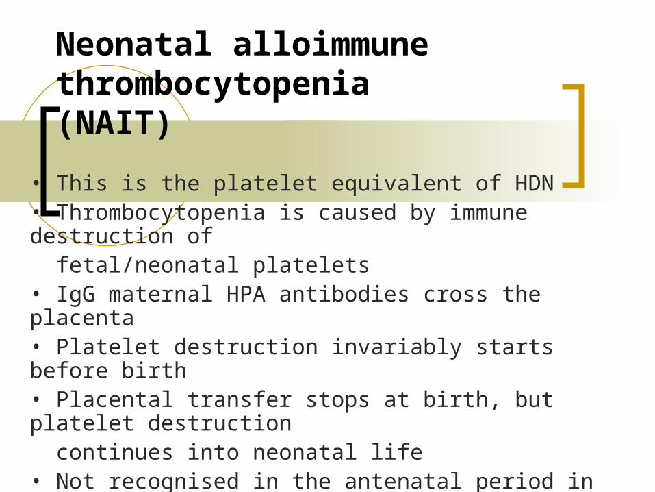
Neonatal alloimmune thrombocytopenia(NAIT)
• This is the platelet equivalent of HDN• Thrombocytopenia is caused by immune destruction of fetal/neonatal platelets• IgG maternal HPA antibodies cross the placenta• Platelet destruction invariably starts before birth• Placental transfer stops at birth, but platelet destruction continues into neonatal life• Not recognised in the antenatal period in women not investigated before
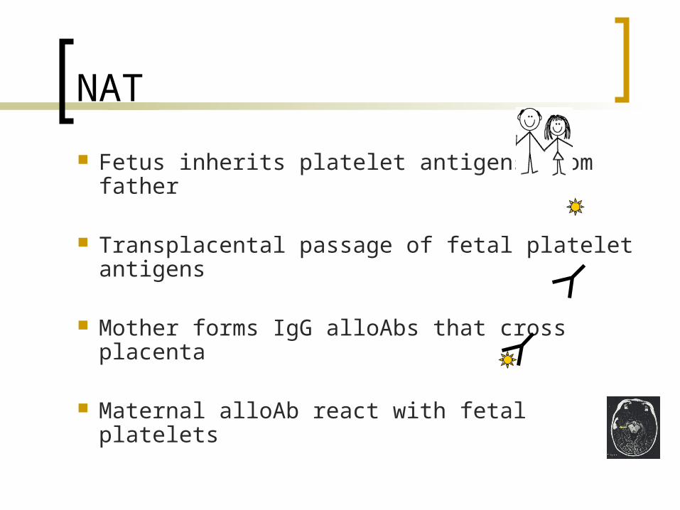
NAT
Fetus inherits platelet antigens from father
Transplacental passage of fetal platelet antigens
Mother forms IgG alloAbs that cross placenta
Maternal alloAb react with fetal platelets
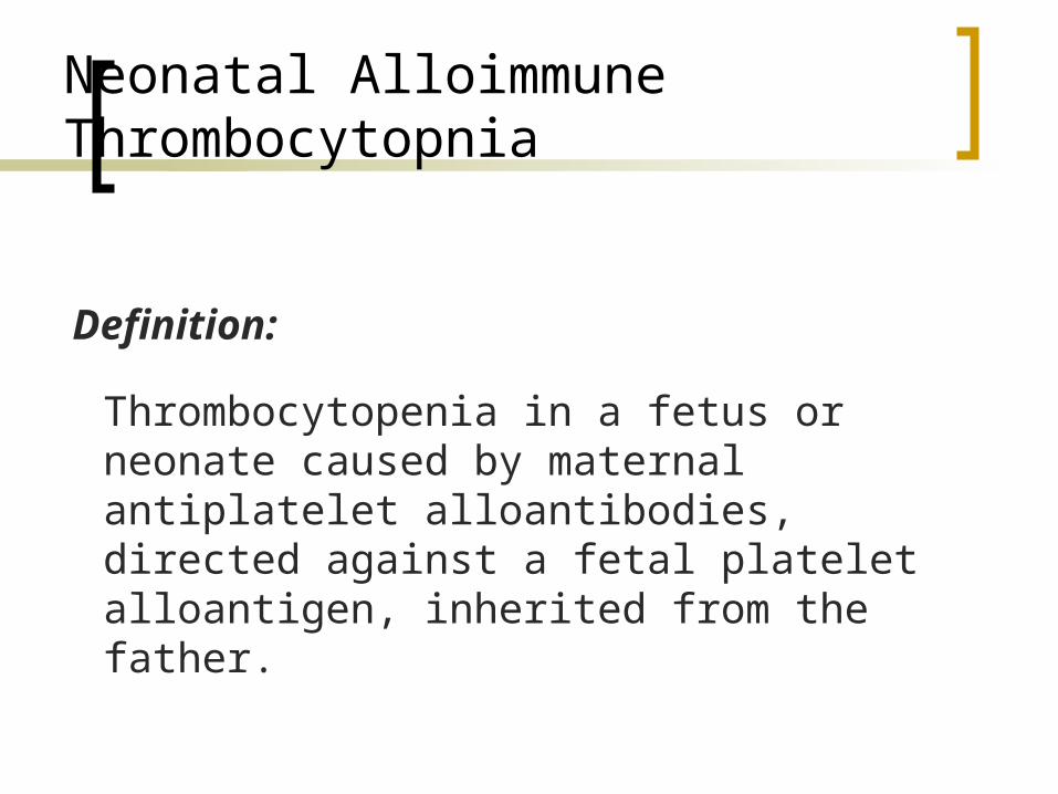
Neonatal Alloimmune Thrombocytopnia
Thrombocytopenia in a fetus or neonate caused by maternal antiplatelet alloantibodies, directed against a fetal platelet alloantigen, inherited from the father.
Definition:
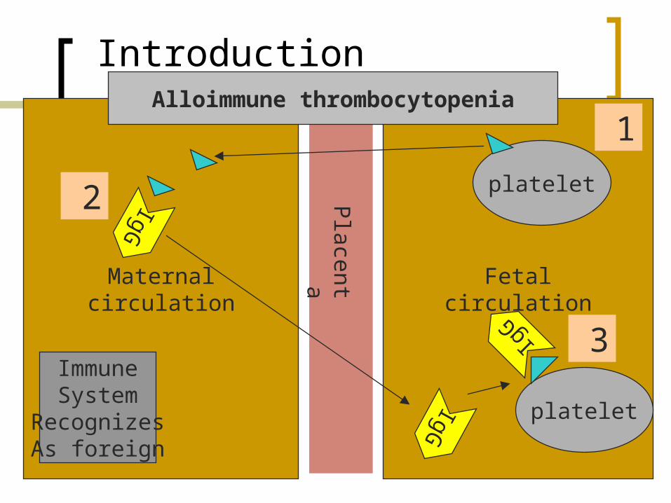
Introduction
Maternalcirculation
Fetalcirculation
Placenta
platelet
IgG
IgG
IgG
platelet
ImmuneSystem
RecognizesAs foreign
1
2
3
Alloimmune thrombocytopenia

HPA alloimmunisation
1. Pregnancy
eg. HPA-1a neg mothercarrying HPA-1a posbaby cc
2. Blood Transfusioneg. HPA-1a neg personreceiving HPA-1a posblood transfusion
Antigen pos platelets enter person’s circulation
Production of platelet antibody (anti-HPA-1a)
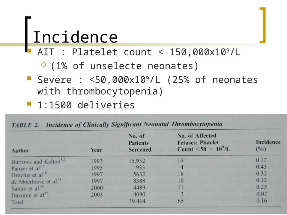
Incidence AIT : Platelet count < 150,000ⅹ109/L
(1% of unselecte neonates) Severe : <50,000ⅹ109/L (25% of neonates with
thrombocytopenia) 1:1500 deliveries
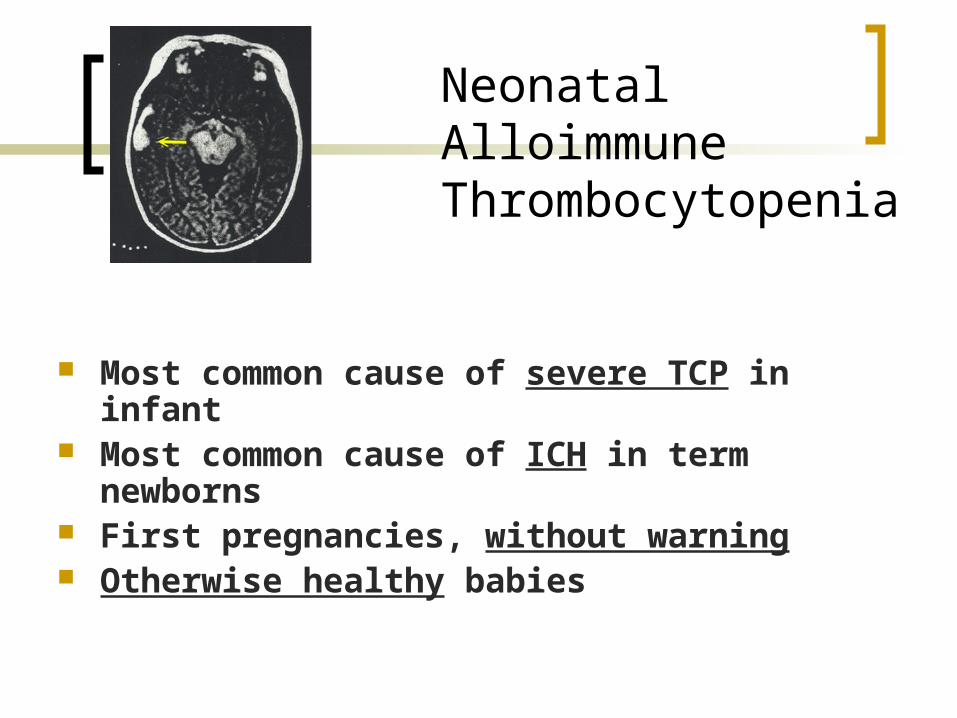
NeonatalAlloimmuneThrombocytopenia
Most common cause of severe TCP in infant Most common cause of ICH in term
newborns First pregnancies, without warning Otherwise healthy babies
Pathophysiology
Definition : fetal/neonatal platelet count < 150ⅹ109/L maternal platelet alloantibodies
Manifestations Concentration of maternal IgG Density of the target antigen on the fetal
platelets Phagocytic activity Compensatory ability of the fetal bone marrow
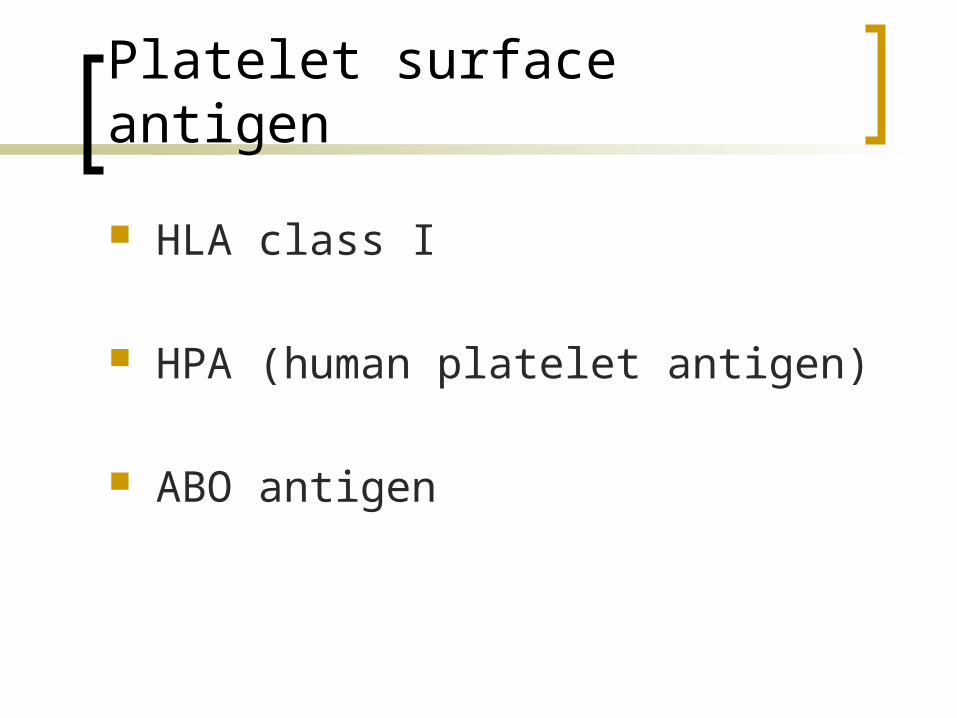
Platelet surface antigen
HLA class I
HPA (human platelet antigen)
ABO antigen
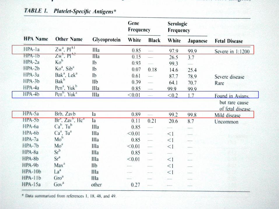
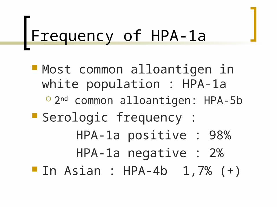
Frequency of HPA-1a
Most common alloantigen in white population : HPA-1a 2nd common alloantigen: HPA-5b
Serologic frequency :
HPA-1a positive : 98%
HPA-1a negative : 2% In Asian : HPA-4b 1,7% (+)
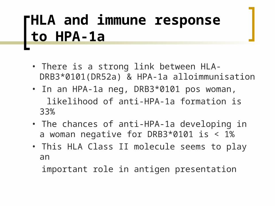
HLA and immune response to HPA-1a
• There is a strong link between HLA-DRB3*0101(DR52a) & HPA-1a alloimmunisation
• In an HPA-1a neg, DRB3*0101 pos woman,
likelihood of anti-HPA-1a formation is 33%
• The chances of anti-HPA-1a developing in a woman negative for DRB3*0101 is < 1%
• This HLA Class II molecule seems to play an
important role in antigen presentation
Listen up!
They say, that the maternal platet count is normal and alloimmunization is not suspected until after the birth of the affected child.Even in the first affected infant , is frequently severe and usually develops before the third trimester. It can thus cause fetal intracranial hemorrhage, even as early as 20 weeks!!!!!!!!!!!!!!!!!!!!!!!!!
Clinical features of NAIT:
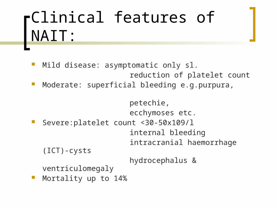
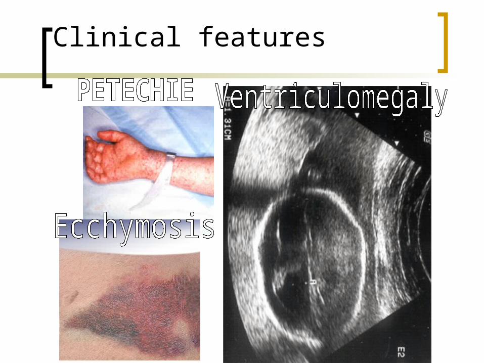
Mild disease: asymptomatic only sl. reduction of platelet count Moderate: superficial bleeding e.g.purpura, petechie, ecchymoses etc. Severe:platelet count <30-50x109/l internal bleeding intracranial haemorrhage (ICT)-cysts hydrocephalus & ventriculomegaly Mortality up to 14%
Clinical features

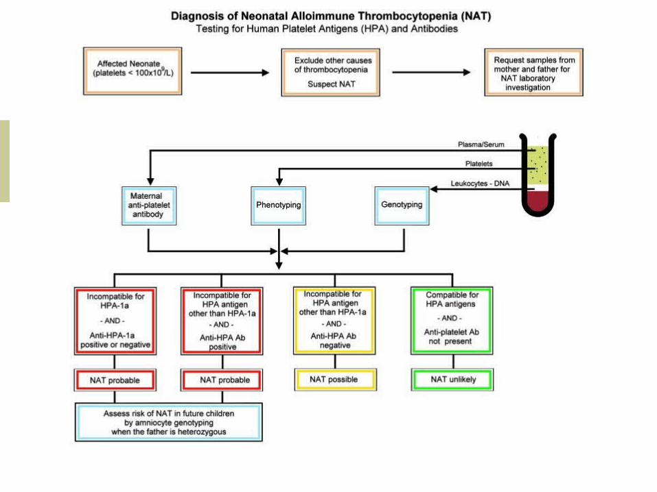
Laboratory Studies
Antiplatelet antibody testing PSIFT (Platelet Suspension
Immunofluorescence Test) MAIPA (Monoclonal Antibody Immobilization of
Platet Antigens)
Platelet antigen typing MAIPA ELISA DNA-based test (PCR-)SPP
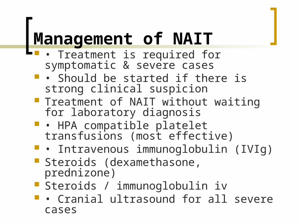
Management of NAIT • Treatment is required for symptomatic &
severe cases • Should be started if there is strong clinical
suspicion Treatment of NAIT without waiting for
laboratory diagnosis • HPA compatible platelet transfusions (most
effective) • Intravenous immunoglobulin (IVIg) Steroids (dexamethasone, prednizone) Steroids / immunoglobulin iv • Cranial ultrasound for all severe cases
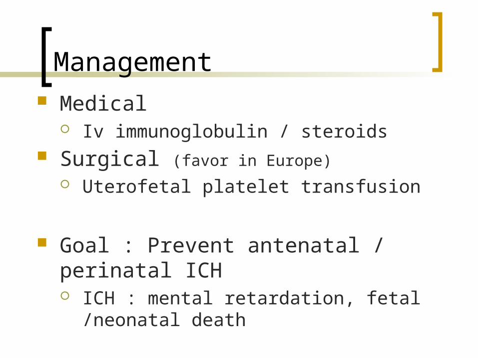
Management Medical
Iv immunoglobulin / steroids Surgical (favor in Europe)
Uterofetal platelet transfusion
Goal : Prevent antenatal / perinatal ICH ICH : mental retardation, fetal /neonatal death
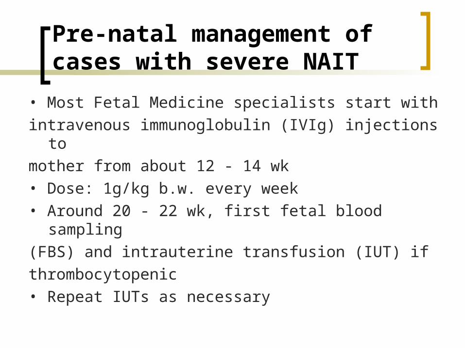
Pre-natal management of cases with severe NAIT
• Most Fetal Medicine specialists start with
intravenous immunoglobulin (IVIg) injections to
mother from about 12 - 14 wk
• Dose: 1g/kg b.w. every week
• Around 20 - 22 wk, first fetal blood sampling
(FBS) and intrauterine transfusion (IUT) if
thrombocytopenic
• Repeat IUTs as necessary
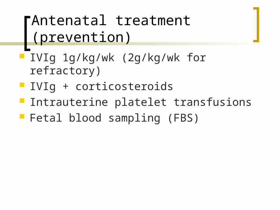
Antenatal treatment (prevention) IVIg 1g/kg/wk (2g/kg/wk for refractory) IVIg + corticosteroids Intrauterine platelet transfusions Fetal blood sampling (FBS)
Future pregnancies
In a mother who has had an affected baby, thechances of having another affected child is high ifthe implicated Ag is inherited• Severe thrombocytopenia may occur as early as 20- 24 weeks of gestation• Chances of inheriting the paternal antigen dependson the zygosity of the father• Both parents should be counselled• All ‘at risk’ pregnancies should be managed inFMUs
Summary
• NAIT is a serious condition with significant morbidity & mortality (ICH: 11% {34/305}; Deaths : 7.2% {22/305})• Most cases are caused by anti-HPA-1a or anti-HPA-5b or both• In severe cases, treatment should be given ASAP before test results• There must be good communication between FMUs and local hospitals when known cases are referred for delivery• BB staff, Haematologist & Neonatal team must be alerted by the Obstetricians
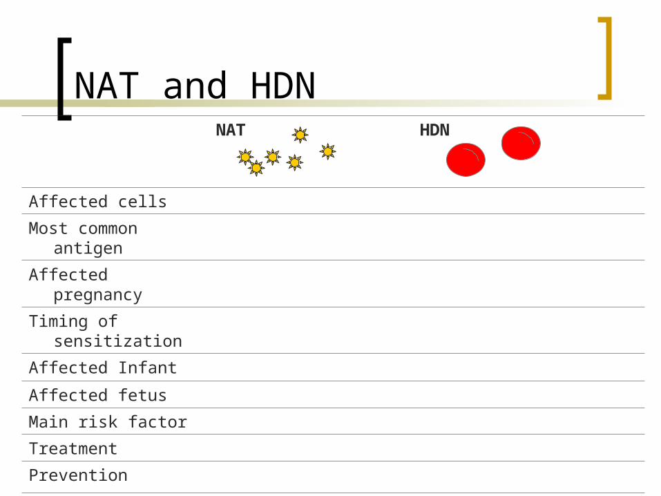
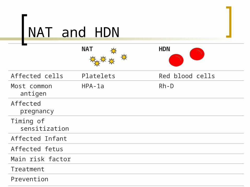
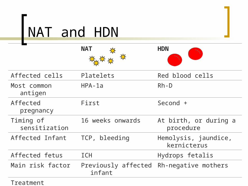
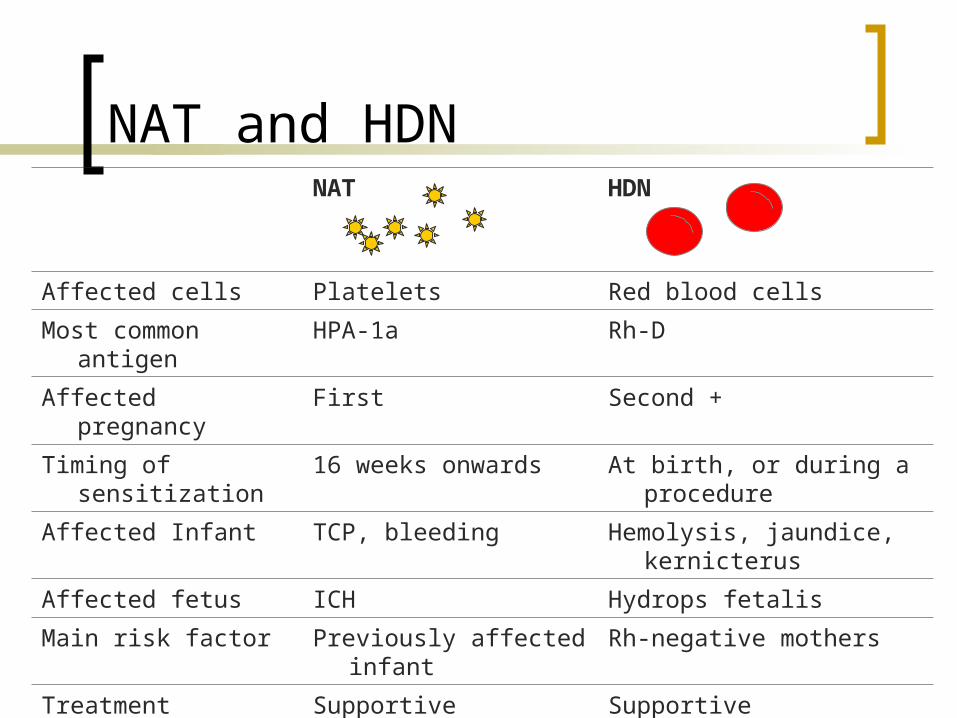
NAT and HDNNAT HDN
Affected cells
Most common antigen
Affected pregnancy
Timing of sensitization
Affected Infant
Affected fetus
Main risk factor
Treatment
Prevention
Efficacy of prevention
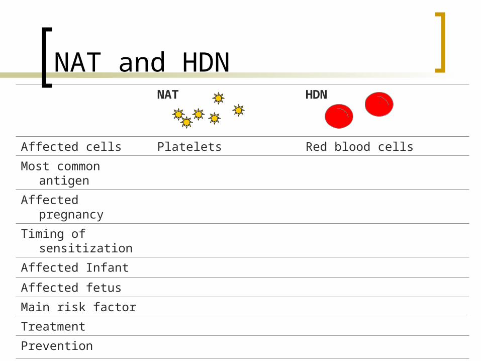
NAT and HDNNAT HDN
Affected cells Platelets Red blood cells
Most common antigen
Affected pregnancy
Timing of sensitization
Affected Infant
Affected fetus
Main risk factor
Treatment
Prevention
Efficacy of prevention
NAT and HDNNAT HDN
Affected cells Platelets Red blood cells
Most common antigen HPA-1a Rh-D
Affected pregnancy
Timing of sensitization
Affected Infant
Affected fetus
Main risk factor
Treatment
Prevention
Efficacy of prevention
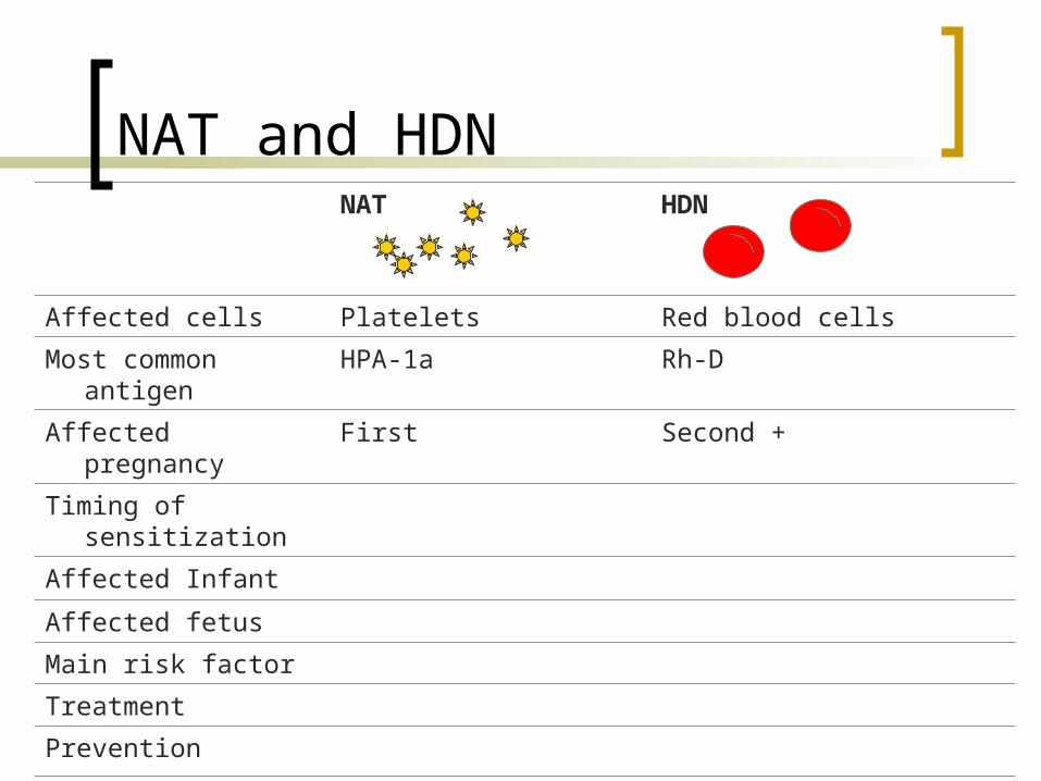
NAT and HDNNAT HDN
Affected cells Platelets Red blood cells
Most common antigen HPA-1a Rh-D
Affected pregnancy First Second +
Timing of sensitization
Affected Infant
Affected fetus
Main risk factor
Treatment
Prevention
Efficacy of prevention
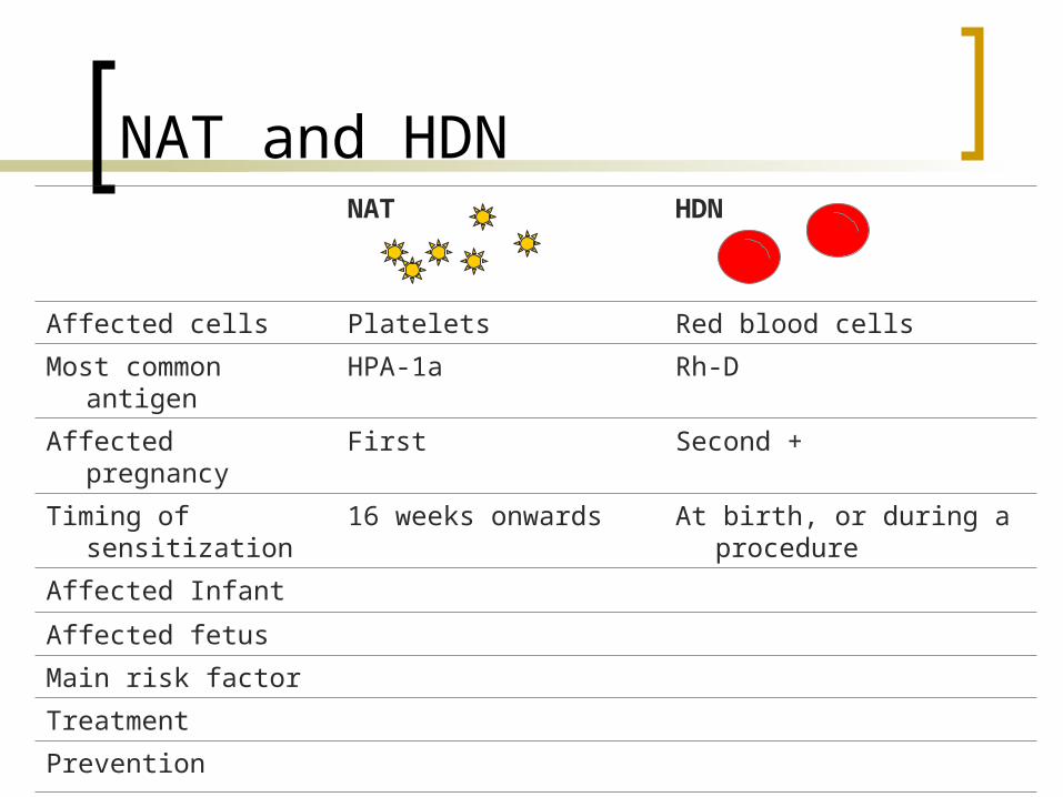
NAT and HDNNAT HDN
Affected cells Platelets Red blood cells
Most common antigen HPA-1a Rh-D
Affected pregnancy First Second +
Timing of sensitization 16 weeks onwards At birth, or during a procedure
Affected Infant
Affected fetus
Main risk factor
Treatment
Prevention
Efficacy of prevention

NAT and HDNNAT HDN
Affected cells Platelets Red blood cells
Most common antigen HPA-1a Rh-D
Affected pregnancy First Second +
Timing of sensitization 16 weeks onwards At birth, or during a procedure
Affected Infant TCP, bleeding Hemolysis, jaundice, kernicterus
Affected fetus
Main risk factor
Treatment
Prevention
Efficacy of prevention
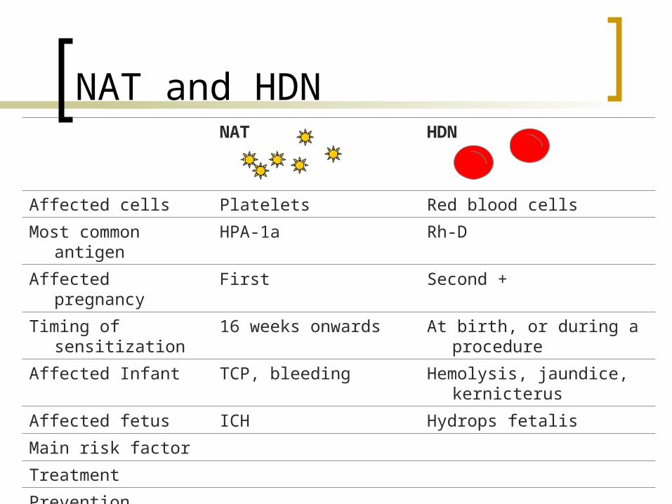
NAT and HDNNAT HDN
Affected cells Platelets Red blood cells
Most common antigen HPA-1a Rh-D
Affected pregnancy First Second +
Timing of sensitization 16 weeks onwards At birth, or during a procedure
Affected Infant TCP, bleeding Hemolysis, jaundice, kernicterus
Affected fetus ICH Hydrops fetalis
Main risk factor
Treatment
Prevention
Efficacy of prevention
NAT and HDNNAT HDN
Affected cells Platelets Red blood cells
Most common antigen HPA-1a Rh-D
Affected pregnancy First Second +
Timing of sensitization 16 weeks onwards At birth, or during a procedure
Affected Infant TCP, bleeding Hemolysis, jaundice, kernicterus
Affected fetus ICH Hydrops fetalis
Main risk factor Previously affected infant Rh-negative mothers
Treatment Supportive Supportive
Prevention
Efficacy of prevention

NAT and HDNNAT HDN
Affected cells Platelets Red blood cells
Most common antigen HPA-1a Rh-D
Affected pregnancy First Second +
Timing of sensitization 16 weeks onwards At birth, or during a procedure
Affected Infant TCP, bleeding Hemolysis, jaundice, kernicterus
Affected fetus ICH Hydrops fetalis
Main risk factor Previously affected infant Rh-negative mothers
Treatment Supportive Supportive
Prevention IVIG Anti-D
Efficacy of prevention ? 99%
AIT
The diagnosis can be made on clinical grounds if the mother has normal platelet count and there is no evidence of any immunological disorder , and her infant has thrombocytopenia without evidence of other disease.
Recurs in 70-90 % of subsequent pregnancies,is ofen severe , and usually develops eariel in with each successive pregnancy.
AIT ( Isoimmune thrombocytopenia)
It is caused by maternal isoimmunisation to fetal platet antigens in a manner similar to D-antigen isoimmunization.
Thus , the maternal platet count is normal and alloimmunization is not suspected until after the birth of the affected child.
Even in the first affected infant , is frequentlysevere and usually develops before the third trimester.
It can thus cause fetal intracranial hemorrhage, even as early as 20 weeks

Questions?