Embed Size (px)
Citation preview

SCREENING AND PARTIAL PURIFICATION OF PROTEASEINHIBITORS FROM SENNA SURATTENSIS LEAVES
AHMAD FIRDHAUS ARHAM
FACULTY OF SCIENCEUNIVERSITY OF MALAYA
KUALA LUMPUR
2011

SCREENING AND PURIFICATION OF PROTEASEINHIBITORS FROM SENNA SURATTENSIS LEAVES
AHMAD FIRDHAUS ARHAM
DISSERTATION SUBMITTED IN FULFILMENT OF THE REQUIREMENT
FOR THE DEGREE OF MASTER OF BIOTECHNOLOGY
FACULTY OF SCIENCEUNIVERSITY OF MALAYA
KUALA LUMPUR
2011

ACKNOWLEDGEMENT
I would like to express my gratitude to all those who gave me the possibility to
complete this thesis. I want to thank my supervisor, Dr Zazali Alias whose help, stimulating
suggestions and encouragement helped me in all the time of research for and writing of this
thesis.
I am also want to thank my colleagues Nora Asyikin Ramli, Rabiatul Adawiyah
Mohd Hairuni and Kamarul Huda Kamaruddin for their supports and helps throughout the
completion of my project.
Lastly, I would like to give my special thanks to my family and all of those who
patient love enabled me to complete this work.
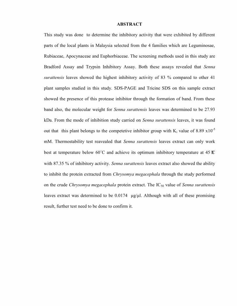
ABSTRACT
This study was done to determine the inhibitory activity that were exhibited by different
parts of the local plants in Malaysia selected from the 4 families which are Leguminosae,
Rubiaceae, Apocynaceae and Euphorbiaceae. The screening methods used in this study are
Bradford Assay and Trypsin Inhibitory Assay. Both these assays revealed that Senna
surattensis leaves showed the highest inhibitory activity of 83 % compared to other 41
plant samples studied in this study. SDS-PAGE and Tricine SDS on this sample extract
showed the presence of this protease inhibitor through the formation of band. From these
band also, the molecular weight for Senna surattensis leaves was determined to be 27.93
kDa. From the mode of inhibition study carried on Senna surattensis leaves, it was found
out that this plant belongs to the competetive inhibitor group with Ki value of 8.89 x10⁻⁵
mM. Thermostability test reavealed that Senna surattensis leaves extract can only work
best at temperature below 60˚C and achieve its optimum inhibitory temperature at 45⁰C
with 87.35 % of inhibitory activity. Senna surattensis leaves extract also showed the ability
to inhibit the protein extracted from Chrysomya megacephala through the study performed
on the crude Chrysomya megacephala protein extract. The IC50 value of Senna surattensis
leaves extract was determined to be 0.0174 μg/μl. Although with all of these promising
result, further test need to be done to confirm it.

ABSTRAK
Kajian ini dijalankan untuk menentukan aktiviti-aktiviti perencatan yang ditunjukkan oleh
beberapa jenis bahagian tumbuh-tumbuhan tempatan di Malaysia yang terdiri daripada 4
famili iaitu Leguminosae, Rubiaceae, Apocynaceae and Euphorbiaceae. Kaedah
penyaringan ujian Bradford dan ujian perencatan tripsin menunjukkan bahawa daun Senna
surattensis mempunyai kebolehan perencatan yang paling tinggi berbanding 41 sampel
tmbuh-tumbuhan yang lain di dalam kajian ini iaitu sebanyak 83 %. Ujian SDS-PAGE dan
Ujian Tricine SDS terhadap sampel ini menunjukkan kehadiran protin perencat melalui
jalur yang terhasil. Melalui jalur ini juga, berat molekular bagi daun Senna surattensis
dianggarkan sebanyak 27.93 kDa. Daripada penentuan Mod perencatan ke atas ekstrak
daun Senna surattensis, didapati ianya tergolong dalam kumpulan perencatan kompetetif
dengan nilai Ki sebanyak of 8.89 x10⁻⁵ mM. Ujian kestabilan suhu yang dijalankan
menunjukkan bahawa ekstrak daun Senna surattensis hanya mampu berfungsi di bawah
suhu 60˚C dan mencapai suhu perencatan optimum pada 45⁰C dengan 87.35 % aktiviti
perencatan. Ekstrak daun Senna surattensis juga mampu merencat protein yang diekstrak
daripada Chrysomya megacephala melalui ujian yang dijalankan ke atas ekstrak protin
mentah Chrysomya megacephala. Nilai IC50 yang diperolehi untuk ekstrak daun Senna
surattensis adalah 0.0174 μg/μl.
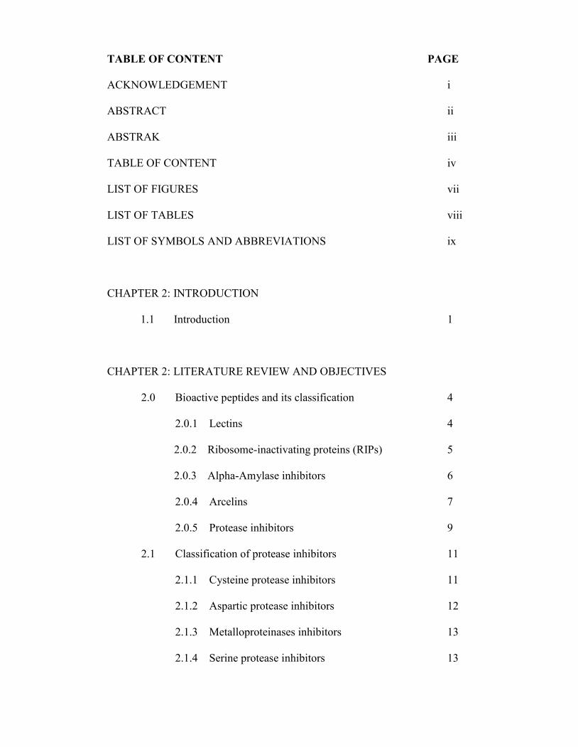
TABLE OF CONTENT PAGE
ACKNOWLEDGEMENT i
ABSTRACT ii
ABSTRAK iii
TABLE OF CONTENT iv
LIST OF FIGURES vii
LIST OF TABLES viii
LIST OF SYMBOLS AND ABBREVIATIONS ix
CHAPTER 2: INTRODUCTION
1.1 Introduction 1
CHAPTER 2: LITERATURE REVIEW AND OBJECTIVES
2.0 Bioactive peptides and its classification 4
2.0.1 Lectins 4
2.0.2 Ribosome-inactivating proteins (RIPs) 5
2.0.3 Alpha-Amylase inhibitors 6
2.0.4 Arcelins 7
2.0.5 Protease inhibitors 9
2.1 Classification of protease inhibitors 11
2.1.1 Cysteine protease inhibitors 11
2.1.2 Aspartic protease inhibitors 12
2.1.3 Metalloproteinases inhibitors 13
2.1.4 Serine protease inhibitors 13

2.1.4.1 Bowman-Birk Inhibitor (BBI) 16
2.1.4.2 Kunitz-type inhibitors 18
2.2 Application of serine protease inhibitors 20
CHAPTER 3: MATERIALS AND METHODS
3.1 Materials 25
3.2 Equipments 26
3.3 Methods
3.3.1 Preparation of Plant Extract 26
3.3.2 Protein content determination 27
3.3.3 Trypsin Inhibitory Assay 27
3.3.4 Trypsin inhibitory Activity detection
in SDS-polyacrylamide gel electrophoresis
and Tricine SDS 28
3.3.5 Preparation of Chrysomya megacephala
crude protease extracts 29
3.3.6 Mode of inhibition and Ki value determination 30
3.3.7 Thermostability determination 30
3.3.8 IC50 estimation 31
CHAPTER 4: RESULTS
4.1 Screening of trypsin inhibitors 32
4.2 Sodium Dodecyl Sulfate Polyacrylamide Gel Electrophoresis
result of sample extracts 35

4.3. Determination of Senna surattensis’s leaves mode of
inhibition and Ki value 38
4.4 Determination of Senna surattensis’s leaves
thermostability 40
4.5 Estimation of Senna surattensis’s leaves IC50 value 41
CHAPTER 5: DISCUSSION 43
CHAPTER 6: CONCLUSION 53
REFERENCES 55
APPENDIXES
APPENDIX 1 Determination of protein standard curve 68
APPENDIX 2 Preparation of Bradford reagents
(Bradford et al., 1976) 69
APPENDIX 3 Determination of plants sample content 69
APPENDIX 4 Preparation of trypsin inhibitor assay buffer 71
APPENDIX 5 Preparation of HI-TRAP G-25 chromatography
buffer 71
APPENDIX 6 HI-TRAP G-25 chromatography 72
APPENDIX 7 Reagents for SDS-PAGE 72
APPENDIX 8 Buffer for Tricine-SDS gel 73
APPENDIX 9 Standard curve of log MW againts
relative mobility determination 73
APPENDIX 10 Trypsin inhibitory assay using Chrysomya
megacephala extracted protease. 76

LIST OF FIGURES
FIGURE TITLE PAGE
Figure 1 Shows SDS-Page gel result of lyophilized sample
extracts of Senna surattensis leaves (A) and
Mimosa diplotricha fruit (B) on 17% acrylamide gel
under two different condition which is with trypsin
incubation and without trypsin incubation. 35
Figure 2 Shows the bands of lyophilized sample extracts of
Senna surattensis leaves (A) and Mimosa diplotricha
fruit (B) on Pre-cast (invitrogen) Tricine SDS gel
at 4-20% acrylamide concentration. 36
Figure 3 Separation of G-25 chromatography eluent of Senna
surattensis leaves (A) and Mimosa diplotricha
fruit (B) on Tricine SDS gel 37
Figure 4 The dixon plot of concentration versus 1/v sample
extract of Senna surattensis’s leaves in the presence
of 1mM and 5mM BapNA in DMSO. 39
Figure 5 The bar chart plotted of Senna surattensis’s leaves
extract temperature versus its reduction of activity
percentage. 40
Figure 6 Shows the Senna surattensis’s leaves percentage of
inhibition and activity of trypsin concentration. 42
Figure 7 Bovine serum albumin standard curve (µg/ml) 68
Figure 8 Standard curve of Log MW against Relative mobility (Rf) 75
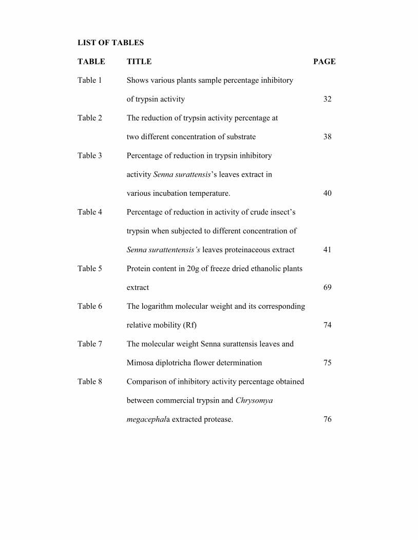
LIST OF TABLES
TABLE TITLE PAGE
Table 1 Shows various plants sample percentage inhibitory
of trypsin activity 32
Table 2 The reduction of trypsin activity percentage at
two different concentration of substrate 38
Table 3 Percentage of reduction in trypsin inhibitory
activity Senna surattensis’s leaves extract in
various incubation temperature. 40
Table 4 Percentage of reduction in activity of crude insect’s
trypsin when subjected to different concentration of
Senna surattentensis’s leaves proteinaceous extract 41
Table 5 Protein content in 20g of freeze dried ethanolic plants
extract 69
Table 6 The logarithm molecular weight and its corresponding
relative mobility (Rf) 74
Table 7 The molecular weight Senna surattensis leaves and
Mimosa diplotricha flower determination 75
Table 8 Comparison of inhibitory activity percentage obtained
between commercial trypsin and Chrysomya
megacephala extracted protease. 76
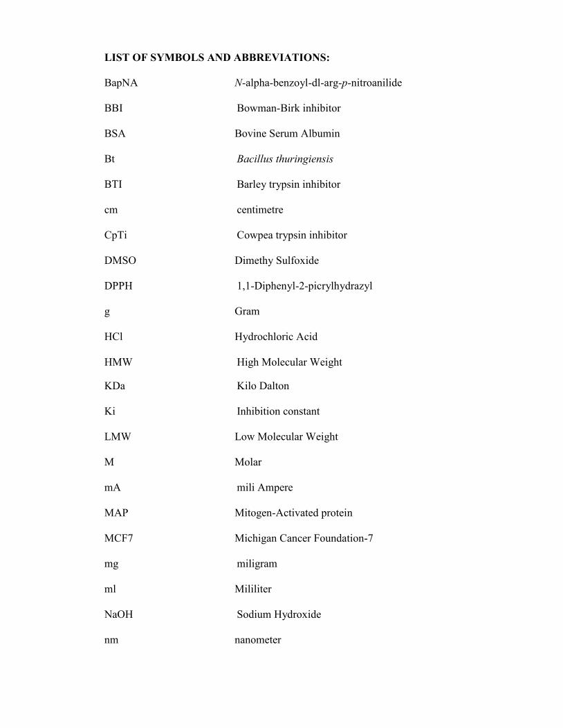
LIST OF SYMBOLS AND ABBREVIATIONS:
BapNA N-alpha-benzoyl-dl-arg-p-nitroanilide
BBI Bowman-Birk inhibitor
BSA Bovine Serum Albumin
Bt Bacillus thuringiensis
BTI Barley trypsin inhibitor
cm centimetre
CpTi Cowpea trypsin inhibitor
DMSO Dimethy Sulfoxide
DPPH 1,1-Diphenyl-2-picrylhydrazyl
g Gram
HCl Hydrochloric Acid
HMW High Molecular Weight
KDa Kilo Dalton
Ki Inhibition constant
LMW Low Molecular Weight
M Molar
mA mili Ampere
MAP Mitogen-Activated protein
MCF7 Michigan Cancer Foundation-7
mg miligram
ml Mililiter
NaOH Sodium Hydroxide
nm nanometer

PIs Protease inhibitors
PVY Potato Virus Y
rpm/min Revolution per minute
SDS Sodium dodecyl Sulfate
SDS PAGE Sodium dodecyl Sulfate-Polyacrylamide
Gel Electrophoresis
SPIs Serine Protease inhibitors
TEMED N,N,N’N’ –tetramethylenediamine
TEV Tobacco Etch Virus
TPCK L-1-tosylamido-2-phenylethyl chloromethyl ketone
UV ultra violet
v/v volume per volume
w/v weight per volume
xg Gravity
α Alpha
β Beta
γ Gamma
⁰C Degree Celcius
% Percent

CHAPTER 1: INTRODUCTION
1.1 Introduction
Proteases have been described in large numbers naturally in animals, plants and
microorganisms and have been extensively studied in order to their structures and
functional properties (Laskowski and Kato, 1980; Hibbetts et al., 1999). Proteases are
known as proteinases, peptidases or proteolytic enzymes and are involved in many
beneficial functions. Many biological functions including food digestion, lysosom
degradation, and signaling cascades rely on proteases (Fear et al., 2006). The proteases are
the enzymes which in vivo catalyzed the hydrolytic breakdown of proteins into specific
peptide fractions and amino acids (Otlewski et al., 1999; Fear et.al, 2006; Habib H. and
Khalid M.F., 2007).
Proteases are divided into endoproteases and exoproteases where function as regulators to
control endogenous enzymes that regulatory activity of proteolytic, whether the target
enzymes are of exogenous or endogenous origin in the presence of the active enzyme
(Barret, 1987; Ryan, 1990; Kato, 2002). There are classified according to their mechanism
of catalysis and the amino acid essential for its activity, as cysteine proteases with a
cysteine, aspartic proteases with an aspartate group, metalloproteases with a metal ions
(Zn2+, Ca2+ or Mn2+) and serine proteases with serine and histidine (Neurath, 1984; Carlini
and Grossi-de-Sa, 2001).
Hans Neurath (1984) was among the first scientist recognized that protease act not only as
digestive enzymes, but also fulfill numerous other functions for many biological organism
including food digestion, lysosome degradation, and signaling cascades rely on proteases
and also functional for others (Neurath, 1984; Laskowski and Qasim, 1999; Fear et al.,
2006). Hans Neurath also recognized that proteinases are highly beneficial and must be
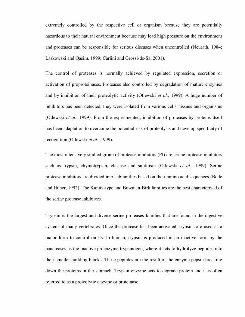
extremely controlled by the respective cell or organism because they are potentially
hazardous to their natural environment because may lead high pressure on the environment
and proteases can be responsible for serious diseases when uncontrolled (Neurath, 1984;
Laskowski and Qasim, 1999; Carlini and Grossi-de-Sa, 2001).
The control of proteases is normally achieved by regulated expression, secretion or
activation of proproteinases. Proteases also controlled by degradation of mature enzymes
and by inhibition of their proteolytic activity (Otlewski et al., 1999). A huge number of
inhibitors has been detected, they were isolated from various cells, tissues and organisms
(Otlewski et al., 1999). From the experimented, inhibition of proteases by proteins itself
has been adaptation to overcome the potential risk of proteolysis and develop specificity of
recognition (Otlewski et al., 1999).
The most intensively studied group of protease inhibitors (PI) are serine protease inhibitors
such as trypsin, chymotrypsin, elastase and subtilisin (Otlewski et al., 1999). Serine
protease inhibitors are divided into subfamilies based on their amino acid sequences (Bode
and Huber, 1992). The Kunitz-type and Bowman-Birk families are the best characterized of
the serine protease inhibitors.
Trypsin is the largest and diverse serine proteases families that are found in the digestive
system of many vertebrates. Once the protease has been activated, trypsins are used as a
major form to control on its. In human, trypsin is produced in an inactive form by the
pancreases as the inactive proenzyme trypsinogen, where it acts to hydrolyze peptides into
their smaller building blocks. These peptides are the result of the enzyme pepsin breaking
down the proteins in the stomach. Trypsin enzyme acts to degrade protein and it is often
referred to as a proteolytic enzyme or proteinase.

Trypsins are bind with inhibitors which as a competitive substrate analog to form an
inactive complex, inactive the digestive enzymes against insect digestive enzymes. The
proteolytic activity of the serine protease stops by trypsin inhibitors when its function is no
longer necessary. This inhibitor is cause depression of growth, nutritional disorders and
pancreatic hypertrophy or hyperplasia (Liener and Kakade, 1980).
The enzymatic mechanism is similar to other serine proteases that contain three residues
that is histidine-57, aspartate-102, and serine-195 (Rawling and Barrett, 1994) form a
charge relay which serves to make the active site serine nucleophilic. This is achieved by
modifying the electrostatic environment of the serine. The enzymatic reaction that trypsins
catalyze is thermodynamically favorable but requires significant activation energy. Trypsin
also contains an "oxyanion hole" formed by the backbone amide hydrogen atoms of Gly-
193 and Ser-195 which serves to stabilize the developing negative charge on the carbonyl
oxygen atom of the cleaved amide. In the catalytic pocket (S1) of trypsins are located the
aspartate residue (Asp 189) which responsible for attracting and stabilizing positively-
charged lysine or arginine, and is thus responsible for the specificity of the enzyme.
Therefore, in this study research is focused on the function of serine proteases inhibitors by
using potential of local plants that are important to control insecticides and thus make them
beneficial in agronomical and health relevance. This research study will be involving a
through screening, isolation, identification and characterization of protease inhibitors.

CHAPTER 2: LITERATURE REVIEW
2.0 Bioactive peptides and its classification
Peptides with biological activities generally contain three to twenty amino acid units, which
are proteins synthesized in the form of large prepropeptides in the cell. Bioactive peptides
in plants supposedly involved in defense mechanisms that confer resistance against
phytophagous predators and infection by viruses, bacteria, fungi, nematodes and other
organisms (Carlini and Grossi-de-Sa, 2001). The best known plant proteins are lectins,
ribosome-inactivating proteins (RIPs), arcelins, alpha-amylase canatoxin and protease
inhibitors.
2.0.1 Lectins
Lectins are class of a protein of non-immune origin that possess at least one non-catalytic
domain that specifically and reversibly binds to mono or oligosaccharide (Lis and Sharon,
1986; Peumans and Van Damme, 1995; Carlini and Grossi-de-Sa, 2001). The seeds of the
Leguminoseae are rich sources of lectins, but the same lectin and homologs are also found
in other parts of the plant, such as the bark, stem, and leaves. This family includes lectins
such as ConA, soybean agglutinin, and lentil lectin. Two other smaller families of plants
whose lectins have been characterized are the Gramineae; cereals, such as wheat germ and
Solanaceae; potatoes and tomatoes.
The function of lectins in plants is still controversies with their biological roles in the parent
organisms (Carlini and Grossi-de-Sa, 2001). The binding site for carbohydrate in some
lectins involves a combination of hydrophobic interactions and van der Waals contacts to
which small plant growth regulators such as adenine can bind to (Roberts and Goldstein,
1983; Carlini and Grossi-de-Sa, 2001). Although plant lectins have an ability to bind

carbohydrate, evidence exists that these proteins may have additional activities. For
example, some lectins, such as Dolichus biflorus, bind adenine residues with high affinity
and specificity in regions of the protein outside the common carbohydrate-binding site.
Some plant lectins have shown entomotoxic effects when fed to insects from Coleoptera,
Homoptera, and Lepidoptera orders (Peumans and Van Damme, 1995; Carlini and Grossi-
de-Sa, 2001). Transgenic expression of the gene encoding G. nivalis agglutinin in rice
plants decreases survival and fecundity of insects attacking the transgenic plants (Schuler et
al., 1998). Therefore this class of protein should be studied more for their potential.
2.0.2 Ribosome-inactivating proteins (RIPs)
Ribosome-inactivating proteins or RIPs are the proteins that are capable of
inactivating ribosomes in many plants (Peumans et al., 2001). RIPs are a group of cytotoxic
N-glycosidases that specifically cleave nucleotide N-C glycosidic bonds. RIPs were first
identified more than 100 years ago. Their biological functions are determined to play a role
in plant defence mechanism.
RIPS have been described into three types, types I is composed of a single polypeptide
chain, whereas type II is a heterodimer consisting of a chain, which is attached to a sugar-
binding B chain that functionally equivalent to a type I while type III is unknown function
that forms single chain containing an extended carboxyl-terminal domain (Park et al.,
2004).
The effect of ribosome show differential sensitivity to RIPs in plant cells and isolated
ribosomes, while ribosomes of protozoans and fungi seem to be highly sensitive. Ricin, the
toxic principle of castor bean was identifried as a protein at the 19th century was shown to
be ineffective to a variety of insects of different orders, although it was able to inhibit

protein synthesis by insect ribosomes in cell-free preparations (Gatehouse et al., 1990;
Carlini and Grossi-de-Sa, 2001). Since the isolation and characterization of ricin, many
structurally and functionally related proteins have been identified in a wide variety of plants
(Peumans et al., 2001).
2.0.3 Alpha-Amylase inhibitors
Alpha-amylases are found in microorganisms, animals and plants, whixh catalyze the initial
hydrolyses of alpha-1,4-linked sugar polymers into shorter oligosaccharides, an important
step towards transforming sugar polymers into single units that can be assimilated by the
organism. The first alpha-amylase inhibitor characterised was that of the monomeric 13 kD
known as 0.31 form, from wheat (Carlini and Grossi-de-Sa, 2001).
This endosperm protein is relatively abundant in seeds that suggesting a role as a storage or
reserve protein, as regulators of endogenous enzyme or as defensive mechanisms against
the attacks of animal predators and insect or microbial pests. These inhibitors were also
relevant in several aspects of human health such as to control of diabetes and obesity,
diagnosis of pancreatic hyperamylasemia disorders and nutritional and toxicological
aspects of foods (Turcotte et al., 1994; Bisschoff et al., 1994; Carlini and Grossi-de-Sa,
2001). The amylase inhibitors present in seeds used as food present some toxicological
significance in the diets of infants who have a lower production of pancreatic alpha-
amylase than adults and for patients with impaired peptic or gastric function (Richardson,
1991).
Alpha-amylase inhibitors also show great interest as potentially important tools of natural
and engineer resistance of crop plant against pests that are improved through the use of
transgenic technology (Gatehouse and Gatehouse, 1998, Carlini and Grossi-de-Sa, 2001).

Focused on lectin-like inhibitors present in common bean Proteus vulgaris seeds have been
shown that toxic effects to several insect pests (Ishimoto and Chrispeels, 1996). The effect
was well determined not only in different oraganisms by enzymatic activity but also in
feeding assay experiments (Ishimoto et al., 1996).
Transgenic pea and azuki bean seeds expressing the inhibitor, Alpha-amylase-1, of the
domesticated common bean Proteus vulgaris was completed resistance against bruchids
(Ishimoto et al., 1996; Morton et al., 2000). Therefore these proteins can be safely
introduced into food plants because the transgenic grains showed minimal effects on
mammalian digestion system (Pusztai et al., 1999; Carlini and Grossi-de-Sa, 2001).
2.0.4 Arcelins
Arcelins are lectin-related proteins detected only in wild beans (Phaseolus vulgaris) and
exists in six electrophoretic variants which supposedly involved in defence mechanisms to
confer resistance against predators such as bruchid beetles (Chrispeels and Raikhel, 1991;
Carlini and Grossi-de-Sa, 2001), although the precise mechanism behind the toxicity of
arcelin is as yet unknown.
Arcelins can be divided into three subgroups,group one are arcelin 1, arcelin 2 and arcelin
6, group two is arcelin 4 and group three are arcelin 5a and arcelin 5b. Sequence data of
arcelin 6 was indicated that this protein is closely related to arcelin 1 and arcelin 2.
Biochemical data indicate that arcelin 3 belongs to the same subgroup of arcelin 4.
Arcelin 5 has been reported to consist of a mixture of two major protein fractions, termed
arcelin 5a, Arc5a, 32.2 kDa and arcelin 5b, Arc5b with 31.5 kDa (Goossens et al., 1994).
Arc5b and Arc5a contain one and two glycans, respectively, while Arc5c is not
glycosylated. Two different arcelin-5 cDNA sequences were reported (Goossens et al.,

1994), called arc5-I and arc5-II. They encode two polypeptides of 240 amino acids (26.8
and 27.0 kDa) with a high identity (96.9%, a difference of 8 residues in the N-terminal part
of the chain).
The arc5-I-encoded protein contains three potential glycosylation sites, while thearc5-
II encoded protein contains only two. Arc5a and Arc5b are encoded by arc5-I and arc5-II,
respectively, while Arc5c could be encoded by arc5-II or by a third copy of the arc5 gene
with a much lower rate of expression. Arcelins are thought to provide resistance against the
bean bruchid pest Zabrotus subfasciatus (Osborn et al., 1988). Among the arcelin variants,
Arc1 and Arc5 appear to be the most promising in conferring insect resistance (Kornegay et
al., 1993) discovery of putative novel enzymatic (Goossens et al., 1994).
2.0.5 Protease inhibitors
Protease inhibitors (PIs) are found in animals, plants and microorganisms (Laskowski and
Kato, 1980). Protease inhibitors adopt many different structures, ranging in size from mini-
proteins to large macromolecular structures, much larger than the target enzyme (Otlewski
et al., 1999). PIs are classified into two large groups based on their structural dichotomy
which include the low molecular weight peptidomimetic inhibitors and protein protease
inhibitors (Fear et al., 2007).
PIs are divided into five groups of serine, cysteine, threonine, aspartyl and metalloprotease
inhibitors according to the mechanism employed at the active site of proteases they inhibit
(Fear et al., 2007). Two mechanisms can occured prroteolytic inhibition by protease
inhibitors by irreversible trapping reactions and reversible tightbinding reactions (Rawlings
et al., 2004).

From previous studies, plants seed are widely distributed and a rich source of protease
(Richardson, 1991; Mello and Silva-Filho, 2002; Chaudhary et al., 2008). Protease
inhibitors (PIs) have evolved to inhibit proteolytic enzymes. They are classified according
to their types of enzyme they inhibit (Mosolov, 1998; Otlewski et al., 1999; Carlini and
Grossi-de-Sa, 2001). The molecular mass of these inhibitors can vary from 4 to 85 kDa,
with majority in the range of 8 to 20 kDa (Hung et al., 2003).
PIs play different roles in their action as storage proteins (Xavier-Filho, 1992), as regulators
of endogenous proteolytic activity (Ryan, 1990), as participants in program cell death, or
as components with extraordinary properties that protective naturally to defense against
pathogens and pests attack such as viral, bacterial, fungal and others, or play regulatory
roles during plant development, involve as markers in studies of plant diversity and
evolution in relation to host co-evolution and other properties (Ryan, 1990; Lu et al., 1998).
PIs also are known to be involved in clinical studies, such as blood coagulation, immune
regulation, platelet aggregation and anti-carcinogenesis (Kennedy, 1998; Chaudhary et al.,
2008).
In the past decade, PIs are used as therapeutic agents for the treatment of human
immunodeficiency virus (HIV) and hypertension. Research done by Hilder et al. In 1987
shown the first success experiment of using the stable genes that encoded the stable
inhibitors was transferred to plants to improve their resistance to pests or fungi (Hilder,
1987; Ryan, 1990). They transferred trypsin inhibitor gene from Vigna unguiculata to
tobacco, which conferred resistance to wide range of insect pests including Lepidopterans,
Coleopterans and Orthoptera to protect the plants.
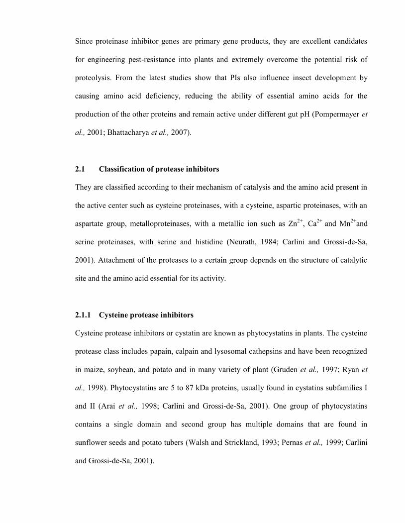
Since proteinase inhibitor genes are primary gene products, they are excellent candidates
for engineering pest-resistance into plants and extremely overcome the potential risk of
proteolysis. From the latest studies show that PIs also influence insect development by
causing amino acid deficiency, reducing the ability of essential amino acids for the
production of the other proteins and remain active under different gut pH (Pompermayer et
al., 2001; Bhattacharya et al., 2007).
2.1 Classification of protease inhibitors
They are classified according to their mechanism of catalysis and the amino acid present in
the active center such as cysteine proteinases, with a cysteine, aspartic proteinases, with an
aspartate group, metalloproteinases, with a metallic ion such as Zn2+, Ca2+ and Mn2+and
serine proteinases, with serine and histidine (Neurath, 1984; Carlini and Grossi-de-Sa,
2001). Attachment of the proteases to a certain group depends on the structure of catalytic
site and the amino acid essential for its activity.
2.1.1 Cysteine protease inhibitors
Cysteine protease inhibitors or cystatin are known as phytocystatins in plants. The cysteine
protease class includes papain, calpain and lysosomal cathepsins and have been recognized
in maize, soybean, and potato and in many variety of plant (Gruden et al., 1997; Ryan et
al., 1998). Phytocystatins are 5 to 87 kDa proteins, usually found in cystatins subfamilies I
and II (Arai et al., 1998; Carlini and Grossi-de-Sa, 2001). One group of phytocystatins
contains a single domain and second group has multiple domains that are found in
sunflower seeds and potato tubers (Walsh and Strickland, 1993; Pernas et al., 1999; Carlini
and Grossi-de-Sa, 2001).

The primary and tertiary structures of cysteine protease inhibitors have been determined
(Kauzuma et al., 2000; Carlini and Grossi-de-Sa, 2001) Homology of cysteine protease
inhibitors are similar with serine protease inhibitors, such as Kunitz-type trypsin inhibitor
family that belong to potato tuber phytocystatins (Ishikawa et al., 1994). The phytocystatins
are displaying high inhibitory activity toward insect gut proteinases making them attractive
to control insect pests (Bode and Huber, 1992; Gatehouse and Gatehouse, 1998; Carlini and
Grossi-de-Sa, 2001).
Cysteine protease inhibitors were reduced fecundity, increased motarlity, decreased weight
and severe deformations when fed to lepidoptera and coleopteran species (Elden, 2000).
Cysteine proteases have been best characterized in the Bruchidae in the Coleoptera, it also
occurs in the Curculionoideae, Meloidae and Silphidae (Xavier-Filho, 1992).
Therefore, in this study research are focusing on the function of serine proteases inhibitors
by using potential of local plants that are important to control insecticides and thus make
them beneficial in agronomical and health relevance.This research study will be involving a
thorough screening, isolation, identification and characterization of bi-functional inhibitor
of α-amylase and protease inhibitors.
2.1.2 Aspartic protease inhibitors
Aspartic proteases are a family of protease enzymes which inhibits the catalytic activity of
an aspartyl protease, a class of proteases that contains active site aspartate residue
(Asp). Aspartic proteases include pepsin and rennin. Members of the aspartic protease
family have been characterised in humans, plants, fungal and retroviruses. Eukaryotic
aspartic proteases include pepsins, cathepsins, and renins.

Aspartic proteases were also involved in defence mechanism. Protease inhibitors active
against serine, cysteine and metallocarboxy-proteases are ubiquitous, while inhibitors
active towards aspartic proteases have not been detected in seeds (Valueva and Mosolov,
1999). Research by Dash and friends in 2001 were recognized that the kinetic studies have
revealed the bifunctional characteristics of a novel bifunctional inhibitor (ATBI) from an
extremophilic Bacillus sp., as it was found to inhibit xylanase and aspartic protease. This
report had shown a novel class of antifungal peptide, exhibiting bifunctional
inhibitory activity (Dash et al., 2001).
2.1.3 Metalloproteinases inhibitors
Tissue inhibitors of metalloproteinases (TIMPs) are the major cellular inhibitors of the
matrix metalloproteinase (MMP). Matrix metalloproteinases are a class of enzymes
involved in degradation of extracellular matrix that can break down proteins, such as
collagen and gelatin. Metalloproteinases include thermolysin and carboxypeptidase A. As
they inhibit cell migration they have antiangiogenic effects. They may be both endogenous
and exogenous. Exogenous matrix metalloproteinases inhibitors include batimastat and
marimastat. The most notorious endogenous metalloproteinases are tissue inhibitor of
metalloproteinases (TIMP). There are also cartilage-derived angiogenesis inhibitors.
2.1.4 Serine protease inhibitors
Serine protease inhibitors and their binding to cognate proteinases have been extremely
well characterized over the years (Bode and Huber, 1992). Serine protease inhibitors are
one of the most diverse families of macromolecules that achieve similar biological
functions with entirely different scaffolds. Serine protease inhibitors have been classified
about 20 subfamilies, based on amino acid sequence and mechanism of interaction
including a-helical, b-sheet and a/b proteins, as well as small disulfide rich proteins. Based
on their mechanisms of action, three types of serine protease inhibitors are now recognized
as canonical inhibitors, non-canonical inhibitors and serpins (Laskowski and Kato, 1980).
They are the group of proteolytic enzymes which are characterized by a catalytically active
serine residue in their active site. Several serine protease inhibitors are effective protect
host plants by against various insect enzymes and therefore have been studied as an
alternative approach to pest control (Reckel et al., 1997; Leo et al., 2001). Serine protease
inhibitors such as trypsin, chymotrypsin and elastase are the most intensively studied
(Otlewski et al., 1999). All three enzymes are synthesized by the pancreatic acinar cells,
secreted in the small intestine, and are responsible for catalyzing the hydrolysis of peptide
bonds. These enzymes are shown similar in structure through their X-ray structures.
Each of these digestive serine proteases targets different regions of polypeptide chain,
based upon the side chains of the amino acid residues surroundinsg the site of cleavage.
Following a positively-charged amino acid residue, trypsin is responsible for cleaving
peptide bonds. Instead of having the hydrophobic pocket of the chymotrypsin, there exists
an aspartic acid residue at the base of the pocket. This can then interact with positively-
charged residues such as arginine and lysine on the substrate peptide to be cleaved.
Chymotrypsin is responsible for cleaving peptide bonds following a bulky hydrophobic
amino acid residue. Preferred residues include phenylalanine, tryptophan, and tyrosine,
which fit into a snug hydrophobic pocket. Elastase is responsible for cleaving peptide
bonds following a small neutral amino acid residue, such as Alanine, glycine, and valine.
These amino acid residues form much of the connective tissues in meat. The pocket that is

in "trypsin" and "chymotrypsin" is now partially filled with valine and threonine, rendering
it a mere depression, which can accommodate these smaller amino acid residues.
Serine proteases have been isolated from various seeds that have been isolated and
characterized from Leguminosae, Cucur bitaceae, Solanaceae Graminae and Rutaceae
families (Garcia-Olmedo et.al., 1987; Oliva et al., 2000; Mello et al., 2002; Oliveira et al.,
2002; Shee and Sharma, 2007) and their physiological roles are extensively studied
including the regulation of endogenous proteases during seed dormancy, the reserve protein
mobilization, the protection against the proteolytic enzymes of parasites and insects and
also as storage or reserve proteins.
Most serine PIs is low-molecular mass molecules from 3 to 25 kDa that inhibit trypsin
and/or chymotrypsin. Kunitz-type inhibitors are proteins of ~20 kDa, with low cysteine
content and a single reactive site, whereas the Bowman–Birk type inhibitors have ~8 to 10
kDa as well as high cysteine content and two reactive sites (Richardson, 1991;
Bhattacharyya and Babu, 2007). Kunitz and Bowman–Birk inhibitors are vary in their
mode of stability but lack a-helix structurally (Bhattacharyya and Babu, 2007). The
linkages of disulfide in the Bowman–Birk inhibitors minimize their conformational entropy
and enhance their stability whereas Kunitz inhibitors are stabilized chiefly by hydrophobic
interactions of short stretches of hydrogen bonded sheets (Sweet et al., 1974; Ramasarma et
al., 1995; Bhattacharyya and Babu, 2007).
The distributions of these two families of inhibitors are in the seeds of Leguminosae that
contain high amounts of protein that suppress proteolytic activities in vivo and in vitro by
forming stable stoichiometric complexes (Richardson, 1991; Bhattacharyya and Babu,
2007). Kunitz type inhibitors are more common in the seeds of highly primitive
Mimosoideae and primitive Caesalpinioideae, in comparison to the recently evolved

Papilionoideae which frequently shows the presence of Bowman-Birk inhibitors (Macedo
et al., 2002).
2.1.4.1 Bowman-Birk Inhibitor (BBI)
The Bowman-Birk Inhibitor (BBI) is a polypeptide that has ability to inhibit both trypsin
and chymotrypsin at independent binding sites. It is characterized by content of high
cystine and the absence of glycine. BBI is a soy-derived protease inhibitor with
anticarcinogenic and anti-inflammatory properties, has been currently shown to be well
tolerated in clinical trials as a human cancer-preventive agent for pre cancerous conditions,
such as oral leukoplakia and the inflammatory disease, ulcerative colitis (Gran et.al, 2006).
In 1963, Bowman and Birk were the earliest scientists that identify and characterise a
member of this family from soybean (Glycine max) (Bowman 1946; Birk et al., 1963). The
most well studied member of this family is the soybean inhibitor (Habib and Fazili, 2007).
BBI can be found in many plant seeds. From the recent researches the inhibitors have been
found in cereals and legumes (Tanaka et al., 1997; Laing and McManus, 2002). The
inhibitors of this family are generally found in seeds, but are also wound-inducible in
leaves (Eckelkamp, 1993) and in the grass family Poaceae (Odani et al., 1986; Habib and
Fazili, 2007). A small cyclic inhibitor has been identified in sunflower (Helianthus annuus)
called sunflower trypsin inhibitor 1 (SFTI-1) (Habib and Fazili, 2007).
BBIs have been classified according to their structural features and inhibitor characteristics.
The first reactive site in these inhibitors is usually specific for trypsin, chymotrypsin and
elastase. The inhibitors have molecular weights ranging from 7000 to 8000, and these
inhibitors are stabilized by the presence of disulfide bridges (Chen et al., 1992; Lin et al.,
1993). The 14 half-cystine residues are conserved in all BBIs and help to maintain their

active conformation, All BBI molecules have two regions of tandem homology and each
has a reactive site. Thus, BBIs can inhibit two proteinases simultaneously and
independently and are considered as “double-headed” inhibitors (Chen et al., 1992).
The inhibitors from dicotyledonous plants consist of a single polypeptide chain have a
molecular mass of approximately 8 kDa and are double-headed, with two homologous
domains each bearing a separate reactive site for the cognate proteases (Birk, 1985). These
inhibitors interact independently, but simultaneously, with two proteases, which may be
same or different (Raj et al., 2002; Birk, 1985). Two types of the inhibitors can be found
from monocotyledonous plants. One group consists of a single polypeptide chain with a
molecular mass of about 8 kDa. They have a single reactive site. Another group has
approximately 16 kDa with two reactive sites (Odani et al., 1986; Tashiro et al., 1987).
The main sturucture of BBI are single polypeptides and comprise a binary arrangement of
two sub-domains with a conserved array of seven disulfide bonds. The BBI family of
protease inhibitors contains a unique of two disulfide-linked nine-residue reactive site loops
that adopts a characteristic canonical conformation (Bode and Huber, 1992) and the
positions of the P1 residues are indicated (Odani and Ikenaka, 1976). The loop is called
protease-binding loop and binds the protease in a substrate-like manner (Lee and Lin, 1995;
Habib and Fazili, 2007).
BBIs are cysteine-rich proteins with inhibitory activity against proteases that are widely
distributed in monocot and dicot species (Lin et al., 2006). Recent studies shown that
proteinase inhibitors of certain types are anticarcinogenic.The soybean derived BBI with a
well-characterized ability to inhibit trypsin and chymotrypsin is particularly effective in
suppressing carcinogenesis in a variety of in vivo and in vitro systems (Kennedy, 1998).
The anticarcinogenic compounds have the ability to reduce the forming of oxygen radicals,
to suppress the growth of chemical-induced colon and anal gland tumors, lung tumor in
mice, and breast tumor in rat to suppress the chemicalor radiation-induced cell
transformation, to reduce spontaneous chromosome abnormality (Kennedy, 1998) and to
prevent tumor invasion and metastasis.
2.1.4.2 Kunitz-type inhibitors
Kunitz-type inhibitors are a type of protein which functions as a protease inhibitor
(Rawlings, 2004) and mostly active against serine proteases, but may also inhibit other
proteases (Ritonja et al., 1990; Laing and McManus, 2002). Kunitz-type inhibitors are
usually in plants and widespread in soybean, legumes seeds, cereals and in solanaceous
species (Laskowski and Kato, 1980; Ishikawa et al., 1994). Kunitz-type PIs have been
found in potato tubers (S. tuberosum) (Plunkett et al., 1982; Park et al., 2005). The
inhibitors with antifungal activity have been located in the roots of punce ginseng
(Pseudostellaria heterophylla) (Wang and Ng, 2006).
Kunitz-type inhibitors usually have molecular weight approximately 18 to 20 kDa proteins,
usually made of two disulfide bridges or contain from 170 to 200 amino acid residues in
one polypeptide chain in their single reactive site with low cysteine content. This family
have been shown to inhibit trypsin, chymotrypsin and subtilisin (Laing and McManus,
2002; Park et al., 2005) and they also inhibit other proteases. Structurally, Kunitz inhibitors
lack a-helix, but vary in their mode of stability. Kunitz inhibitors are stabilized chiefly by
hydrophobic interactions of short stretches of hydrogen bonded sheets (soybean Kunitz
trypsin inhibitor) whereas the disulfide linkages in the Bowman–Birk inhibitors minimize

their conformational entropy and enhance their stability (Sweet et al., 1974; Ramasarma et
al., 1995).
Joubert and others scientist found the source of Kunitz-type typsin inhibitor from Erythrina
seeds to check abilities of inhibition. From the results, the proteins inhibited trypsin
strongly and they were poor inhibitors of chymotrypsin (Joubert et al., 1987). Kunitz
trypsin inhibitors also effectively inhibit the activity of proteolytic of lepidopterans, such as
black cutworm, tobacco, budworm and others.
2.2 Application of serine protease inhibitors
According to research by Bhattacharyya et al., 2007, studied on trypsin and chymotrypsin
inhibitor from Caesalpinia bonduc (CbTI) seeds. They also studied on the effects of CbTI
on insect gut proteases reflect that CbTI is a powerful antifendant of insect herbivores. The
deleterious effects of CbTI on larval gut proteinases of S. litura were similar to previously
observed results with proteinase inhibitors from other leguminous plant (Gomes et al.,
2005).
Another research by Bhattacharyya et al., 2007, researched on the roles of serine proteases
involved in the digestion mechanism of the cutworm Spodoptera litura (Lepidoptera:
Noctuidae) were examined (in vitro and in vivo) following feeding of plant protease
inhibitors. A trypsin inhibitor from Archidendron ellipticum (AeTI) was purified by
ammonium sulfate fractionation, size-exclusion chromatography (HPLC) and ion-exchange
chromatography and its bioinsecticidal properties against S. litura were compared with
Soybean Kunitz trypsin inhibitor (SBTI). AeTI inhibited the trypsin-like activities of the
midgut proteases of fifth instar larvae of S. litura by over 70%. Dixon plot analysis revealed

competitive inhibition of larval midgut trypsin and chymotrypsin by AeTI, with an
inhibition constant (Ki) of 3.5×10⁻⁹ M and 1.5×10⁻⁹ M, respectively.
However, inhibitor kinetics using double reciprocal plots for both trypsin and chymotrypsin
inhibitions demonstrated a mixed inhibition pattern. Feeding experiments conducted on
different (neonate to ultimate) instars suggested a dose-dependent decrease for both the
larval body weight as well as % survival of larva fed on diet containing 50, 100 and 150
μM AeTI. Influence of AeTI on the larval gut physiology indicated a 7-fold decrease of
trypsin-like protease activity and a 5-fold increase of chymotrypsin-like protease activity,
after being fed with a diet supplemented with 150 μM AeTI. This study suggests that
although the early (1st to 3rd) larval instars of S. litura are susceptible to the trypsin
inhibitory action of AeTI, the later instars may facilitate the development of new serine
proteases, insensitive to the inhibitor (Bhattacharyya et al., 2007).
Research on purification and characterization of a highly stable and potent trypsin inhibitor
was purified to homogeneity from the seeds of Putranjiva roxburhii belonging to tree of
tropical India by acid precipitation, cation-exchange and anion-exchange chromatography
(Chaudhary et al., 2008). SDS page analyses showed that protein consist of a single
polypeptide chain with molecular mass of approximately 34 kDa when under reducing
conditions. From the report, the structural stability of inhibitor complete at the high
temperatures and the complete loss of inhibitory activity were observed above 90oC. N-
terminal amino acid sequence of 10 residues did not show any similarities to known serine
protease inhibitors, however, two peptides obtained by internal partial sequencing showed
significant resemblance to Kunitz-type inhibitors.

In another case, research done by Kim and friends in 2005 from Republic of Korea were
described the purification and characterization of the antimicrobial peptide potamin-1 (PT-
1) from potato. A 5.6 kDa trypsin-chymotrypsin protease inhibitor obtained and isolated
from tubers of the potato by extraction of the water-soluble fraction, dialysis, ultrafiltration,
and C18 reversed-phase high performance liquid chromatography. PT-1 strongly inhibited
pathogenic microbial strains, including Candida albicans, Rhizoctonial solani, Clavibacter
michiganense subsp. Michiganinse (Kim et.al., 2005).
The sequence of PT-1 had 62% homology with serine protease inhibitor belonging to the
Kunitz family and the peptide inhibited chymotyrpsin, trypsin and papain. These protease
inhibitors were composed of polypeptide chains joined by disulfide bridges and reduced
PT-1 almost completely lost its activity against fungi. The results suggested that PT-1 is an
excellent candidate as a lead compound for the development of novel oral or other anti-
infective agents (Kim et.al., 2005).
Serine protease inhibitors have been proposed as a strategy against insect pests. Macedo et
al. 2004 have been studied that transgenic grains which expresses gene encoding protease
inhibitors may prevent seed damage against Lepidoptera and Coleopteran species without
side effects which include reduced fecundity, increased mortality, decreased weight gain
and severe deformation though out their developmental phases (Macedo et al., 2002). The
potential of these inhibitors that expressed in transgenic plants showed a higher resistance
to various insects.
Broadway (1995) were researched on six species of Lepidoptera have also been evaluated
for their susceptibility to serine proteinase inhibitors from cabbage. The serine proteinase
activity in the midguts of larval PZutella xylostella was moderately inhibited (40-50%), and
Trichoplusia ni, Lymantria dispar, and Helicoverpa zea were substantially inhibited (55-

85%) by cabbage proteinase inhibitors, while Trypsin and chymotrypsin activity from
larval Pieris rapae and Pieris napi were not significantly inhibited (0-18%), in vitro, by
cabbage proteinase inhibitors.
These results shown that the growth and development of the latter three species should be
reduced following ingestion of these inhibitors but chronic ingestion of cabbage proteinase
inhibitors only reduced the growth and development of T. ni. A shift in the relative
proportion of digestive enzymes was responded to ingestion of proteinase inhibitors
because lack of biological activity of the proteinase inhibitors against the other two species.
Following ingestion of cabbage proteinase inhibitors, the trypsin(s) in T. ni was moderately
susceptible (37% inhibited), while the predominant trypsin-like enzyme(s) in the midgut of
larval L. dispar and H. zea were resistant to inhibition by cabbage trypsin inhibitors (13-
18% inhibited). These results were confirmed for H. zea and T. ni feeding on proteinase
inhibitors in tomato foliage (Broadway et al., 1995).
Atkitson and friends (1993) were reported that protease inhibitors may function to protect
the reproductive tissue against potential pathogens. Their research had shown the isolation
of cDNA clone encoding a protein with sequence similarity to a protease inhibitors type II
of potato and tomato. These protease inhibitors were expressed in ornamental tobacco,
Nicotiana alata stigmas that derived from a precursor protein which is isolated peptide
inhibitors with five homologous inhibitors to six regions of the amino acid sequence
deduced from the cDNA clone (Atkinson et al., 1993).
Brown and Ryan (1984) shown that serine protease inhibitors in leaves of plants are
response to wounding. In leaves of tomato, potato tubers and legume seeds contain
inhibitors that 10% or more of the stored proteins (Green and Ryan, 1972; Richardson,
1977; Atkinson et al., 1993). PIs can accumulate to 2% of the soluble protein within 48

hours of insect attack or other types of wounding (Brown and Ryan, 1984; Graham et al.,
1986).
The main objective of this research was:
1) To screen plant extracts with inhibitory activities
2) To purify to homogeneity proteins with inhibitory activities
3) To characterise the active proteins

CHAPTER 3: MATERIALS AND METHODS
3.1 MATERIALS
All chemicals and materials employed in this study were of the highset grade obtainable,
unless it were otherwise described.
1. PROMEGA-Madison, Wisconsin, USA
-Tris-Base
2. R&M CHEMICAL-Washington, New Jersey
-Dimethy Sulfoxide (DMSO)
3. FISHER SCIENTIFIC-Hampton, New Hampshire
-Triton X-100
4. VIVASCIENCE-Gottingen, Germany
-Centrifugal concentrator
5. SYSTERM-Selangor, Malayisa
-Dichloromethane, 95% Ethanol, Glycerol, Sodium Hydroxide (NaOH), Methanol,
Ammonium Sulfate, 85% Phosphoric Acid, Acetone
6. SIGMA Chemical Corporation-Saint Louis, USA
-Coomassie Brilliant Blue G, Trypsin from bovine pancreas TPCK Treated, Bovine
Serum Albumin, Bromsulfalein
7. BIORAD Laboratories-Hercules, CA, USA
-N-alpha-benzoyl-dl-arg-p-nitroanilide (BapNA), Ammonium Persulfate, 2.5% and
10% Sodium Dodecyl Sulphate, Phenol Red, 10x Tris/Glycine/SDS, N,N,N’N’ –
tetramethylenediamine (TEMED), 30% Acrylamide/Bisacrylamide, 1.5M Tris-HCL
pH 8.8, 0.5M Tris-HCL pH 6.8, 10x Tricine SDS Running Buffer, Tricine buffer,
25mM Tris/HCL buffer pH8.0

8. INVITROGEN-USA
-Novex® 10-12% Tricine Gel (1.0mm, 12 well), Novex® Tris-Glycine SDS Sample
Buffer (2X), Pre-cast Tricine gel
3.2 EQUIPMENTS
1. Shaking incubator (SI-900R)
2. Jusco 6300 spectrophotometer
3. Hi Trap G-25 chromatography
4. Rotary evaporator
5. Fumehood
6. Spectrophotometer
3.3 METHODS
3.3.1 Preparation of Plant Extract
Several of plants from the families Leguminosae, Rubiaceae, Apocynaceae and
Euphorbiaceae were collected from University of Malaya campus ground and the species
were identified according to Boo et al., (2006). All part of the plants such as the leaves,
trees, roots and fruits were subjected to oven drying at temperature less than 55˚C
according to Chen et al., (2005). The dried plants were ground into powder form where
then 20g of these plant were weigh and homogenized with 200ml of dicholoromethane.
These plants mixture were left overnight at room temperature while being shaked on a
shaking incubator (SI-900R) at 100 rpm/min. The homogenate was then filtered to removed
the dichloromathane and the resulted precipitate obtained was dried overnight at room
temperature in fumehood. The dried precipitate were then macerated in 50% (v/v) ethanol

with 200ml of 95% ethanol: distilled water (1:1). These mixture were shaken overnight on
a shaking incubator (SI-900R) at 100 rpm/min. The filtrate were collected through filtration
method and subjected to dicholoromethane partitioning for purification. The purified
filtrates were concentrated using rotary evaporator at 38⁰C by removing the ethanol solvent
in it. The concentrated extract was centrifuged at 5,000 xg, 4⁰C for 15 minutes and cotton
plug were used to discard the plant debris. Supernatant collected were subjected to 90%
ammonium sulphate precipitation and further re-centrifuged at 5,000xg, 4⁰C for 15
minutes. The supernatant were again re-collected and freeze dried for further analysis.
3.3.2 Protein content determination
Bradford assay (Bradford, 1976) were used to determine the total protein content of the
dried extract samples using bovine serum albumin as the stock standard protein. The
resulted protein standard curve determination method and graph result was shown in
Appendix 1. For every plant tested, 1 mg of dried plants ethanolic extracts were diluted in
100µl of distilled water before 5ml of bradford reagent (preparation refer Appendix 2) were
added to the mixture. The mixture was left incubated at dark room at room temperature for
10 minutes before the absorbance was measured at 595 nm using spectrophotometer. The
amount of proteins present in each dried plants ethonolic extracts were estimated from the
standard curve in Appendix 1.

3.3.3 Trypsin Inhibitory Assay
The dried plants extracts were subjected to Trypsin inhibitory assay as described by
Jennings et al., (2001) and Heath et al., (1995). This assay is done in duplicates to screen
the presence of inhibitory activity in each sample towards commercial trypsin. Each dried
plants extracts were added with L-1-tosylamido-2-phenylethyl chloromethyl ketone
(TPCK) trypsin treated from bovine pancreas with concentration of 0.5g in 1 ml 50 mM
Tris-HCL buffer at pH 8.0 (Appendix 4). The mixture was incubated in water bath at 30⁰C
for 10 minutes before 30 µl of N-alpha-benzoyl-dl-arg-p-nitroanilide (BapNA)
(0.0435g/ml) was added into the mixture making the final volume to 3ml. The reactions
were monitored for 10 minutes against blank solution containing substrate solution in the
same buffer used using Jusco 6300 spectrophotometer.
3.3.4 Trypsin inhibitory Activity detection in SDS-polyacrylamide gel
electrophoresis and Tricine SDS
The detection of inhibitor of the dried plants ethanolic extracts were done using the SDS-
PAGE with Laemmli buffer system (Lemmli, 1970) as described by Hanspal et al., (1982).
The electrophoresis was done at room temperature on 17% separating minigel containing
0.1% (w/v) gelatin. Two types of samples were used in this assay, the dried plants ethanolic
extracts samples and the eluent from Hi Trap G-25 chromatography samples (refer
Appendix 5 for preparation). Each type of samples were prepared differently before tested
where 20mg of the dried plants ethanolic extracts samples were diluted with 20 μl aqueous
solution while 1 mg of the G-25 chromatography eluent samples were diluted in 20 μl
aqueous solution from this point each samples were treated the same way where each

samples was dissloved in aqueous solution of 2.5% (w/v) SDS, 10% (v/v) glycerol and
phenol red (4 mg/ml) (Appendix 7).
Electrophoresis for each sample was carried out at at constant current of 20mA with the
presence of running buffer at room temperature. The electrophoresis was stopped once the
tracking dye reached approximately 1 cm from the bottom of the gel.
The resolving gels were washed 3 times with 300ml of 2.5% (w/v) Triton X-100 for 45
minutes each before finally washed with 300ml of ultrapure water for 45 minutes with
gentle rocking on the shaker.
The washed gels were then incubated in 450ml of 0.1M glycine-NaOH buffer pH 8.3 for 16
hours with the addition of 6.4 mg of trypsin at the temperature of 37⁰C to observe the
trypsin effect on the samples.
The sample for the Tricine- Sodium dodecyl sulfate (SDS) were prepared by mixing 20 mg
of dried plants ethonolic extracts sample with 120 µl pre-made buffer (refer Appendix 8).
10μl of each samples prepared and 5 μl of benchmark protein ladder were loaded into the
pre-cast gel (invitrogen) with 4-20% acrylamide concentration; Tricine SDS gels respective
well and elctrophoresis followed by staining were carried out as mentioned above.
3.3.5 Preparation of Chrysomya megacephala crude protease extracts
Adult Chrysomya megacephala were crushed in the ratio of 1 :500 µl ultrapure water in an
eppendorf tube. The extracts were centrifuged and the supernatant was collected and kept in
-20⁰C for further analysis.

3.3.6 Mode of inhibition and Ki value determination
The trypsin inhibitory assay was again applied in determining the plant samples Ki value as
described by Jennings et al., (2001) and Heath et al., (1995) in section 3.2.3. the plants
samples were prepared by mixing 1 μg of the dried plant ethanolic extract in 150 μl of
ultrapure water. Two concentration of substrate were used in this assay which are 1 mM
and 5 mM of BapNA in DMSO. The concentration of plant sample used was 5.0 x 10⁻³
μg/μl, 2.5 x 10⁻³ μg/μl, 1.25 x 10⁻³ μg/μl, 1.0 x 10⁻³ μg/μl, 6.25 x 10⁻⁴ μg/μl, 5.0 x 10⁻⁴ μg/μl,
5.0 x 10⁻⁵ μg/μl and 5.0 x 10⁻⁶ μg/μl. The assays were done in duplicates for each samples
and a control without enzymes for both substrates concentration.
3.2.7 Thermostability determination
The trypsin inhibitory assay as described by Jennings et al., (2001) and Heath et al., (1995)
in section 3.2.3 was again used in determining the lyophilized samples extract thermo
stability. The samples were prepared by diluting lyophilized samples extract in 250μl of
ultrapure water and subjected to the assay with varying incubation period ranging from
15⁰C-90⁰C for 10 minutes. 30 µl of BapNA in DMSO were added into the mixture before
the reaction was monitors with Jusco 6300 spectrophotometer. The experiments were
repeated with two replicates and control without enzymes.
3.2.8 IC50 estimation
The dried plant ethanolic extract IC50 value were estimated by determining the
concentration value at the intersection between percentage of activity and percentage of

inhibition of trypsin by the dried plants ethanolic extracts versus its concentration that were
plotted on the same graph. The readings for the percentage of activity and percentage of
inhibition of trypsin by the dried plants ethanolic extracts were taken from the trypsin
inhibitory assay as described by Jennings et al., (2001) and Heath et al., (1995). The
concentration of plants samples used were 4.0 x 10⁻³ μg/μl, 6.0 x 10⁻³ μg/μl, 8.0 x 10⁻³
μg/μl, 1.0 x 10⁻² μg/μl, 1.2 x 10⁻² μg/μl, 1.4 x 10⁻² μg/μl, 1.6 x 10⁻² μg/μl, 1.8 x 10⁻² μg/μl
and 2.0 x 10⁻² μg/μl.

CHAPTER 4: RESULTS
4.1 Screening of trypsin inhibitors
Table 1: Shows various plants sample percentage inhibitory of trypsin activity
Plants families Plants name Percentage ofReduction Activity
Leguminosae Erythrina fusca Leaves 44 %
Flower 13 %
Cassia floribundaLeaves 17 %
Fruits 8 %
Delonex regiaLeaves nil
Fruits 2 %
Senna surattensis * Leaves 83 % *
Acacia mangiumLeaves 6 %
Caesalpinia pulcherrimaLeaves 12 %
Clitoria ternateaFlower and Fruits 7 %
Cassia alataLeaves nil
Fruits nil
Bauhinia blakeanaLeaves 21 %
Fruits nil
Andira inermisLeaves 7 %
Fruits nil
Mimosa diplotricha * Fruits 92 % *
Pterocarpus indicaLeaves 59 %
Fruits nil
Adenanthera pavominaLeaves 52 %
Acacia auriculiformisLeaves 13 %

Fruits Nil
RubiaceaeIxora finlaysoniana
Leaves nil
Mussaenda erytrophyllaLeaves 18 %
Flower 9 %
Uncaria sppLeaves 10 %
Flower nil
Euclinia longifloraLeaves 7 %
Porterandia anisophyllaLeaves 34 %
Morinda ellipticaLeaves nil
Gardenia carinataLeaves 27 %
Mussaendaphilippica ‘Queen Sirikit'
Leaves10 %
Mussaenda phillipicaLeaves 5 %
Flower 21 %
Morinda citrifolia Fruits 19 %
ApocynaceaeAllamanda oenotherafolia
Flower 36 %
Allamanda catharticaLeaves nil
Plumeria rubra cultivarsLeaves nil
Flower 24 %
EuphorbiaceaeRicinus communis
Leaves 8 %
Fruits 23 %
Jatropha gossypfoliaLeaves 27 %
Fruits 30 %
Macaranga tanarius Flower 75 %
(*) Selected for further analysis for showing higher percentage of trypsin inhibitory activity

Table 1 listed total of 44 lyophilized plant sample extract from the families Leguminosae,
Rubiaceae, Apocynaceae, Euphorbiaceae. From this table, only two plants were selected as
the subject for this experiment for the significantly high reduction activity shown. The
plants were Senna surattensis leaves with 83 % reduction activity and Mimosa diplotricha
fruits with 92% reduction activity.
Almost from each family, there were some plants extract that were labeled as ‘nil’. These
refering to the plants extract that showed no trypsin content reduction activity. Total of
twelve plants extracts were labeled as ‘nil’ from the table 1 which comprise of seven plants
extracts form the Leguminosae family, three plants extracts from Rubiaceae family and two
plants extracts from Apocynaceae family.
Others plants extracts showe varies reduction activity percentage ranging from 5 % to 75 %
such as Mussaenda phillipica leaves from Apocynaceae family and Macaranga tanarius
from Euphorbiaceae family respectively.

4.2 Sodium Dodecyl Sulfate Polyacrylamide Gel Electrophoresis (SDS-PAGE)result of sample extracts
Figure 1: shows SDS-Page gel result of lyophilized sample extracts of Senna surattensis leaves (A)
and Mimosa diplotricha fruit (B) on 17% acrylamide gel under two different conditions which is
with trypsin incubation and without trypsin incubation. Markers used in these gels were Mark 12
from invitrogen and these gels were stained with Coomassie Brilliant Blue staining.
Gel 1 shows the results with the presence of trypsin incubation while gel 2 shows the
results without the presence of trypsin incubation. From Figure 1, In both gels, there were
protein band present (shown by arrow) which indicated no lysis of protein in the band
region due to the the presence of protein inhibitor from Senna surattensis leaves (A) and
Mimosa diplotricha fruit (B).
Marker A B Marker A B
200 kDa
116.3 kDa97.4 kDa
66.3 kDa55.4 kDa
36.5 kDa
31.0 kDa
200 kDa
116.3 kDa97.4 kDa
66.3 kDa55.4 kDa
36.5 kDa
31.0 kDa
Gel 1 Gel 2

Figure 2: shows the bands of lyophilized sample extracts of Senna surattensis leaves (A) and
Mimosa diplotricha fruit (B) on Pre-cast (invitrogen) Tricine SDS gel at 4-20% acrylamide
concentration. Marker used in this gels were Mark 12 from invitrogen and these gels were stained
with Coomassie Brilliant Blue staining.
Figure 2 showed a bands for each sample; Senna surattensis leaves (A) and Mimosa
diplotricha fruit (B). The band formation proven the presence of inhibitor in both samples.
According to band formation and sample migration, the molecular weight for both sample
was estimated to be around 26.0 kDa to 31.0 kDa.
Marker A B
200 kDa
116.3 kDa97.4 kDa
66.3 kDa55.4 kDa
36.5 kDa
31.0 kDa
21.5 kDa
14.4 kDa6.0 kDa

Next, both Senna surattensis leaves (A) and Mimosa diplotricha fruit (B) was subjected to
G-25 chromatography for desalting purpose. The protein was then separated on Tricine
SDS (invitrogen). Based on figure 3, the molecular weight of Senna surattensis leaves (A)
was determine to be 27.93 kDa while Mimosa diplotricha fruit (B) was unable to produce
any band after desalting proses (result not shown here), thus the sample was ommited from
further anylysis.
Figure 3: Separation of G-25 chromatography eluent of Senna surattensis leaves (A) on Tricine
SDS gel. Marker used in these gels was Mark 12 from invitrogen and these gels were stained with
Coomassie Brilliant Blue staining.
Marker A
200 kDa
116.3 kDa97.4 kDa
66.3 kDa55.4 kDa
36.5 kDa31.0 kDa
21.5 kDa14.4 kDa6.0 kDa

4.3 Determination of Senna surattensis’s leaves Mode of inhibition and Ki value
Table 2: The reduction of trypsin activity percentage at two different concentration of
substrate
Initialconcentrationof Sennasurattensis’sleaves
Concentration ofsubstrate(BapNA)
Concentrationof inhibitor(mg/µl)
Concentration ofinhibitor(mM)
1/v(mol/min/ml)-1
6.7 x 10⁻³mg/µl
1mM 5.0 x 10⁻³ 1.8 x 10⁻³ 0.6270
2.5 x 10⁻³ 9.0 x 10⁻⁴ 0.3729
1.3 x 10⁻³ 4.5 x 10⁻⁴ 0.2220
1.0 x 10⁻³ 3.6 x 10⁻⁴ 0.1971
6.3 x 10⁻⁴ 2.2 x 10⁻⁴ 0.1290
5.0 x 10⁻⁴ 1.8 x 10⁻⁴ 0.0875
5.0 x 10⁻⁵ 1.8 x 10⁻⁵ 0.0490
5.0 x 10⁻⁶ 1.8 x 10⁻⁶ 0.0398
5mM 5.0 x 10⁻³ 1.8 x 10⁻³ 0.2379
2.5 x 10⁻³ 9.0 x 10⁻⁴ 0.1087
1.3 x 10⁻³ 4.5 x 10⁻⁴ 0.0673
1.0 x 10⁻³ 3.6 x 10⁻⁴ 0.0611
6.3 x 10⁻⁴ 2.2 x 10⁻⁴ 0.0597
5.0 x 10⁻⁴ 1.8 x 10⁻⁴ 0.0572
5.0 x 10⁻⁵ 1.8 x 10⁻⁵ 0.0488
5.0 x 10⁻⁶ 1.8 x 10⁻⁶ 0.0438

Figure 4: The dixon plot of concentration versus 1/v sample extract of Senna surattensis’s leaves in
the presence of 1mM and 5mM BapNA in DMSO.

4.4 Determination of Senna surattensis’s leaves thermostability
Table 3: Percentage of reduction in trypsin inhibitory activity Senna surattensis’s leaves
extract in various incubation temperature.
Figure 5: The bar chart plotted of Senna surattensis’s leaves extract temperature versus its reduction
of activity percentage.
Temperature Percentage of Reduction
15⁰C 70.4430⁰C 83.5245⁰C 87.3560⁰C 81.3375⁰C 80.1390⁰C 79.32

Based on Table 3, the percentage of reduction of the inhibitory activity of lyophilized
Senna surattensis’s leaves ethanolic extract towards trypsin show a constant increase at the
incubation temperature ranging from 15⁰C to 45⁰C. 45⁰C is shown to be the optimal
temperature for Senna surattensis’s leaves ethanolic extract with 87.35 % reduction of the
inhibitory activity towards trypsin. At temperature more than 45⁰C, a constant decrease of
percentage reduction of the inhibitory activity towards trypsin can be seen.
4.5 Estimation of Senna surattensis’s leaves IC50 value
Table 4: Percentage of reduction in activity of crude insect’s trypsin when subjected to
different concentration of Senna surattensis’s leaves proteinaceous extract
Concentration (µg/µl) Percentage of reduction in activity (%)
0.004 11.9
0.006 12.8
0.008 16.4
0.010 26.4
0.012 34.0
0.014 37.3
0.016 40.1
0.018 59.3
0.020 66.6

Figure 6: shows the Senna surattensis’s leaves percentage of inhibition and activity of trypsin
concentration.

CHAPTER 5: DISCUSSION
DISCUSSION
Protease inhibitors are referring to a group of protein that regulates proteolysis. These
inhibitors have been found in numerous sources, including plant such as soybean, human
pancreas and hemolymph of several arthropods and insect such as Bombyx mori and
Drosophila melanogaster (Cherqui et al., 2001).
However, in this study, we only focus on plant derived protease inhibitor. Protease
inhibitors derived from plants have the ability to reduce insect’s digestive capacity through
specific inhibition of digestive proteases, resulting in insect growth and development arrest
thus can be used as an alternative strategy to control insect pests (Aguirre et al., 2009).
30 different plants were collected in order to study the presence of serine protease inhibitor
and to purify them. All of these plants were from the families of Leguminosae, Rubiaceae,
Apocynaceae and Euphorbiaceae and the species of these plants were determined according
to Boo et al., (2006). All of the plants samples were randomly collected from the University
of Malaya campus ground.
Different parts of the plant such as fruits, trees, leaves and flowers were used in this study.
There were about 43 plants samples obtained from these 30 different plant species.
Leguminosae, Rubiaceae, Apocynaceae and Euphorbiaceae contribute about 21 samples,
13 samples, 4 samples and 5 samples respectively.
As suggested by previous study, the seed and vegetative parts of plant contain various
proteinaceous inhibitors of insect, fungal, mammalian and endogenous proteinases. This
inhibitor mainly involve in plant defense mechanism and nowadays become great interest
to many researcher as the gene coding for this inhibitor can be genetically transfered to
other plants to improve their resistance to pests or fungi (Konarev et al., 2002).
In the screening process, all of these plant were analyzed according to method as described
in section 3.2.1. which comprised of dichloromethane fractionation and sample
concentration using rotary evaporator. This method are widely used by many researcher for
plant crude extracts fractionation such as the fractionation of seven plant materials;
fenugreek seeds, harmal seeds, garlic cloves, cinnamon bark, sticky fleabane leaves, and
nightshade leaves and fruit for detecting the In vitro Antifungal Activities (Kanan and Al-
Najar, 2009).
At the initial stage, some of these plants were treated with ammonium precipitation to
concentrate the samples. Aguirre et al., (2009) has also adopted this method in their
research where the crude extract of their samples; larvae of Prostephanus truncatus was
precipitated with ammonium sulfate at 70% saturation for purification. However during this
step, we figured out that ammonium precipitated samples were most likely to produce an
unknown solution on top of the samples that we later find out as lipid-like solution.
Since, the objective of this study is to screen and purify serine protease inhibitor, we were
more interested in hydrophilic protein, thus we decided to discard the lipid-like solution;
the hydrophobic solution. But in doing so, we found out that the final volume of the
extraction were greatly reduced thus limited our samples extracts for further analysis.
We also feared that by discarding the lipid-like solution will most likely to induce more
contamination to our sample extracts. We finally found a better method which is to freeze
dried our samples. This method was found to be the best method so far for our samples
since it can preserve the quality and quantity of our samples. The powder produced from

the freeze drying methods were stored in -80˚C for long term storage and -20˚C for short
term storage and reconstitute with ultrapure water or appropriate buffer for later use in any
assays.
After all of this plants had been extracted, the protein content for each plant extracts were
determined using the Bradford Assay (Bradford, 1976). The results of this assay were
presented in Table 5 in Appendix 3. The estimated protein content obtained is ranged from
0.24 µg/ml to 34.46 µg/ml.
After the protein content estimation, all of the plant extracts were further tested with
Trypsin Inhibitory Assay to determined the presence of inhibition towards protease namely
trypsin. These methods have been used in determining the total trypsin inhibitor activity of
raw soy milk using N-benzoyl-DL-arginine-p-nitroanilidehydrochloride (BAPNA) as the
substrate (Guerrero-Beltran et al., 2009).
From the results shown in Table 1, it is shown that from the 43 plant samples, about 32
samples shows inhibition towards trypsin activity. The remaining 11 samples did not shows
any inhibition towards trypsin activity thus labelled as ‘nil’. They comprised of 6 ‘nil’
samples, 3 ‘nil’ samples and 2 ‘nil’ samples from Leguminosae, Rubiaceae, families
respectively. These ‘nil’ labeled samples are most likely resulted from plants that have no
protease inhibitor activity ability or loss the protein for protease inhibition ability during the
initial ammonium sulphate precipitation and the lipid-like solution removal.
In the Leguminosae family, the plant samples inhibition towards trypsin activity are
ranging from as low as 2 % to as high as 92 %, which were Delonex regia fruits and
Mimosa diplotricha fruits respectively. A previous study have reported that Kunitz-type
trypsin inhibitors have been isolated from the seeds of Delonex regia (Lin and Ng 2008). 10

samples out of 13 samples in Rubiaceae family shows inhibition towards trypsin activity
are ranging from 7 % to 34 %.
Only two samples in Apocynaceae family shows inhibition towards trypsin activity which
are Plumeria rubra cultivars flowers with 24 % and Allamanda oenotherafolia flower with
36 %. The last family; Euphorbiaceae samples shows inhibition towards trypsin activity in
the range of 8 % to 75 % with no samples labeled as ‘nil’.
From the results presented in Table 1, all the results were narrowed to choose the two
highest inhibition percentages toward trypsin. Since the highest readings for inhibition
towards trypsin activity were from the Leguminosae family, which were Mimosa
diplotricha fruits and Senna surattensis leaves with 92 % and 83 % reaspectively. Both of
these plants were selected for further analysis in this study.
This result is agreeable to many previous studies finding that claimed most serine
proteinase inhibitors have been isolated and characterized from Leguminosae. Other family
that have been also reported to have protease inhibitor as such as Cucurbitaceae,
Solanaceae,Graminae, Rutaceae and Euphorbiaceae (Chaudhary et al., 2008).
Then, the lyophilized plants extract samples for both Mimosa diplotricha fruits and Senna
surattensis leaves were electrophorised on Sodium Dodecyl Sulfate Polyacrylamide Gel
Electrophoresis (SDS-PAGE) (Figure 1) and Pre-cast (invitrogen) Tricine SDS gel (Figure
2) to shows the presence of protease inhibitor in both sample extracts selected.
The SDS-PAGE with 17% resolving gel concentration and 4% stacking gel concentration
were used in this test. We also improvised the SDS-PAGE method by adding an extra steps
after the electrophoresis was done. We added trypsin enzyme into 0.1M glycine-NaOH

buffer pH 8.3 and left the gel in the buffer-trypsin mixture for 16 hours at room temperature
37⁰C.
The improvising was carried out base on our understanding that the trypsin enzyme will
degrade all of the protein presence in the gel. But the degradation will not occur if there
were any protease inhibitor in the plants extract as this protease inhibitor will stop the
protein degradation by trypsin. The presence of these protease inhibitor were proven by the
presence of the band on both gel 1 and gel 2 with trypsin incubation and without trypsin
incubation respectively as shown in Figure 1.
Both sample extracts were then electrophorised on Tricine Pre-cast gel from invitrogen
with 4-20 % acrylamide concentration. We decided to test both sample extracts on tricine
gel to further separate the protein that are less than 30 kDa because the previous Leammli
SDS-PAGE done can only separate protein that are more than 30 kDa in size.
Results in Figure 2 in section 4.2 shows one band respectively for each sample extracts
lane; Senna surattensis leaves (A) and Mimosa diplotricha fruit (B) below the 31.0 kDa
size protein marker.This gel indicates that the protease inhbitor presence may be of low
molecular weight proteins. The molecular weight for both samples was estimated to be
around 26.0 kDa to 31.0 kDa.
The selected Mimosa diplotricha fruits and Senna surattensis leaves were then subjected to
Hi-Trap Sephadex G-25 chromatography as mentioned in appendix 6 to remove the
excessive salt in the plant extracts. The desalted eluent were also freeze dried and kept in -
20˚C to minimize the protein degradation thus, preserve the eluent quality.

The freeze dried samples eluents was then again electrophorised on tricine SDS-PAGE.
The result was shown in Figure 3 where it showed one clear band on the Senna surattensis
leaves (A). From this gel and gel in Figure 2, the molecular weight of Senna surattensis
leaves were calculated and was determined to be 27.93 kDa.
However, the Mimosa diplotricha fruit did not manage to produce any band in all the
attempt done. It will either shows no band formation on the lane if the the extracts
concentration used is low or a thick smear on the samples extract lane if higher
concentration were used (results is not shown here). The smearing may have derived from
sample overloading or simply because of too concentrated protein loaded into the gel. Thus
the Mimosa diplotricha fruit were omited from further analysis and the molecular weight of
Mimosa diplotricha fruit were estimated as to be 29.17 kDa from the Figure 2.
We did further tested the purified protein from Senna surattensis leaves protease inhibitor
activity toward untreated protease namely trypsin that was obtained from
Chrysomya megacephala. These blowflies are also known as the oriental latrine fly belong
to the Calliphoridae family. It is known to spread disease to human and also cause myiasis
in human where it invades an open wound (Sukontason et al., 2005).
The Chrysomya megacephala were crushed and the supernatant collected were tested with
Senna surattensis leaves fortrypsin inhibitory assay. Result are shown in Table 8 in
Appendix 10 where Senna surattensis leaves extract showed 83.52 % inhibitory activity
towards commercial trypsin and 61.74 % inhibitory activity towards
Chrysomya megacephala extracted protease.

Even though the reading for inhibitory activity towards Chrysomya megacephala extracted
protease was lower when compared to commercial trypsin, the results still shows that this
Senna surattensis leaves indeed has protease inhibitor ability towards any source of
protease, be it crude or purified protease. The ability of Senna surattensis leaves to inhibit
protease in Chrysomya megacephala has opened a path to do more research for insecticide
purpose on this plant extract.
The next test carried out was the study of mode of activity of this Senna surattensis leaves.
This study is done to determine the plant Ki value using two concentration of substrate. The
substrate used in this assay was still (BAρNA) but at two different concentrations of 1 mM
and 5 mM respectively. The Ki value of Senna surattensis leaves was determined from the
dixon plot as shown in Figure 4 in section 4.3 with the reciprocal velocity (1/v) is plotted
against concentration of inhibitors for both substrate concentration.
The Ki value of Senna surattensis leaves determined is 8.89 x10⁻⁵ mM. The analysis of
Dixon plot showed that Senna surattensis leaves is a competitive inhibitor because two
lines corresponding to each substrate intersect above the x-axis. A previous study on
Putranjiva roxburghii belonging to Euphorbiaceae family showed that this plant has ki
value of 1.4x 10⁻¹¹ (Chaudhary et al., 2008). By comparing the Ki value in this study with
the Putranjiva roxburghii Ki value, it shows that Putranjiva roxburghii is highly potent
inhibitor of bovin trypsin compared to Senna surattensis leaves.
The next test done on Senna surattensis leaves lyophilized extract was thermo stability
determination. In this test, the trypsin inhibitory activity of the lyophilized extract was
examined at different temperatures ranging from 15⁰C-90⁰C in the water bath. This test

was carried out using Trypsin Inhibitory Assay with N-alpha-benzoyl-dl-arg-p-nitroanilide
(BapNA) as the substrate.
As result shown in Table 3, it shows that the Senna surattensis leaves lyophilized extract
inhibitory activity increased as the temperature increased from 15⁰C-45⁰C. At 15⁰C, the
extracts showed 70.44 % of inhibitory activity and at 30⁰C, the extract showed 83.52 % of
inhibitory activity. The extract reached its optimum inhibitory temperature at 45⁰C with
87.35 % of inhibitory activity.
When the reaction temperature was further increased more than 45˚C, the inhibitory
activity started to decreased. At 60⁰C, the inhibitory activity of the extract decreased to
81.33 %, and further decrease to 80.13 % and 79.32% at 75˚C and 90˚C respectively. This
is due to the sample extract protein thermal degradation at temperature more than 45˚C.
Research done by Asif-Ullah et al., (2006) on Cucumisin claimed to contain subtilisins
which are the second largest class of serine proteases. It is found out through the
thermostablity study that this plant protease increase as the reaction temperature increased
from 20˚C to 70˚C and reach the higher than that of the activities at 37˚C. Due to thermal
denaturation of the protein, the activity rapidly decreased as the reaction reached 80˚C and
higher. By comparing it with our results, it shows that Senna surattensis leaves have
limited range of temperature to works in compared to Cucumisin.
The final test done on Senna surattensis leaves lyophilized extract was IC50 value
determination using trypsin inhibiory assay. Nine concentration of this purified protein
from Senna surattensis leaves were used in this experiment ranged from 0.004 μg/μl to
0.020 μg/μl. Figure 4 shows the effect of Senna surattensis leaves lyophilized extract with

different concentration against insect’s (Chrysomia megacephala) trypsin. The IC50 value
obtained in this study was 0.0174 μg/μl.
These method adopted to determine the IC50 value was also used by other researchers in
many previous study in determining few plant with protease inhibitor such as dull black
soybean that are found out as to contains Kunitz-type trypsin inhibitor (Lin and Ng 2008).
While another study adopted this method towards cinnamon, garlic or sticky fleabane
resulted in IC50 values for these fractions were found to be in the range of 11.2-24; 30.25-
31.50; 25.0-36.0 μg/ml, respectively (Kanan and Al-Najar 2009).

CHAPTER 6: CONCLUSION
CONCLUSION
This study shows that protease inhibitor that derived from Senna surattensis leaves have a
wide potential to be explore in many field especially in producing insecticide. This is due to
the fact that Serine proteinase inhibitors is thought to have phytochemical defenses
against herbivorous insects (Broadway 1995) apart from its other usage in other filed;
biological functions, such as blood coagulation, platelet aggregation and anti-
carcinogenesis (Chaudhary et al., 2008).
The molecular weight of obtained for Senna surattensis leaves were calculated and was
determined to be 27.93 kDa while the molecular weight of Mimosa diplotricha fruit were
estimated as to be 29.17 kDa (Table 7 in Appendix 9). Both plants have the potential serine
protease inhibitor that needs to be explored more. The Ki value of Senna surattensis leaves
determined is 8.89 x10⁻⁵ mM with the mode of activity determined as competitive inhibitor.
Even though Senna surattensis leaves showed stability at a narrow range of temperature,
which is only until 60 ˚C, but it is still should be given a fair chance to be studied even
more because the efficacy of inhibitor is not only dependent on the temperature it react also
dependent on the compatibility of the plant protease inhibitor reactive site and the substrate
binding site in the target organism (Broadway 1995).
Current literature review only support the general hypothesis of the serine proteinase
inhibitors ability in protecting plants against herbivorous insects (Broadway 1995). Thus,
future research should also include the adverse effect of protease inhibitor usage and on
compatibility of the protease inhibitor reactive site with other target binding site to produce
the most effective insecticide toward wide range of insects.

REFERENCES
1. Aguirre, Cesar (2009) Partial characterization of a chymotrypsin-like protease in the
larger grain borer (Prostephanus truncatus (Horn) in relation to activity of Hyptis
suaveolens (L.) trypsin inhibitor. Journal of Stored Products Research 45: 133-138.
2. Arai S., Matsumoto I., Abe K (1998) Phytocystatins and their target enzymes: From
molecular biology to practical application: A review. Journal of Food Biochemistry
22: 287-299.
3. Asif-Ullah, Mufti, Key S.K, and Yeon G.Y (2006) Purification and
characterization of a serine protease from Cucumis trigonus Roxburghi.
Phytochemistry 67: 870-875.
4. Atkinson, A.H., Heath, R.L., Simpson, R., Clarke, A.E., Anderson, M.A (1993)
Proteinase Inhibitors in Nicotiana alata stigmas are derives from a precursor protein
whibh is processed into five homologous inhibitors. The Plant Cell. 5: 203-213.
5. Barret, A.J (1987) The cystatin; a new class of peptidase inhibitors. Trends in
Biochemical Sciences 12: 193-196.
6. Bhattacharyya A., Rai S., Babu C.R (2007) A trypsin and chymotrypsin from
Caesalpinia bonduc seeds: Isolation, partia characterization and insecticidal
properties. Plant Physiology and Biochemistry. 45: 169-177.
7. Birk Y, Gertler A, Khalef S (1963) A pure trypsin inhibitor from soybean.
The Journal of Biochemistry 87: 281-284.
8. Birk Y (1985) The Bowman-Birk inhibitor. Trypsin and chymotrypsin inhibitor from
soybeans International journal of peptide and protein research 25: 113-131

9. Bischoff H., Ahr H.J., Schmidt D., Stoltefuss J (1994) Acarbose-ein neues
Wirkprinzip in der Diabetes-Therapie. Nachr. Chemical Technology Laboratory 42:
1119-1128.
10. Bode W, Huber R. (1992) Natural protein proteinase inhibitors and their interaction
with proteinase inhibitors and their interaction with proteinases. European Journal of
Biochemistry 204: 433-451.
11. Boo C.M, Kartini O.H, Ou Y.C.L (2006) 1001 garden plants in Singapore. National
Parks Board, second edition.
12. Bowman D.E (1946) Differentiation of soybean antitryptic factor. Proceedings of the
Society for Experimental Biology and Medicine 63: 547-550.
13. Bradford M.M (1976) A rapid and sensitive method for the quantitation of
microgram quantities of protein utilizing the principle of protein-dye binding.
Analytical Biochemistry 72: 248-254.
14. Broadway R.M. (1995) Are insects resistant to plant proteinase inhibitors? Journal
of Insect Physiology 41:107-116.
15. Brown W.E., Ryan C.A (1984) Isolation and characterization of a wound-induced
trypsn inhibitor fromalfalfa leaves. Biochemistry 23: 3418-3422.
16. Carlini C.R., Grossi-de-Sa M.F (2001) Plant toxic proteins with insecticidal
properties. A review on their potentialities as bioinsecticides. Toxicon 40: 1515-
1539.
17. Chaudhary N.S., Shee C., Islam A., Ahmad F., Yernool D., Kumar P., Sharma A.K
(2008) Purification and characterization of a trypsin inhibitor from Putranjiva
roxburghii seeds. Phytochemistry. 69:2120-2126.

18. Chen P, Rose J, Love R., Wei C.H, Wang B.C (1992) Reactive sites of an
anticarcinogenic Bowman-Birk proteinase inhibitor are similar to other trypsin
inhibitors. The Journal of Biochemistry 267: 1990-1994.
19. Cherqui A, Norberto C, and Nelson S (2001) Purification and characterization of two
serine protease inhibitors from the hemolymph of Mythimna unipuncta. Insect
Biochemistry and Molecular Biology 31:761-769.
20. Chrispeels M.J, Raikhel N.V (1991) Lectins, lectin genes, and their role in plant
defense. Plant Cell 3:1-9.
21. Dash C, Ahmad A, Nath D, Rao M (2001) Novel Bifunctional Inhibitor of Xylanase
and Aspartic Protease: Implications for Inhibition of Fungal Growth. Antimicrobial
Agents and Chemotherapy, 45: 2008-2017.
22. Eckelkamp C, Ehmann B, Schopfer P (1993) Wound-induced systemic accumulation
of a transcript coding for a Bowman-Birk trypsin inhibitor-related protein in maize
(Zea mays L.) seedlings, Federation of European Biochemical Societies 323: 73-76.
23. Elden T.C (2000) Influence of a cysteine proteinase inhibitor on alfalfa weevil
(Coleoptera: Curculionidae) growth and development over successive generations
Entomologia Experimentalis et. Applicata 72: 1-10.
24. Fear G, Komarnytsky S, Raskin I (2007) Protease inhibitors and their
peptidomimetic derivates as potential drugs. Pharmacology & Therapeutic 113: 354-
368.
25. Garcia-Olmedo F, Salcedo G, Sanchez-Monge R, Gomez L, Royo J, Carbonero P
(1987) Plant proteinaceous inhibitors of proteinases and a-amylases. Oxford Surveys
of Plant Molecular and Cell Biology 4: 275-334.

26. Gatehouse A.M, Barbieri L, Stirpe F, Croy R.R.D (1990) Effects of ribosome
inactivating poeins on insect development differences between Lepidoptera and
Coleoptera. Entomologia Experimentalis et. Applicata 54: 63-74.
27. Gatehouse A.M.R, Gatehouse J.A (1998) Identifying proteins with insecticidal
activity: use of encoding genes to produce insect-resistant transgenic crops. Journal
of Pest Science 52: 165-175.
28. Gomes A.D, Dias S.C, Bloch Jr. C., Melo J.R, Furtado Jr, Monnerat R.G, Grossi-de-
Sa M.F, Franco O.L (2005) Toxicity to cotton boll weevil Anthonomous grandis of
trypsin of a trypsin inhibitor from chickpea seeds. Comparative Biochemistry
and Physiology 140: 313-319.
29. Goosens A, Geremia R, Bauw G, Van Montagu M, Angenon G (1994) Isolation and
characterization of arcelin 5 proteins and cDNA. European Journal of Biochemistry
225: 787- 782.
30. Graham J.S, Half G, Pearce, G, Ryan C.A (1986) Regulation of synthesis of
proteinase inhibitors I and II mRNAs in leavesof wounded tomato plants. Planta
169: 399-405.
31. Gran L, Griffaut B, Debiton E, Vian C, Mustel A, Evray G, Maurizis J.C,
Madelmont, J.C (2006) Analysis of secreted protease inhibitors after water stress in
potato tubers. International Journal of Biological Macromolecules 38: 268-271.
32. Green T.R, Ryan C.A (1972) Wound-induced proteinase inhibitor in leaves: A
possible defense mechanism against insects. Science 175: 776-777.
33. Gruden K, Strukelj B, Ravnikar M, Poljsak-Prijately M, mavric I, Brzin J,
Pungercar J, Kregar I (1997) Potato cysteine proteinase inhibitor gene family:

molecular cloning, characterization and immunocytochemical localization studies.
Plant Molecular Biology 34: 317-323.
34. Guerrero-Beltrán, José A, (2009) Pressure and temperature combination for
inactivation of soymilk trypsin inhibitors. Food Chemistry 116:676-679.
35. Habib H, Fazili K.M (2007) Plant protease inhibitors: a defense strategy in plants
Biotechnology and Molecular Biology Review 2: 68-85.
36. Hanspal J.S, Bushell G.R, Ghosh P (1982) Detection of protease inhibitors using
substrate-containing sodium dodecyl sulfate-polyacrylamide gel electrophoresis.
Analytical Biochemistry 132: 288-293.
37. Heath R.L, Barton P.A, Simpsom R.J, Reid G.E, Lim G, Anderson M.A (1995)
Characterization of the protease processing sites in a multidomain proteinase
inhibitor precursor from Nicotina alata. European Journal of Biochemistry 230:
250-257
38. Hibberts K, Hines B, Williams D (1999) An overview of proteinase inhibitors.
Journal of Veterinary Internal Medicine 13: 302-308.
39. Hilder V.A, Gatehouse A.M.R, Sherman S.E, Barker R.H, Boulter D.A (1987) Novel
mechanism of insect resistance engineered into tobacco. Nature 330:160-163.
40. Hung C.H, Huang C.C, Tsai W.S, Wang H.L, Chen Y.L (2003) Purification and
characterization of a trpsin inhibitor from Brassica campestris seeds. Journal of
Yuanpei University of Science and Technology 10: 13-22.
41. Ishikawa A, Ohta S, Matsuoka K, Hattori T, Nakamura K (1994) A family of potato
genes that encode kunitz-type proteinase inhibitors: Structural comparisons and
differential expression. Plant Cell Physiology 35: 303-312.

42. Ishimoto M, Chrispeels M.J (1996) Protective mechanism of the Mexican bean
weevil against high levels of alpha-amylase inhibitor in the common bean. Plant
Physiology 111: 393 – 401.
43. Ishimoto M, Sato T, Chrispeels M.J, Kitamura K (1996) Bruchid resistance of
transgenic azuki bean expressing seed α-amylase inhibitor of common bean.
Entomologia Experimentalis et. Applicata 79: 309-315.
44. Jennings C, West J, Waine C, Craik D, Anderson M (2001) Biosynthesis and
insecticidal properties of plant cyclotides: The cyclic knotted proteins from
Oldenlandia affinis. Proceedings of the National Academy of Sciences, 98: 10614-
10619
45. Joubert F.J, Merrifield E.H, Dowdle E.B.D (1987) The reactive sites of proteinase
inhibitors from Erythrinaseeds. Int. Journal of Biochemistry 19:601-606.
46. Kanan, Ghassan, and Rasha Al-Najar (2009) In Vitro and In Vivo Activity of
Selected Plant Crude Extracts and Fractions Against Penicillium Italicum. Journal of
Plant Protection Research 49:341-352.
47. Kato H (2002) Regulation of functions of vascular wall cells by tissue factor
pathway inhibitor-basic and clinical aspects. Arteriosclerosis, Thrombosis, and
Vascular Biology 22: 539-548.
48. Kauzuma Y, Inanaga H, Doi-Kawano K, Yamasaki N, Kimura M (2000) Molecular
cloning and functional expression of cDNA encoding the cysteine proteinase
inhibitor with three cystatin domains from sunflower seeds.
The Journal of Biochemistry 128: 161-166.
49. Kennedy A.R (1998) The Bowman-Birk inhibitor from soybeans as an
anticarcinogenic agent. American Journal of Clinical Nutrition, 68: 1406-1412.

50. Kim D, Wang L, Beconi M, Eierman G.J, Fisher M.H, He H (2005) (2R)-4-oxo-4-|3-
(trifuoromethyl)-5,6 dihydrol 1.2, 4 |triazolol 4, 3-alpyrazin-7(8H)-yl|-1-1(2,4,5-
trifluorophenyl) butan-2-amino a potent, orally active dipeptidyl peptidase IV
inhibitor for the treatment of type 2 diabetes. Journal Medical of Chemistry 48:141-
151.
51. Kornegay J, Cardona C, Posso C.E (1993) Inheritance of resistance to Mexican bean
weevil in common bean, determined by bioassay and biochemical tests. Crop
Science 33: 589 – 594.
52. Konarev, Alexander V (2002) Serine proteinase inhibitors in the Compositae:
distribution, polymorphism and properties. Phytochemistry 59:279-291.
53. Laemmli, U.K. (1970) Cleavage of structural proteins during the assembly of the
head bacteriophage T4. Nature, 277: 680-685.
54. Laing W.A, McManus M.T (2002) In Protein Protein interactions in plants. Sheffield
Academic Press. 7: 77-119.
55. Laskowski M. Jr. I, Kato (1980) Protein inhibiors of proteinases. The Annual Review
of Biochemistry 49: 593-626.
56. Laskowski M. Jr, Qasim M.A (1999) What can the structures of enzymeinhibitor
complexes tell us about the structures of enzyme substrate complexes.
Biochimica et Biophysica Acta 1477: 324 -337.
57. Lee C.F, Lin J.Y (1995) Amino acid sequences of trypsin inhibitors from the melon
Cucumis melo. The Journal of Biochemistry 118: 18-22.
58. Leo F, Bonade-Botino M, Ceci R.L, Galerani R, Jouanin L (2001) Effects of mustard
trypsin inhibitors expressed in different plantson three different lepidopteran pests.
Insect Biochemistry and Molecular Biology 31: 593-602.

59. Liener L.E. and Kakade M.L (1980) Protease inhibitors In Toxic Constituents of
Plants Foodstuffs. New York: Academic Press.
60. Lin Peng, and Tzi Bun Ng (2008) A stable trypsin inhibitor from Chinese dull
black soybeans with potentially exploitable activities. Process Biochemistry 43:992-
998.
61. Lin G.D, Bode W, Huber R, Chi C.W, Engh R.A (1993) The 0.25nm X-ray structure
of the Bowman-Birk type inhibitor from mung bean in ternary complex with porcine
trypsin. European Journal of Biochemistry 212:549-555.
62. Lin C, Kwong A.D, Perni R.B (2006) Discovery and development of VX-950, a
novel, covalent, and reversible inhibitor of hepatitis C virus NS3.4A serine protease.
Infectious Disorders Drug Targets 6: 3-16.
63. Lis H, Sharon N (1986) Biological properties of lectins; The Lectins. Properties,
Functions and Applications in Biology and Medicine. Academic Press, New York,
pp. 266-293.
64. Lu X.F, Xia Y.X, Pei Y (1998) Roles of plant proteinase inhibitors in the resistance
of plant against insects and pathogens. Progress in biochemistry biophysics 25: 328-
333.
65. Macedo M.L.R, Mello G.C, Freire M.G.M, Novello J.C, Marangoni S, Matos D.G.G
(2002) Effect of a trypsin inhibitor from Dimorphandra mollis seeds on the
development of Callosobruchus maculatus. Plant Physiology and Biochemistry 40:
891–898.
66. Mello M.O, Silva-Filho M.C (2002) Plant insect interactions:an evolutionary arms
race between two distinct defense mechanisms. Brazilian Journal of Plant
Physiology 14: 71-81.
67. Mosolov V.V (1998) Advances in studies of natural inhibitors of proteolytic
enzymes. Bioorganicheskaya khimiya 24: 332-340.
68. Neurath H (1984) Evolution of proteplytic enzymes. Science 224: 350-357.
69. Odani S, Koide T, Ono T (1986) Wheat gram trypsin inhibitor. Isolation and
structural characterization of single-headed and double-headed inhibitors of the
Bowman-Birk type. The Journal of Biochemistry 100: 975-983.
70. Oliva M.L.V, Souza-Pinto J.C, Batista I.F.C, Araujo M.S.A, Silveira V.F, Auerswald
E.A, Mentele R, Eckerskorn C, Sampaio U.M, Sampaio C.A.M (2000) Leucaena
leucocephala serine proteinase inhibitor: primary structure and action on blood
coagulation, kinin release and rat paw edema. Biochimica et Biophysica Acta 1477:
64–74.
71. Oliveira A.S, Franco O, Melo F, Grossi de Sa´ M.F, Bloch C, Sales M.P (2002)
Activity toward bruchid pest of a Kunitz-type inhibitor from seeds of the Algaroba
tree (Prosopis juliflora D.C.). Pesticide Biochemistry and Physiology 72: 122–132.
72. Osborn T.C, Alexander, D.C, Sun S.S.M, Cardona C, Bliss F.A (1988) Insecticidal
activity and lectin homology of arcelin seed protein. Science 240:207-210.
73. Otlewski J, Krowarsch D, Apostoluk W (1999) Protein inhibitors of serine
proteinases. Acta Biochimica Polonica. 46: 531-565.
74. Peumans W.J, Van Damme E.J.M (1995) Lectin as plant defense proteins Plant
Physiology 109: 347-352.
75. Peumans W.J, Hao Q, Van Damme E.J.M (2001) Ribosome-inactivating proteins
from plants: more than RNA N-glycosidases? Journal of the Federation of American
Societies for Experimental Biology 15: 1493-1506.

76. Park S, Vepachedu R, Sharma N, Vivanco J.M (2004) Ribosome-inactivating
proteins in plant biology. Planta. 219: 1093–1096.
77. Plunkett G, Senear D.F, Zuroske G, Ryan C.A (1982) Proteinase inhibitors I and II
from leaves of wounded tomato plants: Purification and properties. Architecture of
Biochemistry and Biophysic 213:463-472.
78. Pusztai A, Bardocz G.G, Alonso R, Chrispeels M.J, Schroeder H.E, Tabe L.M,
Higgins T.J (1999) Expression of the insecticidal bean alpha-amylase inhibitor
transgene has minimal detrimental effect on the nutritional value of peas fed to rats
at 30% of the diet. The Journal of Nutrition 129: 1597-1603.
79. Raj S.S, Kibushi E, Kurasawa, T, Suzuki A, Yamane T, Odani S, Iwasaki Y, Ashida
T (2002) Crystal structure of bovine trypsin and wheat germ trypsin inhibitor (I-2b)
complex (2:1) at 2.3 a resolution. The Journal of Biochemistry 132: 927-933.
80. Ramasarma P.R, Rao A.G.A, Rao D.R (1995) Role of disulfide linkages in structure
and activity of proteinase inhibitor from horsegram (Dolichos biflorus). Biochimica
et Biophysica Acta 1248: 35–42.
81. Rawlings N.D, Barrett A.J (1993) Evolutionary families of peptidases,
The Journal of Biochemistry 299: 205-218.
82. Rawlings N.D, Tolle D.P, Barrett A.J (2004) Evolutionary families of peptidase
inhibitors. The Journal of Biochemistry 378: 705-716.
83. Reckel G.R.K, Kramer K.J, Baker J.E, Kanost M.R, Fabrick J.A, Behnke G.A
(1997) Proteinase inhibitors and resistance of transgenic plants to insects. In:
Carozzi, N., Koziel, M. (Eds.), Advances in Insect Control. The Role of Transgenic
Plants, London, pp. 157–183.

84. Richardson M (1977) The proteinase inhibitors of plants and micoorganisms.
Phytochemistry 16: 159-169.
85. Richardson M (1991) Seed storage proteins: the enzyme inhibitors. In: Rogers, L.J.
(Eds.), Methods in Plant Biochemistry, Amino Acids, Proteins and Nucleic Acid 5:
241-252.
86. Ritonja A, Krizaj I, Mesko P, Kopitar M, Lucovnik P, Strukelj B, Pungercar J, Buttle
D.J, Barrett A.J, Turk V (1990) The amino acid sequence of a novel inhibitor of
cathepsin D from potato. Federation of the Societies of Biochemistry and Molecular
Biology. Lett, 267:1-15.
87. Roberts, D.D, Goldstein, I.J (1983) Adenine binding sites of the lectin from foreign
beans (Phasseolus lunatus). Journal of Biological Chemistryc258:13820-12824.
88. Ryan C.A (1990) Protease inhibitors in plants: gens for improving defenses against
insects and pathogens. Annual Review of Phytopathology 28:425–449.
89. Ryan S.N, Laing W.A, Mcmanus M.T (1998) A cysteine proteinase inhibitor
purified from apple fruit. Phytochemistry 49: 957-963.
90. Schuler T.H, Poppy G.M, Kerry B.R, Denholm I (1998) Insect-resistant transgenic
plants. Trends in Biotechnology 16: 168-175.
91. Shee C, Sharma A.K (2007) Purification and characterization of a trypsin inhibitor
from seeds of Murraya koenigii. Journal of Enzyme Inhibition and Medicinal
Chemistry. 22: 115-120.
92. Sukontason, Kabkaew L, (2005) First Report of Human Myiasis Caused by
Chrysomya megacephala and Chrysomya rufifacies (Diptera: Calliphoridae) in
Thailand, and Its Implication in Forensic Entomology. Journal of Medical
Entomology 42:702-704.

93. Sweet R.M, Wright H.T, Janin J, Chotio C.H, Blow D.M (1974) Crystal structure of
the complex of porcine trypsin with soybean trypsin inhibitor (Kunitz) at 2.6 A
resolution. Biochemistry 13: 4212-4228.
94. Tanaka AS, Sampaio MU, Marangoni S, de Oliveira B, Novello JC, Oliva ML, Fink
E, Sampaio CA (1997) Purification and primary structure determination of a
Bowman-Birk trypsin inhibitor from Torresea cearensis seeds. The Journal of
Biological Chemistry 378: 273-281.
95. Tashiro M, Hashino K, Shiozaki M, Ibuki F, Maki Z (1987) The complete aminoacid
sequence of rice bran trypsin inhibitor, The Journal of Biochemistry 102: 297-306.
96. Turcotte G.E, Nadeau L, Forest J.C, Douville P, Leclere P, Bergeron J, Delaclos B.F
(1994) A new rapid immunoinhibition pancreatic amylase assay-diagnostic-value for
pancreatic. Clinical Biochemistry 27: 133-139.
97. Valueva T.A, Revina T.A, Gvozdeva E.L, Gerasimova N.G, Ilhinskaia L.I,
Ozretskovakaia O.L (2001) Effects of Elicitors on the Accumulation of Proteinase
Inhibitors in Injured Potato Tubers. Applied Biochemistry and Microbiology 37:512-
516.
98. Walsh T.A, Strickland J.A (1993) The 85-kd crystalline cysteine protease inhibitor
from potato contains 8 cystatin domains. Protein Engineering 6: 53-56.
99. Xavier-Filho J. (1992) The biological roles of serine and cysteine proteinase
inhibitors in plants. Revista Brasileira de Fisiologia Vegetal 4:1-6.

APPENDIXES
APPENDIX 1
DETERMINATION OF PROTEIN STANDARD CURVE
Bradford assay using bovine serum albumin as the stock standard protein was adopted in
determining the total protein content as described by Bradford (1976). 1 mg/ml of Bovine
serum albumine, BSA was diluted to 20-100 μg and 5 ml of Bradford reagent was added to
0.1 ml of standard BSA. 0.9ml of Bradford reagent added to 0.1 ml of distilled water with
the absent of BSA was used as blank. Reactions were monitored at 595 nm after 10 minutes
of incubation at room temperature. A standard curve was plotted from the data gathered and
this standard curve was used to determine the protein concentration in the plant extracts.
Figure 7: Bovine serum albumin standard curve (µg/ml)

APPENDIX 2
PREPARATION OF BRADFORD REAGENT (Bradford et al., 1976)
100mg Coomasive Brilliant Blue G-250 was dissolved in 50 ml 95% ethanol and further
added with100ml 85% (w/v) phosphoric acid and finally diluted to 1 liter with distilled
water. Complete dissolved mixture was obtained by overnight stirring using magnetic
stirrer before and the reagents were filtered through Whatman no.1 paper just before use
APPENDIX 3
DETERMINATION OF PLANTS SAMPLE PROTEIN CONTENT
Table 5: Protein content in 20g of freeze dried ethanolic plants extract
Plants families Plants name ProteinEstimation
(µg/ml)
Leguminosae
Erythrina fusca
Leaves0.25
Flower12.72
Cassia floribunda
Leaves5.52
Fruits12.77
Delonex regia
Leaves7.62
Fruits3.22
Senna surattensis * Leaves34.46
Acacia mangiumLeaves
21.94
Caesalpinia pulcherrimaLeaves
7.41
Clitoria ternateaFlower + Fruits
5.18
Cassia alataLeaves
1.41Fruits
3.41Bauhinia blakeana Leaves
6.44

Fruits0.24
Andira inermis
Leaves16.91
Fruits4.84
Mimosa diplotricha *Fruits
9.86
Pterocarpus indicaLeaves 9.88
Fruits1.08
Adenanthera pavominaLeaves
30.90
Acacia auriculiformisLeaves
13.83Fruits
18.33Rubiaceae
Ixora finlaysonianaLeaves
1.03
Mussaenda erytrophylla
Leaves4.81
Flower11.31
Uncaria spp
Leaves6.01
Flower8.21
Euclinia longifloraLeaves
2.53
Porterandia anisophyllaLeaves
1.06
Morinda ellipticaLeaves
1.63
Gardenia carinataLeaves
0.63
Mussaenda philippica ‘QueenSirikit'
Leaves
2.39
Mussaenda phillipica
Leaves5.21
Flower0.25
Morinda citrifoliaFruits
4.42Apocynaceae
Allamanda oenotherafoliaFlower
11.44
Allamanda catharticaLeaves
6.16
Plumeria rubra cultivars
Leaves18.28
Flower16.93

Euphorbiaceae
Ricinus communis
Leaves4.76
Fruits19.63
Jatropha gossypfolia
Leaves18.30
Fruits2.52
Macaranga tanariusFlower
29.44
(*) indicates higher percentage of trypsin inhibitory activity and selected for further analysis in this
study.
APPENDIX 4
PREPARATION OF TRYPSIN INHIBITORY ASSAY BUFFER
6.057g of Tris-Base was dissolved into 400 ml of ultrapure water to produce 50 mM Tris-
HCl buffer. The buffer was then adjusted the pH to 8.0 using 1M HCl and later added with
ultrapure water making the final volume to 1 Liter.
APPENDIX 5
PREPARATION OF HI-TRAP G-25 CHROMATOGRAPHY BUFFER
6.057g of Tris-Base was dissolved into 400 ml of ultrapure water to produce 25 mM Tris-
HCl buffer. The buffer was then adjusted the pH to 8.0 using 1M HCl and later added with
ultrapure water making the final volume to 2 Liter.
APPENDIX 6
HI-TRAP G-25 CHROMATOGRAPHY
700µl 25mM Tris–HCl buffer, pH 8.0 (refer Appendix 5) was used to resuspend 5mg of
freeze dried crude samples. The mixture was then injected into AktaPrime Plus by

Amersham Bioscinece. Sephadex G-25 column equilibrated with 25mM Tris–HCl, pH 8.0.
was used the the akta prime for the mixture dasaltation purpose. The mixture was desalted
at the flow rate of 2.0 ml/minutes. Eluents for each peak were collected and subjected to
freeze dried. The freeze dried extract were reconstituting with buffer or ultrapure water
before used in SDS PAGE.
APPENDIX 7
REAGENTS FOR SDS-PAGE PREPARATION
1. 10% Sodium Dodecyl Sulphate
Dissolving 10 g of SDS in 100 ml of ultrapure water.
2. 10 ml of aqueous solution stock
was prepared by mixing 0.25ml of 2.5% SDS, 1 ml of 10% gylcerol and 40 mg/ml of
phenol red were mixed until completely dissolved and topped up with ultrapure water to
10 ml.
3. 0.1M glycine-NaOH buffer pH 8.3
7.507g of glycine was dissolved in 400ml of ultratpure water. The pH was adjusted to
8.3 using 1M NaOH prior the solution was topped up with ultratpure water to 1 liter.
4. SDS-PAGE running buffer
900 ml of ultrapure water was added to 100 ml of BIORAD 10x Tris/Glycine/SDS
buffer to make up total volume to 1 liter.
5. Coomassie brilliant blue staining
100 g of ammonium sulphate was dissolved in 400 ml of ultrapure water in a 1L beaker.
1g of Coomassie brilliant blue G-250 was dissolved in 2ml of ultrapure water in another
beaker. Then, 11.8 ml of phosphoric acid was added to the ammonium sulphate solution

prepared in the first before the Coomassie brilliant blue solution prepared from the
second beaker was added into it gradually to make sure it is well mixed. Lastly,
ultrapure water was used to top up the solution to 1 liter.
APPENDIX 8
BUFFER FOR TRICINE-SDS GEL PREPARATION
900ml of ultrapure water was added to 100ml of BIORAD 10x Tricine-SDS buffer to make
up total volume to 1 liter.
APENDIX 9
STANDARD CURVE OF LOG MW AGAIINST RELATIVE MOBILITY
DETERMINATION
The distance of each marker band from the gel and the distance traveled by the dye front
from the well to its final location on the gel were measured in millimeter. The Rf value of
the each marker band were calculated from the data gathered and a standard curve of Log
MW against Relative mobility (Rf) scatter graph was plotted using the Microsoft Excel
with Rf value as the x- axis and log molecular weight as the y-axis.
Rf value for each plants samples were calculated to determine the molecular weight of the
samples by measuring the distance that traveled by the samples. Plants samples molecular
weight were calculated from the equation obtained from the curve of Log MW against
Relative mobility (Rf) scatter graph that was plotted before.
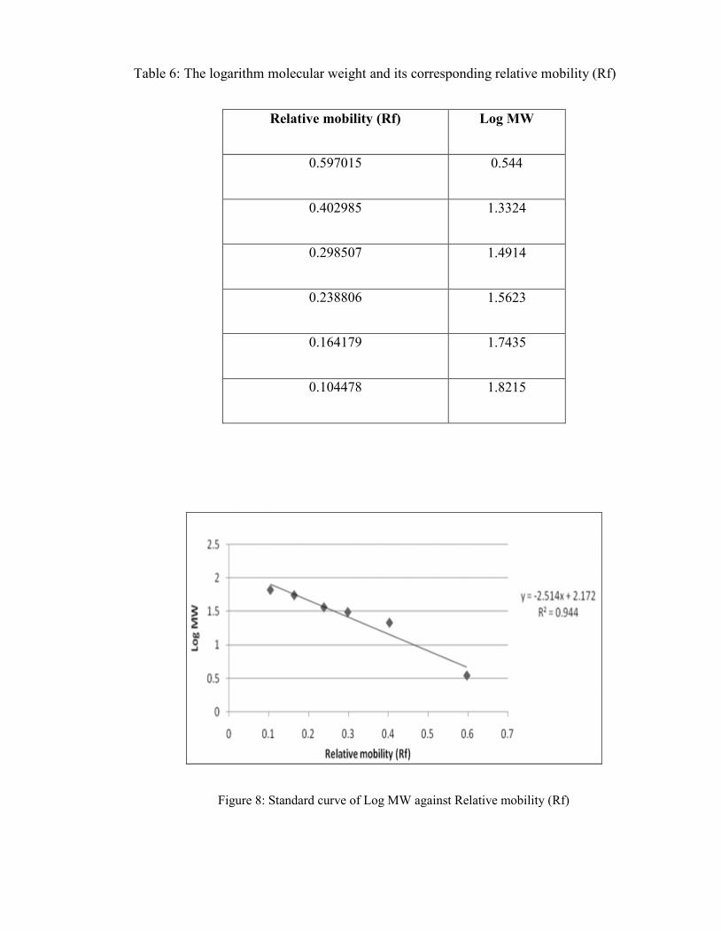
Table 6: The logarithm molecular weight and its corresponding relative mobility (Rf)
Relative mobility (Rf) Log MW
0.597015 0.544
0.402985 1.3324
0.298507 1.4914
0.238806 1.5623
0.164179 1.7435
0.104478 1.8215
Figure 8: Standard curve of Log MW against Relative mobility (Rf)

Table 7: The molecular weight Senna surattensis leaves and Mimosa diplotricha flower
determination
Relative mobility (Rf) Log MW MW (kDa)
Senna surattensis leaves 0.2888 1.446 27.93
Mimosa diplotricha fruits 0.2813 1.465 29.17
APPENDIX 10:
TRYPSIN INHIBITORY ASSAY USING CHRYSOMYA MEGACEPHALA EXTRACTED
PROTEASE
Table 8: Comparison of inhibitory activity percentage obtained between commercial trypsin
and Chrysomya megacephala extracted protease.
Plant sample Commercial trypsin Chrysomya megacephala
extacted protease
Senna surattensis leaves 83.52 % 61.74 %