Embed Size (px)
Citation preview

Agenda 11/27
• Read The Structure of Life– The Genetic Code: pp. 12-13– Worksheet: Genes to Polypeptides
• Four Levels of Protein Structure – Overview – In more detail
• Epigenome – When are proteins produced?
Homework: Thank you notes for Dr. Fisher and ___________Due Friday: rDNA project

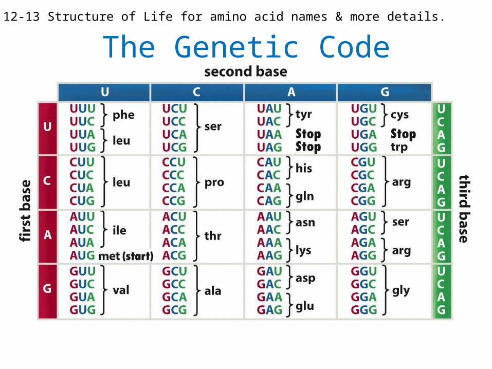
The Genetic CodeSee p.12-13 Structure of Life for amino acid names & more details.
From Genes to Polypeptides
• Complete worksheet
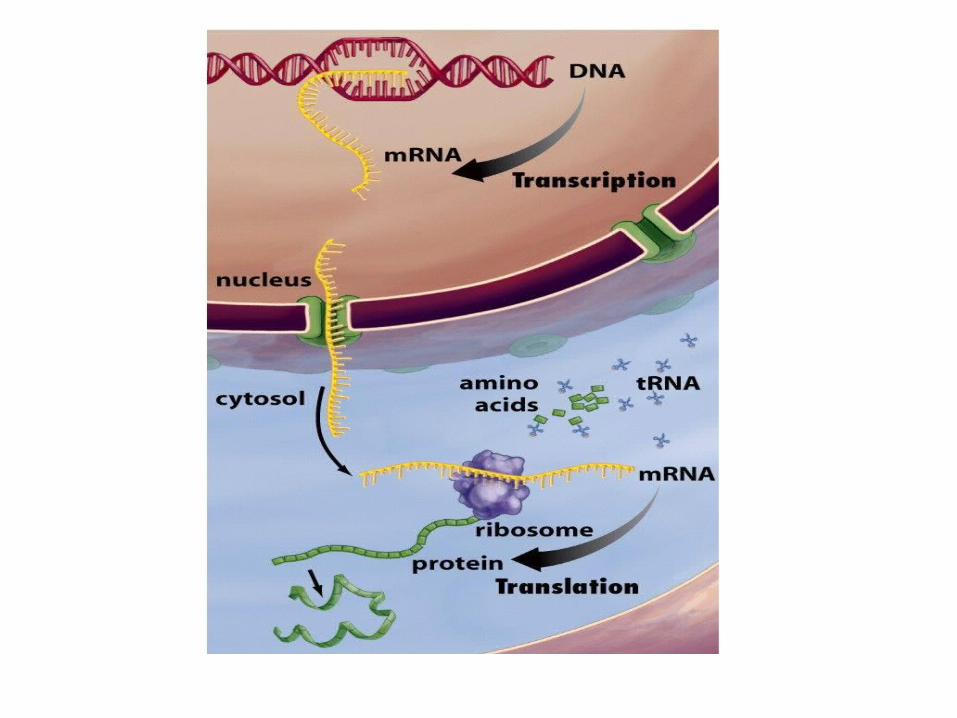
Review: Protein Theater• Setting the scene:
– Room walls are the cell membrane– Nucleus – Ribosome – Cytoplasm
• Transcription starts with RNA polymerase recognizing a promoter
• Gene on the DNA determines the complementary mRNA
• mRNA specified the correct sequence for amino acids

Proteins
• Structure determines function– Or “proteins are shaped to get the job done”
The Structure of Life
• Genetic Code p. 12-13
• Peering into Protein Factories p. 23
• Beyond Drug Design pp. 52-55

Proteins – shape determines function
Structure:PrimarySecondaryTertiaryQuaternary
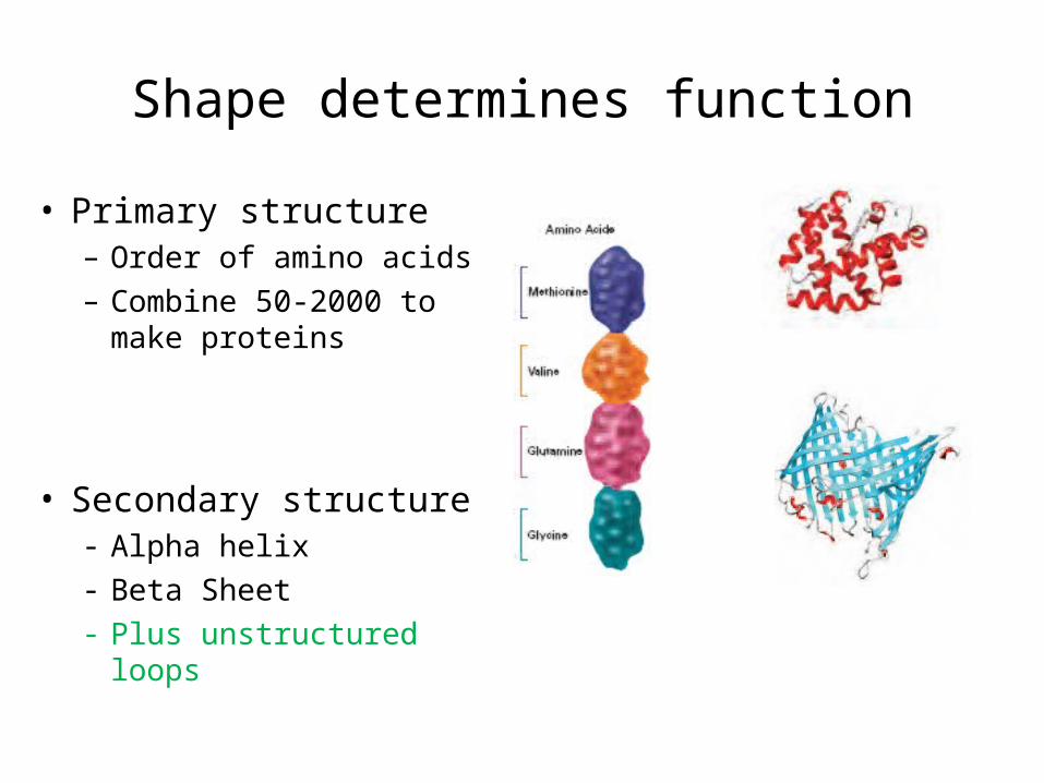
Shape determines function
• Primary structure– Order of amino acids– Combine 50-2000 to
make proteins
• Secondary structure- Alpha helix- Beta Sheet- Plus unstructured loops

Shape determines function
• Tertiary– Globular: compact – Fibrous: linear
• Quaternary– Multiple polypeptides (amino acid chains)
come together

The Structure of Life
• The Problem of Protein Folding
• p. 8 – Read carefully & take notes
• Pp. 10- 11: Slkim– What is the main point?

Why do the amino acids fold in a certain way?

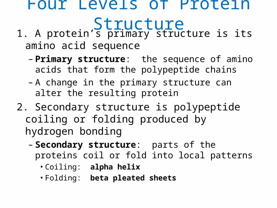
Four Levels of Protein Structure1. A protein’s primary structure is its amino
acid sequence– Primary structure: the sequence of amino
acids that form the polypeptide chains– A change in the primary structure can alter
the resulting protein
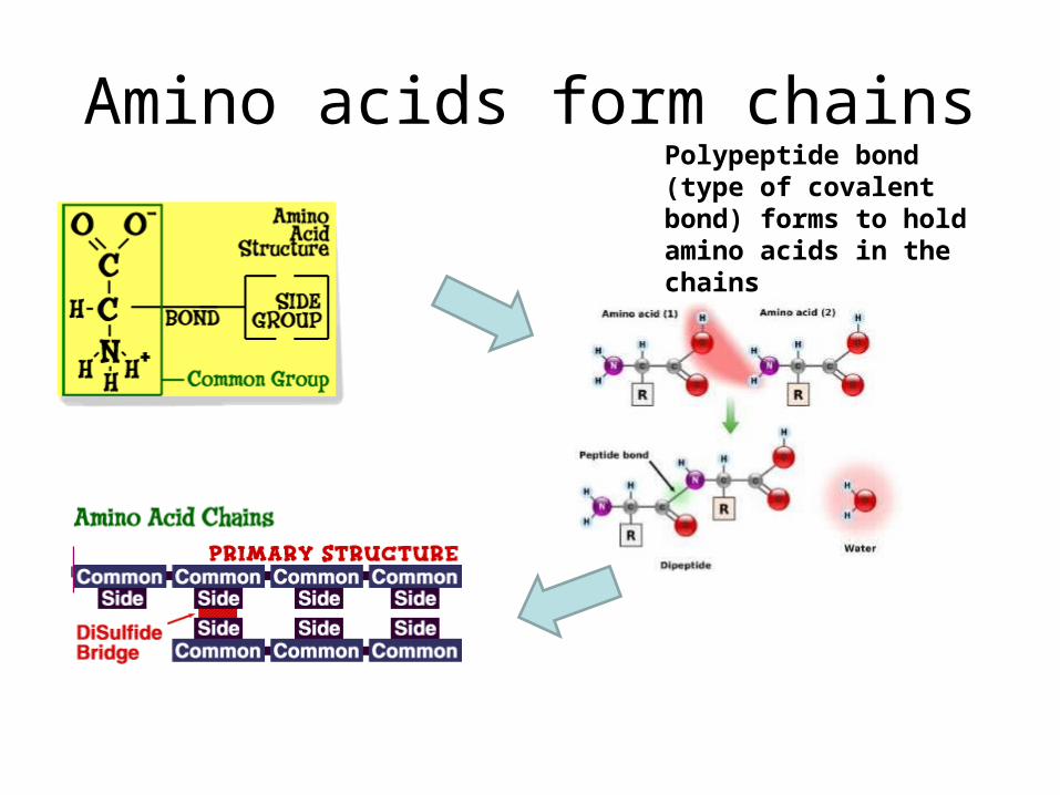
Amino acids form chainsPolypeptide bond (type of covalent bond) forms to hold amino acids in the chains
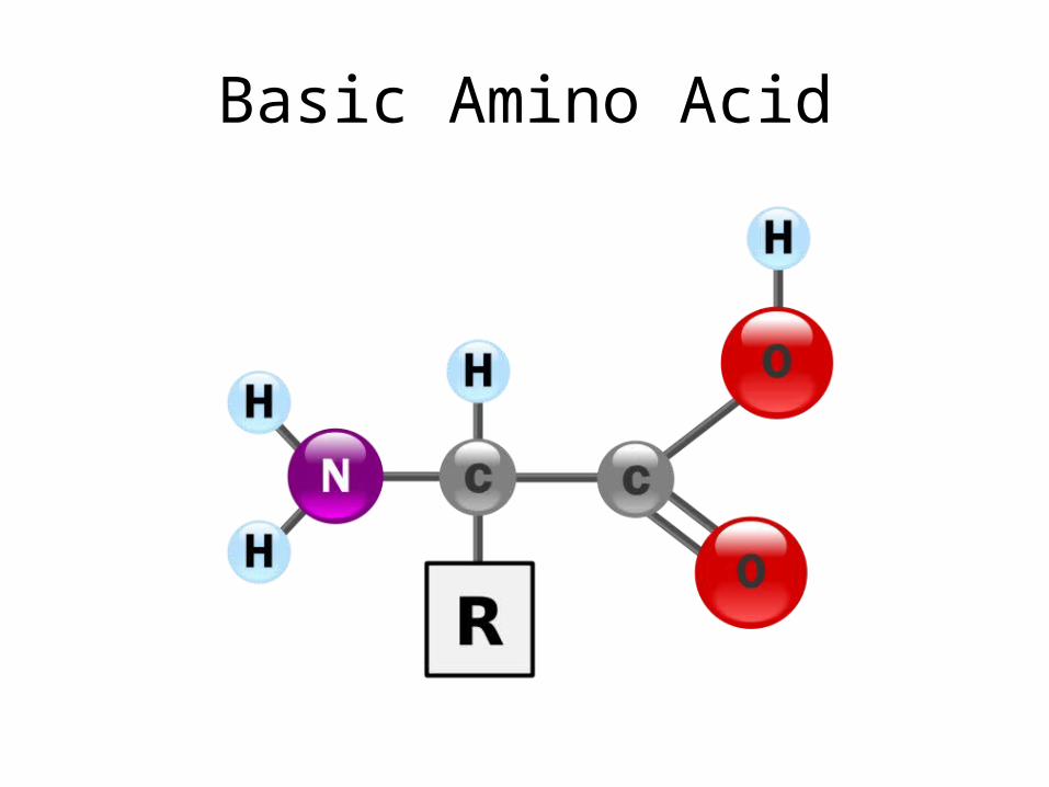
Basic Amino Acid

20 amino acids
• http://www.personal.kent.edu/~cearley/PChem/amino/3d.htm
• http://wbiomed.curtin.edu.au/biochem/tutorials/AAs/AA.html
• http://www.chem4kids.com/files/aminoacids/index.html
• Chem4Kids.com: Biochemistry:Twenty Amino Acids

Four Levels of Protein Structure2. Secondary structure is polypeptide coiling
or folding produced by hydrogen bonding– Secondary structure: parts of the proteins
coil or fold into local patterns• Coiling: alpha helix• Folding: beta pleated sheets
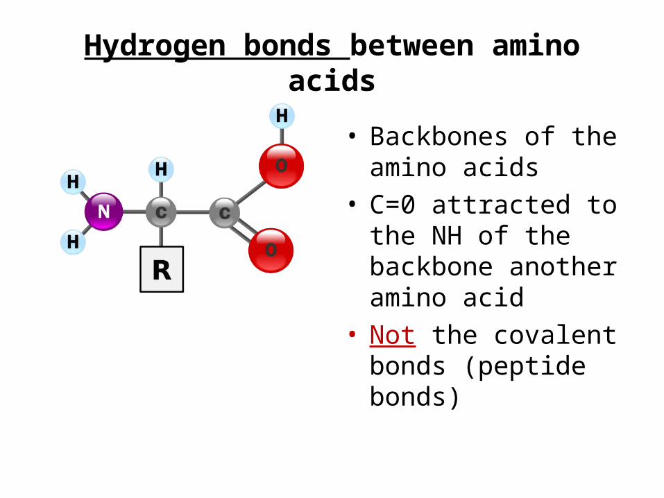
Hydrogen bonds between amino acids
• Backbones of the amino acids
• C=0 attracted to the NH of the backbone another amino acid
• Not the covalent bonds (peptide bonds)

Secondary Structure – Hydrogen Bonding • Alpha helix • Beta Sheet
other type.
(See
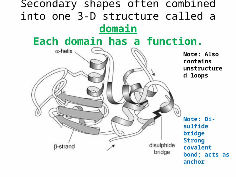
Secondary shapes often combined into one 3-D structure called a domainEach domain has a function.
Note: Di-sulfide bridgeStrong covalent bond; acts as anchor
Note: Also contains unstructured loops

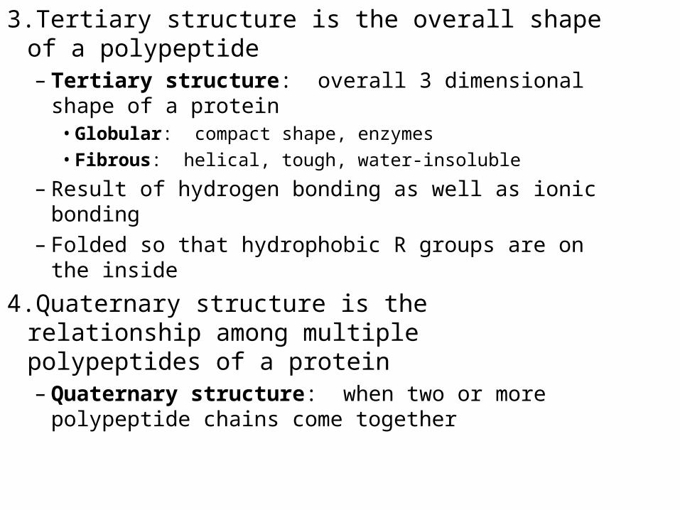
3.Tertiary structure is the overall shape of a polypeptide– Tertiary structure: overall 3 dimensional
shape of a protein• Globular: compact shape, enzymes• Fibrous: helical, tough, water-insoluble
– Result of hydrogen bonding as well as ionic bonding (hydrophilic R groups)
– Folded so that hydrophobic R groups are on the inside
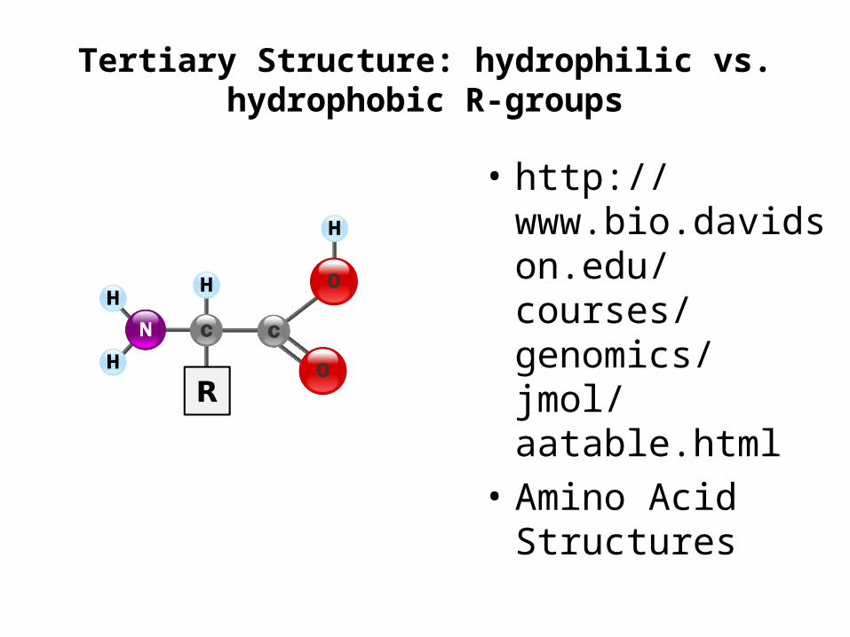
Tertiary Structure: hydrophilic vs. hydrophobic R-groups
• http://www.bio.davidson.edu/courses/genomics/jmol/aatable.html
• Amino Acid Structures

Basic Rules for Structure based on R groups
Hydrophobic
• Non-polar• R groups with only C& H • Side chains fold up into
the interior of the protein
Hydrophilic
• Polar (ionic) • Attracted to water since
water is polar • “Comfortable” in the
watery environment of cytosol (cytoplasm)
• Fold to be on the outside of the protein
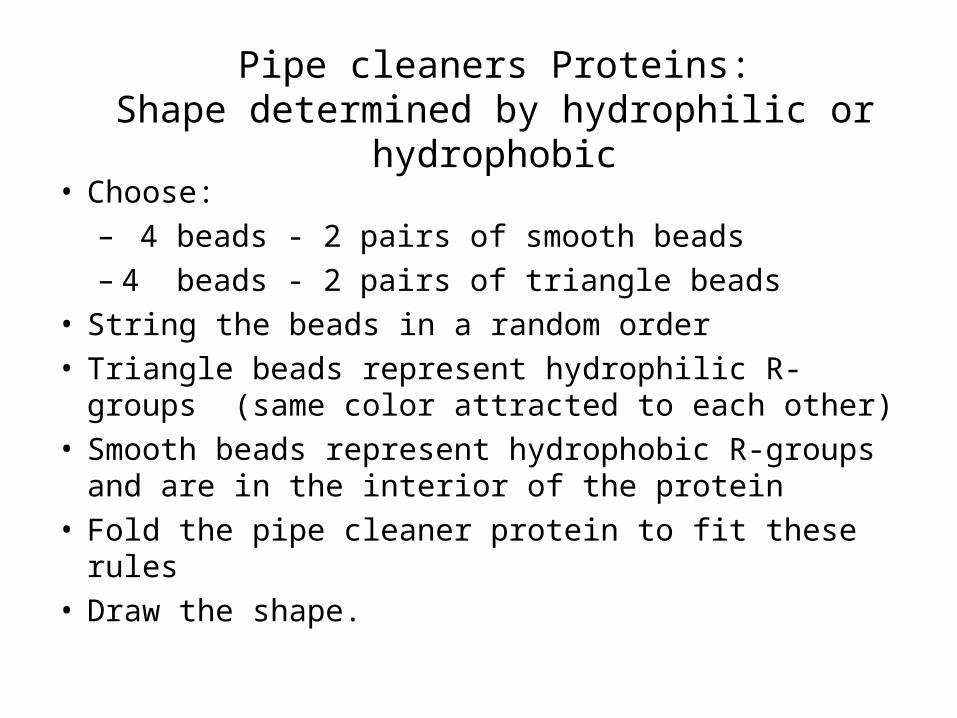
Pipe cleaners Proteins:Shape determined by hydrophilic or hydrophobic
• Choose:– 4 beads - 2 pairs of smooth beads– 4 beads - 2 pairs of triangle beads
• String the beads in a random order• Triangle beads represent hydrophilic R-groups
(same color attracted to each other)• Smooth beads represent hydrophobic R-groups and
are in the interior of the protein• Fold the pipe cleaner protein to fit these rules• Draw the shape.

Pipe cleaner proteins
• Compare your protein’s shape to others at your table.
• How and why are they different?
• What conclusions can you make about folding of proteins?
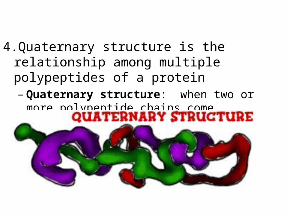
4.Quaternary structure is the relationship among multiple polypeptides of a protein– Quaternary structure: when two or more
polypeptide chains come together
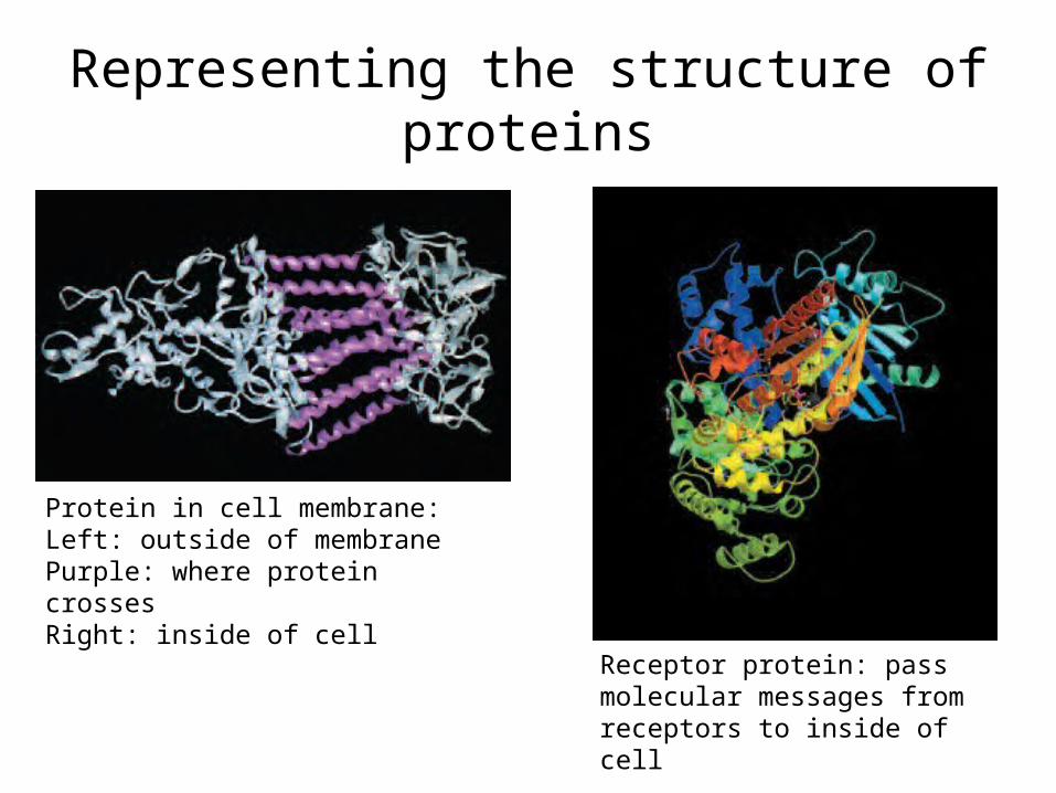
Representing the structure of proteins
Protein in cell membrane: Left: outside of membranePurple: where protein crosses Right: inside of cell
Receptor protein: pass molecular messages from receptors to inside of cell
Major Unsolved Problem“Protein folding problem”
Scientists cannot predict shape & function of a protein based on the gene
•Can determine the amino acid sequence•Can now make rough estimates of shapes
– Compare to known proteins using data bases (bioinformatics)
•Cannot accurately predict the position of each atom

Epigenome
• The Epigenome at a Glance
• Introduction – Nova
• Video: Definition of Epigenetics - YouTube
Supplements
Support:
The Structure of Life• Genetic Code p. 12-13• Peering into Protein Factories p. 23• Beyond Drug Design pp. 52-55
Biotechnology: Science for the New Millenium• Amino acids sequence in insulin pp. 157-158• Protein function p. 157• Amino acids – polar, non-charged & shape p.133
Notes on Transcription• RNA polymerase binds to promoter regions where it
undergoes a conformational change so that transcription can begin. This triggers the opening of the DNA double helix.
• RNA chain growth occurs 5’ to 3’, dNTPs added to the 3’ end, RNA molecule runs antiparallel to the DNA sense strand
• Only one strand of DNA serves as a template (sense strand), the antisense strand is not used
• Nucleotides added at 55/second• Mg++ requirement for polymerization• No pesky introns to worry with in bacteria! No major post-
transcriptional modification needed.

Notes on Translation• Bacterial ribosomes are different from eukaryotic
ribosomes. When assembled they are 70S (Svedberg unit) particles. The have a 30S subunit composed of
16S rRNA and 21 proteins. They also have a 50S subunit composed of 23 rRNA, 55 rRNA and 31 proteins.
• Amino acids added at 17/second.• Bacteria have the unique “coupled” transcription and
translation (polysomes) that does not occur in eukaryotic cells. Why not?
Major Steps of Translation
1. Initiation
2. Elongation
3. Termination

Now what happens to the polypeptide?
• A protein consists of multiple amino acids, these take on a specific shape
• The 3 dimensional shape determines the function of the protein
• Denaturation: the process of altering the specific shape of a protein so that it can no longer function properly = BAD!– This can be influenced by heat or cold, salt
concentrations, changes in pH, etc.
Four Levels of Protein Structure1. A protein’s primary structure is its amino acid
sequence– Primary structure: the sequence of amino acids
that form the polypeptide chains– A change in the primary structure can alter the
resulting protein
2. Secondary structure is polypeptide coiling or folding produced by hydrogen bonding– Secondary structure: parts of the proteins coil or
fold into local patterns• Coiling: alpha helix• Folding: beta pleated sheets
3.Tertiary structure is the overall shape of a polypeptide– Tertiary structure: overall 3 dimensional shape of
a protein• Globular: compact shape, enzymes• Fibrous: helical, tough, water-insoluble
– Result of hydrogen bonding as well as ionic bonding– Folded so that hydrophobic R groups are on the
inside
4.Quaternary structure is the relationship among multiple polypeptides of a protein– Quaternary structure: when two or more
polypeptide chains come together
Mutations• Mutation: a change to the DNA of a cell. This in turn
changes the RNA (codon) and may potentially change the protein produced by that segment of DNA. – Not all mutations are bad!! In fact, some are good.
• Mutations will occur spontaneously and are the only way to generate diversity in bacterial populations that reproduce by binary fission.
• Any substance that can cause a mutation is called a mutagenic agent.
• There are a variety of DNA repair mechanisms that can deal with minor damage.
Types of Mutations
• There are many types of mutations. Two common varieties include:– Point (substitution) mutations:
– Frameshift mutations:

Gene Expression• This is the regulation of transcription and
translation. Eukaryotes and prokaryotes have different ways of doing this. – Remember that bacteria are not going to have to
deal with the intron issue or RNA splicing. – This is something that must be considered when
eukaryotic genes are inserted into prokaryotic cells. One option is to use RT to work backwards into eukaryotic cDNA (which lacks introns)
– A note on differentiation – while this is a major issue for gene expression in eukaryotes (more to come on this later) it is not an issue for bacteria.

Bacterial Regulation of Gene Expression
• Operons: the system bacteria use to regulate their gene expression.– Operons are basically a set of genes under
the control of a single operator and promoter sequence.
– The set of genes produces a single polycistronic mRNA so, in most cases, all the genes in the operon are either “on” or “of”.
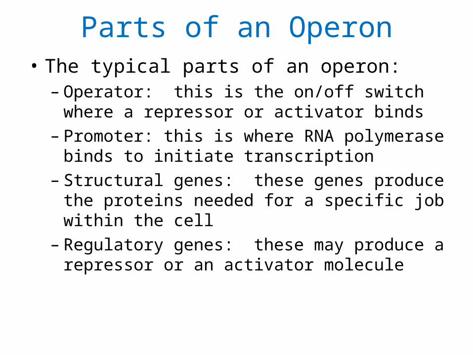
Parts of an Operon• The typical parts of an operon:
– Operator: this is the on/off switch where a repressor or activator binds
– Promoter: this is where RNA polymerase binds to initiate transcription
– Structural genes: these genes produce the proteins needed for a specific job within the cell
– Regulatory genes: these may produce a repressor or an activator molecule

The lac Operon

How Operons are Controlled• Positive control: An activator protein must bind to
the promoter to turn the operon “on”– Positive inducible: The activator normally can’t
get to the promoter. However, under certain conditions, the activator gets access to the promoter, turning the operon “on”
– Positive repressible: The activator is normally bound to the promoter. Under the right conditions, another molecule binds the activator preventing it from binding to the promoter and the operon is turned “off”.

How Operons are Controlled• Negative control: A repressor protein must bind to
the promoter to turn the operon “off”– Negative inducible: The repressor is normally
bound to the promoter keeping the operon “off”. Another molecule can bind to the repressor, preventing it from binding to the operator so that transcription occurs.
– Negative repressible: The repressor is designed such that it cannot bind to the promoter so that transcription occurs. Only when a corepressor is present will the repressor bind to the operator and turn off the operon.