Embed Size (px)
Citation preview
P1: FPX/FOZ/fop/fok P2: FHN/FDR/fgi QC: FhN/fgm T1: FhN
April 6, 2000 16:12 Annual Reviews AR098-11
?Annu. Rev. Biophys. Biomol. Struct. 2000. 29:291–325
Copyright c© 2000 by Annual Reviews. All rights reserved
COMPARATIVE PROTEIN STRUCTURE MODELING
OF GENES AND GENOMES
Marc A. Martı-Renom, Ashley C. Stuart, Andras Fiser,Roberto Sanchez, Francisco Melo, and Andrej SaliLaboratories of Molecular Biophysics, Pels Family Center for Biochemistry andStructural Biology, Rockefeller University, 1230 York Ave, New York, NY 10021;e-mail: [email protected]
Key Words protein structure prediction, fold assignment, alignment, homologymodeling, model evaluation, fully automated modeling, structural genomics
■ Abstract Comparative modeling predicts the three-dimensional structure of agiven protein sequence (target) based primarily on its alignment to one or more pro-teins of known structure (templates). The prediction process consists of fold assign-ment, target–template alignment, model building, and model evaluation. The numberof protein sequences that can be modeled and the accuracy of the predictions are in-creasing steadily because of the growth in the number of known protein structuresand because of the improvements in the modeling software. Further advances are nec-essary in recognizing weak sequence–structure similarities, aligning sequences withstructures, modeling of rigid body shifts, distortions, loops and side chains, as well asdetecting errors in a model. Despite these problems, it is currently possible to modelwith useful accuracy significant parts of approximately one third of all known proteinsequences. The use of individual comparative models in biology is already rewardingand increasingly widespread. A major new challenge for comparative modeling is theintegration of it with the torrents of data from genome sequencing projects as wellas from functional and structural genomics. In particular, there is a need to developan automated, rapid, robust, sensitive, and accurate comparative modeling pipelineapplicable to whole genomes. Such large-scale modeling is likely to encourage newkinds of applications for the many resulting models, based on their large number andcompleteness at the level of the family, organism, or functional network.
CONTENTS
INTRODUCTION . . . . . . . . . . . . . . . . . . . . . . . . . . . . . . . . . . . . . . . . . . . . . . . . 292STEPS IN COMPARATIVE MODELING. . . . . . . . . . . . . . . . . . . . . . . . . . . . . . . 294
Fold Assignment and Template Selection. . . . . . . . . . . . . . . . . . . . . . . . . . . . . . 294Target–Template Alignment. . . . . . . . . . . . . . . . . . . . . . . . . . . . . . . . . . . . . . . 297Model Building . . . . . . . . . . . . . . . . . . . . . . . . . . . . . . . . . . . . . . . . . . . . . . . . 298Loop Modeling . . . . . . . . . . . . . . . . . . . . . . . . . . . . . . . . . . . . . . . . . . . . . . . . 300Sidechain Modeling. . . . . . . . . . . . . . . . . . . . . . . . . . . . . . . . . . . . . . . . . . . . . 302
1056-8700/00/0610-0291$14.00 291
Ann
u. R
ev. B
ioph
ys. B
iom
ol. S
truc
t. 20
00.2
9:29
1-32
5. D
ownl
oade
d fr
om a
rjou
rnal
s.an
nual
revi
ews.
org
by D
uque
sne
Uni
vers
ity o
n 06
/20/
05. F
or p
erso
nal u
se o
nly.
P1: FPX/FOZ/fop/fok P2: FHN/FDR/fgi QC: FhN/fgm T1: FhN
April 6, 2000 16:12 Annual Reviews AR098-11
?292 MARTI-RENOM ET AL
ERRORS IN COMPARATIVE MODELS. . . . . . . . . . . . . . . . . . . . . . . . . . . . . . . 304EVALUATION OF MODELS . . . . . . . . . . . . . . . . . . . . . . . . . . . . . . . . . . . . . . . . 306APPLICATIONS OF COMPARATIVE MODELING. . . . . . . . . . . . . . . . . . . . . . . 309COMPARATIVE MODELING IN STRUCTURAL GENOMICS . . . . . . . . . . . . . . 311CONCLUSION. . . . . . . . . . . . . . . . . . . . . . . . . . . . . . . . . . . . . . . . . . . . . . . . . . 314
INTRODUCTION
The aim of comparative or homology protein structure modeling is to build athree-dimensional (3D) model for a protein of unknown structure (the target) onthe basis of sequence similarity to proteins of known structure (the templates)(10, 17, 80, 145, 155). Two conditions must be met to build a useful model. First,the similarity between the target sequence and the template structure must bedetectable. Second, a substantially correct alignment between the target sequenceand the template structures must be calculated. Comparative modeling is possiblebecause small changes in the protein sequence usually result in small changesin its 3D structure (34). Although considerable progress has been made in abinitio protein structure prediction (92), comparative protein structure modelingremains the most accurate prediction method. The overall accuracy of comparativemodels spans a wide range, from low resolution models with only a correct fold tomore accurate models comparable to medium resolution structures determined bycrystallography or nuclear magnetic resonance (NMR) spectroscopy (155). Evenlow resolution models can be useful in biology because some aspects of functioncan sometimes be predicted only from the coarse structural features of a model.
The 3D structures of proteins in a family are more conserved than their se-quences (101). Therefore, if similarity between two proteins is detectable at thesequence level, structural similarity can usually be assumed. Moreover, even pro-teins that have nondetectable sequence similarity can have similar structures. Ithas been estimated that approximately one third of all sequences are recognizablyrelated to at least one known protein structure (54, 77, 81, 144, 157). Because thenumber of known protein sequences is approximately 500,000 (9), comparativemodeling could in principle be applied to more than 150,000 proteins. This num-ber can be compared to approximately 10,000 protein structures determined byexperiment (1, 211). The usefulness of comparative modeling is steadily increasingbecause the number of unique structural folds that proteins adopt is limited (204)and because the number of experimentally determined new structures is increasingexponentially (70). It is possible that in less than 10 years at least one example ofmost structural folds will be known, making comparative modeling applicable tomost protein sequences (70, 155).
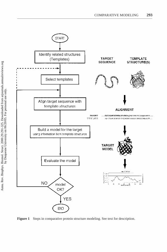
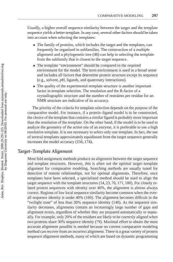
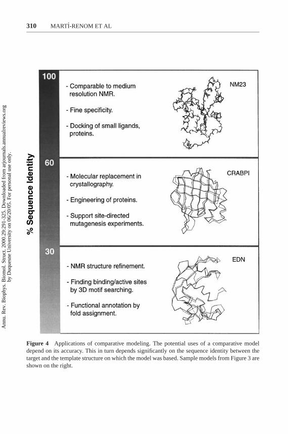
All current comparative modeling methods consist of four sequential steps(Figure 1) (155): fold assignment and template selection, template–target align-ment, model building, and model evaluation. If the model is not satisfactory, tem-plate selection, alignment, and model building can be repeated until a satisfactory
Ann
u. R
ev. B
ioph
ys. B
iom
ol. S
truc
t. 20
00.2
9:29
1-32
5. D
ownl
oade
d fr
om a
rjou
rnal
s.an
nual
revi
ews.
org
by D
uque
sne
Uni
vers
ity o
n 06
/20/
05. F
or p
erso
nal u
se o
nly.
P1: FPX/FOZ/fop/fok P2: FHN/FDR/fgi QC: FhN/fgm T1: FhN
April 6, 2000 16:12 Annual Reviews AR098-11
?COMPARATIVE MODELING 293
Figure 1 Steps in comparative protein structure modeling. See text for description.
Ann
u. R
ev. B
ioph
ys. B
iom
ol. S
truc
t. 20
00.2
9:29
1-32
5. D
ownl
oade
d fr
om a
rjou
rnal
s.an
nual
revi
ews.
org
by D
uque
sne
Uni
vers
ity o
n 06
/20/
05. F
or p
erso
nal u
se o
nly.
P1: FPX/FOZ/fop/fok P2: FHN/FDR/fgi QC: FhN/fgm T1: FhN
April 6, 2000 16:12 Annual Reviews AR098-11
?294 MARTI-RENOM ET AL
model is obtained. For each of the steps in the modeling process, there are manydifferent methods, programs, and World Wide Web servers (Table 1).
We begin this review by describing the techniques used for all the steps in com-parative modeling. We continue by discussing the errors in model structures andmethods for detecting these errors, and we conclude by outlining the applicationsof comparative modeling to individual proteins and to whole genomes. The reviewfocuses on using the methods and tools for comparative modeling, rather than onthe physical principles on which the methods are based. The bibliography is notexhaustive, but an attempt was made to quote the latest papers or reviews in therelevant fields.
STEPS IN COMPARATIVE MODELING
Fold Assignment and Template Selection
The starting point in comparative modeling is to identify all protein structuresrelated to the target sequence, and then to select those that will be used as templates.This step is facilitated by numerous protein sequence and structure databases,and database scanning software available on the World Wide Web (4, 11, 70, 163)(Table 1). Templates can be found using the target sequence as a query for searchingstructure databases such as the Protein Data Bank (1, 211), SCOP (76), DALI (71),and CATH (124). Depending on a genome, the probability of finding a relatedprotein of known structure for a sequence randomly picked from a genome rangesfrom 20% to 50% (54, 77, 81, 144, 157).
There are three main classes of protein comparison methods that are usefulin fold identification. The first class includes the methods that compare the tar-get sequence with each of the database sequences independently, using pairwisesequence–sequence comparison (7). The performance of these methods in search-ing for related protein sequences (128) and structures (22) has been evaluatedexhaustively. Frequently used programs in this class include FASTA (129) andBLAST (5).
The second set of methods relies on multiple sequence comparisons to improvethe sensitivity of the search (6, 63, 67, 94, 144). A widely used program in thisclass is PSI-BLAST (6), which iteratively expands the set of homologs of the targetsequence. For a given sequence, an initial set of homologs from a sequence databaseis collected, a weighted multiple alignment is made from the query sequence andits homologs, a position-specific scoring matrix is constructed from the alignment,and the matrix is used to search the database for new homologs. These steps arerepeated until no new homologs are found. In comparison to BLAST, PSI-BLASTfinds homologs of known structure for approximately twice as many sequences(125, 176). A related approach (144) also begins by finding all sequences clearlyrelated to the target sequence to obtain the target sequence profile. In addition,similar profiles are constructed for all known protein structures. The potentialtemplates are then found by comparing the target sequence profile with each of thesequence profiles for known structures. Another variation uses multiple sequence
Ann
u. R
ev. B
ioph
ys. B
iom
ol. S
truc
t. 20
00.2
9:29
1-32
5. D
ownl
oade
d fr
om a
rjou
rnal
s.an
nual
revi
ews.
org
by D
uque
sne
Uni
vers
ity o
n 06
/20/
05. F
or p
erso
nal u
se o
nly.
P1: FPX/FOZ/fop/fok P2: FHN/FDR/fgi QC: FhN/fgm T1: FhN
April 6, 2000 16:12 Annual Reviews AR098-11
?COMPARATIVE MODELING 295
TABLE 1 Programs and World Wide Web servers useful in comparative modeling
Name Typea World Wide Web addressb Referencec
DatabasesCATH S www.biochem.ucl.ac.uk/bsm/cath/ 124GenBank S www.ncbi.nlm.nih.gov/GenBank 15GeneCensus S bioinfo.mbb.yale.edu/genome 58MODBASE S guitar.rockefeller.edu/modbase/ 159PDB S www.rcsb.org/pdb/ 16PRESAGE S presage.stanford.edu 21SCOP S scop.mrc-lmb.cam.ac.uk/scop/ 76SWISSPROT+TrEMBL S www.ebi.ac.uk/swissprot 9
Template search123D S www.lmmb.ncifcrf.gov/∼nicka/123D.html 2BLAST S www.ncbi.nlm.nih.gov/BLAST/ 5DALI S www2.ebi.ac.uk/dali/ 71FastA S www2.ebi.ac.uk/fasta3 127MATCHMAKER P bioinformatics.burnham-inst.org 59PHD, TOPITS S www.embl-heidelberg.de/predictprotein/ 139
predictprotein.htmlPROFIT P www.came.sbg.ac.at 57THREADER P globin.bio.warwick.ac.uk/∼jones/threader.html 82UCLA-DOE FRSVR S www.doe-mbi.ucla.edu/people/frsvr/frsvr.html 53
Sequence alignmentBCM SERVER S dot.imgen.bcm.tmc.edu:9331/ 170BLAST S www.ncbi.nlm.nih.gov/BLAST 6BLOCK MAKER S blocks.fhcrc.org/blocks/blockmkr/ 68
makeblocks.htmlCLUSTAL S www2.ebi.ac.uk/clustalw/ 78FASTA3 S www2.ebi.ac.uk/fasta3/ 127MULTALIN S pbil.ibcp.fr/ 41
ModelingCOMPOSER P www-cryst.bioc.cam.ac.uk 179CONGEN P www.congenomics.com/congen/congen.html 29CPH models S www.cbs.dtu.dk/services/CPHmodels/ 206DRAGON P www.nimr.mrc.ac.uk/∼mathbio/a-aszodi/ 8
dragon.htmlICM P www.molsoft.com (a)
InsightII P www.msi.com (b)MODELLER P guitar.rockefeller.edu/modeller/modeller.html 148LOOK P www.mag.com 102QUANTA P www.msi.com (b)SYBYL P www.tripos.com (c)SCWRL P www.cmpharm.ucsf.edu/∼bower/scrwl/ 19
scrwl.htmlSWISS-MOD S www.expasy.ch/swissmod 131WHAT IF P www.sander.embl-heidelberg.de/whatif/ 194
(continued)
Ann
u. R
ev. B
ioph
ys. B
iom
ol. S
truc
t. 20
00.2
9:29
1-32
5. D
ownl
oade
d fr
om a
rjou
rnal
s.an
nual
revi
ews.
org
by D
uque
sne
Uni
vers
ity o
n 06
/20/
05. F
or p
erso
nal u
se o
nly.
P1: FPX/FOZ/fop/fok P2: FHN/FDR/fgi QC: FhN/fgm T1: FhN
April 6, 2000 16:12 Annual Reviews AR098-11
?296 MARTI-RENOM ET AL
TABLE 1 (Continued)
Name Typea World Wide Web addressb Referencec
Model evaluationANOLEA S www.fundp.ac.be/pub/ANOLEA.html 113AQUA P www-nmr.chem.ruu.nl/users/rull/aqua.html 98BIOTECHd S biotech.embl-ebi.ac.uk:8400/ 73, 96ERRAT S www.doe-mbi.ucla.edu/erratserver.html 40PROCHECK P www.biochem.ucl.ac.uk/∼roman/procheck/ 96
procheck.htmlProCeryone P www.proceryon.com/ (d)ProsaIIe P www.came.sbg.ac.at 169PROVE S www.ucmb.ulb.ac.be/UCMB/PROVE 134SQUID P www.yorvic.york.ac.uk/∼oldfield/squid 121VERIFY3D S www.doe-mbi.ucla.edu/verify3d.html 105WHATCHECK P www.sander.embl-heidelberg.de/whatcheck/ 73
aS, server; P, program.bSome of the sites are mirrored on additional computers.c(a) MolSoft Inc., San Diego. (b) Molecular Simulations Inc., San Diego. (c) Tripos Inc., St Louis. (d) ProCeryon BiosciencesInc. New York.dThe BIOTECH server uses PROCHECKand WHATCHECK for structure evaluation.eProCyon is a new software package that includes PeeP, PROFIT and PROSUP, a structure comparison program.
alignments combined with structural information predicted from the sequence ofthe target (54). The multiple sequence methods for fold identification are especiallyuseful for finding significant structural relationships when the sequence identifybetween the target and the template drops below 25%. This class of methodsappears to be one of the most sensitive fully-automated approaches for detectingremote sequence–structure relationships (6, 77, 203, 213).
The third class of methods are the so-called threading or 3D template matchingmethods (20, 59, 82), reviewed in (83, 103, 173, 185). These methods rely on pair-wise comparison of a protein sequence and a protein of known structure. Whetheror not a given target sequence adopts any one of the many known 3D folds is pre-dicted by an optimization of the alignment with respect to a structure-dependentscoring function, independently for each sequence–structure pair. That is, the targetsequence is threaded through a library of 3D folds. These methods are especiallyuseful when there are no sequences clearly related to the modeling target, and thusthe search cannot benefit from the increased sensitivity of the sequence profilemethods.
A useful fold assignment approach is to accept an uncertain assignment providedby any of the methods, build a full-atom comparative model of the target sequencebased on this match, and make the final decision about whether or not the matchis real by evaluating the resulting comparative model (64, 115, 156).
Once a list of all related protein structures has been obtained, it is necessaryto select those templates that are appropriate for the given modeling problem.
Ann
u. R
ev. B
ioph
ys. B
iom
ol. S
truc
t. 20
00.2
9:29
1-32
5. D
ownl
oade
d fr
om a
rjou
rnal
s.an
nual
revi
ews.
org
by D
uque
sne
Uni
vers
ity o
n 06
/20/
05. F
or p
erso
nal u
se o
nly.
P1: FPX/FOZ/fop/fok P2: FHN/FDR/fgi QC: FhN/fgm T1: FhN
April 6, 2000 16:12 Annual Reviews AR098-11
?COMPARATIVE MODELING 297
Usually, a higher overall sequence similarity between the target and the templatesequence yields a better template. In any case, several other factors should be takeninto account when selecting the templates:
• The family of proteins, which includes the target and the templates, canfrequently be organized in subfamilies. The construction of a multiplealignment and a phylogenetic tree (46) can help in selecting the templatefrom the subfamily that is closest to the target sequence.
• The template “environment” should be compared to the requiredenvironment for the model. The term environment is used in a broad senseand includes all factors that determine protein structure except its sequence(e.g., solvent,pH, ligands, and quaternary interactions).
• The quality of the experimental template structure is another importantfactor in template selection. The resolution and the R-factor of acrystallographic structure and the number of restraints per residue for anNMR structure are indicative of its accuracy.
The priority of the criteria for template selection depends on the purpose of thecomparative model. For instance, if a protein–ligand model is to be constructed,the choice of the template that contains a similar ligand is probably more importantthan the resolution of the template. On the other hand, if the model is to be used toanalyze the geometry of the active site of an enzyme, it is preferable to use a highresolution template. It is not necessary to select only one template. In fact, the useof several templates approximately equidistant from the target sequence generallyincreases the model accuracy (156, 174).
Target–Template Alignment
Most fold assignment methods produce an alignment between the target sequenceand template structures. However, this is often not the optimal target–templatealignment for comparative modeling. Searching methods are usually tuned fordetection of remote relationships, not for optimal alignments. Therefore, oncetemplates have been selected, a specialized method should be used to align thetarget sequence with the template structures (14, 23, 70, 171, 180). For closely re-lated protein sequences with identity over 40%, the alignment is almost alwayscorrect. Regions of low local sequence similarity become common when the over-all sequence identity is under 40% (160). The alignment becomes difficult in the“twilight zone” of less than 30% sequence identity (140). As the sequence sim-ilarity decreases, alignments contain an increasingly large number of gaps andalignment errors, regardless of whether they are prepared automatically or manu-ally. For example, only 20% of the residues are likely to be correctly aligned whentwo proteins share 30% sequence identity (79). Maximal effort to obtain the mostaccurate alignment possible is needed because no current comparative modelingmethod can recover from an incorrect alignment. There is a great variety of proteinsequence alignment methods, many of which are based on dynamic programming
Ann
u. R
ev. B
ioph
ys. B
iom
ol. S
truc
t. 20
00.2
9:29
1-32
5. D
ownl
oade
d fr
om a
rjou
rnal
s.an
nual
revi
ews.
org
by D
uque
sne
Uni
vers
ity o
n 06
/20/
05. F
or p
erso
nal u
se o
nly.
P1: FPX/FOZ/fop/fok P2: FHN/FDR/fgi QC: FhN/fgm T1: FhN
April 6, 2000 16:12 Annual Reviews AR098-11
?298 MARTI-RENOM ET AL
techniques (120, 172). A frequently used program for multiple sequence align-ment is CLUSTAL (78), which is also available as a World Wide Web server(Table 1).
In the more difficult alignment cases, it is frequently beneficial to rely onmultiple structure and sequence information (12, 181). First, the alignment of thepotential templates is prepared by superposing their structures. Next, the sequencesthat are clearly related to the templates and are easily aligned with them are addedto the alignment. The same is done for the target sequence. Finally, the two profilesare aligned with each other, taking structural information into account as much aspossible (78, 93, 152). In principle, most sequence alignment and structure compar-ison methods can be used for these tasks (11, 70, 104, 171). The information fromstructures helps to avoid gaps in secondary structure elements, in buried regions,or between two residues that are far in space. It is generally necessary to checkand edit the alignment by inspecting the template structures visually, especiallyif the target–template sequence identity is low. Secondary structure predictionsfor the target sequence and its profile are also frequently useful in obtaining amore accurate alignment (3, 141). Because evaluation of a model is more reliablethan an evaluation of an alignment, a good way to proceed in the difficult cases isto generate 3D models for all alternative alignments, evaluate the correspondingmodels, and pick the best model according to the 3D model evaluation rather thanthe alignment score (64, 115, 156).
Model Building
Once an initial target–template alignment has been built, a variety of methods canbe used to construct a 3D model for the target protein. The original and still widelyused method is modeling by rigid-body assembly (17, 27, 61). Another family ofmethods, modeling by segment matching, relies on the approximate positions ofconserved atoms in the templates (38, 85, 102, 187). The third group of methods,modeling by satisfaction of spatial restraints, uses either distance geometry oroptimization techniques to satisfy spatial restraints obtained from the alignment(8, 24, 66, 148, 175). Accuracies of the various model building methods are rela-tively similar when used optimally. Other factors such as template selection andalignment accuracy usually have a larger impact on the model accuracy, especiallyfor models based on less than 40% sequence identity to the templates. However, itis important that a modeling method allows a degree of flexibility and automation,making it easier and faster to obtain better models. For example, a method shouldpermit an easy recalculation of a model when a change is made in the alignment; itshould be straightforward to calculate models based on several templates; and themethod should provide the tools to incorporate prior knowledge about the target(e.g., experimental data or predicted features such as secondary structure). Thereare many reviews of comparative model building methods (10, 17, 80, 145, 155).Several programs and World Wide Web servers for comparative modeling are listedin Table 1.
Ann
u. R
ev. B
ioph
ys. B
iom
ol. S
truc
t. 20
00.2
9:29
1-32
5. D
ownl
oade
d fr
om a
rjou
rnal
s.an
nual
revi
ews.
org
by D
uque
sne
Uni
vers
ity o
n 06
/20/
05. F
or p
erso
nal u
se o
nly.
P1: FPX/FOZ/fop/fok P2: FHN/FDR/fgi QC: FhN/fgm T1: FhN
April 6, 2000 16:12 Annual Reviews AR098-11
?COMPARATIVE MODELING 299
Modeling by Assembly of Rigid BodiesThis method assembles a model froma small number of rigid bodies obtained from aligned protein structures. The ap-proach is based on the natural dissection of the protein folds into conserved coreregions, variable loops, and side chains. For example, the following semiauto-mated procedure is implemented in the computer program COMPOSER(179). First,the template structures are selected and superposed. Second, the frame-work iscalculated by averaging the coordinates of the Cα atoms of structurally conservedregions in the template structures. Third, the main chain atoms of each core regionin the target model are generated by superposing the core segment from the tem-plate with the highest sequence similarity to the target on the framework. Fourth,the loops are generated by scanning a database of all known protein structures toidentify the structurally variable regions that fit the anchor core regions and havea compatible sequence. Fifth, the side chains are modeled based on their intrinsicconformational preferences and on the conformation of the equivalent side chainsin the template structures. And finally, the stereochemistry of the model is improvedeither by a restrained energy minimization or a molecular dynamics refinement.The accuracy of a model can be somewhat increased if more than one templatestructure is used to construct the framework and if the templates are averagedinto the framework using weights corresponding to their sequence similarities tothe target sequence (174). For example, differences between the model and X-raystructures may be slightly smaller than the differences between the X-ray struc-ture of the modeled protein and the best template used to build the model. Futureimprovements in modeling by rigid body assembly may include the incorporationof rigid body shifts, such as the relative shifts in the packing ofα-helices.
Modeling by Segment Matching or Coordinate ReconstructionThe basis ofmodeling by coordinate reconstruction is the finding that most hexapeptide seg-ments of protein structure can be clustered into approximately 100 structuralclasses (187). Thus, comparative models can be constructed by using a subsetof atomic positions from template structures as “guiding” positions, and by iden-tifying and assembling short, all-atom segments that fit these guiding positions.Usually the Cα atoms of the segments, which are conserved in the alignment be-tween the template structure and the target sequence, are taken as the guiding po-sitions. The all-atom segments that fit the guiding positions can be obtained eitherby scanning all the known protein structures, including those that are not related tothe sequence being modeled (38, 69), or by a conformational search restrained byan energy function (13, 190). For example, a general method for modeling by seg-ment matching is guided by the positions of some atoms (usually Cα atoms) to findthe matching segments in a representative database of all known protein structures(102). This method can construct both main chain and side chain atoms and canalso model insertions and deletions. It is implemented in the program SEGMOD
(Table 1). Some side chain modeling methods (195) and loop construction meth-ods based on finding suitable fragments in the database of known structures (85)can be seen as segment matching or coordinate reconstruction methods.
Ann
u. R
ev. B
ioph
ys. B
iom
ol. S
truc
t. 20
00.2
9:29
1-32
5. D
ownl
oade
d fr
om a
rjou
rnal
s.an
nual
revi
ews.
org
by D
uque
sne
Uni
vers
ity o
n 06
/20/
05. F
or p
erso
nal u
se o
nly.
P1: FPX/FOZ/fop/fok P2: FHN/FDR/fgi QC: FhN/fgm T1: FhN
April 6, 2000 16:12 Annual Reviews AR098-11
?300 MARTI-RENOM ET AL
Modeling by Satisfaction of Spatial RestraintsThe methods in this class gener-ate many constraints or restraints on the structure of the target sequence, using itsalignment to related protein structures as a guide. The restraints are generally ob-tained by assuming that the corresponding distances and angles between alignedresidues in the template and the target structures are similar. These homology-derived restraints are usually supplemented by stereochemical restraints on bondlengths, bond angles, dihedral angles, and nonbonded atom–atom contacts ob-tained from a molecular mechanics force field. The model is then derived by min-imizing the violations of all the restraints. This can be achieved either by distancegeometry or real–space optimization. A distance geometry approach constructsall-atom models from lower and upper bounds on distances and dihedral angles(66). A real–space optimization method, such as that implemented in the com-puter program MODELLER (148), starts by building the model using the distanceand dihedral angle restraints on the target sequence derived from its alignment withtemplate 3D structures. Then, the spatial restraints and the CHARMM22 force fieldterms, which enforce proper stereochemistry (106), are combined into an objec-tive function. Finally, the model is generated by optimizing the objective functionin Cartesian space. Because modeling by satisfaction of spatial restraints can usemany different types of information about the target sequence, it is perhaps themost promising of all comparative modeling techniques. One of the strengths ofmodeling by satisfaction of spatial restraints is that constraints or restraints derivedfrom a number of different sources can easily be added to the homology-derived re-straints. For example, restraints might be obtained from NMR experiments, cross-linking experiments, fluorescence spectroscopy, image reconstruction in electronmicroscopy, site-directed mutagenesis, etc. In this way, a comparative model, es-pecially in the difficult cases, could be improved by making it consistent withavailable experimental data.
Loop Modeling
In a given fold family, structural variability is a result of substitutions, insertions,and deletions of residues between members of the family. Such changes frequentlycorrespond to exposed loop regions that connect elements of secondary structurein the protein fold. Thus, loops often determine the functional specificity of a givenprotein framework. They contribute to active and binding sites. Examples includebinding of metal ions by metal-binding proteins, small protein toxins by their re-ceptors, antigens by immunoglobulins, nucleotides by a variety of proteins, proteinsubstrates by serine proteases, and DNA by DNA-binding proteins. Consequently,the accuracy of loop modeling is a major factor determining the usefulness ofcomparative models in studying interactions between the protein and its ligands.This includes the use of models for recognizing ligand binding sites (47, 84) andfor ligand docking computations (87). Unfortunately, no generally reliable methodis available for constructing loops longer than 5 residues (108), although recentlysome progress has been made and occasionally longer loops can be modeled
Ann
u. R
ev. B
ioph
ys. B
iom
ol. S
truc
t. 20
00.2
9:29
1-32
5. D
ownl
oade
d fr
om a
rjou
rnal
s.an
nual
revi
ews.
org
by D
uque
sne
Uni
vers
ity o
n 06
/20/
05. F
or p
erso
nal u
se o
nly.
P1: FPX/FOZ/fop/fok P2: FHN/FDR/fgi QC: FhN/fgm T1: FhN
April 6, 2000 16:12 Annual Reviews AR098-11
?COMPARATIVE MODELING 301
correctly (122, 135, 142, 153, 191). An exhaustive set of references for loop mod-eling papers can be found in (55).
Loop modeling can be seen as a mini–protein folding problem. The correctconformation of a given segment of a polypeptide chain has to be calculatedmainly from the sequence of the segment itself. However, loops are generally tooshort to provide sufficient information about their local fold. Segments of up to9 residues sometimes have entirely unrelated conformations in different proteins(114). Thus, the conformation of a given segment is also influenced by the corestem regions that span the loop and by the structure of the rest of a protein thatcradles the loop.
Many loop modeling procedures have been described. Similarly to the predic-tion of whole protein structures, there are ab initio methods (30, 50, 119), databasesearch techniques (35, 60, 85), and procedures that combine these two basic ap-proaches (107, 191).
The ab initio loop prediction is based on a conformational search or enumerationof conformations in a given environment, guided by a scoring or energy function.There are many such methods, exploiting different protein representations, energyfunction terms, and optimization or enumeration algorithms. The search algo-rithms include sampling of main chain dihedral angles biased by their distributionsin known protein structures (119), minimum perturbation random tweak method(165), systematic conformational search (26, 29), global energy minimization bymapping a trajectory of local minima (43), importance-sampling by local mini-mization of randomly generated conformations (95), local energy minimization(110), molecular dynamics simulations (28, 55), genetic algorithms (111, 136),biased probability Monte Carlo search (45, 183), Monte Carlo with simulatedannealing (33, 39, 192), Monte Carlo and molecular dynamics (135), extended-scaled-collective-variable Monte Carlo (88), scaling relaxation and multiple copysampling (138, 205), searching through discrete conformations by dynamic pro-gramming (51, 188), random sampling of conformations relying on dimers fromknown protein structures (177), self-consistent field optimization (91), and an enu-meration based on the graph theory (153). A variety of representations have beenused, such as unified atoms, all nonhydrogen atoms, nonhydrogen and “polar”hydrogen atoms, all atoms, as well as implicit and explicit solvent models. Theoptimized degrees of freedom include Cartesian coordinates and internal coordi-nates, such as dihedral angles, optimized in continuous or discrete spaces. Loopprediction by optimization is in principle applicable to simultaneous modeling ofseveral loops and loops interacting with ligands, which is not straightforward forthe database search approaches.
The database approach to loop prediction consists of finding a segment of mainchain that fits the two stem regions of a loop (36, 60, 85, 122, 142, 166, 178, 198).The stems are defined as the main chain atoms that precede and follow the loop butare not part of it. They span the loop and are part of the core of the fold. The searchis performed through a database of many known protein structures, not only ho-mologs of the modeled protein. Usually, many different alternative segments that
Ann
u. R
ev. B
ioph
ys. B
iom
ol. S
truc
t. 20
00.2
9:29
1-32
5. D
ownl
oade
d fr
om a
rjou
rnal
s.an
nual
revi
ews.
org
by D
uque
sne
Uni
vers
ity o
n 06
/20/
05. F
or p
erso
nal u
se o
nly.
P1: FPX/FOZ/fop/fok P2: FHN/FDR/fgi QC: FhN/fgm T1: FhN
April 6, 2000 16:12 Annual Reviews AR098-11
?302 MARTI-RENOM ET AL
fit the stem residues are obtained and possibly sorted according to geometric cri-teria or sequence similarity between the template and target loop sequences. Theselected segments are then superposed and annealed on the stem regions. Theseinitial crude models are often refined by optimization of some energy function.The database search approach to loop modeling is accurate and efficient when aspecific set of loops is created to address the modeling of that class of loops, suchasβ-hairpins (167) and the hypervariable regions in immunoglobulins (36). Forimmunoglobulins, an analysis of the hypervariable regions in known immunoglob-ulin structures resulted in rules with high prediction accuracy for other membersof the family. These rules are possible because of the relatively small number ofconformations for each loop and because of the dependence of loop conformationon loop length and certain key residues. There are attempts to classify loop confor-mations into more general categories, thus extending the impressive performanceof the key residues approach to more cases (122, 142, 198). However, the databasemethods are limited by the exponential increase in the number of geometricallypossible conformations as a function of loop length. Consequently, only segmentsof 7 residues or less have most of their conceivable conformations present in thedatabase of known protein structures (48).
The problem of database completeness has recently been ameliorated by re-strained energy minimization of the candidate loops obtained from a databasesearch (191). Both the internal conformation and the global orientation relative tothe rest of the protein have been optimized. It was concluded that the candidatesegments from a database were suitable starting points for modeling loops up to9 residues long, but extensive optimization was required for loops longer than 4residues.
Sidechain Modeling
As with loops, side chain conformations are predicted from similar structuresand from steric or energetic considerations (145, 193). Disulphide bridges can betreated as a special case in side-chain modeling. They are modeled using structuralinformation from proteins in general (86) and from equivalent disulfide bridges inrelated structures (150).
Vasquez reviewed and commented on various approaches to side-chain model-ing (193). The importance of two effects on sidechain conformation was empha-sized: The first effect was the coupling between the main chain and side chains,and the second effect was the continuous nature of the distributions of side-chaindihedral angles; for example, 5–30% of side chains in crystal structures are sig-nificantly different from their rotamer conformations (162) and 6% of theχ1 orχ2 values are not within±40◦ of any rotamer conformation (19). Both effectsappear to be important when correlating packing energies and stability (100). Thecorrect energetics may be obtained for the incorrect reasons; i.e., the side chainsmay adopt distorted conformations to compensate for the rigidity of the backbone.
Ann
u. R
ev. B
ioph
ys. B
iom
ol. S
truc
t. 20
00.2
9:29
1-32
5. D
ownl
oade
d fr
om a
rjou
rnal
s.an
nual
revi
ews.
org
by D
uque
sne
Uni
vers
ity o
n 06
/20/
05. F
or p
erso
nal u
se o
nly.
P1: FPX/FOZ/fop/fok P2: FHN/FDR/fgi QC: FhN/fgm T1: FhN
April 6, 2000 16:12 Annual Reviews AR098-11
?COMPARATIVE MODELING 303
Correspondingly, the backbone shifts may hinder the use of these methods whenthe template structures are related at less than 50% sequence identity (37). Some at-tempts to include backbone flexibility in side-chain modeling have been described(65, 75, 91) but are not yet generally applicable.
Significant correlations were found between side-chain dihedral angle proba-bilities and backbone8, 9 values (19). These correlations go beyond the depen-dence of side chain conformation on the secondary structure (112). For example,the preferred rotamers can vary within the same secondary structure, even when8, 9 dihedral angles change by as little as 20◦ (19). Because these changes aresmaller than the differences between closely related homologs, the prediction ofthe side-chain conformations generally cannot be uncoupled from the backboneprediction. This partly explains why the conformation of equivalent side chains inhomologous structures is useful in side-chain modeling (148).
Chung & Subbiah gave an elegant structural explanation for the rapid decreasein the conservation of sidechain packing as the sequence identity falls below 30%(37). Although the fold is maintained, the pattern of side-chain interactions isgenerally lost in this range of sequence similarity (143). Two sets of computa-tions were done for two sample protein sequences: The side-chain conforma-tion was predicted by maximizing packing on the fixed native backbone and ona fixed backbone with approximately 2A RMSD from the native backbone; the2 A RMSDgenerally corresponds to the differences between the conserved cores oftwo proteins related at 25–30% sequence identity. The side-chain predictions onthe two backbones turned out to be unrelated. Thus, inasmuch as packing reflectsthe true laws determining side-chain conformation, a backbone with less than 30%sequence identity to the sequence being modeled is not sufficient to produce thecorrect packing of the buried side chains.
The solvation term is important for the modeling of exposed sidechains (42, 133,161, 196), many of which are expected to be highly flexible without a singledominant conformation. It was also demonstrated that treating hydrogen bondsexplicitly could significantly improve side-chain predictions (44, 49). Calculationsthat do not account for the solvent, either implicitly or explicitly, may introduceerrors in the hydrogen-bonding patterns even in the core regions of a protein (133).
A recent survey analyzed the accuracy of three different side-chain predictionmethods (75). These methods were tested by predicting side-chain conformationson near-native protein backbones with<4 A RMSD to the native structures. Thethree methods include the packing of backbone-dependent rotamers (19), the self-consistent mean-field approach to positioning rotamers based on their van derWaals interactions (89), and the segment-matching method of Levitt (102). Theaccuracies of the methods were surprisingly similar. All were able to correctlypredict approximately 50% ofχ1 angles and 35% of bothχ1 andχ2 angles. Intypical comparative modeling applications where the backbone is closer to thenative structures (<2 A RMSD), these numbers increase by approximately 20%(151).
Ann
u. R
ev. B
ioph
ys. B
iom
ol. S
truc
t. 20
00.2
9:29
1-32
5. D
ownl
oade
d fr
om a
rjou
rnal
s.an
nual
revi
ews.
org
by D
uque
sne
Uni
vers
ity o
n 06
/20/
05. F
or p
erso
nal u
se o
nly.
P1: FPX/FOZ/fop/fok P2: FHN/FDR/fgi QC: FhN/fgm T1: FhN
April 6, 2000 16:12 Annual Reviews AR098-11
?304 MARTI-RENOM ET AL
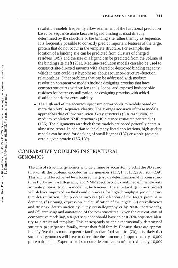
ERRORS IN COMPARATIVE MODELS
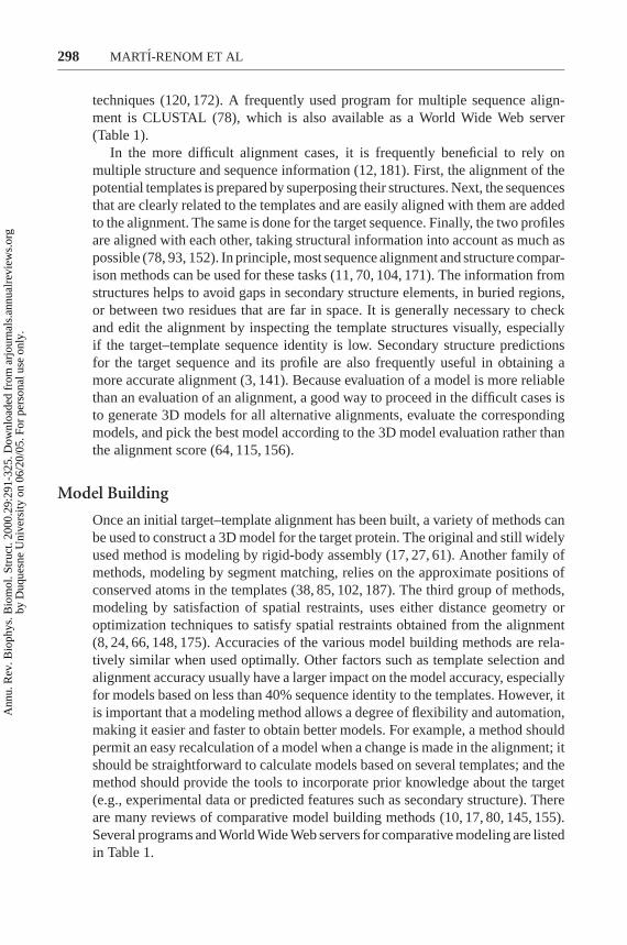
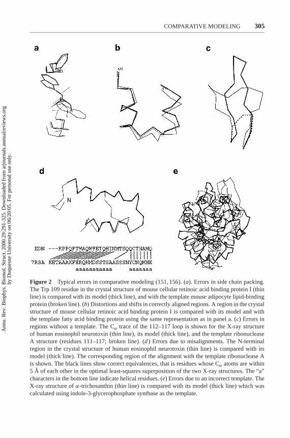
As the similarity between the target and the templates decreases, the errors in themodel increase. Errors in comparative models can be divided into five categories(151, 156) (Figure 2):
• Errors in side-chain packing. As the sequences diverge, the packing of sidechains in the protein core changes. Sometimes even the conformation ofidentical side chains is not conserved, a pitfall for many comparativemodeling methods. Side-chain errors are critical if they occur in regionsthat are involved in protein function, such as active sites andligand-binding sites.
• Distortions and shifts in correctly aligned regions. As a consequence ofsequence divergence, the main chain conformation changes, even if theoverall fold remains the same. Therefore, it is possible that in somecorrectly aligned segments of a model, the template is locally different(<3 A) from the target, resulting in errors in that region. The structuraldifferences are sometimes not due to differences in sequence but are aconsequence of artifacts in structure determination or structuredetermination in different environments (e.g., packing of subunits in acrystal). The simultaneous use of several templates can minimize this kindof error (156, 174).
• Errors in regions without a template. Segments of the target sequence thathave no equivalent region in the template structure (i.e., insertions orloops) are the most difficult regions to model. If the insertion is relativelyshort, less than 9 residues long, some methods can correctly predict theconformation of the backbone (191). Conditions for successful predictionare the correct alignment and an accurately modeled environmentsurrounding the insertion.
• Errors due to misalignments. The largest source of errors in comparativemodeling are misalignments, especially when the target–template sequenceidentity decreases below 30%. However, alignment errors can beminimized in two ways. First, it is usually possible to use a large number ofsequences to construct a multiple alignment, even if most of thesesequences do not have known structures. Multiple alignments are generallymore reliable than pairwise alignments (12, 181). The second way ofimproving the alignment is to iteratively modify those regions in thealignment that correspond to predicted errors in the model (156).
• Incorrect templates. This is a potential problem when distantly relatedproteins are used as templates (i.e., less than 25% sequence identity).Distinguishing between a model based on an incorrect template and amodel based on an incorrect alignment with a correct template is difficult.In both cases, the evaluation methods will predict an unreliable model. The
Ann
u. R
ev. B
ioph
ys. B
iom
ol. S
truc
t. 20
00.2
9:29
1-32
5. D
ownl
oade
d fr
om a
rjou
rnal
s.an
nual
revi
ews.
org
by D
uque
sne
Uni
vers
ity o
n 06
/20/
05. F
or p
erso
nal u
se o
nly.
P1: FPX/FOZ/fop/fok P2: FHN/FDR/fgi QC: FhN/fgm T1: FhN
April 6, 2000 16:12 Annual Reviews AR098-11
?COMPARATIVE MODELING 305
Figure 2 Typical errors in comparative modeling (151, 156). (a). Errors in side chain packing.The Trp 109 residue in the crystal structure of mouse cellular retinoic acid binding protein I (thinline) is compared with its model (thick line), and with the template mouse adipocyte lipid-bindingprotein (broken line). (b) Distortions and shifts in correctly aligned regions. A region in the crystalstructure of mouse cellular retinoic acid binding protein I is compared with its model and withthe template fatty acid binding protein using the same representation as in panel a. (c) Errors inregions without a template. The Cα trace of the 112–117 loop is shown for the X-ray structureof human eosinophil neurotoxin (thin line), its model (thick line), and the template ribonucleaseA structure (residues 111–117; broken line). (d) Errors due to misalignments. The N-terminalregion in the crystal structure of human eosinophil neurotoxin (thin line) is compared with itsmodel (thick line). The corresponding region of the alignment with the template ribonuclease Ais shown. The black lines show correct equivalences, that is residues whose Cα atoms are within5 A of each other in the optimal least-squares superposition of the two X-ray structures. The “a”characters in the bottom line indicate helical residues. (e) Errors due to an incorrect template. TheX-ray structure ofα-trichosanthin (thin line) is compared with its model (thick line) which wascalculated using indole-3-glycerophosphate synthase as the template.
Ann
u. R
ev. B
ioph
ys. B
iom
ol. S
truc
t. 20
00.2
9:29
1-32
5. D
ownl
oade
d fr
om a
rjou
rnal
s.an
nual
revi
ews.
org
by D
uque
sne
Uni
vers
ity o
n 06
/20/
05. F
or p
erso
nal u
se o
nly.
P1: FPX/FOZ/fop/fok P2: FHN/FDR/fgi QC: FhN/fgm T1: FhN
April 6, 2000 16:12 Annual Reviews AR098-11
?306 MARTI-RENOM ET AL
conservation of the key functional or structural residues in the targetsequence increases the confidence in a given fold assignment.
Comparative modeling has been criticized for its inability to provide a finalmodel closer to the target-experimental structure than the template used to generatethe model (108). This is only the case when there are errors in the template–targetalignment used for modeling. When the evaluation of the template–target similarityis based on the template–target alignment used for modeling the model is generallycloser to the target structure than is any of the templates (156).
An informative way to test protein structure modeling methods as well as themodelers using them is provided by the biannual meetings on Critical Assessmentof Techniques for Protein Structure Prediction (CASP) (92, 118). The last meetingwas held in December of 1998 and is summarized in the special issue ofProteinsSuppl. 3, 1999 (212). Protein modelers are challenged to model sequences withunknown 3D structure and to submit their models to the organizers before themeeting. At the same time, the 3D structures of the prediction targets are being de-termined by X-ray crystallography or NMR methods. They only become availableafter the models are calculated and submitted. Thus, a bona fide evaluation of pro-tein structure modeling methods is possible. An important extension of the CASPmeetings is a completely automated and online evaluation of protein structuremodeling servers on the World Wide Web. The idea has so far been implementedin the threading category only (52). It is likely to be extended to other kinds ofstructural prediction.
EVALUATION OF MODELS
The quality of the predicted model determines the information that can be ex-tracted from it. Thus, estimating the accuracy of 3D protein models is essentialfor interpreting them. The model can be evaluated as a whole as well as in indi-vidual regions. There are many model evaluation programs and servers (97, 197)(Table 1).
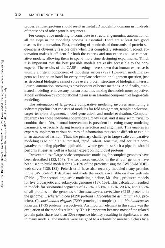
The first step in model evaluation is to assess if the model has the correctfold (157). A model will have the correct fold if the correct template is pickedand if that template is aligned at least approximately correctly with the targetsequence. The fold of a model can be assessed by a high sequence similarity withthe closest template, an energy based Z-score (157, 169), or by conservation of thekey functional or structural residues in the target sequence.
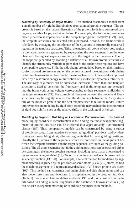
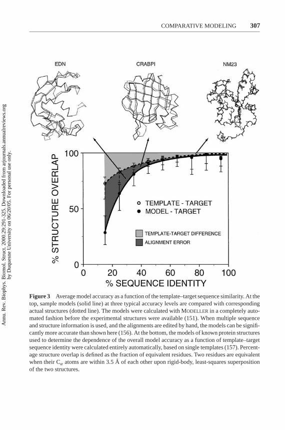
Once the fold of a model is assessed, a more detailed evaluation of the overallmodel accuracy can be obtained based on the similarity between the target and tem-plate sequences (Figure 3) (157). Sequence identity above 30% is a relatively goodpredictor of the expected accuracy. The reasons are the well-known relationshipbetween structural and sequence similarities of two proteins (34), the ‘geometrical’nature of modeling that forces the model to be as close to the template as possible
Ann
u. R
ev. B
ioph
ys. B
iom
ol. S
truc
t. 20
00.2
9:29
1-32
5. D
ownl
oade
d fr
om a
rjou
rnal
s.an
nual
revi
ews.
org
by D
uque
sne
Uni
vers
ity o
n 06
/20/
05. F
or p
erso
nal u
se o
nly.
P1: FPX/FOZ/fop/fok P2: FHN/FDR/fgi QC: FhN/fgm T1: FhN
April 6, 2000 16:12 Annual Reviews AR098-11
?COMPARATIVE MODELING 307
Figure 3 Average model accuracy as a function of the template–target sequence similarity. At thetop, sample models (solid line) at three typical accuracy levels are compared with correspondingactual structures (dotted line). The models were calculated with MODELLER in a completely auto-mated fashion before the experimental structures were available (151). When multiple sequenceand structure information is used, and the alignments are edited by hand, the models can be signifi-cantly more accurate than shown here (156). At the bottom, the models of known protein structuresused to determine the dependence of the overall model accuracy as a function of template–targetsequence identity were calculated entirely automatically, based on single templates (157). Percent-age structure overlap is defined as the fraction of equivalent residues. Two residues are equivalentwhen their Cα atoms are within 3.5A of each other upon rigid-body, least-squares superpositionof the two structures.
Ann
u. R
ev. B
ioph
ys. B
iom
ol. S
truc
t. 20
00.2
9:29
1-32
5. D
ownl
oade
d fr
om a
rjou
rnal
s.an
nual
revi
ews.
org
by D
uque
sne
Uni
vers
ity o
n 06
/20/
05. F
or p
erso
nal u
se o
nly.
P1: FPX/FOZ/fop/fok P2: FHN/FDR/fgi QC: FhN/fgm T1: FhN
April 6, 2000 16:12 Annual Reviews AR098-11
?308 MARTI-RENOM ET AL
(148), and the inability of any current modeling procedure to recover from anincorrect alignment (156). The dispersion of the model–target structural overlapincreases with the decrease in sequence identity. If the target–template sequenceidentity falls below 30%, the sequence identity becomes unreliable as a measureof expected accuracy of a single model. Models that deviate significantly from theaverage accuracy are frequent. It is in such cases that model evaluation methodsare particularly useful.
In addition to the target–template sequence similarity, the environment canstrongly influence the accuracy of a model. For instance, some calcium-bindingproteins undergo large conformational changes when bound to calcium. If a cal-cium-free template is used to model the calcium-bound state of the target, it is likelythat the model will be incorrect irrespective of the target–template similarity oraccuracy of the template structure (126). This also applies to the experimental de-termination of protein structure; a structure must be determined in the functionallymeaningful environment.
A basic requirement for a model is to have good stereochemistry. Some usefulprograms for evaluating stereochemistry are PROCHECK (96), PROCHECK-NMR (98), AQUA (98), SQUID (121), and WHATCHECK (72). The features ofa model that are checked by these programs include bond lengths, bond angles,peptide bond and side-chain ring planarities, chirality, main-chain and side-chaintorsion angles, and clashes between nonbonded pairs of atoms.
Distributions of many spatial features have been compiled from high resolutionprotein structures, and large deviations from the most likely values have been in-terpreted as strong indicators of errors in the model. Such features include packing(62), formation of a hydrophobic core (31), residue and atomic solvent acces-sibilities (90), spatial distribution of charged groups (32), distribution of atom–atom distances (40), atomic volumes (134), and main-chain hydrogen bonding(96).
There are also methods for testing 3D models that implicitly take into accountmany of the criteria listed above. These methods are based on 3D profiles and sta-tistical potentials of mean force (105, 168). Programs implementing this approachinclude VERIFY3D (105), PROSAII (169), HARMONY (184), and ANOLEA(113). The programs evaluate the environment of each residue in a model with re-spect to the expected environment as found in the high-resolution X-ray structures.There is a concern about the theoretical validity of the energy profiles for detect-ing regional errors in models. It is likely that the contributions of the individualresidues to the overall free energy of folding vary widely, even when normalizedby the number of atoms or interactions made. If this is correct, the correlationbetween the prediction errors and energy peaks is greatly weakened, resulting inthe loss of predictive power of the energy profile. Despite these concerns, errorprofiles have been useful in some applications (115).
Recently, a physics-based approach to deriving energy functions has beentested for use in protein structure evaluation. Lazaridis & Karplus (99) used an
Ann
u. R
ev. B
ioph
ys. B
iom
ol. S
truc
t. 20
00.2
9:29
1-32
5. D
ownl
oade
d fr
om a
rjou
rnal
s.an
nual
revi
ews.
org
by D
uque
sne
Uni
vers
ity o
n 06
/20/
05. F
or p
erso
nal u
se o
nly.
P1: FPX/FOZ/fop/fok P2: FHN/FDR/fgi QC: FhN/fgm T1: FhN
April 6, 2000 16:12 Annual Reviews AR098-11
?COMPARATIVE MODELING 309
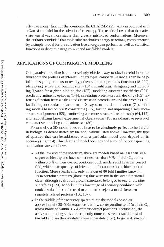
effective energy function that combined the CHARMM (25) vacuum potential witha Gaussian model for the solvation free energy. The results showed that the nativestate was always more stable than grossly misfolded conformations. Moreover,the authors concluded that molecular mechanics energy functions, complementedby a simple model for the solvation free energy, can perform as well as statisticalfunctions in discriminating correct and misfolded models.
APPLICATIONS OF COMPARATIVE MODELING
Comparative modeling is an increasingly efficient way to obtain useful informa-tion about the proteins of interest. For example, comparative models can be help-ful in designing mutants to test hypotheses about a protein’s function (18, 200),identifying active and binding sites (164), identifying, designing and improv-ing ligands for a given binding site (137), modeling substrate specificity (201),predicting antigenic epitopes (149), simulating protein–protein docking (189), in-ferring function from a calculated electrostatic potential around the protein (109),facilitating molecular replacement in X-ray structure determination (74), refin-ing models based on NMR constraints (116), testing and improving a sequence–structure alignment (199), confirming a remote structural relationship (64, 115),and rationalizing known experimental observations. For an exhaustive review ofcomparative modeling applications see (80).
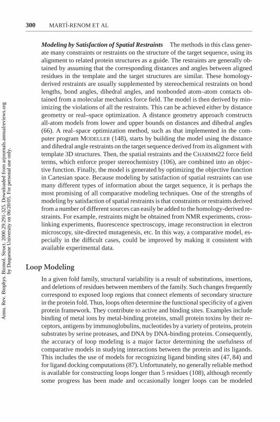
Fortunately, a 3D model does not have to be absolutely perfect to be helpfulin biology, as demonstrated by the applications listed above. However, the typeof question that can be addressed with a particular model does depend on itsaccuracy (Figure 4). Three levels of model accuracy and some of the correspondingapplications are as follows.
• At the low end of the spectrum, there are models based on less than 30%sequence identity and have sometimes less than 50% of their Cα atomswithin 3.5A of their correct positions. Such models still have the correctfold, which is frequently sufficient to predict approximate biochemicalfunction. More specifically, only nine out of 80 fold families known in1994 contained proteins (domains) that were not in the same functionalclass, although 32% of all protein structures belonged to one of the ninesuperfolds (123). Models in this low range of accuracy combined withmodel evaluation can be used to confirm or reject a match betweenremotely related proteins (156, 157).
• In the middle of the accuracy spectrum are the models based onapproximately 30–50% sequence identity, corresponding to 85% of the Cα
atoms modeled within 3.5A of their correct positions. Fortunately, theactive and binding sites are frequently more conserved than the rest ofthe fold and are thus modeled more accurately (157). In general, medium
Ann
u. R
ev. B
ioph
ys. B
iom
ol. S
truc
t. 20
00.2
9:29
1-32
5. D
ownl
oade
d fr
om a
rjou
rnal
s.an
nual
revi
ews.
org
by D
uque
sne
Uni
vers
ity o
n 06
/20/
05. F
or p
erso
nal u
se o
nly.
P1: FPX/FOZ/fop/fok P2: FHN/FDR/fgi QC: FhN/fgm T1: FhN
April 6, 2000 16:12 Annual Reviews AR098-11
?310 MARTI-RENOM ET AL
Figure 4 Applications of comparative modeling. The potential uses of a comparative modeldepend on its accuracy. This in turn depends significantly on the sequence identity between thetarget and the template structure on which the model was based. Sample models from Figure 3 areshown on the right.
Ann
u. R
ev. B
ioph
ys. B
iom
ol. S
truc
t. 20
00.2
9:29
1-32
5. D
ownl
oade
d fr
om a
rjou
rnal
s.an
nual
revi
ews.
org
by D
uque
sne
Uni
vers
ity o
n 06
/20/
05. F
or p
erso
nal u
se o
nly.
P1: FPX/FOZ/fop/fok P2: FHN/FDR/fgi QC: FhN/fgm T1: FhN
April 6, 2000 16:12 Annual Reviews AR098-11
?COMPARATIVE MODELING 311
resolution models frequently allow refinement of the functional predictionbased on sequence alone because ligand binding is most directlydetermined by the structure of the binding site rather than by its sequence.It is frequently possible to correctly predict important features of the targetprotein that do not occur in the template structure. For example, thelocation of a binding site can be predicted from clusters of chargedresidues (109), and the size of a ligand can be predicted from the volume ofthe binding site cleft (201). Medium-resolution models can also be used toconstruct site-directed mutants with altered or destroyed binding capacity,which in turn could test hypotheses about sequence–structure–functionrelationships. Other problems that can be addressed with mediumresolution comparative models include designing proteins that havecompact structures without long tails, loops, and exposed hydrophobicresidues for better crystallization; or designing proteins with addeddisulfide bonds for extra stability.
• The high end of the accuracy spectrum corresponds to models based onmore than 50% sequence identity. The average accuracy of these modelsapproaches that of low resolution X-ray structures (3A resolution) ormedium resolution NMR structures (10 distance restraints per residue)(156). The alignments on which these models are based generally containalmost no errors. In addition to the already listed applications, high qualitymodels can be used for docking of small ligands (137) or whole proteinsonto a given protein (186, 189).
COMPARATIVE MODELING IN STRUCTURALGENOMICS
The aim of structural genomics is to determine or accurately predict the 3D struc-ture of all the proteins encoded in the genomes (117, 147, 182, 202, 207–209).This aim will be achieved by a focused, large-scale determination of protein struc-tures by X-ray crystallography and NMR spectroscopy, combined efficiently withaccurate protein structure modeling techniques. The structural genomics projectwill deliver improved methods and a process for high-throughput protein struc-ture determination. The process involves (a) selection of the target proteins ordomains, (b) cloning, expression, and purification of the targets, (c) crystallizationand structure determination by X-ray crystallography or by NMR spectroscopy,and (d) archiving and annotation of the new structures. Given the current state ofcomparative modeling, a target sequence should have at least 30% sequence iden-tity to a structural template. This corresponds to one experimentally determinedstructure per sequence family, rather than fold family. Because there are approx-imately five times more sequence families than fold families (70), it is likely thatstructural genomics will have to determine the structure of approximately 10,000protein domains. Experimental structure determination of approximately 10,000
Ann
u. R
ev. B
ioph
ys. B
iom
ol. S
truc
t. 20
00.2
9:29
1-32
5. D
ownl
oade
d fr
om a
rjou
rnal
s.an
nual
revi
ews.
org
by D
uque
sne
Uni
vers
ity o
n 06
/20/
05. F
or p
erso
nal u
se o
nly.
P1: FPX/FOZ/fop/fok P2: FHN/FDR/fgi QC: FhN/fgm T1: FhN
April 6, 2000 16:12 Annual Reviews AR098-11
?312 MARTI-RENOM ET AL
properly chosen proteins should result in useful 3D models for domains in hundredsof thousands of other protein sequences.
For comparative modeling to contribute to structural genomics, automation ofall the steps in the modeling process is essential. There are at least five goodreasons for automation. First, modeling of hundreds of thousands of protein se-quences is obviously feasible only when it is completely automated. Second, au-tomation makes it efficient for both the experts and non-experts to use compar-ative models, allowing them to spend more time designing experiments. Third,it is important that the best possible models are easily accessible to the non-experts. The results of the CASP meetings have shown that human expertise isusually a critical component of modeling success (92). However, modeling ex-perts will not be on hand for every template selection or alignment question, justas structural biologists cannot solve every protein structure of biological interest.Fourth, automation encourages development of better methods. And finally, auto-mated modeling removes any human bias, thus making the models more objective.Model evaluation by computational means is an essential component of large-scalemodeling.
The automation of large-scale comparative modeling involves assembling asoftware pipeline that consists of modules for fold assignment, template selection,target–template alignment, model generation, and model evaluation. Computerprograms for these individual operations already exist, and it may seem trivial tocombine them. Yet, manual intervention is prevalent in setting many modelingparameters, especially during template selection and alignment. This enables anexpert to implement various sources of information that can be difficult to exploitin an automated fashion. Thus, the primary challenge in large-scale comparativemodeling is to build an automated, rapid, robust, sensitive, and accurate com-parative modeling pipeline applicable to whole genomes; such a pipeline shouldperform at least as well as a human expert on individual proteins.
Two examples of large-scale comparative modeling for complete genomes havebeen described (132, 157). The sequences encoded in theE. coli genome havebeen used to build models for 10–15% of the proteins using the SWISS-MODELweb server (130, 132). Peitsch et al have also recently modeled many proteinsin the SWISS-PROT database and made the models available on their web site(Table 1). The second large-scale modeling pipeline, MODPIPE, produced modelsfor five procaryotic and eukaryotic genomes (157, 159). This calculation resultedin models for substantial segments of 17.2%, 18.1%, 19.2%, 20.4%, and 15.7%of all proteins in the genomes ofSaccharomyces cerevisiae(6218 proteins inthe genome);Escherichia coli(4290 proteins),Mycoplasma genitalium(468 pro-teins),Caenorhabditis elegans(7299 proteins, incomplete), andMethanococcusjanaschii(1735 proteins), respectively. An important element in this study was theevaluation of the model’s reliability. This is important because most of the relatedprotein pairs share less than 30% sequence identity, resulting in significant errorsin many models. The models were assigned to a reliable or unreliable class by a
Ann
u. R
ev. B
ioph
ys. B
iom
ol. S
truc
t. 20
00.2
9:29
1-32
5. D
ownl
oade
d fr
om a
rjou
rnal
s.an
nual
revi
ews.
org
by D
uque
sne
Uni
vers
ity o
n 06
/20/
05. F
or p
erso
nal u
se o
nly.
P1: FPX/FOZ/fop/fok P2: FHN/FDR/fgi QC: FhN/fgm T1: FhN
April 6, 2000 16:12 Annual Reviews AR098-11
?COMPARATIVE MODELING 313
procedure (157) that depends on the statistical potential function from PROSAII(169). This allowed the identification of those models that were likely to be basedon correct templates and approximately correct alignments. As a result, 236 yeastproteins lacking any prior structural information were assigned to a fold family;40 of these proteins had no prior functional annotations. A more precise evaluationwas used to calibrate the relationship between model accuracy and the percent-age of sequence identity on which the model was based (157). Almost half ofthe 1071 reliably modeled proteins in the yeast genome share more than approx-imately 35% sequence identity with their templates. All the alignments, models,and model evaluations are available in the MODBASEdatabase of comparative pro-tein structure models (159, 210). Most recently, the MODPIPEpipeline software hasbeen improved by using PSI-BLAST (6) for fold assignment, multiple templatesand sequences for target–template alignment, and a complex statistical potentialof mean force for model evaluation. This resulted in models for approximately17,000 proteins, covering substantial segments of 18–45% of the proteins in 12complete genomes (210).
Large-scale comparative modeling will extend opportunities to tackle a myr-iad of problems by providing many protein models for many genomes. A largedatabase of experimental structures leveraged by comparative models will arousequestions about protein evolution, such as the physical origins of protein structurestability and protein activity, regulatory differences among similar enzymes, andthe specificity and plasticity of ligand binding sites. Structural genomics will alsoaid in the process of drug design. A collection of experimentally determined com-plexes of proteins with their ligands, aligned with comparative models for the restof the family members, will permit a facile comparison of ligand binding require-ments and also reveal permitted substitutions in and around important residues.Structural genomics will provide an obvious resource for many questions and,hopefully, will provoke new ones.
A specific example of a new opportunity for tackling existing problems byvirtue of providing many protein models from many genomes is the selection ofa target protein for drug development. A protein that is likely to have high ligandspecificity is a good choice; specificity is important because specific drugs areless likely to be toxic. Large-scale modeling facilitates imposing the specificityfilter in target selection by enabling a structural comparison of the ligand bindingsites of many proteins, from human or other organisms. Such comparisons maymake it possible to rationally select a target whose binding site is structurally mostdifferent from the binding sites of all the other proteins that may potentially reactwith the same drug. For example, when a human pathogenic organism needs to beinhibited, it may be possible to select a pathogen target that is structurally mostdifferent from all the human homologs. Alternatively, when a human metabolicpathway needs to be regulated, the target identification could focus on the particularprotein in the pathway that has the binding site most dissimilar from its humanhomologs.
Ann
u. R
ev. B
ioph
ys. B
iom
ol. S
truc
t. 20
00.2
9:29
1-32
5. D
ownl
oade
d fr
om a
rjou
rnal
s.an
nual
revi
ews.
org
by D
uque
sne
Uni
vers
ity o
n 06
/20/
05. F
or p
erso
nal u
se o
nly.
P1: FPX/FOZ/fop/fok P2: FHN/FDR/fgi QC: FhN/fgm T1: FhN
April 6, 2000 16:12 Annual Reviews AR098-11
?314 MARTI-RENOM ET AL
CONCLUSION
Over the past few years, there has been a gradual increase in both the accuracyof comparative models and the fraction of protein sequences that can be mod-eled with useful accuracy. The magnitude of errors in fold assignment, alignment,and the modeling of sidechains, loops, distortions, and rigid body shifts has de-creased measurably. This is a consequence of both better techniques and a largernumber of known protein sequences and structures. Nevertheless, all the errorsremain significant and demand future methodological improvements. In addition,there is a great need for more accurate detection of errors in a given protein struc-ture model. Error detection is useful both for refinement and interpretation of themodels.
It is now possible to predict by comparative modeling significant segments ofapproximately one third of all known protein sequences. One half of these modelsare in the least accurate class, based on less than 30% sequence identity to knownprotein structures. The remaining 35 and 15% of the models are in the medium(<50% sequence identity) and high (>50% identity) accuracy classes. The fractionof protein sequences that can be modeled by comparative modeling is currentlyincreasing by approximately 4% per year (158). It has been estimated that globularprotein domains cluster in only a few thousand fold families, approximately 900of which have already been structurally defined (70, 76). Assuming the currentgrowth rate in the number of known protein structures, the structure of at least onemember of most of the globular folds will be determined in less than 10 years (70).According to this argument, comparative modeling will be applicable to most ofthe globular protein domains soon after the expected completion of the humangenome project. However, there are some classes of proteins, such as membraneproteins, that will not be amenable to modeling without improvements in structuredetermination and modeling techniques. To maximize the number of proteins thatcan be modeled reliably, a concerted effort toward structure determination of newfolds by X-ray crystallography and nuclear magnetic resonance spectroscopy isin order, as envisioned by structural genomics (117, 147, 182, 202, 207–209). Thefull potential of the genome sequencing projects will only be realized once allprotein functions are assigned and understood. This will be facilitated by inte-grating genomic sequence information with databases arising from functional andstructural genomics. Comparative modeling will play an important bridging rolein these efforts.
ACKNOWLEDGMENTS
We are grateful to Dr. Azat Badretdinov and Mr. Eric Feyfant for many discussionsabout comparative protein structure modeling. MAM-R and AF are BurroughsWellcome Fellows. RS is a Howard Hughes Medical Institute predoctoral fellow.FM is a Norman and Rosita Winston Biomedical Research Foundation Fellow. ASis a Sinsheimer Scholar and an Alfred P. Sloan Research Fellow. The investigations
Ann
u. R
ev. B
ioph
ys. B
iom
ol. S
truc
t. 20
00.2
9:29
1-32
5. D
ownl
oade
d fr
om a
rjou
rnal
s.an
nual
revi
ews.
org
by D
uque
sne
Uni
vers
ity o
n 06
/20/
05. F
or p
erso
nal u
se o
nly.
P1: FPX/FOZ/fop/fok P2: FHN/FDR/fgi QC: FhN/fgm T1: FhN
April 6, 2000 16:12 Annual Reviews AR098-11
?COMPARATIVE MODELING 315
have also been aided by grants from NIH (GM 54762) and NSF (BIR-9601845).This review is based on (55, 56, 145, 146, 154, 156, 158).
Visit the Annual Reviews home page at www.AnnualReviews.org
LITERATURE CITED
1. Abola EE, Bernstein FC, Bryant SH, Koet-zle TF, Weng J. 1987. Protein data bank. InCrystallographic Databases—Information,Content, Software Systems, Scientific Appli-cations, ed. FH Allen, G Bergerhoff, R Siev-ers, pp. 107–132. Bonn/Cambridge/Chester.Data Commission Int. Union of Crystallog-raphy.
2. Alexandrov NN, Nussinov R, Zimmer RM.1995. Fast protein fold recognition via se-quence to structure alignment and contactcapacity potentials. InPacific Symposium onBiocomputing ’96, ed. L Hunter, TE Klein,pp. 53–72, Singapore: World Sci. Pub.
3. Aloy P, Mas JM, Mart´ı-Renom MA, QuerolE, Aviles FX, Oliva B. 2000. Human a2pro-carboxypeptidase model: secondarystructure prediction as a powerful toolfor homology modelling improvement.J.Computer-Aided Molec. Design.14:83–92
4. Altschul SF, Boguski MS, Gish W, Woot-ton JC. 1994. Issues in searching molecularsequence databases.Nat. Genet.6:119–29
5. Altschul SF, Gish W, Miller W, Myers EW,Lipman DJ. 1990. Basic local alignmentsearch tool.J. Mol. Biol.215:403–10
6. Altschul SF, Madden TL, Schaffer AA,Zhang J Zhang, Miller W, Lipman DJ.1997. Gapped BLAST and PSI-BLAST: anew generation of protein database searchprograms. Nucleic Acids Res.25:3389–402
7. Apostolico A, Giancarlo R. 1998. Sequencealignment in molecular biology.J. Comput.Biol. 5:173–96
8. Aszodi A, Taylor WR. 1996. Homologymodelling by distance geometry.FoldingDesign1:325–34
9. Bairoch A, Apweiler R. 1999. The SWISS-PROT protein sequence data bank and its
supplement TrEMBL in 1999.Nucleic AcidsRes.27:49–54
10. Bajorath J, Stenkamp R, Aruffo A. 1994.Knowledge-based model building of pro-teins: Concepts and examples.Protein Sci.2:1798–810
11. Barton GJ. 1998. Protein sequence align-ment and database scanning. InProteinStructure Prediction: A Practical Approach,ed. MJE Sternberg. Oxford, UK: OxfordUniv. Press
12. Barton GJ, Sternberg MJE. 1987. A strat-egy for the rapid multiple alignment of pro-tein sequences; confidence levels from ter-tiary structure comparisons.J. Mol. Biol.198:327–37
13. Bassolino-Klimas D, Bruccoleri RE. 1992.Application of a directed conformationalsearch for generating 3-D coordinates forprotein structures fromα-carbon coordi-nates.Proteins14:465–74
14. Baxevanis AD. 1998. Practical aspects ofmultiple sequence alignment.Methods Bio-chem. Anal.39:172–88
15. Benson DA, Karsch-Mizrachi I, Lipman DJ,Ostell J, Rapp BA, Wheeler DL. 2000. Gen-Bank.Nucleic Acid Res.28:15–18
16. Bernstein FC, Koetzle TF, Williams GJB,Meyer EF, Brice MD, Rodgers JR, Ken-nard O, Shimanouchi T, Tasumi M. 1977.The protein data bank: a computer-basedarchival file for macromolecular structures.J. Mol. Biol.112:535–42
17. Blundell TL, Sibanda BL, Sternberg MJE,Thornton JM. 1987. Knowledge-based pre-diction of protein structures and the de-sign of novel molecules.Nature 326:347–52
18. Boissel JP, Lee WR, Presnell SR, CohenFE, Bunn HF. 1993. Erythropoietin
Ann
u. R
ev. B
ioph
ys. B
iom
ol. S
truc
t. 20
00.2
9:29
1-32
5. D
ownl
oade
d fr
om a
rjou
rnal
s.an
nual
revi
ews.
org
by D
uque
sne
Uni
vers
ity o
n 06
/20/
05. F
or p
erso
nal u
se o
nly.
P1: FPX/FOZ/fop/fok P2: FHN/FDR/fgi QC: FhN/fgm T1: FhN
April 6, 2000 16:12 Annual Reviews AR098-11
?316 MARTI-RENOM ET AL
structure-function relationships. Mutantproteins that test a model of tertiary struc-ture.J. Biol. Chem.268:15983–93
19. Bower MJ, Cohen FE, Dunbrack RL Jr.1997. Prediction of protein side-chain ro-tamers from a backbone-dependent ro-tamer library: a new homology modelingtool. J. Mol. Biol.267:1268–82
20. Bowie JU, Luthy R, Eisenberg D. 1991. Amethod to identify protein sequences thatfold into a known three-dimensional struc-ture.Science253:164–70
21. Brenner SE, Barken D, Levitt M. 1999. ThePRESAGEdatabase for structural genomics.Nucleic Acids Res.27:251–53
22. Brenner SE, Chothia C, Hubbard TJ. 1998.Assessing sequence comparison methodswith reliable structurally identified dis-tant evolutionary relationships.Proc. Natl.Acad. Sci. USA95:6073–78
23. Briffeuil P, Baudoux G, Lambert C, BolleX De, Vinals C, Feytmans E, Depiereux E.1998. Comparative analysis of seven mul-tiple protein sequence alignment servers:clues to enhance reliability of predictions.Bioinformatics14:357–66
24. Brocklehurst SM, Perham RN. 1993. Pre-diction of the three-dimensional structuresof the biotinylated domain from yeast pyru-vate carboxylase and of the lipolyated H-protein from the pea leaf glycine cleavagesystem: a new automated methods for theprediction of protein tertiary structure.Pro-tein Sci.2:626–39
25. Brooks BR, Bruccoleri RE, Olafson BD,States DJ, Swaminathan S, Karplus M.1983. CHARMM: a program for macro-molecular energy minimization and dy-namics calculations.J. Comp. Chem.4:187–217
26. Brower RC, Vasmatzis G, Silverman M,DeLisi C. 1993. Exhaustive conforma-tional search and simulated annealing formodels of lattice peptides.Biopolymers33:329–34
27. Browne WJ, North ACT, Phillips DC, BrewK, Vanaman TC, Hill RC. 1969. A possi-
ble three-dimensional structure of bovineα-lactalbumin based on that of hen’s egg-white lysozyme.J. Mol. Biol.42:65–86
28. Bruccoleri BR, Karplus M. 1990. Con-formational sampling using high temper-ature molecular dynamics.Biopolymers29:1847–62
29. Bruccoleri RE. 1993. Application of sys-tematic conformational search to proteinmodeling.Molec. Simulat.10:151–74
30. Bruccoleri RE, Karplus M. 1987. Predic-tion of the folding of short polypeptidesegments by uniform conformational sam-pling. Biopolymers26:137–68
31. Bryant SH, Amzel LM. 1987. Correctlyfolded proteins make twice as many hy-drophobic contacts.Int. J. Peptide ProteinRes.29:46–52
32. Bryant SH, Lawrence CE. 1991. The fre-quency of ion-pair substructures in proteinsis quantitatively related to electrostatic po-tential: a statistical model for nonbondedinteractions.Proteins9:108–19
33. Carlacci L, Englander SW. 1996. Loopproblem in proteins: developments onthe Monte Carlo simulated annealing ap-proach.Comp. Chem.17:1002–12
34. Chothia C, Lesk AM. 1986. The relationbetween the divergence of sequence andstructure in proteins.EMBO J.5:823–26
35. Chothia C, Lesk AM. 1987. Canonicalstructures for the hypervariable regions ofimmunoglobulins.J. Mol. Biol. 196:901–17
36. Chothia C, Lesk AM, Tramontano A,Levitt M, Smith-Gill SJ, Air G, SheriffS, Padlan EA, Davies D, Tulip WR, Col-man PM, Spinelli S, Alzari PM, PoljakRJ. 1989. Conformation of immunoglobu-lin hypervariable regions.Nature342:877–83
37. Chung SY, Subbiah S. 1996. A structuralexplanation for the twilight zone of proteinsequence homology.Structure 4:1123–27
38. Claessens M, Cutsem EV, Lasters I, WodakS. 1989. Modelling the polypeptide back-
Ann
u. R
ev. B
ioph
ys. B
iom
ol. S
truc
t. 20
00.2
9:29
1-32
5. D
ownl
oade
d fr
om a
rjou
rnal
s.an
nual
revi
ews.
org
by D
uque
sne
Uni
vers
ity o
n 06
/20/
05. F
or p
erso
nal u
se o
nly.
P1: FPX/FOZ/fop/fok P2: FHN/FDR/fgi QC: FhN/fgm T1: FhN
April 6, 2000 16:12 Annual Reviews AR098-11
?COMPARATIVE MODELING 317
bone with ‘spare parts’ from known proteinstructures.Protein Eng.4:335–45
39. Collura V, Higo J, Garnier J. 1993. Model-ing of protein loops by simulated anneal-ing. Protein Sci.2:1502–10
40. Colovos C, Yeates TO. 1993. Verificationof protein structures: patterns of non-bonded atomic interactions.Protein Sci.2:1511–19
41. Corpet F. 1988. Multiple sequence align-ment with hierarchical clustering.NucleicAcids Res.16:10881–90
42. Cregut D, Liautard J-P, Chiche L. 1994.Homology modeling of annexin I: implicitsolvation improves side-chain predictionand combination of evaluation criteria al-lows recognition of different types of con-formational error.Protein Eng.7:1333–44
43. Dudek MJ, Ramnarayan K, Ponder JW.1998. Protein structure prediction using acombination of sequence homology andglobal energy minimization: II. Energyfunctions.J. Comp. Chem.19:548–73
44. Dunbrack RL, Karplus M. 1993. Predictionof protein side-chain conformations from abackbone conformation dependent rotamerlibrary. J. Mol. Biol.230:543–71
45. Evans JS, Mathiowetz AM, Chan SI, God-dard WA III. 1995. De novo prediction ofpolypeptide conformations using dihedralprobability grid Monte Carlo methodology.Protein Sci.4:1203–16
46. Felsenstein J. 1985. Confidence limits onphylogenies: an approach using the boot-strap.Evolution39:783–91
47. Fetrow JS, Godzik A, Skolnick J. 1998.Functional analysis of theEscherichiacoli genome using the sequence-to-struc-ture-to-function paradigm: identificationof proteins exhibiting the glutaredoxin/thioredoxin disulfide oxidoreductase activ-ity. J. Mol. Biol.282:703–11
48. Fidelis K, Stern PS, Bacon D, Moult J.1994. Comparison of systematic searchand database methods for constructing seg-ments of protein structure.Protein Eng.
7:953–6049. Filippis V De, Sander C, Vriend G. 1994.
Predicting local structural changes that re-sult from point mutations.Protein Eng.7:1203–8
50. Fine RM, Wang H, Shenkin PS, YarmushDL, Levinthal C. 1986. Predicting antibodyhypervariable loop conformations. II: Min-imization and molecular dynamics studiesof MCP603 from many randomly gener-ated loop conformations.Proteins1:342–62
51. Finkelstein AV, Reva BA. 1992. Search forthe stable state of a short chain in a molec-ular field.Protein Eng.5:617–24
52. Fischer D, Barret C, Bryson K, ElofssonA, Godzik A, Jones D, Karplus KJ, KelleyLA, MacCallum RM, Pawowski K, RostB, Rychlewski L, Sternberg MJE. 1999.CAFASP-1: critical assessment of fullyautomated structure prediction methods.Proteins.Suppl. 3, pp. 209–17
53. Fischer D, Eisenberg D. 1996. Fold recog-nition using sequence-derived predictions.Protein Sci.5:947–55
54. Fischer D, Eisenberg D. 1997. Assign-ing folds to the proteins encoded by thegenome ofMycoplasma genitalium. Proc.Natl. Acad. Sci. USA94:11929–34
55. Fiser A, Do RKG,Sali A. 2000. Modelingof loops in protein structures. Submitted
56. Fiser A, S´anchez R, Melo F,Sali A. 2000.Comparative protien structure modeling.In Computational Biochemistry and Bio-physics, ed. M Watanabe, B Roux, AMacKerell, O Backer. New York: MarcelDekker. In press
57. Flockner H, Braxenthaler M, Lackner P,Jaritz M, Ortner M, Sippl MJ. 1995. Pro-gress in fold recognition.Proteins23:376–86
58. Gerstein M, Levitt M. 1997. A structuralcensus of the current population of pro-tein sequences.Proc. Natl. Acad. Sci. USA94:11911–16
59. Godzik A, Kolinski A, Skolnick J. 1992.Topology fingerprint approach to the
Ann
u. R
ev. B
ioph
ys. B
iom
ol. S
truc
t. 20
00.2
9:29
1-32
5. D
ownl
oade
d fr
om a
rjou
rnal
s.an
nual
revi
ews.
org
by D
uque
sne
Uni
vers
ity o
n 06
/20/
05. F
or p
erso
nal u
se o
nly.
P1: FPX/FOZ/fop/fok P2: FHN/FDR/fgi QC: FhN/fgm T1: FhN
April 6, 2000 16:12 Annual Reviews AR098-11
?318 MARTI-RENOM ET AL
inverse protein folding problem.J. Mol.Biol. 227:227–38
60. Greer J. 1980. Model for haptoglobinheavy chain based upon structural homol-ogy. Proc. Natl. Acad. Sci. USA77:3393–97
61. Greer J. 1990. Comparative modelling me-thods: application to the family of themammalian serine proteases.Proteins 7:317–34
62. Gregoret LM, Cohen FE. 1991. Effect ofpacking density on chain conformation.J.Mol. Biol. 219:109–22
63. Gribskov M. 1994. Profile analysis.Meth.Mol. Biol. 25:247–66
64. Guenther B, Onrust R,Sali A, O’DonnellM, Kuriyan J. 1997. Crystal structure of theδ′ subunit of the clamp-loader complex ofE. coli DNA polymerase III.Cell 91:335–45
65. Harbury PB, Tidor B, Kim PS. 1995.Repacking proteins cores with backbonefreedom: Structure prediction for coiledcoils.Proc. Natl. Acad. Sci. USA92:8408–12
66. Havel TF, Snow ME. 1991. A new methodfor building protein conformations fromsequence alignments with homologues ofknown structure.J. Mol. Biol.217:1–7
67. Henikoff S, Henikoff JG. 1994. Proteinfamily classification based on searching adatabase of blocks.Genomics19:97–107
68. Henikoff S, Henikoff JG, Alford WJ,Pietrokovski S. 1995. Automated construc-tion and graphical presentation of proteinblocks from unaligned sequences.Gene163:17–26
69. Holm L, Sander C. 1991. Database algo-rithm for generating protein backbone andside-chain co-ordinates from Cα trace: ap-plication to model building and detection ofco-ordinate errors.J. Mol. Biol. 218:183–94
70. Holm L, Sander C. 1996. Mapping the pro-tein universe.Science273:595–602
71. Holm L, Sander C. 1999. Protein foldsand families: sequence and structure align-
ments.Nucleic Acids Res.27:244–4772. Hooft RWW, Sander C, Vriend G. 1996.
Verification of protein structures: side-chain planarity.J. Appl. Crystallogr.29:714–16
73. Hooft RWW, Vriend G, Sander C, AbolaEE. 1996. Errors in protein structures.Na-ture381:272
74. Howell PL, Almo SC, Parsons MR, HajduJ, Petsko GA. 1992. Structure determina-tion of turkey egg-white lysozyme usingLaue diffraction data.Acta Crystallogr. B48:200–7
75. Huang ES, Koehl P, Levitt M, Pappu RV,Ponder JW. 1998. Accuracy of side-chainprediction upon near-native protein back-bones generated by ab initio folding meth-ods.Proteins33:204–17
76. Hubbard TJP, Ailey B, Brenner SE, MurzinAG, Chothia C. 1999. SCOP: A structuralclassification of proteins database.NucleicAcids Res.27:254–56
77. Huynen M, Doerks T, Eisenhaber F, Ore-ngo C, Sunyaev S, Yuan Y, Bork P. 1998.Homology-based fold predictions forMy-coplasma genitaliumproteins.J. Mol. Biol.280:323–26
78. Jeanmougin F, Thompson JD, Gouy M,Gibson DG, Higgins TJ. 1998. Multiplesequence alignment with CLUSTAL X.Trends Biochem. Sci.23:403–5
79. Johnson MS, Overington JP. 1993. A struc-tural basis for sequence comparisons: anevaluation of scoring methodologies.J.Mol. Biol. 233:716–38
80. Johnson MS, Srinivasan N, Sowdhamini R,Blundell TL. 1994. Knowledge-based pro-tein modelling.CRC Crit. Rev. Biochem.Mol. Biol. 29:1–68
81. Jones DT. 1999. GenTHREADER: An ef-ficient and reliable protein fold recogni-tion method for genomic sequences.J. Mol.Biol. 287:797–815
82. Jones DT, Taylor WR, Thornton JM. 1992.A new approach to protein fold recogni-tion. Nature358:86–89
83. Jones DT. 1997. Progress in protein struc-
Ann
u. R
ev. B
ioph
ys. B
iom
ol. S
truc
t. 20
00.2
9:29
1-32
5. D
ownl
oade
d fr
om a
rjou
rnal
s.an
nual
revi
ews.
org
by D
uque
sne
Uni
vers
ity o
n 06
/20/
05. F
or p
erso
nal u
se o
nly.
P1: FPX/FOZ/fop/fok P2: FHN/FDR/fgi QC: FhN/fgm T1: FhN
April 6, 2000 16:12 Annual Reviews AR098-11
?COMPARATIVE MODELING 319
ture prediction.Curr. Opin. Struct. Biol.7:377–87
84. Jones S, Thornton JM. 1997. Predictionof protein-protein interaction sites usingpatch analysis.J. Mol. Biol.272:133–43
85. Jones TH, Thirup S. 1986. Using knownsubstructures in protein model building andcrystallography.EMBO J.5:819–22
86. Jung S-H, Pastan I, Lee B. 1994. Designof interchain disulfide bonds in the frame-work region of the Fv fragment of themonoclonal antibody B3.Proteins19:35–47
87. Kick EK, Roe DC, Skillman AG, Liu G,Ewing TJ, Sun Y, Kuntz ID, Ellman JA.1997. Structure-based design and combi-natorial chemistry yield low nanomolar in-hibitors of cathepsin D.Chem. Biol.4:297–307
88. Kidera A. 1995. Enhanced conformationalsampling in Monte Carlo simulations ofproteins: applications to a constrained pep-tide.Proc. Natl. Acad. Sci. USA92:9886–89
89. Koehl P, Delarue M. 1994. Application of aself-consistent mean field theory to predictprotein side-chains conformation and esti-mate their conformational entropy.J. Mol.Biol. 239:249–75
90. Koehl P, Delarue M. 1994. Polar and non-polar atomic environments in the proteincore: implication for folding and binding.Proteins20:264–78
91. Koehl P, Delarue M. 1995. A self consis-tent mean field approach to simultaneousgap closure and side-chain positioning inprotein homology modelling.Nat. Struct.Biol. 2:163–70
92. Koehl P, Levitt M. 1999. A brighter futurefor protein structure prediction.Nat. Struct.Biol. 6:108–11
93. Koretke KK, Luthey-Schulten Z, WolynesPG. 1996. Self-consistently optimized sta-tistical mechanical energy functions for se-quence structure alignment.Protein Sci.5:1043–59
94. Krogh A, Brown M, Mian IS, Sjolander K,
Haussler D. 1994. Hidden Markov modelsin computational biology: applications toprotein modeling.J. Mol. Biol.235:1501–31
95. Lambert MH, Scheraga HA. 1989. Pat-tern recognition in the prediction of pro-tein structure. I. Tripeptide conformationalprobabilities calculated from the aminoacid sequence.J. Comp. Chem.10:770–97
96. Laskowski RA, McArthur MW, Moss DS,Thornton JM. 1993. PROCHECK: a pro-gram to check the stereochemical qualityof protein structures.J. Appl. Crystalogr.26:283–91
97. Laskowski RA, MacArthur MW, ThorntonJM. 1998. Validation of protein models de-rived from experiment.Curr. Opin. Struct.Biol. 5:631–39
98. Laskowski RA, Rullmann JAC, MacArthurMW, Kaptein R, Thornton JM. 1996.AQUA and PROCHECK-NMR: programsfor checking the quality of protein struc-tures solved by NMR.J. Biomol. NMR8:477–86
99. Lazaridis T, Karplus M. 1999. Discrimina-tion of the native from misfolded proteinmodels with an energy function includingimplicit solvation.J. Mol. Biol. 288:477–87
100. Lee C. 1995. Testing homology modelingon mutant proteins: predicting structuraland thermodynamic effects in the Ala98→Val mutants of T4 lysozyme.Folding De-sign1:1–12
101. Lesk AM, Chothia C. 1980. How differ-ent amino acid sequences determine sim-ilar protein structures: The structure andevolutionary dynamics of the globins.J.Mol. Biol. 136:225–70
102. Levitt M. 1992. Accurate modeling of pro-tein conformation by automatic segmentmatching.J. Mol. Biol.226:507–33
103. Levitt M. 1997. Competitive assessment ofprotein fold recognition and alignment ac-curacy.Proteins(Suppl.) 1:92–104
104. Levitt M, Gerstein M. 1998. A uni-fied statistical framework for sequence
Ann
u. R
ev. B
ioph
ys. B
iom
ol. S
truc
t. 20
00.2
9:29
1-32
5. D
ownl
oade
d fr
om a
rjou
rnal
s.an
nual
revi
ews.
org
by D
uque
sne
Uni
vers
ity o
n 06
/20/
05. F
or p
erso
nal u
se o
nly.
P1: FPX/FOZ/fop/fok P2: FHN/FDR/fgi QC: FhN/fgm T1: FhN
April 6, 2000 16:12 Annual Reviews AR098-11
?320 MARTI-RENOM ET AL
comparison and structure comparison.Proc. Natl. Acad. Sci. USA95:5913–20
105. Luthy R, Bowie JU, Eisenberg D. 1992.Assessment of protein models with three-dimensional profiles.Nature356:83–85
106. MacKerell AD, Bashford D, Bellott M,Dunbrack RL Jr., Evanseck JD, FieldMJ, Fischer S, Gao J, Guo H, Ha S,Joseph-McCarthy D, Kuchnir L, Kucz-era K, Lau FTK, Mattos C, Michnick S,Ngo T, Nguyen DT, Prodhom B, ReiherWE, Roux M., Smith JC, Stote J, Watan-abe M., Wiorkiewicz-Kuczera J, Yin D,Karplus M. 1998. All-atom empirical po-tential for molecular modeling and dy-namics studies of proteins.J. Phys. Chem.B 102:3586–16
107. Martin ACR, Cheetham JC, Rees AR.1989. Modeling antibody hypervariableloops: a combined algorithm.Proc. Natl.Acad. Sci. USA86:9268–72
108. Martin ACR, MacArthur MW, ThorntonJM. 1997. Assessment of comparativemodeling in CASP2.Proteins (Suppl.)1:14–28
109. Matsumoto R,Sali A, Ghildyal N,Karplus M, Stevens RL. 1995. Packag-ing of proteases and proteoglycans inthe granules of mast cells and otherhematopoietic cells. A cluster of his-tidines in mouse mast cell protease-7 reg-ulates its binding to heparin serglycin pro-teoglycan.J. Biol. Chem.270:19524–31
110. Mattos C, Petsko GA, Karplus M. 1994.Analysis of two-residue turns in proteins.J. Mol. Biol.238:733–47
111. McGarrah DB, Judson RS. 1993. Anal-ysis of the genetic algorithm method ofmolecular conformation determination.J.Comp. Chem.14:1385–95
112. McGregor MJ, Islam SA, Sternberg MJE.1987. Analysis of the relationship be-tween side-chain conformation and sec-ondary structure in globular proteins.J.Mol. Biol. 198:295–310
113. Melo F, Feytmans E. 1998. Assessing pro-tein structures with a non-local atomic in-
teraction energy.J. Mol. Biol.277:1141–52
114. Mezei M. 1998. Chameleon sequences inthe PDB.Protein Eng.11:411–14
115. Miwa JM, Ibanez-Tallon I, Crabtree GW,Sanchez R,Sali A, Role LW, Heintz N.1999. lynx1, an endogenous toxin-likemodulator of nicotinic acetylcholine re-ceptors in the mammalian CNS.Neuron23:105–14
116. Modi S, Paine MJ, Sutcliffe MJ, Lian L-Y,Primrose WU, Wolfe CR, Roberts GCK.1996. A model for human cytochromeP450 2D6 based on homology modelingand NMR studies of substrate binding.Biochemistry35:4540–50
117. Montelione GT, Anderson S. 1999. Struc-tural genomics: keystone for a humanproteome project.Nat. Struct. Biol.6:11–12
118. Moult J, Hubbard T, Bryant SH, FidelisK, Pedersen JT. 1997. Critical assessmentof methods of protein structure prediction(CASP): round II.Proteins(Suppl.) 1:2–6
119. Moult J, James MNG. 1986. An algo-rithm for determining the conformation ofpolypeptide segments in proteins by sys-tematic search.Proteins1:146–63
120. Needleman SB, Wunsch CD. 1970. Ageneral method applicable to the searchfor similarities in the amino acid sequenceof two proteins.J. Mol. Biol.48:443–53
121. Oldfield TJ. 1992. Squid: a program forthe analysis and display of data from crys-tallography and molecular dynamics.J.Mol. Graphics10:247–52
122. Oliva B, Bates PA, Querol E, Aviles FX,Sternberg MJE. 1997. An automated clas-sification of the structure of protein loops.J. Mol. Biol.266:814–30
123. Orengo CA, Jones DT, Thornton JM.1994. Protein superfamilies and domainsuperfolds.Nature372:631–34
124. Orengo CA, Pearl FMG, Bray JE, ToddAE, Martin AC, Conte L. Lo, ThorntonJM. 1999. The CATH database providesinsights into protein structure/function re-
Ann
u. R
ev. B
ioph
ys. B
iom
ol. S
truc
t. 20
00.2
9:29
1-32
5. D
ownl
oade
d fr
om a
rjou
rnal
s.an
nual
revi
ews.
org
by D
uque
sne
Uni
vers
ity o
n 06
/20/
05. F
or p
erso
nal u
se o
nly.
P1: FPX/FOZ/fop/fok P2: FHN/FDR/fgi QC: FhN/fgm T1: FhN
April 6, 2000 16:12 Annual Reviews AR098-11
?COMPARATIVE MODELING 321
lationship.Nucleic Acids Res.27:275–79125. Park J, Karplus K, Barret C, Hughey R,
Haussler D, Hubbard T, Chothia C. 1998.Sequence comparisons using multiple se-quences detect three times as many re-mote homologues as pairwise methods.J. Mol. Biol.284:1201–10
126. Pawlowski K, Bierzy´nski A, Godzik A.1996. Structural diversity in a familyof homologous proteins.J. Mol. Biol.258:349–66
127. Pearson WR. 1990. Rapid and sensi-tive comparison with FASTA and FASTP.Methods Enzymol.183:63–98
128. Pearson WR. 1995. Comparison of meth-ods for searching protein sequencedatabases.Protein Sci.4:1145–60
129. Pearson WR. 1998. Empirical statisticalestimates for sequence similaritysearches.J. Mol. Biol.276:71–84
130. Peitsch MC. 1996. PROMOD andSWISS-MODEL—Internet-based toolsfor automated comparative protein mod-eling.Biochem. Soc. Trans24:274–79
131. Peitsch MC, Jongeneel CV. 1993. A 3-Dmodel for the CD40 ligand predicts thatit is a compact trimer similar to the tumornecrosis factors.Int. Immunol.5:233–38
132. Peitsch MC, Wilkins MR, Tonella L,Sanchez JC, Appel RD, Hochstrasser DF.1997. Large-scale protein modelling andintegration with the SWISS-PROT andSWISS-2DPAGE databases: the exam-ple of Escherichia coli. Electrophoresis18:498–501
133. Petrella RJ, Lazardis T, Karplus M. 1998.Protein side chain conformer prediction:a test of the energy function.Folding De-sign3:353–77
134. Pontius J, Richelle J, Wodak SJ. 1996. De-viations from standard atomic volumes asa quality measure for protein crystal struc-tures.J. Mol. Biol.264:121–36
135. Rapp CS, Friesner RA. 1999. Predictionof loop geometries using a generalizedBorn model of solvation effect.Proteins35:173–83
136. Ring CS, Cohen FE. 1994. Conforma-tional sampling of loop structures usinggenetic algorithm.Isr. J. Chem.34:245–52
137. Ring CS, Sun E, McKerrow JH, Lee GK,Rosenthal PJ, Kuntz ID, Cohen FE. 1993.Structure-based inhibitor design by usingprotein models for the development of an-tiparasitic agents.Proc. Natl. Acad. Sci.USA90:3583–87
138. Rosenbach D, Rosenfeld R. 1995. Simul-taneous modeling of multiple loops inproteins.Protein Sci.4:496–505
139. Rost B. 1995. Topits: Threading one-dimensional predictions into three-dimensional structures. InThe Third Int.Conf. on Intelligent Systems for Molecu-lar Biology (ISMB), ed. C Rawlings, DClark, R Altman, L Hunter, T Lengauer,S Wodak, pp. 314–21. Menlo Park, CA;AAAI Press
140. Rost B. 1999. Twilight zone of protein se-quence alignments.Protein Eng.12:85–94
141. Rost B, Sander C. 1993. Prediction of pro-tein structure at better than 70% accuracy.J. Mol. Biol.232:584–99
142. Rufino SD, Donate LE, Canard LHJ,Blundell TL. 1997. Predicting the confor-mational class of short and medium sizeloops connecting regular secondary struc-tures: application to comparative model-ing. J. Mol. Biol.267:352–67
143. Russell RB, Barton GJ. 1994. Structuralfeatures can be unconserved in proteinswith similar folds. An analysis of side-chain to side-chain contacts secondarystructure and accessibility.J. Mol. Biol.244:332–50
144. Rychlewski L, Zhang B, Godzik A. 1998.Fold and function predictions forMy-coplasma genitaliumproteins. FoldingDesign3:229–38
145. Sali A. 1995. Modelling mutations andhomologous proteins.Curr. Opin. Bio-tech.6:437–51
146. Sali A. 1995. Protein modeling by
Ann
u. R
ev. B
ioph
ys. B
iom
ol. S
truc
t. 20
00.2
9:29
1-32
5. D
ownl
oade
d fr
om a
rjou
rnal
s.an
nual
revi
ews.
org
by D
uque
sne
Uni
vers
ity o
n 06
/20/
05. F
or p
erso
nal u
se o
nly.
P1: FPX/FOZ/fop/fok P2: FHN/FDR/fgi QC: FhN/fgm T1: FhN
April 6, 2000 16:12 Annual Reviews AR098-11
?322 MARTI-RENOM ET AL
satisfaction of spatial restraints.Mol.Med. Today1:270–77
147. Sali A. 1998. 100,000 protein structuresfor the biologist. Nat. Struct. Biol.5:1029–32
148. Sali A, Blundell TL. 1993. Compara-tive protein modelling by satisfaction ofspatial restraints.J. Mol. Biol. 234:779–815
149. Sali A, Matsumoto R, McNeil HP,Karplus M, Stevens RL. 1993. Three-dimensional models of four mousemast cell chymases. Identification ofproteoglycan-binding regions and prote-ase-specific antigenic epitopes.J. Biol.Chem.268:9023–34
150. Sali A, Overington JP. 1994. Derivationof rules for comparative protein model-ing from a database of protein structurealignments.Protein Sci.3:1582–96
151. Sali A, Potterton L, Yuan F, Vlijmen H,Karplus M. 1995. Evaluation of compara-tive protein structure modeling by MOD-ELLER. Proteins23:318–26
152. Sali A, Sanchez R, Badretdinov AY, Fi-ser A, Melo F, Overington JP, FeyfantE, Martı-Renom MA. 1999.MODELLER,A Protein Structure Modeling Program,Release 5.URL http://guitar. rockefeller.edu/
153. Samudrala R, Moult J. 1998. A graph-theoretic algorithm for comparative mod-eling of protein structure.J. Mol. Biol.279:287–302
154. Sanchez R, Badretdinov A. Ya, FeyfantE, Sali A. 1997. Homology protein struc-ture modeling.Trans. Am. Cryst. Assoc.32:81–91
155. Sanchez R,Sali A. 1997. Advances incomparative protein-structure modeling.Curr. Opin. Struct. Biol.7:206–14
156. Sanchez R,Sali A. 1997. Evaluationof comparative protein structure model-ing by MODELLER-3.Proteins(Suppl.)1:50–58
157. Sanchez R,Sali A. 1998. Large-scale pro-tein structure modeling of theSaccha-
romyces cerevisiaegenome.Proc. Natl.Acad. Sci. USA95:13597–602
158. Sanchez R,Sali A. 1999. Comparativeprotein structure modeling in genomics.J. Comp. Phys.151:388–401
159. Sanchez R,Sali A. 1999. The MODBASE
database of comparative protein structuremodels.Bioinformatics.In press
160. Saqi MAS, Russell RB, Sternberg MJE.1999. Misleading local sequence align-ments: implications for comparative pro-tein modelling.Protein Eng.11:627–30
161. Schiffer CA, Caldwell JW, Kollman PA,Stroud RM. 1990. Prediction of homol-ogous protein structures based on con-formational searches and energetics.Pro-teins8:30–43
162. Schrauber H, Eisenhaber F, Argos P.1993. Rotamers: To be or not to be? Ananalysis of amino acid side-chain confor-mations in globular proteins.J. Mol. Biol.230:592–612
163. Schuler GD. 1998. Sequence alignmentand database searching.Meth. Biochem.Anal.39:145–71
164. Sheng Y,Sali A, Herzog H, Lahnstein J,Krilis S. 1996. Modelling, expression andsite-directed mutagenesis of humanβ2-glycoprotein I. Identification of the ma-jor phospholipid binding site.J. Immunol.157:3744–51
165. Shenkin PS, Yarmush DL, Fine RM,Wang H, Levinthal C. 1987. Predictingantibody hypervariable loop conforma-tion. I. Ensembles of random conforma-tions for ring-like structures.Biopolymers26:2053–85
166. Shepherd AJ, Gorse D, Thornton JM.1999. Prediction of the location and typeof β-turns in proteins using neural net-works.Protein Sci.8:1045–55
167. Sibanda BL, Blundell TL, Thornton JM.1989. Conformation ofβ-hairpins in pro-tein structures: a systematic classificationwith applications to modelling by homol-ogy, electron deosity fitting and proteinengineering.J. Mol. Biol.206:759–77
Ann
u. R
ev. B
ioph
ys. B
iom
ol. S
truc
t. 20
00.2
9:29
1-32
5. D
ownl
oade
d fr
om a
rjou
rnal
s.an
nual
revi
ews.
org
by D
uque
sne
Uni
vers
ity o
n 06
/20/
05. F
or p
erso
nal u
se o
nly.
P1: FPX/FOZ/fop/fok P2: FHN/FDR/fgi QC: FhN/fgm T1: FhN
April 6, 2000 16:12 Annual Reviews AR098-11
?COMPARATIVE MODELING 323
168. Sippl MJ. 1990. Calculation of conforma-tional ensembles from potentials of meanforce. An approach to the knowledge-based prediction of local structures inglobular proteins.J. Mol. Biol.213:859–83
169. Sippl MJ. 1993. Recognition of errors inthree-dimensional structures of proteins.Proteins17:355–62
170. Smith RF, Wiese BA, Wojzynski MK,Davison DB, Worley KC. 1996. Bcmsearch launcher–an integrated interface tomolecular biology data base search andanalysis services available on the worldwide web.Genome Res.6:454–62
171. Smith TF. 1999. The art of matchmaking:sequence alignment methods and theirstructural implications.Structure7:R7–R12
172. Smith TF, Waterman MS. 1981. Iden-tification of common molecular subse-quences.J. Mol. Biol.147:195–97
173. Smith TF, Conte L. Lo, BienkowskaJ, Gaitatzes C, Rogers RGJ, LathropR. 1997. Current limitations to proteinthreading approaches.J. Comput. Biol.4:217–25
174. Srinivasan N, Blundell TL. 1993. An eval-uation of the performance of an auto-mated procedure for comparative mod-elling of protein tertiary structure.ProteinEng.6:501–12
175. Srinivasan S, March CJ, Sudarsanam S.1993. An automated method for model-ing proteins on known templates usingdistance geometry.Protein Sci.2:227–89
176. Sternberg MJE, Bates PA, Kelley LA,MacCallum RM. 1999. Progress in pro-tein structure prediction: assessment ofCASP3.Curr. Opin. Struct. Biol.9:368–73
177. Sudarsanam S, DuBose RF, March CJ,Srinivasan S. 1995. Modeling proteinloops using aφi + 1, ψ i dimer database.Protein Sci.4:1412–20
178. Summers NL, Karplus M. 1990. Model-ing of globular proteins: a distance-based
search procedure for the construction ofinsertion/deletion regions and pro→non-pro mutations.J. Mol. Biol. 216:991–1016
179. Sutcliffe MJ, Haneef I, Carney D, Blun-dell TL. 1987. Knowledge based mod-elling of homologous proteins. Part I.Three dimensional frameworks derivedfrom the simultaneous superposition ofmultiple structures.Protein Eng.1:377–84
180. Taylor WR. 1996. Multiple protein se-quence alignment: algorithms and gap in-sertion.Methods Enzymol.266:343–67
181. Taylor WR, Flores TP, Orengo CA. 1994.Multiple protein structure alignment.Pro-tein Sci.3:1858–70
182. Terwilliger TC, Waldo G, Peat TS, New-man JM, Chu K, Berendzen J. 1998.Class-directed structure determination:foundation for a protein structure initia-tive. Protein Sci.7:1851–56
183. Thanki N, Zeelen JP, Mathieu M,Jaenicke R, Abagyan RA, WierengaRK, Schliebs W. 1997. Protein engineer-ing with monomeric triosephosphate iso-merase (monoTIM): the modelling andstructure verification of a seven-residueloop.Protein Eng.10:159–67
184. Topham CM, Srinivasan N, Thorpe CJ,Overington JP, Kalsheker NA. 1994.Comparative modelling of major housedust mite allergender p I: structure val-idation using an extended environmentalamino acid propensity table.Protein Eng.7:869–94
185. Torda AE. 1997. Perspectives in protein-fold recognition.Curr. Opin. Struct. Biol.7:200–5
186. Totrov M, Abagyan R. 1994. Detailedab initioprediction of lysozyme-antibodycomplex with 1. 6A accuracy.Nat. Struct.Biol. 1:259–63
187. Unger R, Harel D, Wherland S, SussmanJL. 1989. A 3-D building blocks approachto analyzing and predicting structure ofproteins.Proteins5:355–73
Ann
u. R
ev. B
ioph
ys. B
iom
ol. S
truc
t. 20
00.2
9:29
1-32
5. D
ownl
oade
d fr
om a
rjou
rnal
s.an
nual
revi
ews.
org
by D
uque
sne
Uni
vers
ity o
n 06
/20/
05. F
or p
erso
nal u
se o
nly.

P1: FPX/FOZ/fop/fok P2: FHN/FDR/fgi QC: FhN/fgm T1: FhN
April 6, 2000 16:12 Annual Reviews AR098-11
?324 MARTI-RENOM ET AL
188. Vajda S, DeLisi C. 1990. Determin-ing minimum energy conformations ofpolypeptides by dynamic programming.Biopolymers29:1755–72
189. Vakser IA. 1997. Evaluation of GRAMMlow-resolution docking methodology onthe hemagglutinin-antibody complex.Proteins(Suppl.) 1:226–30
190. Gelder CWG, Leusen FJJ, LeunissenJAM, Noordik JH. 1994. A moleculardynamics approach for the generationof complete protein structures from lim-ited coordinate data.Proteins 18:174–85
191. Vlijmen HWT, Karplus M. 1997. PDB-based protein loop prediction: parametersfor selection and methods for optimiza-tion. J. Mol. Biol.267:975–1001
192. Vasmatzis G, Brower RC, DeLisi C. 1994.Predicting immunoglobulin-like hyper-variable loops.Biopolymers34:1669–80
193. Vasquez M. 1996. Modeling side-chainconformation.Curr. Opin. Struct. Biol.6:217–21
194. Vriend G. 1990. WHAT IF: A molecu-lar modeling and drug design program.J.Mol. Graph.8:52–56
195. Vriend G, Sander C, Stouten PFW.1994. A novel search method for proteinsequence-structure relations using prop-erty profiles.Protein Eng.7:23–29
196. Wilson C, Gregoret LM, Agard DA.1993. Modeling side-chain conforma-tion for homologous proteins using anenergy-based rotamer search.J. Mol. Biol.229:996–1006
197. Wilson KS, Dauter Z, Lamsin VS,Walsh M, Wodak S, Richelle J, Pon-tius J, Vaguine A, Sander RWW Hooft,Vriend G, Thornton JM, Laskowski RA,MacArthur MW, Dodson EJ, Murshu-dov G, Oldfield TJ, Kaptein RR, Rull-man JAC. 1998. Who checks the check-ers? Four validation tools applied to eightatomic resolution structures.J. Mol. Biol.276:417–36
198. Wojcik J, Mornon J-P, Chomilier J.
1999. New efficient statistical sequence-dependent structure prediction of shortto medium-sized protein loops based onan exhaustive loop classification.J. Mol.Biol. 289:1469–90
199. Wolf E, Vassilev A, Makino Y,SaliA, Nakatani Y, Burley SK. 1998.Crystal structure of a GCN5-related N-acetyltransferase:Serratia marcescensaminoglycoside 3-N-acetyltransferase.Cell 94:51–61
200. Wu G, Fiser A, Kuile B,Sali A, MullerM. 1999. Convergent evolution ofTri-chomonas vaginalislactate dehydroge-nase from malate dehydrogenase.Proc.Natl. Acad. Sci. USA96:6285–90
201. Xu LZ, Sanchez R, Sali A, HeintzN. 1996. Ligand specificity of brainlipid binding protein. J. Biol. Chem.271:24711–19
202. Zarembinski TI, Hung LW, Mueller-Dieckmann HJ, Kim KK, Yokota H, KimR, Kim SH. 1998. Structure-based assign-ment of the biochemical function of a hy-pothetical protein: a test case of struc-tural genomics.Proc. Nat. Acad. Sci. USA95:15189–93
203. Zhang B, Jaroszewski L, Rychlewski L,Godzik A. 1998. Similarities and differ-ences between nonhomologous proteinswith similar folds: evaluation of thread-ing strategies.Folding Design 2:307–17
204. Zhang ZT. 1997. Relations of the numbersof protein sequences, families and folds.Protein Eng.10:757–61
205. Zheng Q, Kyle DJ. 1996. Accuracyand reliability of the scaling-relaxationmethod for loop closure: an evaluationbased on extensive and multiple copy con-formational samplings.Proteins24:209–17
206. Lund O, Frimand K, Gorodkiu J, BohrH, Bohr J, Brunak S. 1997. Protein dis-tance constraints predicted by neural net-works and probability density functions.Prot. Eng.10:1241–48
Ann
u. R
ev. B
ioph
ys. B
iom
ol. S
truc
t. 20
00.2
9:29
1-32
5. D
ownl
oade
d fr
om a
rjou
rnal
s.an
nual
revi
ews.
org
by D
uque
sne
Uni
vers
ity o
n 06
/20/
05. F
or p
erso
nal u
se o
nly.
P1: FPX/FOZ/fop/fok P2: FHN/FDR/fgi QC: FhN/fgm T1: FhN
April 6, 2000 16:12 Annual Reviews AR098-11
?COMPARATIVE MODELING 325
207. Sali A, Kuriyan J. 1999. Challenges atthe frontiers of structural biology.TrendsBiochem. Sci.22:M20–M24
208. Burley SK, Almo SC, Bonanno JB, CapelM, Chance MR, Gaasterland T, Lin D,Sali A, Studier FW, Swaminathan S.1999. Structural genomics: beyond theHuman Genome Project.Nat. Genet.23:151–57
209. Cort JR, Koonin EV, Bash PA, KennedyMA. 1999. A phylogenetic approach totarget selection for structural genomics:solution structure of YciH.Nucl. AcidsRes.27:4018–27
210. Sanchez R, Pieper U, Mirkovi´c N, deBakker PIW, Wittenstein E,Sali A, 2000.
MODBASE a database of annotated com-parative protein structure models.Nucl.Acids Res.28:250–53
211. Berman HM, Westbrook J, Feng Z,Gilliland G, Bhat TN, Weissig H,Shindyalov IN, Bourne PE. 2000. TheProtein Data Bank.Nucleic Acids Res.28:235–42
212. Jones TA, Kleywegt GJ. 1999. CASP3.Comperative modeling evoluation.Pro-teins: Struct., Funct. & Gen.Suppl. 3 pp.30–46
213. Muller A. MacCallum RM, SternbergMJ. 1999. Benchmarking PSI-BLAST ingenome anotationJ. Mol. Biol.293:1257–41
Ann
u. R
ev. B
ioph
ys. B
iom
ol. S
truc
t. 20
00.2
9:29
1-32
5. D
ownl
oade
d fr
om a
rjou
rnal
s.an
nual
revi
ews.
org
by D
uque
sne
Uni
vers
ity o
n 06
/20/
05. F
or p
erso
nal u
se o
nly.
Annual Review of Biophysics and Biomolecular Structure Volume 29, 2000
CONTENTSMEASURING THE FORCES THAT CONTROL PROTEIN INTERACTIONS, Deborah Leckband 1STRUCTURE AND FUNCTION OF LIPID-DNA COMPLEXES FOR GENE DELIVERY, S. Chesnoy, L. Huang 27SIGNALING AND SUBCELLULAR TARGETING BY MEMBRANE-BINDING DOMAINS, James H. Hurley, Saurav Misra 49GCN5-RELATED N-ACETYLTRANSFERASES: A Structural Overview, Fred Dyda, David C. Klein, Alison Burgess Hickman 81STRUCTURAL SYMMETRY AND PROTEIN FUNCTION, David S. Goodsell, Arthur J. Olson 105
ELECTROKINETICALLY CONTROLLED MICROFLUIDIC ANALYSIS SYSTEMS, Luc Bousse, Claudia Cohen, Theo Nikiforov, Andrea Chow, Anne R. Kopf-Sill, Robert Dubrow, J. Wallace Parce
155DNA RECOGNITION BY Cys2His2 ZINC FINGER PROTEINS, Scot A. Wolfe, Lena Nekludova, Carl O. Pabo 183PROTEIN FOLDING INTERMEDIATES AND PATHWAYS STUDIED BY HYDROGEN EXCHANGE, S. Walter Englander 213QUANTITATIVE CHEMICAL ANALYSIS OF SINGLE CELLS, D. M. Cannon Jr, N. Winograd, A. G. Ewing 239THE STRUCTURAL BIOLOGY OF MOLECULAR RECOGNITION BY VANCOMYCIN, Patrick J. Loll, Paul H. Axelsen 265COMPARATIVE PROTEIN STRUCTURE MODELING OF GENES AND GENOMES, Marc A. Martí-Renom, Ashley C. Stuart, András Fiser, Roberto Sánchez, Francisco Melo, Andrej Sali 291FAST KINETICS AND MECHANISMS IN PROTEIN FOLDING, William A. Eaton, Victor Muñoz, Stephen J. Hagen, Gouri S. Jas, Lisa J. Lapidus, Eric R. Henry, James Hofrichter 327ATOMIC FORCE MICROSCOPY IN THE STUDY OF MACROMOLECULAR CRYSTAL GROWTH, A. McPherson, A. J. Malkin, Yu. G. Kuznetsov 361A DECADE OF CLC CHLORIDE CHANNELS: Structure, Mechanism, and Many Unsettled Questions, Merritt Maduke, Christopher Miller, Joseph A. Mindell 411DESIGNED SEQUENCE-SPECIFIC MINOR GROOVE LIGANDS, David E. Wemmer 439PULSED AND PARALLEL-POLARIZATION EPR CHARACTERIZATION OF THE PHOTOSYSTEM II OXYGEN-EVOLVING COMPLEX, R. David Britt, Jeffrey M. Peloquin, Kristy A. Campbell 463
Ann
u. R
ev. B
ioph
ys. B
iom
ol. S
truc
t. 20
00.2
9:29
1-32
5. D
ownl
oade
d fr
om a
rjou
rnal
s.an
nual
revi
ews.
org
by D
uque
sne
Uni
vers
ity o
n 06
/20/
05. F
or p
erso
nal u
se o
nly.
ELECTROSTATIC MECHANISMS OF DNA DEFORMATION, Loren Dean Williams, L. James Maher III 497STRESS-INDUCED STRUCTURAL TRANSITIONS IN DNA AND PROTEINS, T. R. Strick, J.-F. Allemand, D. Bensimon, V. Croquette 523MOLECULAR MECHANISMS CONTROLLING ACTIN FILAMENT DYNAMICS IN NONMUSCLE CELLS, Thomas D. Pollard, Laurent Blanchoin, R. Dyche Mullins 545
UNNATURAL LIGANDS FOR ENGINEERED PROTEINS: New Tools for Chemical Genetics, Anthony Bishop, Oleksandr Buzko, Stephanie Heyeck-Dumas, Ilyoung Jung, Brian Kraybill, Yi Liu, Kavita Shah, Scott Ulrich, Laurie Witucki, Feng Yang, Chao Zhang, Kevan M. Shokat
577
Ann
u. R
ev. B
ioph
ys. B
iom
ol. S
truc
t. 20
00.2
9:29
1-32
5. D
ownl
oade
d fr
om a
rjou
rnal
s.an
nual
revi
ews.
org
by D
uque
sne
Uni
vers
ity o
n 06
/20/
05. F
or p
erso
nal u
se o
nly.