Embed Size (px)
Citation preview
2018SYSTEMATIC REVIEW:
Age estimation by ossification stages of the medial clavicular epiphysis
REPORT

1
Title
Norwegian title
Publisher
Age estimation by ossification stages of the medial clavicular epiphysis: a systematic review Estimering av alder ved hjelp av utviklingsstadier av det mediale kragebeinet: en systematisk oversikt Norwegian Institute of Public Health(Folkehelseinstituttet) Camilla Stoltenberg, Director-General
Authors
Kristoffer Yunpeng Ding, project leader, Norwegian Institute of Public Health Veslemøy Rolseth, Oslo University Hospital Pål Skage Dahlberg, Oslo University Hospital Annhild Mosdøl, Norwegian Institute of Public Health Gyri Hval Straumann, Norwegian Institute of Public Health Øyvind Bleka, Oslo University Hospital Gunn Elisabeth Vist, Norwegian Institute of Public Health
ISBN 978-82-8082-949-8
Type of report Systematic review
No. of pages 40 (63 including appendices)
Client Norwegian Institute of Public Health
Subject heading (MeSH)
Age determination by clavicle
Citation
Ding KY, Rolseth V, Dahlberg PS, Mosdøl A, Straumann GH, Bleka Ø, Vist GE. Age estimation by ossification stages of the medial clavicular epiphysis: a systematic review (Estimering av alder ved hjelp av utviklingsstadier av det mediale kragebeinet: en systematisk oversikt). Report 2018. Oslo: Norwegian Institute of Public Health, 2018.
2
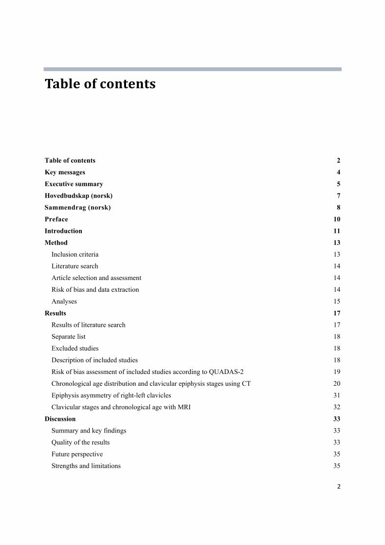
Tableofcontents
Table of contents 2
Key messages 4
Executive summary 5
Hovedbudskap (norsk) 7
Sammendrag (norsk) 8
Preface 10
Introduction 11
Method 13
Inclusion criteria 13
Literature search 14
Article selection and assessment 14
Risk of bias and data extraction 14
Analyses 15
Results 17
Results of literature search 17
Separate list 18
Excluded studies 18
Description of included studies 18
Risk of bias assessment of included studies according to QUADAS-2 19
Chronological age distribution and clavicular epiphysis stages using CT 20
Epiphysis asymmetry of right-left clavicles 31
Clavicular stages and chronological age with MRI 32
Discussion 33
Summary and key findings 33
Quality of the results 33
Future perspective 35
Strengths and limitations 35

3
Conclusion 37
References 38
Appendices 41
Appendix 1. Literature search strategy 41
Appendix 2. Description of included studies with quality assessment 45
Appendix 3. Separate list 55
Appendix 4. Excluded studies 57
Appendix 5. Project protocol 61

4
Keymessages
This systematic review summarizes the evidence on the distribution of chronological age from pre-defined stages of clavicular ossification studied by MRI or CT in living individuals. We included ten observational studies, all published within the last five years. The studies were conducted in five different countries, involving 4190 participants. Nine studies used CT and one used MRI. The QUADAS-2 checklist was used to assess risk of bias. The mean chronological age and its 95 % confidence
interval (95% CI) were presented for males and females separately in each stage and substage of medial clavicle epiphysis. Most of the studies showed a high risk of selection bias due to age mimicry, which could influence the distribution of age from each clavicular stage. Due to the limitations, we are unable to provide a reliable distribution of chronological age from each stage of medial clavicular development.
Title: Age estimation by ossification stages of the medial clavicular epiphysis: a systematic review ------------------------------------------------ Type of publication:
Systematic review A review of a clearly formulated question that uses systematic and explicit methods to identify, select, and critically appraise relevant research, and to collect and analyse data from the studies that are included in the review. Statistical methods (meta-analysis) may or may not be used to analyse and summarise the results of the included studies. ------------------------------------------------ Doesn’t answer everything:
- Excluded studies are not evaluated - No recommendation - No cost-effectiveness evaluation
------------------------------------------------ Publisher: Norwegian Institute of Public Health ------------------------------------------------ Updated: Last search for studies: April 2017 ------------------------------------------------ Peer review: - Signe Agnes Flottorp (internal) - Kjetil Gundro Brurberg (internal) - Andreas Schmeling (Professor, Dr. Med and Director of the Institute of Legal Medicine, University of Munster)

5
Executivesummary
Introduction
Every year, young asylum seekers come to Norway without legal documentation of their chronological age. To ensure that children receive their entitled rights and that adults are not treated as children, it is necessary to estimate their chronological age. Assessments of hand skeleton and third molar teeth using X-ray have been used for age estimation in Norway for years. We have previously published systematic reviews assessing age distribution using radiographs based on the Greulich & Pyle atlas for hand-wrist skeleton and the Demirjian’s stages for the third molar teeth. Here we present a systematic review to evaluate the use of computed tomography (CT) and magnetic resonance imaging (MRI) on the medial clavicle epiphysis for age estimation in the living.
Method We searched for studies in the Cochrane Central Register of Controlled Trials (CENTRAL), MEDLINE, Embase and Google Scholar. This was a joint search conducted for studies using X-ray of teeth and hand, CT and MRI of the clavicle, knee and ankle in both males and females. An update search was conducted in April 2017 for clavicle, knee and ankle only. Two people screened the literature independently and assessed the risk of the included studies based on the QUADAS-2 checklist. The mean chronological age and the standard deviation in the included studies were extracted for each stage and substage for each sex separately.
Results We found 10059 abstracts in the first search and 663 in the second search. In total 63 potentially relevant publications were forwarded for full-text screening. Eventually ten studies were included, all published
within the last five years. Nine studies used CT and one study used MRI. The sample sizes in the included studies ranged from 152 to 752 participants. Six studies were from Turkey, and one from Australia, China, Germany and Thailand, respectively.
The age distribution for each clavicular stage varied largely in and between the included studies. According to the QUADAS-2 checklist, all the included studies were associated with high risk of age mimicry bias. This particular bias lead to unreliable estimates of chronological age within each stage of medial clavicular ossification. In addition, we summarized information on the developmental asymmetry of right and left clavicles. Five relevant studies were found and 11% of included cases showed different epiphysis stages of clavicular ossification.

6
Discussion These findings are consistent with our previous systematic reviews regarding estimation of chronological age by development of the hand-wrist skeleton and third molar teeth, which also revealed high risks of age mimicry bias. Our findings raise concern about the study design and sample selection process used in age estimation research. When studying the direct probability of chronological age from stage we highlight the need to include populations with uniform distribution and sufficiently wide spectrum of chronological age, or using statistical techniques like transition analysis to overcome the age mimicry bias. Another suggested possibility is to observe the minimum age for each stage (minimum age concept), which is data that is not affected by the age mimicry bias. However, this cannot give an estimate of the individual’s probable age (which is centred on the mean value).
Conclusion We are unable to assess the validity of using CT or MRI as a method to show the distribution of chronological age from each clavicular stage due to the high risk of age mimicry bias in the included studies and the observed high heterogeneity among included studies. None of the studies included a study population with a uniform distribution of chronological age, which leads to potential age mimicry bias of the results (the distribution of chronological age from each clavicular stage). Studies avoiding the age mimicry selection bias are warranted for a better description of the age distribution of the medial clavicular epiphysis development stages.

7
Hovedbudskap(norsk)
Denne systematiske oversikten oppsummerer den forskningsbaserte dokumentasjonen på fordelingen av kronologisk alder fra forhåndsdefinerte stadier av den mediale kragebeinsutviklingen hos levende personer med CT eller MR. Ti studier oppfylte inklusjonskriteriene våre, alle publisert de siste fem årene. Studiene var fra fem land og omfattet totalt 4190 deltakere. Ni studier brukte CT og én MR. QUADAS-2 sjekklisten ble brukt for å vurdere risikoen for systematiske skjevheter. Gjennomsnittlig kronologisk alder og 95 % konfidensintervall (95 % CI) ble presentert separat for menn og kvinner for hvert stadium av den mediale kragebeinsutviklingen. Alle de inkluderte studiene viste høy risiko for seleksjonsskjevhet knyttet til aldersmimikering, som kan påvirke presisjonen og påliteligheten av den estimerte aldersfordelingen. Vi kan derfor ikke angi pålitelige fordelinger av kronologisk alder fra hvert stadium av den mediale kragebeinsutviklingen.
Tittel: Estimering av alder ved hjelp av utviklingsstadier av det mediale kragebeinet: en systematisk oversikt
------------------------------------------ Publikasjonstype:
Systematisk oversikt En systematisk oversikt er resultatet av å - innhente, kritisk vurdere og - sammenfatte relevante forskningsresultater ved hjelp av forhåndsdefinerte og eksplisitte metoder. ------------------------------------------ Svarer ikke på alt:
- Ingen studier utenfor de eksplisitte inklusjonskriteriene
- Ingen helseøkonomisk evaluering - Ingen anbefalinger ------------------------------------------ Hvem står bak denne publikasjonen? Folkehelseinstituttet
------------------------------------------
Når ble litteratursøket utført? Siste søket: April 2017
------------------------------------------ Fagfeller: - Signe Agnes Flottorp (internal) - Kjetil Gundro Brurberg (internal) - Andreas Schmeling ((Professor, Dr. Med og Direktør ved Rettsmedisinsk institutt, Universitet i Munster))
8
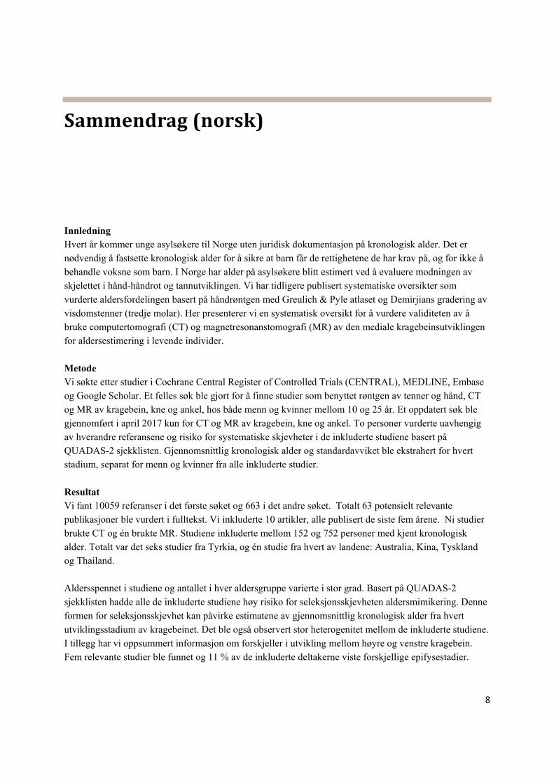
Sammendrag(norsk)
Innledning Hvert år kommer unge asylsøkere til Norge uten juridisk dokumentasjon på kronologisk alder. Det er nødvendig å fastsette kronologisk alder for å sikre at barn får de rettighetene de har krav på, og for ikke å behandle voksne som barn. I Norge har alder på asylsøkere blitt estimert ved å evaluere modningen av skjelettet i hånd-håndrot og tannutviklingen. Vi har tidligere publisert systematiske oversikter som
vurderte aldersfordelingen basert på håndrøntgen med Greulich & Pyle atlaset og Demirjians gradering av visdomstenner (tredje molar). Her presenterer vi en systematisk oversikt for å vurdere validiteten av å bruke computertomografi (CT) og magnetresonanstomografi (MR) av den mediale kragebeinsutviklingen for aldersestimering i levende individer.
Metode Vi søkte etter studier i Cochrane Central Register of Controlled Trials (CENTRAL), MEDLINE, Embase og Google Scholar. Et felles søk ble gjort for å finne studier som benyttet røntgen av tenner og hånd, CT og MR av kragebein, kne og ankel, hos både menn og kvinner mellom 10 og 25 år. Et oppdatert søk ble gjennomført i april 2017 kun for CT og MR av kragebein, kne og ankel. To personer vurderte uavhengig av hverandre referansene og risiko for systematiske skjevheter i de inkluderte studiene basert på QUADAS-2 sjekklisten. Gjennomsnittlig kronologisk alder og standardavviket ble ekstrahert for hvert stadium, separat for menn og kvinner fra alle inkluderte studier.
Resultat Vi fant 10059 referanser i det første søket og 663 i det andre søket. Totalt 63 potensielt relevante publikasjoner ble vurdert i fulltekst. Vi inkluderte 10 artikler, alle publisert de siste fem årene. Ni studier brukte CT og én brukte MR. Studiene inkluderte mellom 152 og 752 personer med kjent kronologisk alder. Totalt var det seks studier fra Tyrkia, og én studie fra hvert av landene: Australia, Kina, Tyskland og Thailand. Aldersspennet i studiene og antallet i hver aldersgruppe varierte i stor grad. Basert på QUADAS-2 sjekklisten hadde alle de inkluderte studiene høy risiko for seleksjonsskjevheten aldersmimikering. Denne
formen for seleksjonsskjevhet kan påvirke estimatene av gjennomsnittlig kronologisk alder fra hvert utviklingsstadium av kragebeinet. Det ble også observert stor heterogenitet mellom de inkluderte studiene. I tillegg har vi oppsummert informasjon om forskjeller i utvikling mellom høyre og venstre kragebein. Fem relevante studier ble funnet og 11 % av de inkluderte deltakerne viste forskjellige epifysestadier.

9
Diskusjon Disse resultatene er i samsvar med funnene fra våre tidligere systematiske oversikter om estimering av kronologisk alder ved hjelp av røntgenbilder av håndskjelettet og visdomstenner. Funnene våre viser problemer knyttet til studiedesign og utvelgelse av deltakere til aldersestimeringsstudier. Når man studerer den direkte sannsynligheten av alder fra stadium er det viktig å fordele antall deltakere jevnt mellom hver aldersgruppe og inkludere en populasjon med tilstrekkelig bredt aldersspenn, eller å benytte statistiske teknikker slik som transisjonsanalyse for å redusere konsekvensen av aldersmimikering. En annen mulighet er å observere den laveste observerte alder for hvert stadium («minimum age concept»), som vil være upåvirket av aldersmimikeringen i studiene. Ulempen er at en slik bruk av dataene ikke vil gi et estimat av personens sannsynlige alder (som gjenspeiles i gjennomsnittsverdiene av kronologisk alder fra hvert stadium).
Konklusjon Vi kan ikke vurdere validiteten knyttet til å bruke CT og MR som metode for å angi fordelingen av
kronologisk alder for hvert stadium av kragebeinsutviklingen på grunn av aldersmimikering i studiene og høy heterogenitet mellom studiene. Ingen av studiene inkluderte en studiepopulasjon med jevn aldersfordeling, noe som fører til potensielle systematiske skjevheter i de ekstraherte resultatene (fordelingen av kronologisk alder for hvert steg av kragebeinsutviklingen). Studier som unngår seleksjonsskjevheten aldersmimikering er nødvendig for å få bedre mål på aldersfordelingen for hvert stadium av kragebeinsutviklingen.

10
Preface
This systematic review summarizes evidence of age distribution according to the ossification stages of medial clavicular epiphysis using CT or MRI. We have previously published two systematic reviews on age assessment by skeletal hand-wrist maturation using the Greulich & Pyle atlas and wisdom teeth formation using Demirjian’s grading. In parallel with the current study, we have carried out another systematic review on age assessment of knee and ankle by CT and MRI. We have chosen to write these
systematic reviews as separate documents, but we use consistent texts throughout the documents where relevant. The project group consisted of:
Kristoffer Y. Ding, project leader, Norwegian Institute of Public Health
Veslemøy Rolseth, Oslo University Hospital
Pål Skage Dahlberg, Oslo University Hospital
Annhild Mosdøl, Norwegian Institute of Public Health
Gyri H. Straumann, Norwegian Institute of Public Health
Øyvind Bleka, Oslo University Hospital
Gunn E. Vist, Norwegian Institute of Public Health
We thank Signe Agnes Flottorp and Kjetil Gundro Brurberg for being the internal reviewers, and Professor Dr. Med. and Director of the Institute of Legal Medicine at University of Munster, Andreas Schmeling for conducting the external review of our research protocol and final report. We also want to thank Marit Johansen as peer reviewer for our literature search. Notably, Figure 1 is reprinted from “The value of sub-stages and thin slices for the assessment of the medial clavicular epiphysis: a prospective multi-center CT study”, 2013, Daniel Wittschieber, Ronald Schulz, Volker Vieth et al, with permission from Springer Nature [Forensic Science, Medicine, and Pathology]. License no: 4382961293227.
Signe Flottorp Department director
Gunn E. Vist Unit leader
Kristoffer Y. Ding Project leader

11
Introduction
Age estimation of individuals with unknown age has been of considerable interest in forensic practice and research, especially for young, unaccompanied asylum-seekers (1). From 1 January 2016, the Division of Forensic Sciences at the Norwegian Institute of Public Health, now at the Oslo University Hospital, received the national assignment to take the scientific responsibility for medical age assessment. It was
decided to conduct systematic reviews of various methods used for medical age assessment.
There are a number of biological changes as a person grows and develops. The assessment of specific developmental stages constitutes the basis for medical age assessments. Currently, the most widely used methods (2) are based on evaluation of radiographs of hand-wrist and teeth, of which we recently reviewed the methods based on the Greulich and Pyle atlas (3) and Demirjian’s development stages of third molar tooth (4). In the latter review, we observed high heterogeneity in results between studies. Part of the variation may be due to the study characteristics, particularly uneven number of participants in each age group or the age range in the studies. This is known to introduce “age mimicry” bias in this type of studies. This particular form of selection bias in forensic age assessment has been discussed in detail elsewhere (4). In short, age mimicry occurs when the results are given as age from stage, and the numbers of enrolled participants in each age group are uneven, or the age range do not cover all ages that can fall into the stages analyzed. Hence, the results “mimic” the distribution of age in the reference sample (study
population) (5).
Since the skeleton of hand and wrist is fully developed close to 18 years of age, it has been suggested to use regions of interest that mature at later ages, such as the clavicle or third molar teeth. Assessment of medial clavicular bone can e.g. be performed by X-ray, computed tomography (CT), magnetic resonance imaging (MRI) or sonography. We chose to review the CT and MRI method as CT is recommended over conventional radiography due to superimposition of anatomy (6). MRI is the most advanced radiation-free
method and we also chose to review this method.
The purpose of this systematic review is to get an overview of how chronological age is distributed within different medial clavicular epiphysis stages, and to explore, if possible, variations between different populations. The current study, together with others, constitutes part of a basis for further discussion and
recommendation on how medical age assessments should be conducted in Norway.

12
Descriptionofclaviculardevelopmentalstages
There is 4 common staging systems for ossification of the clavicle: A 4-stage classification system (7, 8), Schmeling et al. 5-stage classification (9), Kellinghaus et al. 9-stage classification (10) and Ufuk et al. modified 5-stage classification (11). The studies often use one of these staging systems or a combination of these. The classification methods of the medial clavicular epiphysis defined by Schmeling et al. and Kellinghaus et al. is shown in Figure 1. The stages 1-5 illustrate the classification system by Schmeling et
al. (9), while stages followed by a letter (from a to c) are the Kellinghaus substages.
Figure 1. Assessment of the medial clavicular epiphysis. Stage 1, Ossification center has not ossified yet; Stage 2, Ossification center has ossified. Epiphyseal cartilage has not ossified; Stage 3, Epiphyseal cartilage has partially ossified; Stage 4, Epiphyseal cartilage has completely ossified. Epiphyseal scar is visible; Stage 5, Epiphyseal cartilage has completely ossified. Epiphyseal scar is not visible any more. The figure is reprinted with permission from Springer Nature (12).
For further sub-classification of the stages 2 and 3 (2a-3c), the sub-staging scheme by Kellinghaus et al. (10) is often applied. In stage 2a, the lengthwise epiphyseal measurement is one third or less compared to the widthwise measurement of the metaphyseal ending. In stage 2b, the lengthwise epiphyseal measurement is over one third to two thirds compared to the widthwise measurement of the metaphyseal ending. In stage 2c, the lengthwise epiphyseal measurement is over two thirds compared to the widthwise measurement of the metaphyseal ending. In stage 3a, the epiphyseal-metaphyseal fusion completes one third or less of the former gap between epiphysis and metaphysis. In stage 3b, the epiphyseal-metaphyseal fusion completes over one third to two thirds of the former gap between epiphysis and metaphysis. In stage 3c, the epiphyseal-metaphyseal fusion completes over two thirds of the former gap between epiphysis and metaphysis (12). The 4-stage system has a stage 4 that is a combination of the 4 and 5 in the Schmeling et al. staging system. The Ufuk et al. staging system (11) is a modified version of Schmeling et
al. 5 stages.

13
Method
The current project included systematic literature searches on studies focusing on age estimation of the clavicle using computed tomography (CT) or magnetic resonance imaging (MRI) techniques. This systematic review is conducted following the guideline “Slik oppsummerer vi forskning” published by the
former Norwegian Knowledge Center (13). We used the following specifications:
Inclusion criteria
Study design: Studies that summarized data on age estimation from clavicular development stages using CT or MRI techniques
Population: Living persons between 10 and 25 years old with no pathological clavicular findings
Index test: Maturity stages of medial clavicular epiphysis
Reference: Chronological age
Outcome: Chronological age summary in each stage
Language: Language limits was not applied to the searches. However, project members only read Chinese, Danish, English, German, Japanese, Norwegian, and Swedish. Relevant publications that we could not read due to language limitation is listed in a separate table
Exclusion Criteria:
Studies without full-text (conference abstracts)
Studies that is not an empirical study
Studies using techniques other than CT or MRI
Studies that have included remains instead of living human beings
Studies that focused on osteometric parameters instead of age estimation
Studies with less than 50 participants between 10 and 25 years old
Separate list
After reading full-text articles, we made a separate list for future reference to track studies that might have relevant data, but did not present the data in a way that we could utilize.

14
Literaturesearch
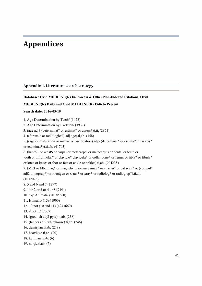
Research librarian Gyri Hval Straumann created and conducted the literature searches and Marit Johansen peer-reviewed the search strategies. We searched for primary studies with no limit on study design, publication time, or language in the following databases:
Cochrane Central Register of Controlled Trials (CENTRAL)
MEDLINE (Ovid) and Pubmed [sb]
Embase (Ovid)
Google Scholar
The first search was carried out on 19 May 2016. This was a joint search conducted for studies using X-ray of teeth and hand, CT and MRI of the clavicle, knee and ankle in both males and females. An update search was conducted in April 2017 only for clavicle, knee and ankle. The search strategies are presented in Appendix 1.
Articleselectionandassessment
For the first search, six review authors (AH, GEV, GHS, KYD, PSD and VR) independently screened abstracts identified by the searches; for the second search, two review authors screened abstracts independently (GEV and KYD). All abstracts were screened in duplicates via the systematic reviews web-application Rayyan (14). Articles were excluded if the title and/or abstract did not meet the inclusion criteria. For potentially relevant studies, the full-text articles were obtained and screened by two reviewers independently, with discrepancies resolved by consensus of reviewers. Studies that were considered as relevant to the review topic but did not meet all the inclusion criteria for the review were listed in the ‘Characteristics of excluded studies’ table, with the reason for their exclusion described. We recorded the selection process in
sufficient detail to complete a Preferred Reporting Items for Systematic Reviews and Meta-Analyses (PRISMA) flow diagram.
Riskofbiasanddataextraction
To evaluate the risk of bias (methodical quality) of included studies, we adopted a revised QUADAS-2 checklist (15) that has been described in detail in the previous age estimation projects on hands and teeth. KYD extracted the following information from articles, and GEV double-checked the data accordingly:
Where and when the study was carried out (country and year)
Scoring method (e.g. Schmeling’s stages and/or Kellinghaus’ substages)
Age range, sex, and sample size
Study design
15
Sample selection method
Age estimation method In addition, we extracted the following data for analysis
Mean age and its standard deviation (SD) of chronological age for each stage
Total number of participants in each stage
Left and right clavicle epiphysis asymmetry
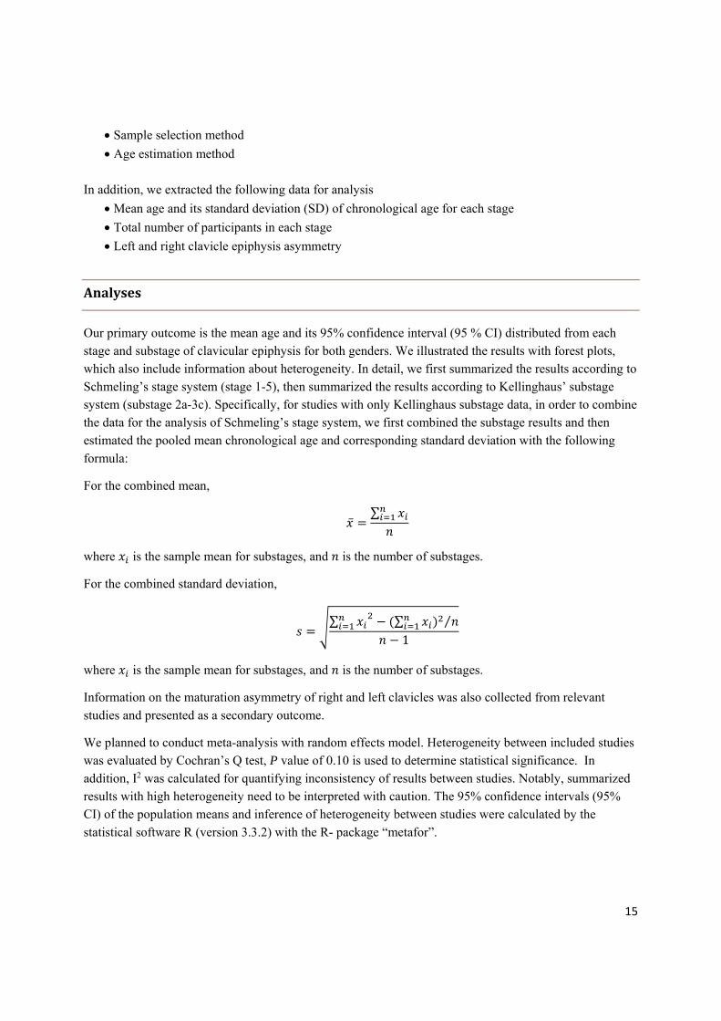
Analyses
Our primary outcome is the mean age and its 95% confidence interval (95 % CI) distributed from each stage and substage of clavicular epiphysis for both genders. We illustrated the results with forest plots, which also include information about heterogeneity. In detail, we first summarized the results according to Schmeling’s stage system (stage 1-5), then summarized the results according to Kellinghaus’ substage system (substage 2a-3c). Specifically, for studies with only Kellinghaus substage data, in order to combine the data for the analysis of Schmeling’s stage system, we first combined the substage results and then estimated the pooled mean chronological age and corresponding standard deviation with the following
formula:
For the combined mean,
∑
where is the sample mean for substages, and is the number of substages.
For the combined standard deviation,
∑ ∑ ⁄
1
where is the sample mean for substages, and is the number of substages.
Information on the maturation asymmetry of right and left clavicles was also collected from relevant
studies and presented as a secondary outcome.
We planned to conduct meta-analysis with random effects model. Heterogeneity between included studies was evaluated by Cochran’s Q test, P value of 0.10 is used to determine statistical significance. In addition, I2 was calculated for quantifying inconsistency of results between studies. Notably, summarized results with high heterogeneity need to be interpreted with caution. The 95% confidence intervals (95% CI) of the population means and inference of heterogeneity between studies were calculated by the
statistical software R (version 3.3.2) with the R- package “metafor”.

16
GRADEframework
Notably, the Grading of Recommendations Assessment, Development and Evaluation (GRADE) tool (16) is often used in systematic reviews to rate the quality and certainty of the included evidence. However, the current systematic review is not a typical diagnostic accuracy assessment study, where one presents positive/negative results with sensitivity and specificity analysis. Therefore, evaluating evidence quality
by GRADE could not be conducted in the current systematic review.

17
Results
Resultsofliteraturesearch
We initially searched electronic databases and registries in May 2016 (search I), and found 10059
potential relevant publications on age estimation with hands, teeth, clavicles, knees and ankles after removing duplicates. Among those publications, we found 52 potential relevant articles on age estimation using the medial clavicle epiphysis. In addition, we carried out an update search in April 2017 (search II) and found 663 articles, in which 11 articles were of interest and were read in full-text. We eventually
included ten studies. Process in detail is described below in Figure 2.
Figure 2. Flow chart of literature selection
References identified through database searching (n=10722)
References excluded on the basis of title and abstract (n=10669)
Full-text articles assessed for eligibility (n=63)
Articles excluded on the basis of full-text assessment (n=41)
Included studies (n=10)
Articles with relevant data but was not fully presented (n=12)
18
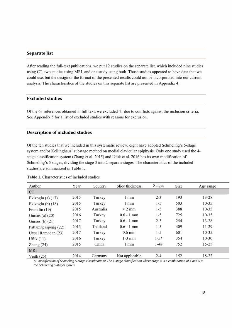
Separatelist
After reading the full-text publications, we put 12 studies on the separate list, which included nine studies using CT, two studies using MRI, and one study using both. Those studies appeared to have data that we could use, but the design or the format of the presented results could not be incorporated into our current analysis. The characteristics of the studies on this separate list are presented in Appendix 4.
Excludedstudies
Of the 63 references obtained in full text, we excluded 41 due to conflicts against the inclusion criteria. See Appendix 5 for a list of excluded studies with reasons for exclusion.
Descriptionofincludedstudies
Of the ten studies that we included in this systematic review, eight have adopted Schmeling’s 5-stage system and/or Kellinghaus’ substage method on medial clavicular epiphysis. Only one study used the 4-stage classification system (Zhang et al. 2015) and Ufuk et al. 2016 has its own modification of
Schmeling’s 5 stages, dividing the stage 3 into 2 separate stages. The characteristics of the included
studies are summarized in Table 1.
Table 1. Characteristics of included studies
Author Year Country Slice thickness Stages Size Age range
CT
Ekizoglu (a) (17) 2015 Turkey 1 mm 2-3 193 13-28
Ekizoglu (b) (18) 2015 Turkey 1 mm 1-5 503 10-35
Franklin (19) 2015 Australia < 2 mm 1-5 388 10-35
Gurses (a) (20) 2016 Turkey 0.6 - 1 mm 1-5 725 10-35
Gurses (b) (21) 2017 Turkey 0.6 - 1 mm 2-3 254 13-28
Pattamapaspong (22) 2015 Thailand 0.6 - 1 mm 1-5 409 11-29
Uysal Ramadan (23) 2017 Turkey 0.6 mm 1-5 601 10-35
Ufuk (11) 2016 Turkey 1-3 mm 1-5* 354 10-30
Zhang (24) 2015 China 1 mm 1-4# 752 15-25
MRI
Vieth (25) 2014 Germany Not applicable 2-4 152 18-22 *A modification of Schmeling 5-stage classification# The 4-stage classification where stage 4 is a combination of 4 and 5 in the Schmeling 5-stages system
19
Studies included in the project were published during the last five years. There was one study from Germany, China, Thailand and Australia, and six studies from Turkey. Among those studies, there were nine studies using CT and one study using MRI. Most CT studies were conducted using slice thickness of 1 mm or less, which is recommended by Kellinghaus (10). The complete sample size for the CT studies was 4179 individuals, covering both genders from 10-35 years old. The MRI study was comprised of 152 male football players from 18-22 years old.
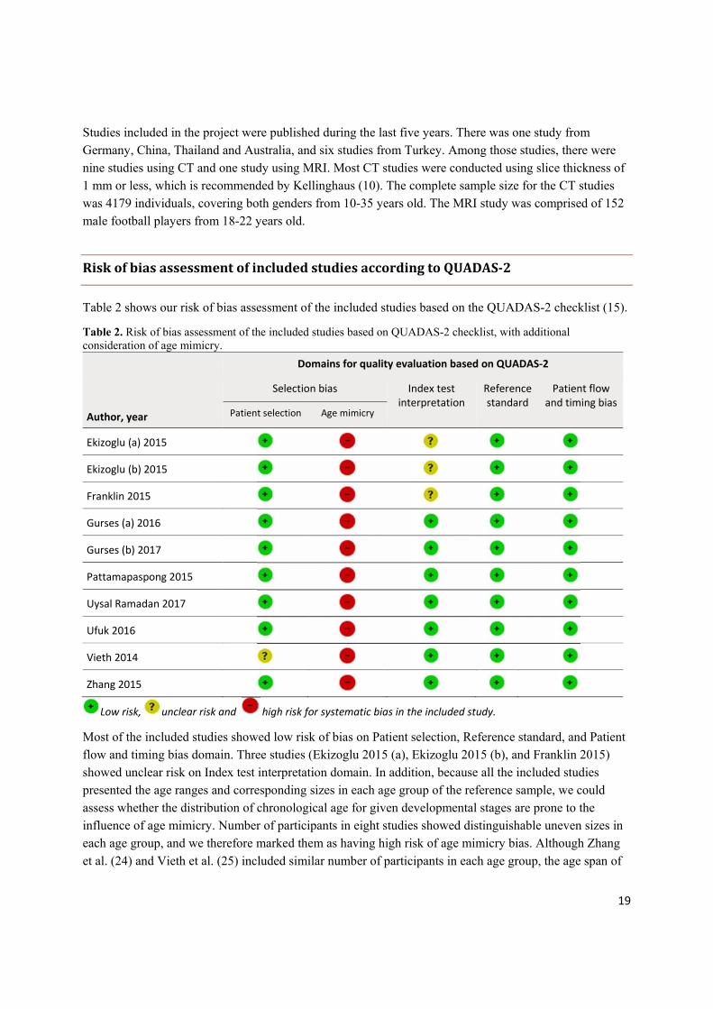
RiskofbiasassessmentofincludedstudiesaccordingtoQUADAS‐2
Table 2 shows our risk of bias assessment of the included studies based on the QUADAS-2 checklist (15).
Table 2. Risk of bias assessment of the included studies based on QUADAS-2 checklist, with additional consideration of age mimicry.
Domains for quality evaluation based on QUADAS‐2
Author, year
Selection bias Index test interpretation
Reference standard
Patient flow and timing bias
Patient selection Age mimicry
Ekizoglu (a) 2015
Ekizoglu (b) 2015
Franklin 2015
Gurses (a) 2016
Gurses (b) 2017
Pattamapaspong 2015
Uysal Ramadan 2017
Ufuk 2016
Vieth 2014
Zhang 2015
Low risk, unclear risk and high risk for systematic bias in the included study.
Most of the included studies showed low risk of bias on Patient selection, Reference standard, and Patient flow and timing bias domain. Three studies (Ekizoglu 2015 (a), Ekizoglu 2015 (b), and Franklin 2015) showed unclear risk on Index test interpretation domain. In addition, because all the included studies presented the age ranges and corresponding sizes in each age group of the reference sample, we could assess whether the distribution of chronological age for given developmental stages are prone to the influence of age mimicry. Number of participants in eight studies showed distinguishable uneven sizes in each age group, and we therefore marked them as having high risk of age mimicry bias. Although Zhang
et al. (24) and Vieth et al. (25) included similar number of participants in each age group, the age span of

20
the reference sample did not cover all possible ages for the analysed stages. We therefore marked these
two studies as high risk of age mimicry bias due to narrow age range.
ChronologicalagedistributionandclavicularepiphysisstagesusingCT
Nine studies that used CT were included. We first assessed whether it was appropriate to summarize the results of these studies in meta-analyses. QUADAS-2 checklist showed that all the included studies were at high risk of being affected by age mimicry. We believe that the effect of age mimicry is so large that it could severely bias the pooled results and lead to inaccurate and unreliable estimates of chronological age in each clavicular stage. Therefore, we concluded that it is not appropriate to combine and utilize the results in meta-analyses. However, we present the findings for each study and stage graphically with estimated heterogeneity. Figures 3-7 show the results for each development stage 1-5 using Schmeling’s stages for males, while figures 8-12 show the results for females. We have included the stages 1-3 from
Zhang et al. 2015 using the 4-stage system as these stages corresponds to Schmeling’s stages 1-3.
Figure 3. Age distribution of clavicular ossification stage 1 in males

21
Figure 4. Age distribution of clavicular ossification stage 2 in males
Figure 5. Age distribution of clavicular ossification stage 3 in males

22
Figure 6. Age distribution of clavicular ossification stage 4 in males
Figure 7. Age distribution of clavicular ossification stage 5 in males

23
Figure 8. Age distribution of clavicular ossification stage 1 in females
Figure 9. Age distribution of clavicular ossification stage 2 in females

24
Figure 10. Age distribution of clavicular ossification stage 3 in females
Figure 11. Age distribution of clavicular ossification stage 4 in females

25
Figure 12. Age distribution of clavicular ossification stage 5 in females
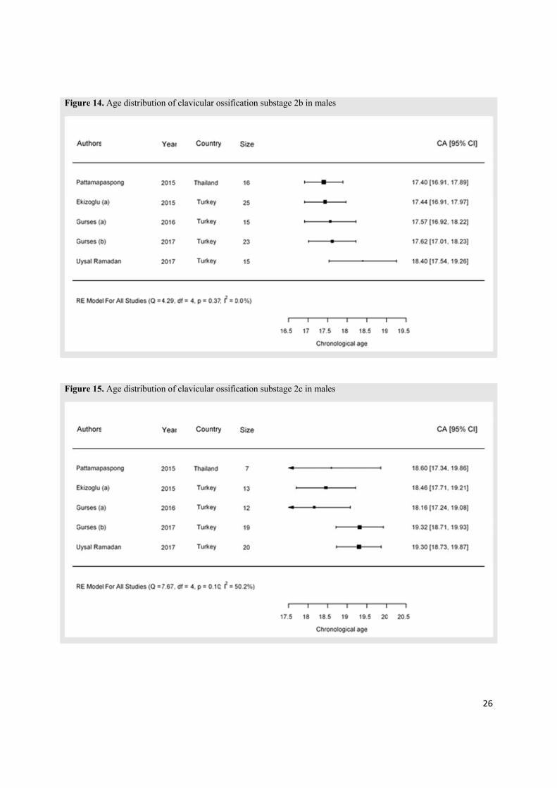
We found five studies presenting results of Kellinghaus’ substage. Below we present the results of age distribution in each substage in males (Figures 13-18) and females (Figures 19-24), respectively.
Figure 13. Age distribution of clavicular ossification substage 2a in males
26
Figure 14. Age distribution of clavicular ossification substage 2b in males
Figure 15. Age distribution of clavicular ossification substage 2c in males

27
Figure 16. Age distribution of clavicular ossification substage 3a in males
Figure 17. Age distribution of clavicular ossification substage 3b in males

28
Figure 18. Age distribution of clavicular ossification substage 3c in males
Figure 19. Age distribution of clavicular ossification substage 2a in females

29
Figure 20. Age distribution of clavicular ossification substage 2b in females
Figure 21. Age distribution of clavicular ossification substage 2c in females

30
Figure 22. Age distribution of clavicular ossification substage 3a in females
Figure 23. Age distribution of clavicular ossification substage 3b in females

31
Figure 24. Age distribution of clavicular ossification substage 3c in females
Epiphysisasymmetryofright‐leftclavicles
Bilateral clavicle asymmetry is the difference in the fusion timing of the medial epiphysis between the two clavicular bones of the same individual. Several studies (Franklin 2015, Gurses 2016, Pattamapaspong 2015, Uysal Ramadan 2017 and Ufuk 2016) presented information on the right-left asymmetry of
clavicular epiphysis development (Table 4).
Table 4. Epiphysis asymmetry of right-left clavicles
Study Size Differed 1 Percentage
Franklin 2015 332 27 8.1%
Gurses 2016 725 94 13.0%
Pattamapaspong 2015 409 62 15.2%
Uysal Ramadan 2017 601 64 10.7%
Ufuk 2016 300 18 6.0%
Total 2367 265 11.2% 1 Differed indicates the number of participants who appeared to have bilateral clavicle asymmetry in the assessment of medial clavicular epiphysis.
The asymmetry rate varied from 6.0% to 15.2%, the weighted average is 11.2%, suggesting a possible developmental difference between left and right clavicles in the included populations. However, it is not
clear if this difference is systematic for left/right orientation and by gender.

32
ClavicularstagesandchronologicalagewithMRI
Only one study (25) fulfilling the inclusion criteria has explored the age distribution within clavicular stages using MRI. This study included 152 German football players from 18 to 22 years (30 men in each of the 18-20 age groups, and 31 men in the 21 and 22 age groups). The detailed age distribution is
presented in Table 5.
Table 5. Statistical parameters for Schmeling stages from Vieth et al. (25)
Stage Size Mean±SD Min Max
2 15 20.4±1.1 18.4 22.3
3 111 20.6±1.5 18.1 23.0
4 1 21.2 21.2 21.2
In stage 2, there were 15 participants, with a mean age of 20.4 years (SD=1.1). The majority of participants were in stage 3, with a mean age of 20.6 years (SD=1.5). There was only one participant in stage 4, aged 21.2 years.
33
Discussion
Summaryandkeyfindings
In this systematic review, we have summarized evidence on the distribution of chronological age from
pre-defined stages of medial clavicular ossification measured by CT or MRI in living individuals. We
included ten studies (nine using CT and one using MRI) that met the inclusion criteria.
Most of the CT studies covered a relatively wide age spectrum, but included uneven numbers of participants in each age group, leading to an inaccurate estimation of age distribution due to age mimicry bias. The right-left asymmetry analysis revealed a possible developmental difference of right and left
clavicles in around 11% of the included populations.
Only one MRI study was included, which included a relatively uniform sample size in each age group, but
the age spectrum was too narrow, which is also associated with high risk of age mimicry bias.
Taken together, our pooled results suggested high risk of age mimicry bias in the data we extracted from all of the included studies, which is characterized by uneven number of participants in each age group or an age range not covering all ages that can fall into the analyzed ossification stages. This bias could lead to skewed and inaccurate estimates of age distribution. Future studies need to take into account the
potential age mimicry bias in study design when presenting the probability of age from stage.
Qualityoftheresults
The results of age distribution within Schmeling’s stages 1-5 in both males and females showed considerable heterogeneity. This is caused by large variation in mean age between the included studies. Part of the heterogeneity might be caused by age mimicry, which is a special type of selection bias found in the data we extracted from all included studies. In addition, there might be other sources for the
observed heterogeneity.
There has been an extensive debate on the causes of the observed heterogeneity between studies of skeletal maturation over the last decades. Obviously, the critical lack of well-conducted studies we have pointed at in this systematic review makes it even more difficult to identify what is truly causing the
heterogeneity between studies on the ossification of the medial clavicle.

34
Several studies point at socio-economic status as an important variable affecting skeletal maturation (reviewed by Buckley and Clark (6)). However, the number of studies supporting this is small and the results are conflicting (6). Also, age assessment studies only occasionally report the socio-economic level of the included individuals. Buckley and Clark summarized all methods used for medial clavicle ossification in both living and remains, and conclude with CT as the method of choice for forensic age estimation. They also point to multiple factors that may influence the age assessment. These are interpreters experience, socio-economic class/regional differences and limitations of the methods applied
(6).
According to the QUADAS-2 checklist, extracted data from all the included studies showed high risk of age mimicry. Among those studies, eight out of ten showed uneven numbers of participants in each age group. In addition, the other source of age mimicry bias is caused by different age range in the reference sample. Several studies included populations from 10 to 35 years old, while others used a narrower range. This systematic difference also leads to skewness when pooling the mean age from included studies according to the ossification stages, and appears to be more obvious at both the early and the late stages in both genders. As an example, Figure 3 shows the mean chronological age of clavicular ossification stage 1 in men among included studies. The mean age of Zhang et al. (24) was 1 year older than the rest of studies, most likely due to a minimum inclusion age of 15 years. Another example is shown in Figure 12 (results for stage 5 for females), in which Pattamapaspong et al. (22) and Ufuk et al. (11) only included participants younger than 30 years, while other studies included participants up to 35 years. This latter example is what is called the “end stage problem”, as the last stage of all skeletal development measurements is everlasting. The mean age within the last stage is highly dependent on the highest included ages in the reference sample. The pooled mean age (95% CI) in Figure 12 is 29.7 (27.6 – 31.8) years old (results not shown). If the included studies extended the upper boundary of age range by including more participants older than 35 years old, the pooled mean age of this stage would be driven up. In fact, this issue has also been observed in several studies (26-28) and is discussed in our consortium papers on age estimation by hand-wrist and teeth development (3, 4). The end stage problem is avoided when using the minimum age concept (29), since this is not a probability distribution of age. However, it is equally important to include a reference sample that is able to reflect a population’s true minimum age
of a stage (in terms of size and age range).
The analysis of the Kellinghaus’ substages (2a-3c) revealed relatively lower heterogeneity in our analysis. The age span of the relevant substages was from 15 to 23 years, which was similar in all the included studies. This may explain the relatively lower level of heterogeneity. However, we need to keep in mind that the sample size in each substage was relatively small, resulting in larger variation in each study and hence wider confidence intervals. Therefore, the substantial within-study error in each study probably
masked the heterogeneity between studies.
Regarding the right-left asymmetry of medial clavicular epiphysis, the results are relatively consistent and showed that approximately 10% of the population in the analysis might have different development stages
between the right and left medial clavicle. However, it is not clear if this difference is systematic for left/right orientation and by gender, or at random. The difference in ossification of the right and left medial clavicle is not a problem as long as the same procedure is used when the method is applied in
practical age assessment work as in the reference study.

35
Futureperspective
In our opinion, age mimicry is an important source of selection bias in age estimation studies when mean age within stage is used as outcome, which also makes it less sensible to summarize individual studies that did not take this issue into account. Future studies are warranted to use uniform number of participants in each age group to estimate the age distribution, and also covering a wide age range with large sample size. An alternative approach is to collect the original research data from current studies, re-distribute and
analyze them, which can yield more reliable results.
In practice, to determine age from developmental stages of skeleton or teeth a reference sample from a selected age estimation study is often applied. Therefore, to assess the most probable age interval it is crucial to have a reliable and representative reference age distribution for each stage for the individuals
tested. Several age estimation studies (16, 29) have proposed transition analysis as an analytical technique to obtain reference samples. This approach estimates probability-based age of a population according to Bayes’ theorem, which is known to be less sensitive to age mimicry bias. The detailed explanation of
transition analysis can be found elsewhere (5).
The minimum age concept is proposed as a measurement to avoid assessing children as adults (29). As ossification of the medial clavicle is suggested as an additional method when skeletal hand is fully matured, the age prediction is focused on the question if the individual is under or over 18 years. In this matter the minimum age concept may be a preferred measurement compared to probability of an age range within a stage. Several studies indicate that ossification stage 3c and 4 represent an age over 18 years (6, 12). However, further studies assessing chronological age by clavicle ossification from several
populations are warranted.
Strengthsandlimitations
The strength of this systematic review is the systematic and transparent approach that we have used to review the question. We have implemented systematic literature search in many electronic databases, with clear inclusion and exclusion criteria. Two people have independently considered each reference according to these criteria, and assessed the risk of bias in the included studies. These independent assessments are one of the strengths of this systematic review. Although we have conducted a thorough literature search, potential relevant studies might not have been identified. A built-in weakness with systematic review is that they may become outdated when new studies are published. This systematic
review is up-to-date as of April 2017.
As the purpose of this systematic review was to evaluate age assessment by the medial clavicle ossification in the living, we excluded studies of corpses. Several reviews (6, 12) have chosen to include studies on corpses as well, and this might be regarded as a limitation of our systematic review. However,
36
we argue that the differences between the two groups are not fully understood (e.g. movement artifacts in
the study of living persons), which is the reason for our choice.
The current systematic review was undertaken to evaluate different age estimation methods in order to improve the Norwegian system for age assessment of minor asylum seekers. The assignment given was to estimate a probable age, as well as prediction values at threshold ages of 16 and 18 years. Hence, we chose to focus on the most probable age range (mean values) given the observed development stages. A
different approach is to focus on the minimum age (ever observed for an individual with a certain observed development stage of e.g. the clavicle). With this approach, one may reach a high degree of certainty as to whether the individual is above eighteen. One concern with this method is that it often relies on single or few observations instead of the trend of the whole reference sample (as for the mean
age of stage).
In the end-phase of the writing of this paper another systematic review on age estimation by CT of the medial clavicle epiphysis was published (30). This review included 13 papers, of which 7 overlap with our systematic review. The papers included in this study and excluded in ours do not contain numbers of individuals in each stage, which is necessary for calculation of 95 % CI. Hermetet et al. 2018 present box plots for the stages from each of the studies and focus on whether the participants are under or over 18 years old. They conclude that all individuals from all studies in stage 4 and 5 are over the age of 18 years. This is also the case for the stage 3c, except for some females in one study (22). The systematic review from Hermetet et al. (30) provides an interesting alternative way to portray results compared to ours. The minimum age concept avoids the pitfall of age mimicry bias that haunts the probability distributions of
age from stage.
37
Conclusion
In this systematic review, we have summarized evidence that describes the use of CT and MRI to assess the distribution of chronological age from pre-defined stages of medial clavicular epiphysis. In all of the
ten studies, the age estimates are likely to be influenced by a specific selection bias called age mimicry.
To get more reliable results of mean age within ossification stages, studies need to have an evenly distributed number of participants in each age group of the reference population covering a sufficiently wide age spectrum, or using statistical techniques like transition analysis to overcome the age mimicry bias. In addition, 11% of the population included in the analysis showed right-left asymmetry of medial clavicular development, which also needs to be considered when carrying out age estimation based on clavicular development stages. Nevertheless, future studies with particular consideration of age mimicry and a large reference sample are warranted for forensic age estimation. Alternatively, the studies can be used to determine the minimum age observed for a certain stage, and apply this principle to the 18 years threshold. Although those data are not subjected to the age mimicry bias, they answer a somewhat
different question.

38
References
1. Bassed RB, Drummer OH, Briggs C, Valenzuela A. Age estimation and the medial clavicular epiphysis: analysis of the age of majority in an Australian population using computed tomography. Forensic science, medicine, and pathology. 2011;7:148‐54. 2. EASO. Practical guide on age asessment. : European Asylum Support Office; 2018. Available from: https://www.easo.europa.eu/sites/default/files/easo‐practical‐guide‐qualification‐for‐international‐protection‐2018.pdf. 3. Dahlberg PS, Mosdøl A, Ding KY, Bleka Ø, Rolseth V, Straumann GH, Skjerven‐Martinsen M, Delaveris GJM, Vist GE. Samsvar mellom kronologisk alder og skjelettalder basert på Greulich og Pyle‐atlaset for aldersestimering: en systematisk oversikt [Agreement Between Chronological Age and Bone Age Based on the Greulich and Pyle Atlas for Age Estimation: A Systematic Review]. Oslo: The Norwegian Institute of Public Health (NIPH); 2017 [Available from: https://www.fhi.no/en/publ/2017/samsvar‐mellom‐kronologisk‐alder‐og‐skjelettalder‐basert‐pa‐greulich‐og‐pyl/ 4. Rolseth V, Mosdøl A, Dahlberg PS, Ding KY, Bleka Ø, Skjerven‐Martinsen M, Straumann GH, Møller GJ, Vist GE. Demirjians utviklingsstadier på visdomstenner for estimering av kronologisk alder: en systematisk oversikt. [Demirjian’s Development Stages on Wisdom Teeth for Estimation of Chronological Age: A Systematic Review] Oslo: The Norwegian Institute of Public Health (NIPH); 2017 [Available from: https://www.fhi.no/en/publ/2017/demirjians‐utviklingsstadier‐pa‐visdomstenner‐for‐estimering‐av‐kronologisk/. 5. Boldsen JL, Milner GR, Konigsberg LW, Wood JW. Transition analysis: a new method for estimating age from skeletons. In: Vaupel JW, Hoppa RD, editors. Paleodemography: Age Distributions from Skeletal Samples. Cambridge Studies in Biological and Evolutionary Anthropology. Cambridge: Cambridge University Press; 2002. p. 73‐106. 6. Buckley MB, Clark KR. Forensic Age Estimation Using the Medial Clavicular Epiphysis: A Study Review. Radiologic technology. 2017;88:482‐98. 7. Webb PAO, Suchey JM. Epiphyseal union of the anterior iliac crest and medial clavicle in a modern multiracial sample of American males and females. American Journal of Physical Anthropology. 1985;68:457‐66. 8. Kreitner K‐F, Schweden FJ, Riepert T, Nafe B, Thelen M. Bone age determination based on the study of the medial extremity of the clavicle. European Radiology. 1998;8:1116‐22. 9. Schmeling A, Schulz R, Reisinger W, Muhler M, Wernecke KD, Geserick G. Studies on the time frame for ossification of the medial clavicular epiphyseal cartilage in conventional radiography. International journal of legal medicine. 2004;118:5‐8. 10. Kellinghaus M, Schulz R, Vieth V, Schmidt S, Pfeiffer H, Schmeling A. Enhanced possibilities to make statements on the ossification status of the medial clavicular epiphysis using an amplified staging scheme in evaluating thin‐slice CT scans. International journal of legal medicine. 2010;124:321‐5.

39
11. Ufuk F, Agladioglu K, Karabulut N. CT evaluation of medial clavicular epiphysis as a method of bone age determination in adolescents and young adults. Diagnostic and interventional radiology (Ankara, Turkey). 2016;22:241‐6. 12. Wittschieber D, Schulz R, Vieth V, Kuppers M, Bajanowski T, Ramsthaler F, Puschel K, Pfeiffer H, Schmidt S, Schmeling A. The value of sub‐stages and thin slices for the assessment of the medial clavicular epiphysis: a prospective multi‐center CT study. Forensic science, medicine, and pathology. 2014;10:163‐9. 13. Slik oppsummerer vi forskning. Håndbok for Nasjonalt kunnskapssenter for helsetjenesten. Nylenna M, editor. Oslo: 4. reviderte utg; 2015. 14. Ouzzani M, Hammady H, Fedorowicz Z, Elmagarmid A. Rayyan‐a web and mobile app for systematic reviews. Systematic reviews. 2016;5:210. 15. Whiting PF, Rutjes AW, Westwood ME, Mallett S, Deeks JJ, Reitsma JB, Leeflang MM, Sterne JA, Bossuyt PM. QUADAS‐2: a revised tool for the quality assessment of diagnostic accuracy studies. Annals of internal medicine. 2011;155:529‐36. 16. Guyatt G, Oxman AD, Akl EA, Kunz R, Vist G, Brozek J, Norris S, Falck‐Ytter Y, Glasziou P, deBeer H, et al. GRADE guidelines: 1. Introduction—GRADE evidence profiles and summary of findings tables. Journal of Clinical Epidemiology. 2011;64:383‐94. 17. Ekizoglu O, Hocaoglu E, Inci E, Can IO, Aksoy S, Sayin I. Estimation of forensic age using substages of ossification of the medial clavicle in living individuals. International journal of legal medicine. 2015;129:1259‐64. 18. Ekizoglu O, Hocaoglu E, Inci E, Sayin I, Solmaz D, Bilgili MG, Can IO. Forensic age estimation by the Schmeling method: computed tomography analysis of the medial clavicular epiphysis. International journal of legal medicine. 2015;129:203‐10. 19. Franklin D, Flavel A. CT evaluation of timing for ossification of the medial clavicular epiphysis in a contemporary Western Australian population. International journal of legal medicine. 2015;129:583‐94. 20. Gurses MS, Inanir NT, Gokalp G, Fedakar R, Tobcu E, Ocakoglu G. Evaluation of age estimation in forensic medicine by examination of medial clavicular ossification from thin‐slice computed tomography images. International journal of legal medicine. 2016;130:1343‐52. 21. Gurses MS, Inanir NT, Soylu E, Gokalp G, Kir E, Fedakar R. Evaluation of the ossification of the medial clavicle according to the Kellinghaus substage system in identifying the 18‐year‐old age limit in the estimation of forensic age‐is it necessary? International journal of legal medicine. 2017;131:585‐92. 22. Pattamapaspong N, Madla C, Mekjaidee K, Namwongprom S. Age estimation of a Thai population based on maturation of the medial clavicular epiphysis using computed tomography. Forensic science international. 2015;246:123.e1‐5. 23. Uysal Ramadan S, Gurses MS, Inanir NT, Hacifazlioglu C, Fedakar R, Hizli S. Evaluation of the medial clavicular epiphysis according to the Schmeling and Kellinghaus method in living individuals: A retrospective CT study. Legal Medicine. 2017;25:16‐22. 24. Zhang K, Chen XG, Zhao H, Dong XA, Deng ZH. Forensic Age Estimation Using Thin‐Slice Multidetector CT of the Clavicular Epiphyses Among Adolescent Western Chinese. Journal of forensic sciences. 2015;60:675‐8. 25. Vieth V, Schulz R, Brinkmeier P, Dvorak J, Schmeling A. Age estimation in U‐20 football players using 3.0 tesla MRI of the clavicle. Forensic science international. 2014;241:118‐22. 26. Lee S‐H, Lee J‐Y, Park H‐K, Kim Y‐K. Development of third molars in Korean juveniles and adolescents. Forensic science international. 2009;188:107‐11. 27. Olze A, Pynn BR, Kraul V, Schulz R, Heinecke A, Pfeiffer H, Schmeling A. Studies on the chronology of third molar mineralization in First Nations people of Canada. International journal of legal medicine. 2010;124:433‐7.
40
28. Roberts GJ, McDonald F, Andiappan M, Lucas VS. Dental Age Estimation (DAE): Data management for tooth development stages including the third molar. Appropriate censoring of Stage H, the final stage of tooth development. Journal of forensic and legal medicine. 2015;36:177‐84. 29. Schmeling A, Dettmeyer R, Rudolf E, Vieth V, Geserick G. Forensic Age Estimation: Methods, Certainty, and the Law. Deutsches Ärzteblatt International. 2016;113:44‐50. 30. Hermetet C, Saint‐Martin P, Gambier A, Ribier L, Sautenet B, Rérolle C. Forensic age estimation using computed tomography of the medial clavicular epiphysis: a systematic review. International journal of legal medicine. 2018.
41
Appendices
Appendix1.Literaturesearchstrategy
Database: Ovid MEDLINE(R) In-Process & Other Non-Indexed Citations, Ovid
MEDLINE(R) Daily and Ovid MEDLINE(R) 1946 to Present
Search date: 2016-05-19
1. Age Determination by Teeth/ (1422)
2. Age Determination by Skeleton/ (3937)
3. (age adj3 (determinat* or estimat* or assess*)).ti. (2851)
4. ((forensic or radiological) adj age).ti,ab. (158)
5. ((age or maturation or mature or ossification) adj5 (determinat* or estimat* or assess*
or examinat*)).ti,ab. (41703)
6. (hand$1 or wrist$ or carpal or metacarpal or metacarpus or dental or teeth or
tooth or third molar* or clavicle* clavicula* or collar bone* or femur or tibia* or fibula*
or knee or knees or foot or feet or ankle or ankles).ti,ab. (904235)
7. (MRI or MR imag* or magnetic resonance imag* or ct scan* or cat scan* or (comput*
adj2 tomograp*) or roentgen or x‐ray* or xray* or radiolog* or radiograp*).ti,ab.
(1032026)
8. 5 and 6 and 7 (1297)
9. 1 or 2 or 3 or 4 or 8 (7491)
10. exp Animals/ (20185560)
11. Humans/ (15941900)
12. 10 not (10 and 11) (4243660)
13. 9 not 12 (7007)
14. (greulich adj2 pyle).ti,ab. (238)
15. (tanner adj2 whitehouse).ti,ab. (246)
16. demirjian.ti,ab. (218)
17. haavikko.ti,ab. (20)
18. kullman.ti,ab. (6)
19. nortje.ti,ab. (5)

42
20. liversidge.ti,ab. (10)
21. kvaal.ti,ab. (13)
22. or/14‐21 (674)
23. 13 or 22 (7178)
Database: Embase 1974 to 2016 May 18
Search date: 2016-05-19
1. age determination/ (5176)
2. (age adj3 (determinat* or estimat* or assess*)).ti. (3291)
3. ((forensic or radiological) adj age).ti,ab. (199)
4. ((age or maturation or mature or ossification) adj5 (determinat* or estimat* or assess*
or examinat*)).ti,ab. (57474)
5. (hand$1 or wrist$ or carpal or metacarpal or metacarpus or dental or teeth or
tooth or third molar* or clavicle* clavicula* or collar bone* or femur or tibia* or fibula*
or knee or knees or foot or feet or ankle or ankles).ti,ab. (1087091)
6. (MRI or MR imag* or magnetic resonance imag* or ct scan* or cat scan* or (comput*
adj2 tomograp*) or roentgen or x‐ray* or xray* or radiolog* or radiograp*).ti,ab.
(1334461)
7. 4 and 5 and 6 (1656)
8. 1 or 2 or 3 or 7 (8121)
9. exp animals/ or exp invertebrate/ or animal experiment/ or animal model/ or animal
tissue/ or animal cell/ or nonhuman/ (23089391)
10. human/ or normal human/ or human cell/ (17222575)
11. 9 not (9 and 10) (5913580)
12. 8 not 11 (7315)
13. (greulich adj2 pyle).ti,ab. (338)
14. (tanner adj2 whitehouse).ti,ab. (279)
15. demirjian.ti,ab. (208)
16. haavikko.ti,ab. (19)
17. kullman.ti,ab. (7)
18. nortje.ti,ab. (4)
19. liversidge.ti,ab. (18)
20. kvaal.ti,ab. (11)
21. or/13‐20 (794)
22. 12 or 21 (7692)
Database: Central
Search date: 2016-05-19
#1 MeSH descriptor: [Age Determination by Skeleton] explode all trees (99)

43
#2 MeSH descriptor: [Age Determination by Teeth] explode all trees (5)
#3 (age near/3 (determinat* or estimat* or assess*)):ti (30)
#4 ((forensic or radiological) next age) (0)
#5 ((age or maturation or mature or ossification) near/5 (determinat* or estimat* or
assess* or examinat*)) (3474)
#6 (hand or hands or wrist or wrists or carpal or metacarpal or metacarpus or dental
or teeth or tooth or third molar* or clavicle* clavicula* or collar bone* or femur or tibia*
or fibula* or knee or knees or foot or feet or ankle or ankles) (78361)
#7 (MRI or (MR next imag*) or (magnetic next resonance next imag*) or ct‐scan* or
cat‐scan* or (comput* near/2 tomograp*) or roentgen or x‐ray* or xray* or radiolog* or
radiograp*) (52159)
#8 #5 and #6 and #7 (236)
#9 (greulich near/2 pyle) (6)
#10 (tanner near/2 whitehouse) (12)
#11 demirjian (11)
#12 haavikko (1)
#13 kullman (17)
#14 nortje (9)
#15 liversidge (9)
#16 kvaal (5)
#17 #1 or #2 or #3 or #4 or #8 or #9 or #10 or #11 or #12 or #13 or #14 or #15 or
#16 in Trials (197)
Database: PubMed
Search date: 2016‐03‐14
Search (((publisher [sb]) OR pubstatusaheadofprint)) AND (((age determinat*[Title/Abstract]) OR age estimat*[Title/Abstract]) OR age assess*[Title/Abstract])
46
Database: Google Scholar
Search date: 2016‐03‐23
"age estimation" OR "estimation of age" OR "estimating age" OR "age determination" OR "determination of age" OR "determining age" OR "age assessment" OR "assessing age" OR "assessment of age"
We screened the first 100 references
Database: Clinicaltrials.gov
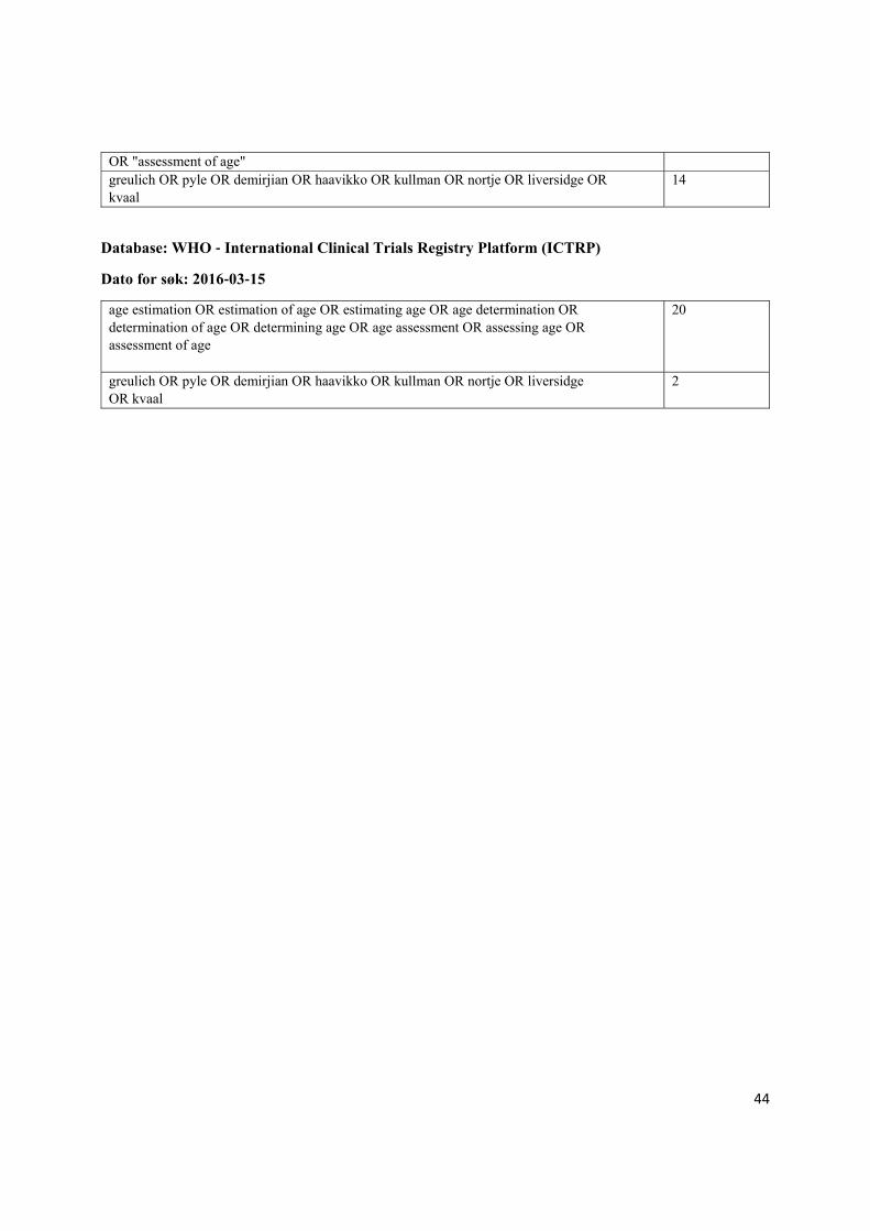
Dato for søk: 2016‐03‐15
"age estimation" OR "estimation of age" OR "estimating age" OR "age determination" OR "determination of age" OR "determining age" OR "age assessment" OR "assessing age"
16
44
OR "assessment of age" greulich OR pyle OR demirjian OR haavikko OR kullman OR nortje OR liversidge OR kvaal
14
Database: WHO ‐ International Clinical Trials Registry Platform (ICTRP)
Dato for søk: 2016‐03‐15
age estimation OR estimation of age OR estimating age OR age determination OR determination of age OR determining age OR age assessment OR assessing age OR assessment of age
20
greulich OR pyle OR demirjian OR haavikko OR kullman OR nortje OR liversidge OR kvaal
2

45
Appendix2.Descriptionofincludedstudieswithqualityassessment
Ekizoglu O, Hocaoglu E, Inci E, Can IO, Aksoy S, Sayin I. Estimation of forensic age using substages of ossification of the medial clavicle in living individuals. Int J Legal Med. 2015;129(6):1259-64.
Population: Country, ethnicity, place and year
Turkish patients who presented for trauma and other conditions at Istanbul between January 2014 and August 2014
Age and sex, sample 193 patients, 13-28 years, 64 (33.2%) females
Design of the study Retrospective
Index test Kellinghaus stage 2a-3c, slice thickness 1 mm
Aim of the study "we explored the utility of the subclassification of Kellinghaus et al. in such a population and compared our data to those of previous studies"
QUADAS-2 assessment
Patient selection method: Consecutive
Rating Comment
- Consecutive or random sample of patients?
Yes
- Avoid inappropriate exclusions? Yes "Patients with a history of any surgery or pathological bone conditions were also excluded"
DOMAIN 1: Patient selection Low risk
DOMAIN 1: Extra questions on age cohorts and age range
High risk
- Index test interpreted without knowledge of CA? Unclear No relevant description
DOMAIN 2: Index test interpretation Unclear risk
- CA interpreted without knowledge of SA?
Yes "Age was concealed from observers using a blind data feature of the software from the hospital"
DOMAIN 3: Reference standard Low risk
- All patients included in analysis? Yes DOMAIN 4: Patient flow and timing bias
Low risk
Ekizoglu O, Hocaoglu E, Inci E, Sayin I, Solmaz D, Bilgili MG, Can IO. Forensic age estimation by the Schmeling method: computed tomography analysis of the medial clavicular epiphysis. Int J Legal Med. 2015;129(1):203-10.
Population: Country, ethnicity, place and year
Turkish patients who presented for trauma and other conditions at Istanbul between January 2013 and November 2013
Age and sex, sample 503 patients, 10-35 years, 141 (28.0%) females

46
Design of the study Retrospective
Index test Schmeling stage 1-5, slice thickness 1 mm
Aim of the study "to enlarge the database by studying and reporting the results of staging the degree of ossification of the medial clavicle."
QUADAS-2 assessment
Patient selection method: Consecutive
Rating Comment
- Consecutive or random sample of patients?
Yes
- Avoid inappropriate exclusions? Yes
"Patients with a history of craniofacial or thoracic surgery were excluded from the study. CT scans that demonstrated clavicular fractures (45 cases) or developmental abnormalities (2 cases of clavicular aplasia) were also excluded."
DOMAIN 1: Patient selection Low risk
DOMAIN 1: Extra questions on age cohorts and age range
High risk
- Index test interpreted without knowledge of CA? Unclear No relevant description
DOMAIN 2: Index test interpretation Unclear risk
- CA interpreted without knowledge of SA?
Yes We assume age was recorded in the medical records when patients went to the hospital prior to this study
DOMAIN 3: Reference standard Low risk
- All patients included in analysis? Yes DOMAIN 4: Patient flow and timing bias
Low risk
Franklin D, Flavel A. CT evaluation of timing for ossification of the medial clavicular epiphysis in a contemporary Western Australian population. Int J Legal Med. 2015;129(3):583-94.
Population: Country, ethnicity, place and year
The sample comprises individuals who presented for clinical thoracic evaluation in the Western Australian hospital system between 2006 and 2013, 80 % of those patients were admitted between 2010 and 2013
Age and sex, sample 388 patients, 10-35 years, 178 (45.9%) females
Design of the study Retrospective
Index test Schmeling stage 1-5, slice thickness < 2 mm
Aim of the study "The primary objective of the study is to statistically quantify ossification and fusion timing in the medial clavicle epiphysis as visualized in high-resolution multislice CT scans"

47
QUADAS-2 assessment
Patient selection method: Consecutive
Rating Comment
- Consecutive or random sample of patients?
Yes
- Avoid inappropriate exclusions? Yes
"those presenting obvious abnormal morphologies were accordingly removed; exclusions were most commonly made on the basis of anatomical shape variants associated with the medial end"
DOMAIN 1: Patient selection Low risk
DOMAIN 1: Extra questions on age cohorts and age range
High risk
- Index test interpreted without knowledge of CA? Unclear No relevant description
DOMAIN 2: Index test interpretation Unclear risk
- CA interpreted without knowledge of SA?
Yes We assume age was recorded in the medical records when patients went to the hospital prior to this study
DOMAIN 3: Reference standard Low risk
- All patients included in analysis? Yes DOMAIN 4: Patient flow and timing bias
Low risk
Gurses MS, Inanir NT, Gokalp G, Fedakar R, Tobcu E, Ocakoglu G. Evaluation of age estimation in forensic medicine by examination of medial clavicular ossification from thin-slice computed tomography images. Int J Legal Med. 2016;130(5):1343-52.
Population: Country, ethnicity, place and year
CT scans were taken from patients between January 2012 and February 2014 at the Uludag University, Turkey
Age and sex, sample 725 patients, 10-35 years, 340 (46.9%) females
Design of the study Retrospective
Index test Schmeling stage 1-5 and Kellinghaus 2a-3c, slice thickness 0.6 - 1 mm
Aim of the study "The aim of our study was to use CT scans with 0.6 mm thick and 1 mm thick slices to assess the stage of clavicle ossification in the Turkish population"
QUADAS-2 assessment
Patient selection method: Consecutive
Rating Comment
- Consecutive or random sample of patients?
Yes
48
- Avoid inappropriate exclusions? Yes
Patients with endocrine disorders (36 cases), constitutional growth retardation or cerebral palsy (41 cases), clavicular fracture (17 cases), chronic illness (43 cases), developmental abnormalities (20 cases), artifacts due to motion or contrast medium (21 cases), missing or doubtful data on chronological age (7 cases), and anatomic shape variants (131 cases) were excluded from the study.
DOMAIN 1: Patient selection Low risk
DOMAIN 1: Extra questions on age cohorts and age range
High risk
- Index test interpreted without knowledge of CA? Yes "Prior to and during the image assessments, neither of the examiners knew the age of the patients."
DOMAIN 2: Index test interpretation Low risk
- CA interpreted without knowledge of SA?
Yes We assume age was recorded in the medical records when patients went to the hospital prior to this study
DOMAIN 3: Reference standard Low risk
- All patients included in analysis? Yes DOMAIN 4: Patient flow and timing bias
Low risk
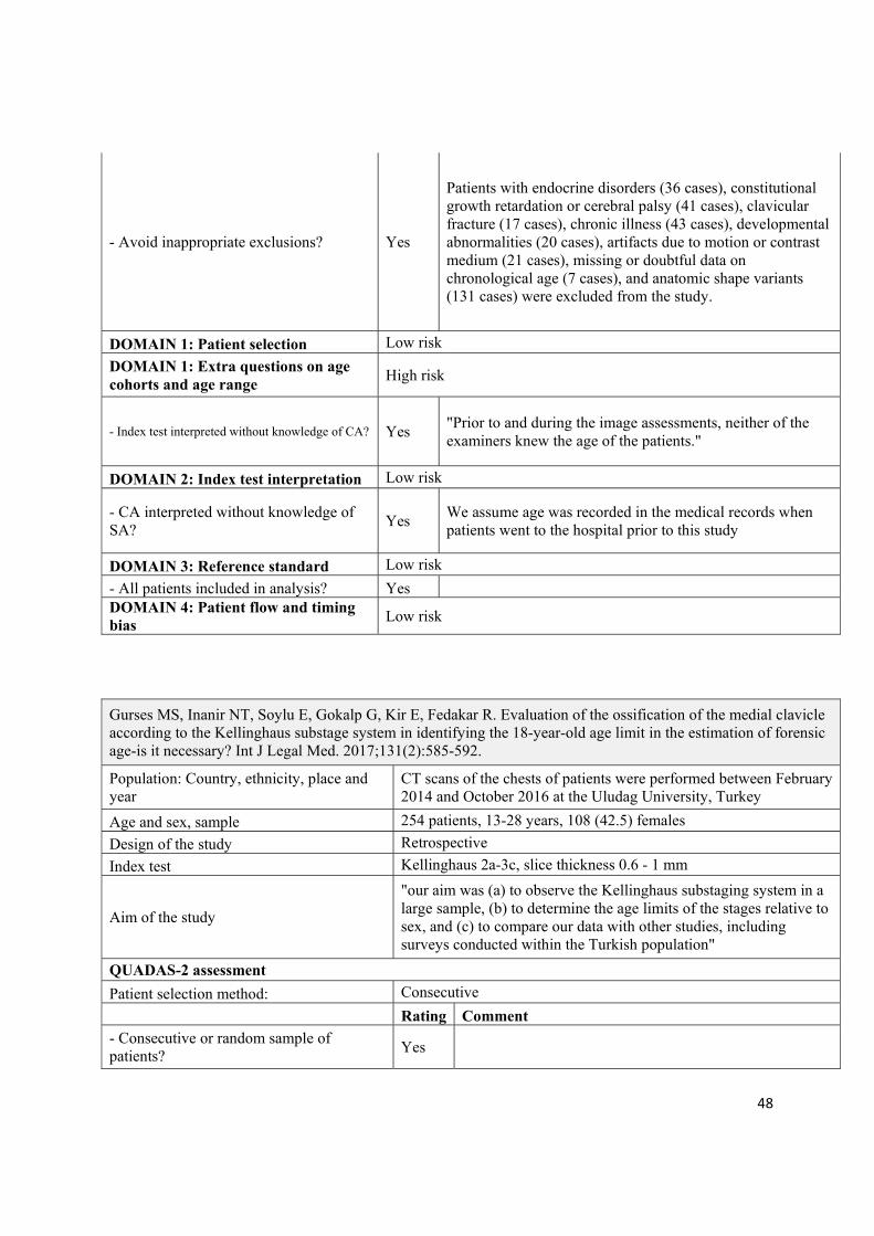
Gurses MS, Inanir NT, Soylu E, Gokalp G, Kir E, Fedakar R. Evaluation of the ossification of the medial clavicle according to the Kellinghaus substage system in identifying the 18-year-old age limit in the estimation of forensic age-is it necessary? Int J Legal Med. 2017;131(2):585-592.
Population: Country, ethnicity, place and year
CT scans of the chests of patients were performed between February 2014 and October 2016 at the Uludag University, Turkey
Age and sex, sample 254 patients, 13-28 years, 108 (42.5) females
Design of the study Retrospective
Index test Kellinghaus 2a-3c, slice thickness 0.6 - 1 mm
Aim of the study
"our aim was (a) to observe the Kellinghaus substaging system in a large sample, (b) to determine the age limits of the stages relative to sex, and (c) to compare our data with other studies, including surveys conducted within the Turkish population"
QUADAS-2 assessment
Patient selection method: Consecutive
Rating Comment
- Consecutive or random sample of patients?
Yes

49
- Avoid inappropriate exclusions? Yes
Patients with endocrine disorders (17 cases), constitutional growth retardation or cerebral palsy (26 cases), clavicular fracture (7 cases), chronic illness (25 cases), developmental abnormalities (9 cases), single clavicle (1 case, excision of clavicle), and those with missing or doubtful data on chronological age (2 cases) were excluded from the study.
DOMAIN 1: Patient selection Low risk
DOMAIN 1: Extra questions on age cohorts and age range
High risk
- Index test interpreted without knowledge of CA? Yes "Prior to and during the image assessments, the ages of the individuals were not known to the two radiologists."
DOMAIN 2: Index test interpretation Low risk
- CA interpreted without knowledge of SA? Yes We assume age was recorded in the medical records when patients went to the hospital prior to this study
DOMAIN 3: Reference standard Low risk
- All patients included in analysis? Yes DOMAIN 4: Patient flow and timing bias
Low risk
Pattamapaspong N, Madla C, Mekjaidee K, Namwongprom S. Age estimation of a Thai population based on maturation of the medial clavicular epiphysis using computed tomography. Forensic Sci Int. 2015;246:123.e1-5.
Population: Country, ethnicity, place and year
CT scans were taken between January 2007 and 2014 in patients of Thai nationals at Chiang Mai, Thailand.
Age and sex, sample 409 patients, 11-29 years, 160 (39.1%) females
Design of the study Retrospective
Index test Schmeling stage 1-5 and Kellinghaus 2a-3c, slice thickness 0.6 - 1 mm
Aim of the study "To assess the relationship between development of the medial clavicular epiphysis and age in a Thai population"
QUADAS-2 assessment
Patient selection method: Consecutive
Rating Comment
- Consecutive or random sample of patients?
Yes
50
- Avoid inappropriate exclusions? Yes "Patients with diseases that may affect bone development including previous fractures of the clavicle, chronic illness, and patients who were treated by steroids, chemotherapy, or immunosuppressive drugs were excluded from the study."
DOMAIN 1: Patient selection Low risk
DOMAIN 1: Extra questions on age cohorts and age range
High risk
- Index test interpreted without knowledge of CA?
Low risk
"Both readers were blinded for patient’s ages"
DOMAIN 2: Index test interpretation Low risk
- CA interpreted without knowledge of SA?
Yes We assume age was recorded in the medical records when patients went to the hospital prior to this study
DOMAIN 3: Reference standard Low risk
- All patients included in analysis? Yes DOMAIN 4: Patient flow and timing bias
Low risk
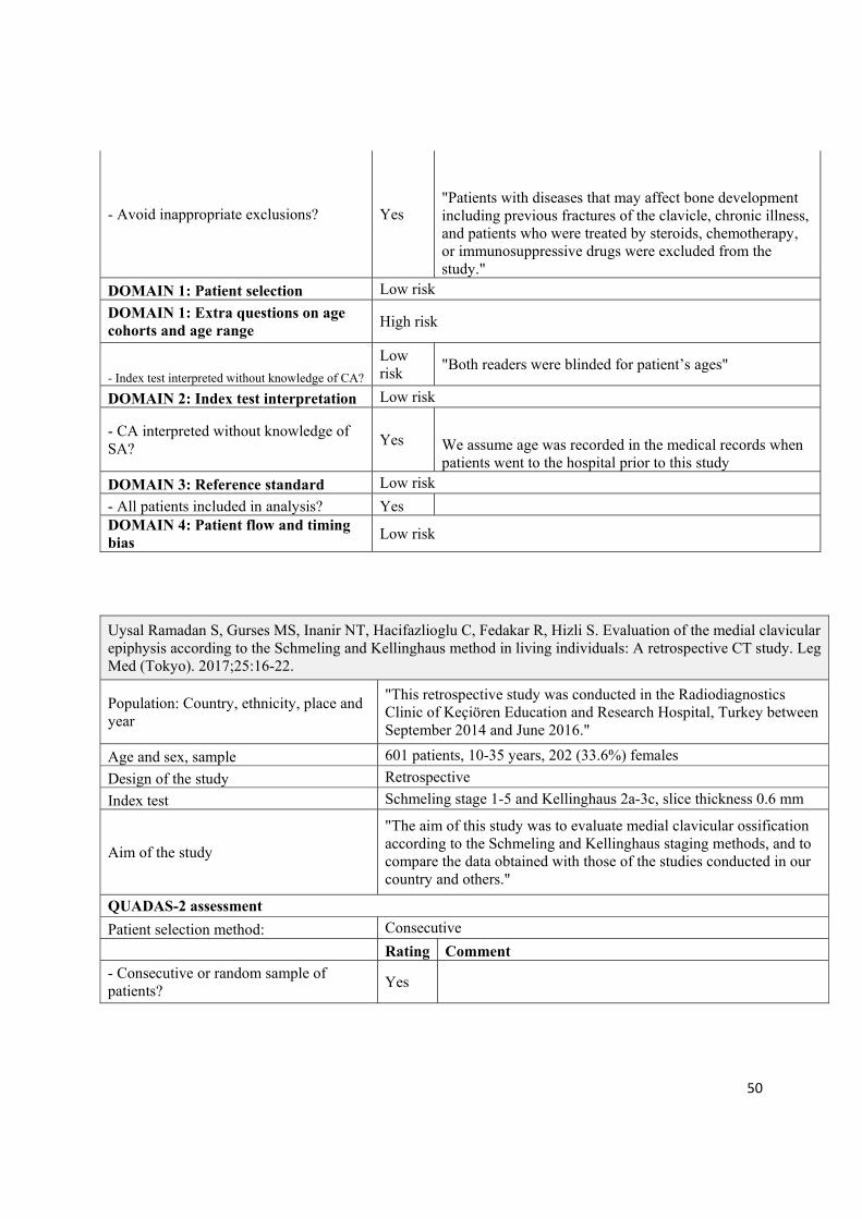
Uysal Ramadan S, Gurses MS, Inanir NT, Hacifazlioglu C, Fedakar R, Hizli S. Evaluation of the medial clavicular epiphysis according to the Schmeling and Kellinghaus method in living individuals: A retrospective CT study. Leg Med (Tokyo). 2017;25:16-22.
Population: Country, ethnicity, place and year
"This retrospective study was conducted in the Radiodiagnostics Clinic of Keçiören Education and Research Hospital, Turkey between September 2014 and June 2016."
Age and sex, sample 601 patients, 10-35 years, 202 (33.6%) females
Design of the study Retrospective
Index test Schmeling stage 1-5 and Kellinghaus 2a-3c, slice thickness 0.6 mm
Aim of the study
"The aim of this study was to evaluate medial clavicular ossification according to the Schmeling and Kellinghaus staging methods, and to compare the data obtained with those of the studies conducted in our country and others."
QUADAS-2 assessment
Patient selection method: Consecutive
Rating Comment
- Consecutive or random sample of patients?
Yes

51
- Avoid inappropriate exclusions? Yes
"Patients with endocrine disorders (36 cases), developmental abnormalities (37 cases), genetic disorder (40 cases), clavicular fracture (17 cases), artifacts due to motion or contrast medium (21 cases), and anatomic shape variants (107 cases) were excluded from the study"
DOMAIN 1: Patient selection Low risk
DOMAIN 1: Extra questions on age cohorts and age range
High risk
- Index test interpreted without knowledge of CA? Yes "Prior to and during the image assessments, neither of the examiners knew the age of the patients."
DOMAIN 2: Index test interpretation Low risk
- CA interpreted without knowledge of SA?
Yes We assume age was recorded in the medical records when patients went to the hospital prior to this study
DOMAIN 3: Reference standard Low risk
- All patients included in analysis? Yes DOMAIN 4: Patient flow and timing bias
Low risk
Ufuk F, Agladioglu K, Karabulut N. CT evaluation of medial clavicular epiphysis as a method of bone age determination in adolescents and young adults. Diagno Interv Radio. 2016;22(3):241-6.
Population: Country, ethnicity, place and year
The chest CT and pulmonary CT angiography examinations of patients acquired from September 2012 to June 2013 in Turkey were reviewed for this study
Age and sex, sample 300 patients, 10-30, 119 (39.7%) females
Design of the study Retrospective
Index test Modified Schmeling 5 stages, slice thickness 1 - 3 mm
Aim of the study "we aimed to assess the medial clavicular epiphysis ossification stage in Turkish population and find a computed tomography (CT) criterion to establish whether an individual is adult or not."
QUADAS-2 assessment
Patient selection method: Consecutive
Rating Comment
- Consecutive or random sample of patients?
Yes

52
- Avoid inappropriate exclusions? Yes
"Fifty-four subjects (15.2%) were excluded from the study, due to history of malignancy (n=40, 11.2%), rickets (n=2, 0.5%), growth hormone deficiency (n=1, 0.3%), thyroid hormone deficiency (n=3, 0.8%), and insufficient documentation of the sternoclavicular joints because of artifacts or significant anatomic variations (n=8, 2.3%)"
DOMAIN 1: Patient selection Low risk
DOMAIN 1: Extra questions on age cohorts and age range
High risk
- Index test interpreted without knowledge of CA? Yes
"The age of the individuals was blinded to the observers"
DOMAIN 2: Index test interpretation Low risk
- CA interpreted without knowledge of SA? Yes We assume age was recorded in the medical records when patients went to the hospital prior to this study
DOMAIN 3: Reference standard Low risk
- All patients included in analysis? Yes DOMAIN 4: Patient flow and timing bias
Low risk
Vieth V, Schulz R, Brinkmeier P, Dvorak J, Schmeling A. Age estimation in U-20 football players using 3.0 tesla MRI of the clavicle. Forensic Sci Int. 2014;241:118-22.
Population: Country, ethnicity, place and year
German males football players took the MRI examination at Munster, Germany from February 2011 to June 2011
Age and sex, sample 152 males football players, 18-22 year
Design of the study Prospective
Index test MRI Schmeling stage 1-5, Kellinghaus 2a-3c
Aim of the study "the present pilot study aims to verify whether an MRI examination of the clavicles appears a promising means of proving completion of the 20th year of life"
QUADAS-2 assessment
Patient selection method: Selective
Rating Comment
- Consecutive or random sample of patients?
Unclear "The study population comprised 30 test persons in each of the 18-, 19- and 21-year-old age categories and 31 test persons in each of the 20- and 22-year-old age categories"
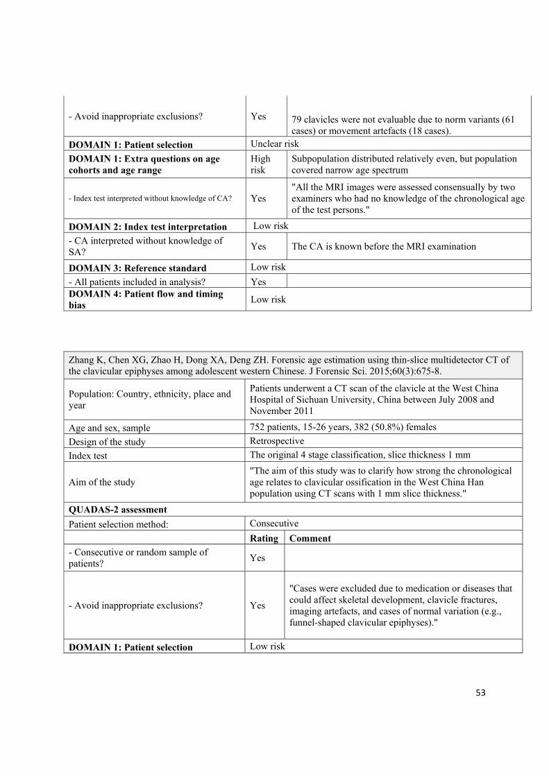
53
- Avoid inappropriate exclusions? Yes 79 clavicles were not evaluable due to norm variants (61 cases) or movement artefacts (18 cases).
DOMAIN 1: Patient selection Unclear risk
DOMAIN 1: Extra questions on age cohorts and age range
High risk
Subpopulation distributed relatively even, but population covered narrow age spectrum
- Index test interpreted without knowledge of CA? Yes "All the MRI images were assessed consensually by two examiners who had no knowledge of the chronological age of the test persons."
DOMAIN 2: Index test interpretation Low risk
- CA interpreted without knowledge of SA?
Yes The CA is known before the MRI examination
DOMAIN 3: Reference standard Low risk
- All patients included in analysis? Yes DOMAIN 4: Patient flow and timing bias
Low risk
Zhang K, Chen XG, Zhao H, Dong XA, Deng ZH. Forensic age estimation using thin-slice multidetector CT of the clavicular epiphyses among adolescent western Chinese. J Forensic Sci. 2015;60(3):675-8.
Population: Country, ethnicity, place and year
Patients underwent a CT scan of the clavicle at the West China Hospital of Sichuan University, China between July 2008 and November 2011
Age and sex, sample 752 patients, 15-26 years, 382 (50.8%) females
Design of the study Retrospective
Index test The original 4 stage classification, slice thickness 1 mm
Aim of the study "The aim of this study was to clarify how strong the chronological age relates to clavicular ossification in the West China Han population using CT scans with 1 mm slice thickness."
QUADAS-2 assessment
Patient selection method: Consecutive
Rating Comment
- Consecutive or random sample of patients?
Yes
- Avoid inappropriate exclusions? Yes
"Cases were excluded due to medication or diseases that could affect skeletal development, clavicle fractures, imaging artefacts, and cases of normal variation (e.g., funnel-shaped clavicular epiphyses)."
DOMAIN 1: Patient selection Low risk

54
DOMAIN 1: Extra questions on age cohorts and age range
High risk
Population distributed relatively even, but population covered narrow age spectrum
- Index test interpreted without knowledge of CA? Yes "Prior to and during analysis of the MDCT images, the age of the individuals was not known to the examiner."
DOMAIN 2: Index test interpretation Low risk
- CA interpreted without knowledge of SA?
Yes We assume age was recorded in the medical records when patients went to the hospital prior to this study
DOMAIN 3: Reference standard Low risk
- All patients included in analysis? Yes DOMAIN 4: Patient flow and timing bias
Low risk

55
Appendix3.Separatelist
In addition to the 10 included studies above, we identified 12 studies with relevant study design but no available data. These studies were listed below.
Reference Country Scoring
tool Scoring
scale Sample
size Sex
Age range
Wang YH, Wei H, Ying CL, Wan L, Zhu GY. The staging method of sternal end of clavicle epiphyseal growth by thin layer CT scan and imaging reconstruction. Fa Yi Xue Za Zhi. 2013;29(3):168-71.
China CT 1-5 460 Both 15-25
Kellinghaus M, Schulz R, Vieth V, Schmidt S, Schmeling A. Forensic age estimation in living subjects based on the ossification status of the medial clavicular epiphysis as revealed by thin-slice multidetector computed tomography. Int J Legal Med. 2010;124(2):149-54.
Germany CT 1-5 592 Both 10-35
Kreitner KF, Schweden F, Schild HH, Riepert T, Nafe B. Computerized tomography of the epiphyseal union of the medial clavicle: an auxiliary method of age determination during adolescence and the 3d decade of life?. Rofo. 1997;166(6):481-6.
Germany CT 1-4 279 Both 0-29
Kreitner KF, Schweden FJ, Riepert T, Nafe B, Thelen M. Bone age determination based on the study of the medial extremity of the clavicle. Eur Radiol. 1998;8(7):1116-22.
Germany CT 1-4 248 Both 0-29
Milenkovic P, Djuric M, Milovanovic P, Djukic K, Zivkovic V, Nikolic S. The role of CT analyses of the sternal end of the clavicle and the first costal cartilage in age estimation. Int J Legal Med. 2014;128(5):825-39.
Serbia CT 1-5 154 Both 15-35
Schulz R, Mühler M, Mutze S, Schmidt S, Reisinger W, Schmeling A. Studies on the time frame for ossification of the medial epiphysis of the clavicle as revealed by CT scans. Int J Legal Med. 2005;119(3):142-5.
Germany CT 1-5 556 Both 15-30
Schulze D, Rother U, Fuhrmann A, Richel S, Faulmann G, Heiland M. Correlation of age and ossification of the medial clavicular epiphysis using computed tomography. Forensic Sci Int. 2006;158(2-3):184-9.
Germany CT 2-4 100 Both 16-25
Vieth V, Kellinghaus M, Schulz R, Pfeiffer H, Schmeling A, Beurteilung des Ossifikationsstadiums der medialen Klavikulaepiphysenfuge. Rechtsmedizin. 2010;20(6):483-8.
Germany CT, MRI
1-5 Unclear Both Unclear
Hillewig E, De Tobel J, Cuche O, Vandemaele P, Piette M, Verstraete K. Magnetic resonance imaging of the medial extremity of the clavicle in forensic bone age determination: a new four-minute approach. Eur Radiol. 2011;21(4):757-67.
Belgium MRI 1-5 121 Both 11-30
Tangmose S, Jensen KE, Villa C, Lynnerup N. Forensic age estimation from the clavicle using 1.0T MRI--preliminary results. Forensic Sci Int. 2014;234:7-12.
Danmark MRI 1-4 74 Both 12-33
Houpert T, Rérolle C, Savall F, Telmon N, Saint-Martin P. Is a CT-scan of the medial clavicle epiphysis a good exam to attest to the 18-year threshold in forensic age estimation? Forensic Sci Int. 2016;260:103.e1-3.
France CT 1-5 319 Both 15-30

56
Hua W, Guang-you Z, Lei W, Chong-liang Y, Ya-hui W. Correlation between age and the parameters of medial epiphysis and metaphysis of the clavicle using CT volume rendering images. Forensic Sci Int. 2014;244:316.e1-7.
China CT Not
applicable 795 Both 15-25
57
Appendix4.Excludedstudies
Author Reason
Benito M, Sánchez JA, Codinha S. Age-at-death estimation based on radiological and image analysis methods in clavicle in a current Spanish population. Int J Legal Med. 2014;128(3):523-33.
This study used X-ray approach, not CT approach
Brough AL, Bennett J, Morgan B, Black S, Rutty GN. Anthropological measurement of the juvenile clavicle using multi-detector computed tomography--affirming reliability. J Forensic Sci. 2013;58(4):946-51.
Population is not living people
Cameriere R, De Luca S, De Angelis D, Merelli V, Giuliodori A, Cingolani M, Cattaneo C, Ferrante L. Reliability of Schmeling's stages of ossification of medial clavicular epiphyses and its validity to assess 18 years of age in living subjects. Int J Legal Med. 2012;126(6):923-32.
This study used X-ray approach, not CT or MR approach
Cao Z. The comparative study on girls skeletal age estimation methods. Chine J Forensic Med. 2012; 27(6): 438-41.
This study used X-ray approach, not CT or MR approach
Doorenbost H, Van Rijn RR, Robben SGF. X-rays of the medial clavicle for age determination according to the method of the immigration and naturalizaiton service: Unsuitable for the determination of adulthood (multiple letters). Nederlands Tijdschrift voor Geneeskunde. 2005;149(6): 323-4.
This is not an empirical study
Fujita T, Orimo H, Ohata M, Yoshikawa M. Changes in the cortical thickness of the clavicle according to age. J Am Geriatr Soc. 1968;16(4): 458-62.
This study used X-ray approach, not CT or MR approach
Garamendi PM, Landa MI, Botella MC, Alemán I. Forensic age estimation on digital X-ray images: Medial epiphyses of the clavicle and first rib ossification in relation to chronological age. J Forensic Sci. 2011;56 Suppl 1:S3-12.
This study used X-ray approach, not CT or MR approach
Gonsior M, Ramsthaler F, Gehl A, Verhoff MA. Morphology as a cause for different classification of the ossification stage of the medial clavicular epiphysis by ultrasound, computed tomography, and macroscopy. Int J Legal Med. 2013;127(5):1013-21.
Less than 50 people in total
Bassed RB, Drummer OH, Briggs C, Valenzuela A. Age estimation and the medial clavicular epiphysis: analysis of the age of majority in an Australian population using computed tomography. Forensic Sci Med Pathol. 2011;7(2):148-54.
Population is not living people
Hassan Q, Memon MU, Khalil IU, Ashraf T, Arif MM, Iqbal AS. Radiological Mean Age of Fusion of Medial End of the Clavicle: AParameter of Age. J Coll Physicians Surg Pak. 2016;26(1):18-22.
This study used X-ray approach, not CT or MR approach
Ji L, Terazawa K, Tsukamoto T, Haga K. Estimation of age from epiphyseal union degrees of the sternal end of the clavicle. Hokkaido Igaku Zasshi. 1994;69(1):104-11.
This study used anatomical approach to check osteometric parameters directly from remains
58
Jit I, Kulkarni M. Times of appearance and fusion of epiphysis at the medial end of the clavicle. Indian J Med Res. 1976;64(5):773-82.
This study used X-ray approach, not CT or MR approach
Lai X, Tang J, Tan Q, Lin H, Fu Y, Chen R. Skeletal age estimation in Dongguan males adolescence by digital shoulder X-ray. Chinese Journal of Forensic Medicine. 2013;28(6): 464-8.
This study used X-ray approach, not CT or MR approach
Langley NR. The lateral clavicular epiphysis: fusion timing and age estimation. Int J Legal Med. 2016;130(2):511-7.
This study used anatomical approach to check osteometric parameters directly from remains
Langley-Shirley N, Jantz RL. A Bayesian approach to age estimation in modern Americans from the clavicle. J Forensic Sci. 2010;55(3):571-83.
This study used anatomical approach to check osteometric parameters directly from remains
Mansour H, Fuhrmann A, Paradowski I, van Well EJ, Püschel K. The role of forensic medicine and forensic dentistry in estimating the chronological age of living individuals in Hamburg, Germany. Int J Legal Med. 2017;131(2):593-601.
This is not an empirical study
Mateen A, Afridi HK, Malik AR, Aziz K. Age estimation from medial end of clavicle by x-ray examination. PJMHS. 2013;7(4):1106-8.
This study used X-ray approach, not CT or MR approach
Meijerman L, Maat GJ, Schulz R, Schmeling A. Variables affecting the probability of complete fusion of the medial clavicular epiphysis. Int J Legal Med. 2007;121(6):463-8.
This is not an empirical study
Memon N, Memon MU, Memon K, Memon A. Radiological indicators to serve the purpose of juvenile justice system ordinance, 2000. J liaquat uni med helth sci. 2015;14(1):06-11.
This study used X-ray approach, not CT or MR approach
Milenkovic P, Djukic K, Djonic D, Milovanovic P, Djuric M. Skeletal age estimation based on medial clavicle--a test of the method reliability. Int J Legal Med. 2013;127(3):667-76.
This study used anatomical approach to check osteometric parameters directly from remains
Sarkar NR, Mukhopadhyay PP. Determination of age from the length of clavicle using digital X-ray in adolescent subjects: A preliminary study in Indian Bengali. Indian Journal of Forensic Medicine and Toxicology. 2015;9(1): 165-8.
This study used X-ray approach, not CT or MR approach
Schmeling A, Schulz R, Reisinger W, Mühler M, Wernecke KD, Geserick G. Studies on the time frame for ossification of the medial clavicular epiphyseal cartilage in conventional radiography. Int J Legal Med. 2004;118(1):5-8.
This study used X-ray approach, not CT or MR approach
Singh J, Chavali KH. Age estimation from clavicular epiphyseal union sequencing in a Northwest Indian population of the Chandigarh region. J Forensic Leg Med. 2011;18(2):82-7.
This study used anatomical approach to check osteometric parameters directly from remains

59
Sironi E, Gallidabino M, Weyermann C, Taroni F. Probabilistic graphical models to deal with age estimation of living persons. Int J Legal Med. 2016;130(2):475-88.
This study focused on mathematical validity for age estimation
Stout SD, Paine RR. Brief communication: histological age estimation using rib and clavicle. Am J Phys Anthropol. 1992;87(1):111-5.
Less than 50 people in total
Wan L. The analysis of variation of han females adolescent bone development in Henan and Zhejiang province. J Forensic Med. 2010;26(2):97-9.
This study used X-ray approach, not CT or MR approach
Wan L. Analysis of variation of Han males adolescent bone development in Hainan, Henan and Zhejiang provinces. J Forensic Med. 2012;28(1):21-3.
This study used X-ray approach, not CT or MR approach
Wang YH, Wei H, Ying CL, Wan L, Zhu GY. Progress in thin layer CT scan technology in estimating skeletal age of sternal end of clavicle. Fa Yi Xue Za Zhi. 2013;29(2):130-3.
This is not an empirical study
Wei H. Mathematical models for teenager's living age evaluation based on CT image of medial clavicular epiphysis.Fa Yi Xue Za Zhi. 2013;29(4):248-51.
This study is a duplicate of Hua (2014), which is included in the project
Wittschieber D, Ottow C, Schulz R, Püschel K, Bajanowski T, Ramsthaler F, Pfeiffer H, Vieth V, Schmidt S, Schmeling A. Forensic age diagnostics using projection radiography of the clavicle: a prospective multi-center validation study. Int J Legal Med. 2016;130(1):213-9.
Population is not living people
Wittschieber D, Schulz R, Pfeiffer H, Schmeling A, Schmidt S. Systematic procedure for identifying the five main ossification stages of the medial clavicular epiphysis using computed tomography: a practical proposal for forensic age diagnostics. Int J Legal Med. 2017;131(1):217-224.
This is not an empirical study
Yoon SH, Yoo HJ, Yoo R-E, et al. Ossification of the Medial Clavicular Epiphysis on Chest Radiographs: Utility and Diagnostic Accuracy in Identifying Korean Adolescents and Young Adults under the Age of Majority. Journal of Korean Medical Science. 2016;31(10):1538-1545.
This study used X-ray approach, not CT or MR approach
Zhang JZ, Shu YK, Chen SX. Age determination by the Chinese males clavicle. Chinese Journal of Forensic Medicine. 1989;4(3): 134-7.
This study used anatomical approach to check osteometric parameters directly from remains
60
Tangmose S, Jensen KE, Lynnerup N. Comparative study on developmental stages of the clavicle by postmortem MRI and CT imaging. J Forensic Radio Imag. 2013;1(3):102-6.
Population is not living people
Wittschieber D, Schulz R, Vieth V, Küppers M, Bajanowski T, Ramsthaler F, Püschel K, Pfeiffer H, Schmidt S, Schmeling A. The value of sub-stages and thin slices for the assessment of the medial clavicular epiphysis: a prospective multi-center CT study. Forensic Sci Med Pathol. 2014;10(2):163-9.
Population is not living people
Schmidt S, Henke CA, Wittschieber D, Vieth V, Bajanowski T, Ramsthaler F, Püschel K, Pfeiffer H, Schmeling A, Schulz R. Optimising magnetic resonance imaging-based evaluation of the ossification of the medial clavicular epiphysis: a multi-centre study. Int J Legal Med. 2016;130(6):1615-1621.
Population is not living people
Schulz R, Mühler M, Reisinger W, Schmidt S, Schmeling A. Radiographic staging of ossification of the medial clavicular epiphysis. Int J Legal Med. 2008;122(1):55-8.
No population age presented
Schmidt S, Mühler M, Schmeling A, Reisinger W, Schulz R. Magnetic resonance imaging of the clavicular ossification. Int J Legal Med. 2007;121(4):321-4.
Eligible sample size is less than 50.
Wittschieber D, Schulz R, Vieth V, Küppers M, Bajanowski T, Ramsthaler F, Püschel K, Pfeiffer H, Schmidt S, Schmeling A. Influence of the examiner's qualification and sources of error during stage determination of the medial clavicular epiphysis by means of computed tomography. Int J Legal Med. 2014;128(1):183-91.
Population is not living people

61
Appendix5.Projectprotocol
Note: This protocol is published on December 2016, which is a combination for several projects,
including age estimation with hand, third molar teeth, clavicle and knee and ankle.
We will conduct systematic reviews of the precision and accuracy of methods for age estimation of young people (10 to 25 years of age).
Literature search
Research librarian, Gyri Hval Straumann (GHS), will plan the searches in collaboration with the project
group. Another research librarian will peer review the search strategy. GHS will execute the electronic
searches for literature in the following databases:
MEDLINE
Embase
Cochrane Central Register of Controlled Trials (CENTRAL)
Google scholar
In addition, we will search for ongoing trials in Clinicaltrials.gov and WHO International Clinical Trials
Registry Platform, and check references and citations of relevant articles in Web of Science.
Inclusion criteria
Study design: We will include studies that compare an age indicator of one of the index tests
specified below with known chronological age.
Population: Living persons between the age of 10 and 25 years old
Index test: Age estimation tests including:
X-ray of the hand-wrist
dental X-ray
CT or MR of the clavicle, knee or ankle
Reference test: Known chronological age
Outcome: For x-ray of the hand: The difference between the estimated age
and chronological age, or the distribution of chronological age

62
given a specific age indicator (typically a pre-defined
development stage).
For x-ray of the teeth or CT or MR of the clavicle, knee or ankle:
The distribution of chronological age given a specific age
indicator (typically a pre-defined development stage).
Language: No restrictions in the search
Exclusion criteria
We will exclude studies that have fewer than 50 living persons that qualify for our analysis.
Article selection
At least two reviewers will independently screen each of the identified references. We will conduct one
combined literature search. Because we expect many studies relevant for inclusion, we plan to separate the
potentially relevant references into three groups for full-text screening where we will handle each index
test separately, resulting in three systematic reviews. At least two authors will independently screen each
of the full-text references considered potentially relevant.
Assessment of methodological quality and data extraction
At least two authors will independently use QUADAS-2 to assess methodological quality of each of the
included studies. We will resolve any disagreement by discussion or by consulting a third reviewer. One
person will collect data about population (age, ethnicity and sex), where and when the studies were
conducted, the reference and index tests used, and the results. Another person will check the extraction.
Statistical methods
In the current project, we would like to estimate the mean chronological age and its variation given a
certain pre-defined developments stage. Therefore, we will present the mean chronological age and its
95% confidence interval separately in both genders (male/female) for all relevant development stages or
categories of age (when mean difference is the outcome).
.
63
We will use random effects models to pool summary data at trial level. Potential heterogeneity of country
and publication date will be explored in Meta-regression. Publication bias will be assessed with Funnel
plots and Egger's regression model (1).
Diagnostic test accuracy methods
All results will be checked for statistical heterogeneity using the Cochrane Q statistic (P<0.10) and I2
methodology (I2>50%). Statistical significance is set at a P value<0.05. The meta-analysis will be
performed using R software (R Core Team, Vienna, Austria; version 3.3.2; using packages “glm”, “lme4”
and “metafor”).
References
1. Egger, M., et al., Bias in meta-analysis detected by a simple, graphical test. BMJ, 1997. 315(7109): p. 629-34.

Published by the Norwegian Institute of Public Health September 2018P.O.B 4404 NydalenNO-0403 OsloTelefon: + 47-21 07 70 00The report can be downloaded as pdf at www.fhi.no/en/publ/


![Transcriptional Network Controlling Endochondral Ossification · branous ossification and endochondral ossification.[1] During intramembranous ossification, osteoblasts produce type](https://img.pdfslide.us/doc/110x75/5e8cf0c24763783dcf0d78ef/transcriptional-network-controlling-endochondral-ossification-branous-ossification.jpg)