Embed Size (px)
Citation preview
Copyright © 2006 Pearson Education, Inc., publishing as Benjamin Cummings
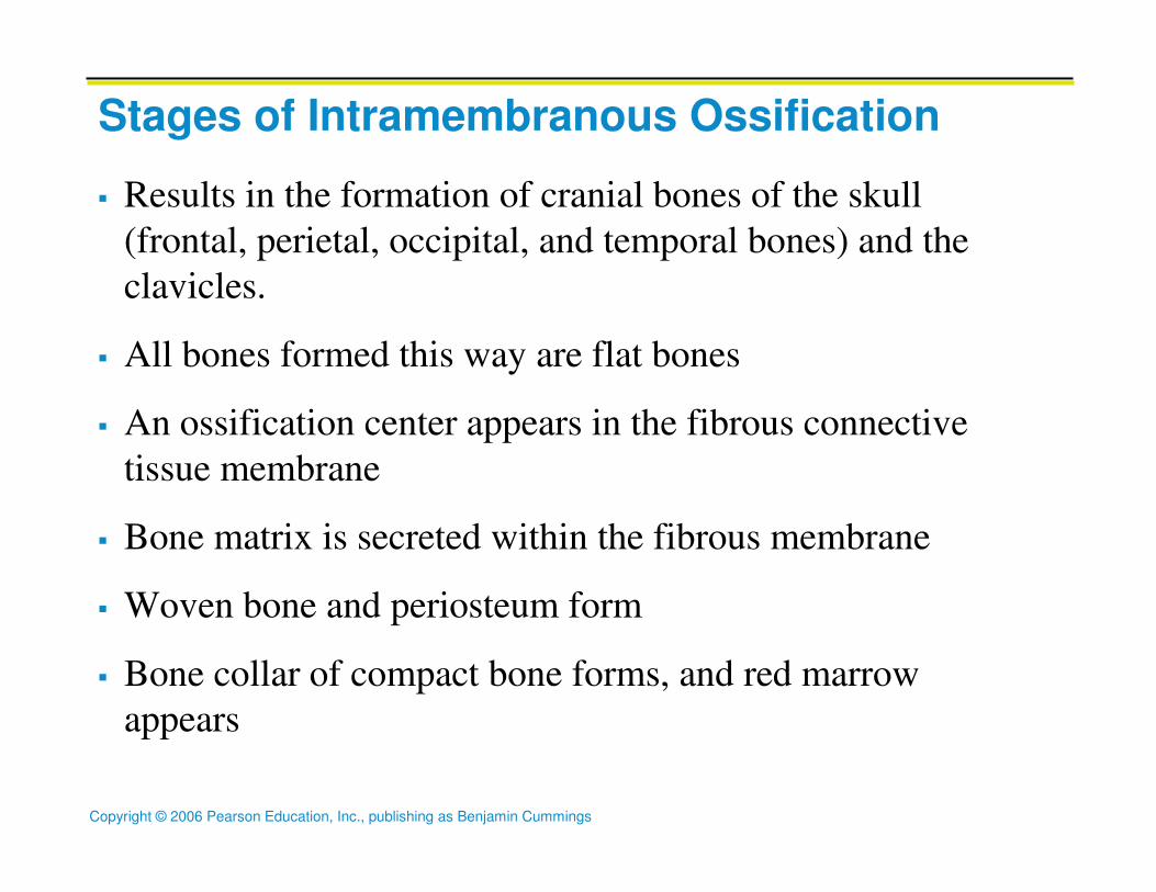
Stages of Intramembranous Ossification
� Results in the formation of cranial bones of the skull
(frontal, perietal, occipital, and temporal bones) and the
clavicles.
� All bones formed this way are flat bones
� An ossification center appears in the fibrous connective
tissue membrane
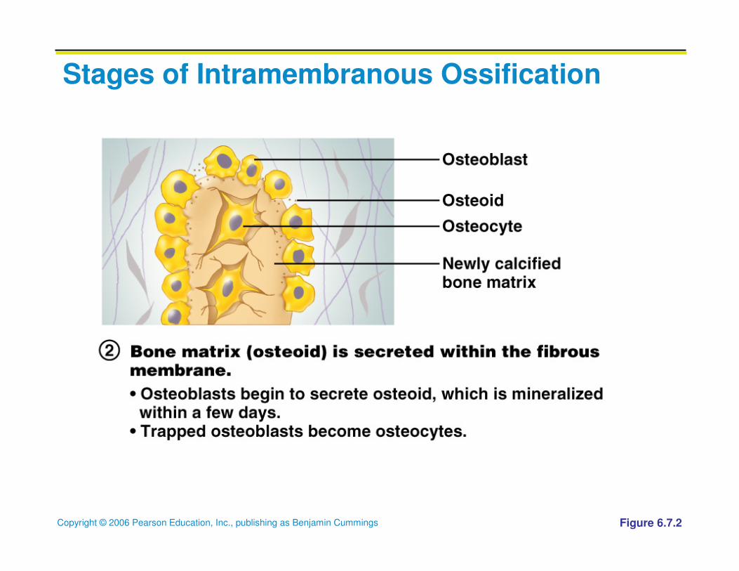
� Bone matrix is secreted within the fibrous membrane
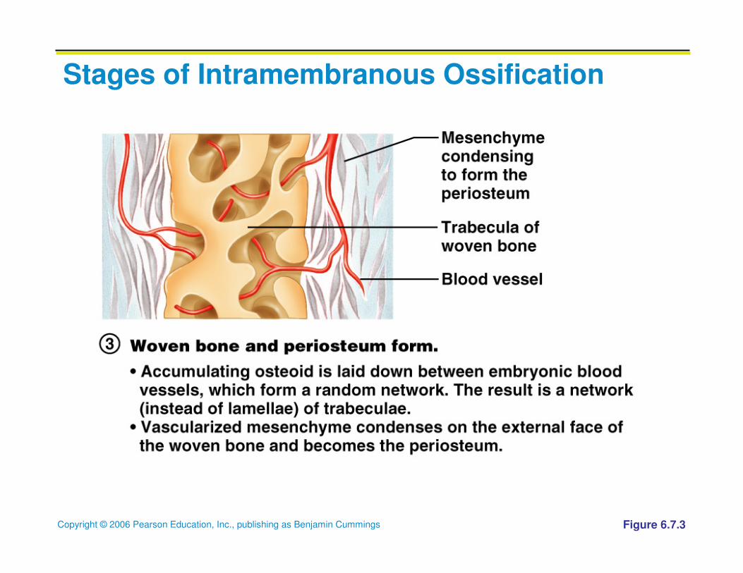
� Woven bone and periosteum form
� Bone collar of compact bone forms, and red marrow
appears
Copyright © 2006 Pearson Education, Inc., publishing as Benjamin Cummings
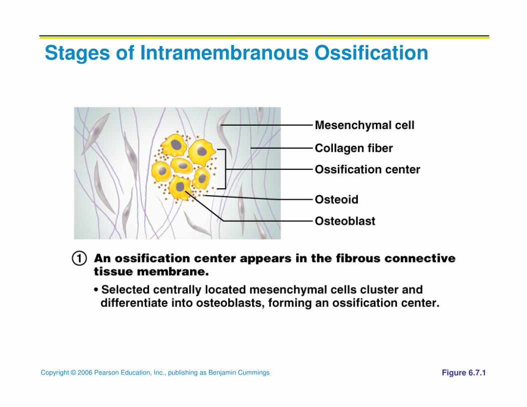
Stages of Intramembranous Ossification
Figure 6.7.1
Copyright © 2006 Pearson Education, Inc., publishing as Benjamin Cummings
Stages of Intramembranous Ossification
Figure 6.7.2
Copyright © 2006 Pearson Education, Inc., publishing as Benjamin Cummings
Stages of Intramembranous Ossification
Figure 6.7.3
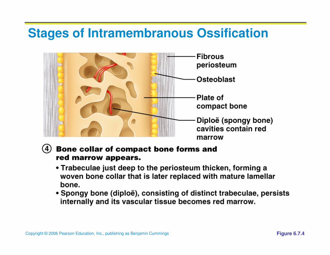
Copyright © 2006 Pearson Education, Inc., publishing as Benjamin Cummings
Stages of Intramembranous Ossification
Figure 6.7.4
Copyright © 2006 Pearson Education, Inc., publishing as Benjamin Cummings
Endochondral Ossification
� Results in the formation of all of the rest of the
bones
� Begins in the second month of development
� Uses hyaline cartilage “bones” as models for bone
construction
� Requires breakdown of hyaline cartilage prior to
ossification
� Formation begins at the primary ossification center
Copyright © 2006 Pearson Education, Inc., publishing as Benjamin Cummings
Endochondral Ossification
� The perichondrium covering the hyaline cartilage
“bone” is infiltrated with blood vessels converting
it to vascularized periosteum.
� This change in nutrition causes the underlying
mesenchymal cells to specialize into osteoblasts
Copyright © 2006 Pearson Education, Inc., publishing as Benjamin Cummings
Stages of Endochondral Ossification
� Formation of bone collar
� Cavitation of the hyaline cartilage
� Invasion of internal cavities by the periosteal bud,
and spongy bone formation
� Formation of the medullary cavity; appearance of
secondary ossification centers in the epiphyses
� Ossification of the epiphyses, with hyaline
cartilage remaining only in the epiphyseal plates
Copyright © 2006 Pearson Education, Inc., publishing as Benjamin Cummings
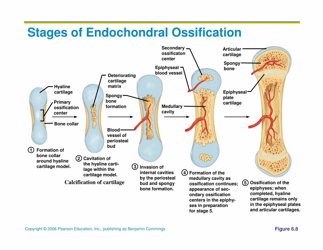
Stages of Endochondral Ossification
Figure 6.8
Formation of
bone collararound hyaline
cartilage model.
Hyalinecartilage
Cavitation of
the hyaline carti-lage within the
cartilage model.
Invasion of
internal cavitiesby the periosteal
bud and spongybone formation.
Formation of themedullary cavity as
ossification continues;appearance of sec-
ondary ossificationcenters in the epiphy-
ses in preparationfor stage 5.
Ossification of theepiphyses; when
completed, hyalinecartilage remains only
in the epiphyseal platesand articular cartilages.
Deterioratingcartilagematrix
Epiphyseal
blood vessel
Spongybone
formation
Epiphyseal
platecartilage
Secondary
ossificatoncenter
Bloodvessel of
periostealbud
Medullarycavity
Articular
cartilage
Spongybone
Primary
ossificationcenter
Bone collar
1
2
3
4
5Calcification of cartilage
Copyright © 2006 Pearson Education, Inc., publishing as Benjamin Cummings
Postnatal Bone Growth
� Growth in length of long bones
� Cartilage on the side of the epiphyseal plate closest
to the epiphysis is relatively inactive
� Cartilage abutting the shaft of the bone organizes
into a pattern that allows fast, efficient growth
� Cells of the epiphyseal plate proximal to the resting
cartilage form three functionally different zones:
growth, transformation, and osteogenic
Copyright © 2006 Pearson Education, Inc., publishing as Benjamin Cummings
Details of Stages of Endochondral Ossification
� 1) Bone collar forms around the diaphysis of the
hyaline cartilage model
� Osteoblasts of the converted periosteum secrete
osteoid against the hyaline cartilage diaphysis
encasing it in a bone collar
Copyright © 2006 Pearson Education, Inc., publishing as Benjamin Cummings
Details of Stages of Endochondral Ossification
� 2) cartilage in the center of the diaphysis calcifies
and then cavitates
� Chondrocytes w/I the shaft hypertrophy &
signal surrounding cartilage matrix to calcify.
� Chondrocytes die due to lack of nutrients
(impermeability of calcified matrix)
� Matrix deteriorates thus opening up cavities
Copyright © 2006 Pearson Education, Inc., publishing as Benjamin Cummings
Details of Stages of Endochondral Ossification
� 3) Periosteal bud invades the internal cavities and spongy
bone forms
� The forming cavities are invaded by a collection of
elements
� Periosteal bud contains a nutrient artery and vein,
lymphatics, nerve fibers, red marrow elements,
osteoblasts, and osteoclasts
� Osteoclasts erode the calcified cartilage matrix &
osteoblasts secrete osteoid around the remaining hyaline
cartilage forming bone-covered cartilage trabuculae (the
formation of spongy bone)
Copyright © 2006 Pearson Education, Inc., publishing as Benjamin Cummings
Details of Stages of Endochondral Ossification
� 4) The diaphysis elongates and a medullary cavity
forms
� Osteoclasts open up a medullary cavity by
breaking down the newly formed spongy bone
� Cartilage is growing, bones being calcified and
eroded and then replaced by bony spicules on
the epiphyseal surfaces facing the medullary
cavity
Copyright © 2006 Pearson Education, Inc., publishing as Benjamin Cummings
Details of Stages of Endochondral Ossification
� 5) The epiphysis ossify
� Secondary ossification centers appear in one or
both epiphyses.
� Steps 1-4 occur there except no medullary
cavity forms
Copyright © 2006 Pearson Education, Inc., publishing as Benjamin Cummings
Details of Stages of Endochondral Ossification
� Finally, hyaline cavity remains at:
� Epiphyseal surface (articular cartilage)
� Epiphyseal plates (junction of the diaphysis and
the epiphysis)
Copyright © 2006 Pearson Education, Inc., publishing as Benjamin Cummings
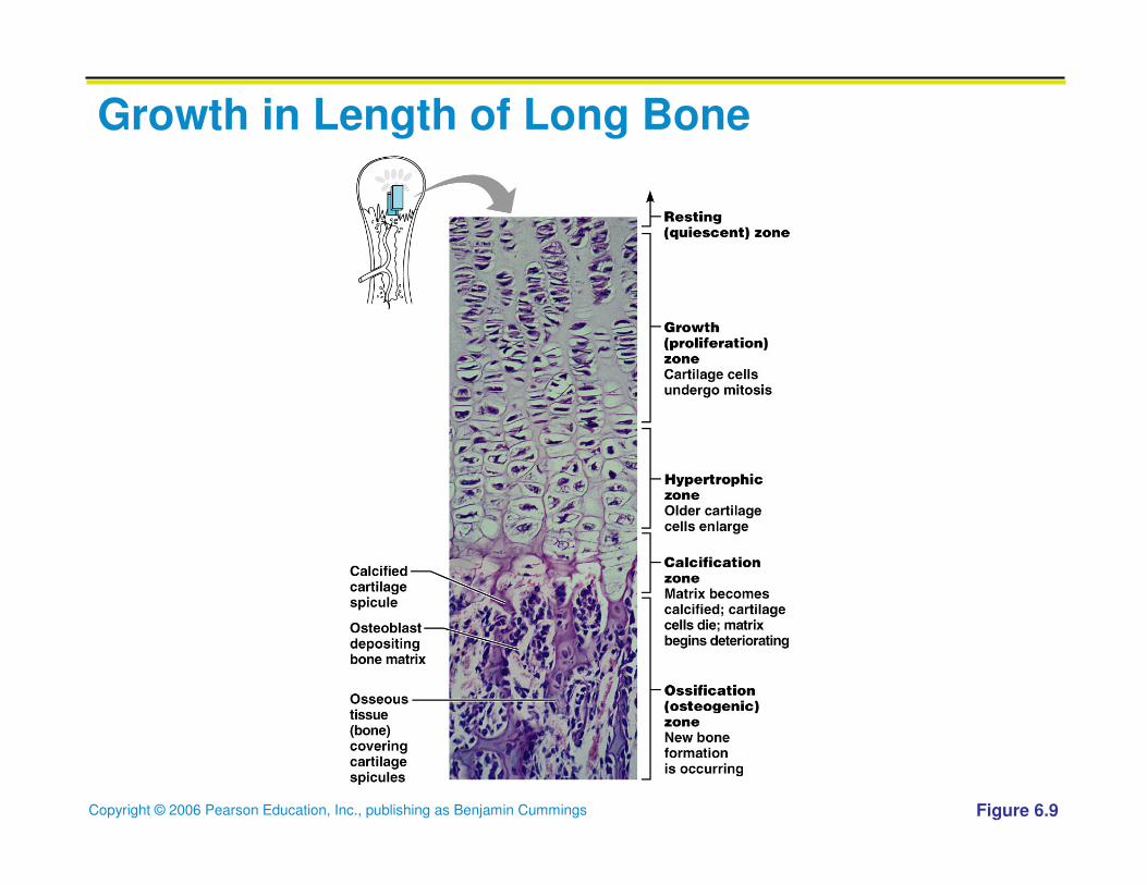
Functional Zones in Long Bone Growth
� Growth zone – cartilage cells undergo mitosis,
pushing the epiphysis away from the diaphysis
� Transformation zone – older cells enlarge, the
matrix becomes calcified, cartilage cells die, and
the matrix begins to deteriorate
� Osteogenic zone – new bone formation occurs
Copyright © 2006 Pearson Education, Inc., publishing as Benjamin Cummings
Postnatal Bone Growth
� Long bones lengthen by interstial growth of the
epiphyseal plates, and increase thickness by
appositional growth
Copyright © 2006 Pearson Education, Inc., publishing as Benjamin Cummings
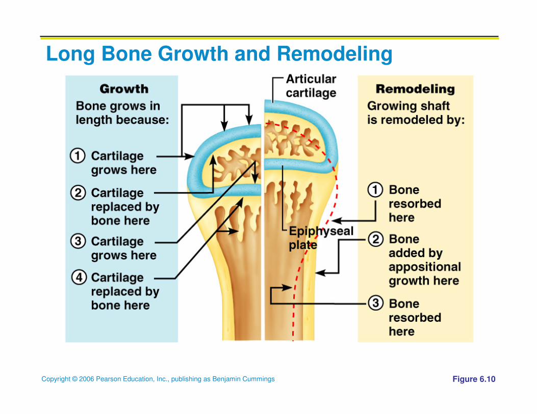
Long Bone Growth and Remodeling
� Growth occurs at the epiphyseal plate (the cartilage
abutting the diaphysis) called the Growth Zone
� Cartilage cells stack and divide quickly pushing the
epiphysis away from the diaphysis causing bone to
lengthen
� The older chondrocytes die & deteriorate forming the
Calcification Zone
� The resulting calcified spicules become part of the
Ossification Zone and are invaded by marrow elements
from the medullary cavity
Copyright © 2006 Pearson Education, Inc., publishing as Benjamin Cummings
Growth in Length of Long Bone
Figure 6.9
Copyright © 2006 Pearson Education, Inc., publishing as Benjamin Cummings
Long Bone Growth and Remodeling
Figure 6.10
Copyright © 2006 Pearson Education, Inc., publishing as Benjamin Cummings
Long Bone Growth and Remodeling
� Longitudinal growth is accompanied by
remodelling which includes appositional growth to
thicken bone
� Includes bone formation & reabsorption
� Bone growth stops around age 21 for males and 18
for females when the epiphysis & diaphysis fuse
(epiphyseal plate closure)
Copyright © 2006 Pearson Education, Inc., publishing as Benjamin Cummings
Long Bone Growth and Remodeling
� Growth in width (thickness) via appositional
growth
� Osteoblasts beneath the periosteum secrete bone
matrix on the external bone surface as osteoclasts
on the endosteal surface of the dyaphysis remove
bone
Copyright © 2006 Pearson Education, Inc., publishing as Benjamin Cummings
� During infancy and childhood, epiphyseal plate activity is stimulated by growth hormone (released by the anterior pituitary)
� During puberty, testosterone and estrogens:
� Initially promote adolescent growth spurts
� Cause masculinization and feminization of specific parts of the skeleton
� Later induce epiphyseal plate closure, ending longitudinal bone growth
Hormonal Regulation of Bone Growth During Youth
Copyright © 2006 Pearson Education, Inc., publishing as Benjamin Cummings
Bone Remodeling
� Remodeling units – adjacent osteoblasts and
osteoclasts deposit and resorb bone at periosteal
and endosteal surfaces
Copyright © 2006 Pearson Education, Inc., publishing as Benjamin Cummings
Bone Deposition
� Occurs where bone is injured or added strength is
needed
� Requires a diet rich in protein, vitamins C, D, and
A, calcium, phosphorus, magnesium, and
manganese
� Alkaline phosphatase is essential for
mineralization of bone
Copyright © 2006 Pearson Education, Inc., publishing as Benjamin Cummings
Bone Deposition
� Sites of new matrix deposition are revealed by the:
� Osteoid seam – unmineralized band of bone matrix
� Calcification front – abrupt transition zone between
the osteoid seam and the older mineralized bone
Copyright © 2006 Pearson Education, Inc., publishing as Benjamin Cummings
Bone Resorption
� Accomplished by osteoclasts (giant, multinucleated cells that arise from the same stem cells that produce macrophages)
� Resorption bays – grooves formed by osteoclasts as they break down bone matrix
� The osteoclast membrane seals off the bone that is to be broken down
� Resorption involves osteoclast secretion of:
� Lysosomal enzymes that digest organic matrix
� Hydrochloric acid that converts calcium salts into soluble forms
� The broken down products are endocytosed (transcytosed) and released into the interstitial fluid and blood
Copyright © 2006 Pearson Education, Inc., publishing as Benjamin Cummings
Control of Remodeling
� Two control loops regulate bone remodeling
� Hormonal mechanism maintains calcium
homeostasis in the blood (negative feedback)
� Mechanical and gravitational forces acting on the
skeleton
Copyright © 2006 Pearson Education, Inc., publishing as Benjamin Cummings
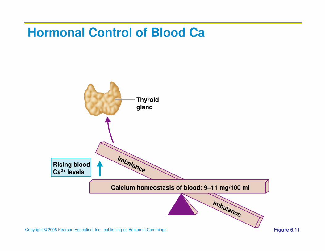
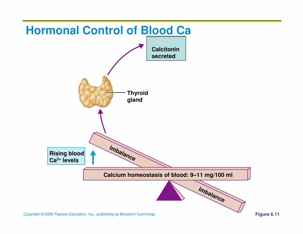
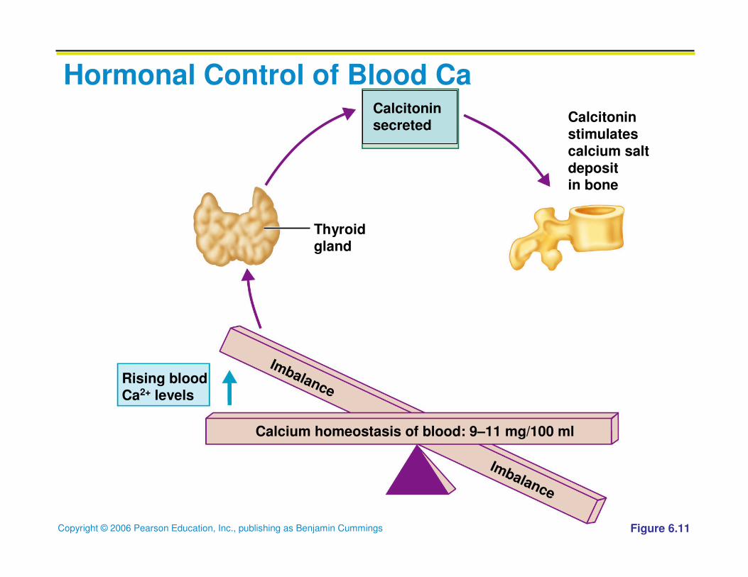
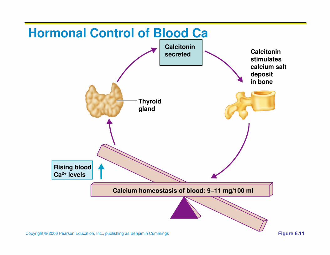
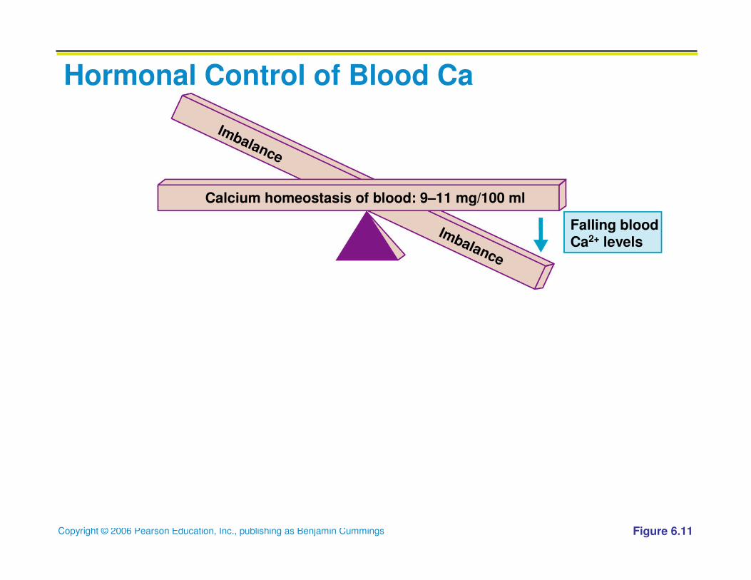
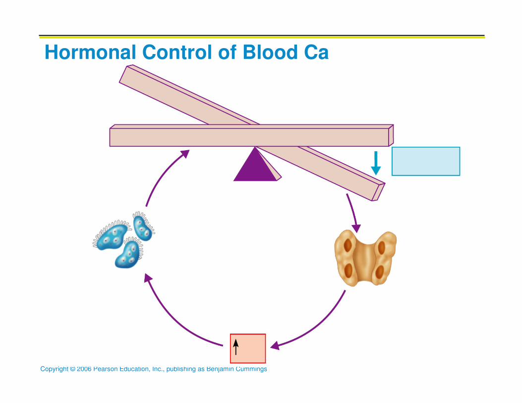
Hormonal Mechanism
� Rising blood Ca2+ levels trigger the thyroid to
release calcitonin
� Calcitonin inhibits bone resorption and stimulates
calcium salt deposit in bone
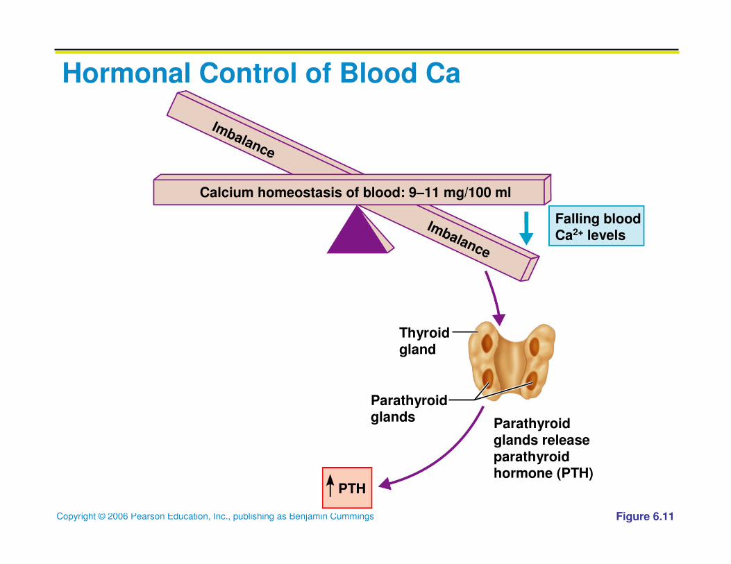
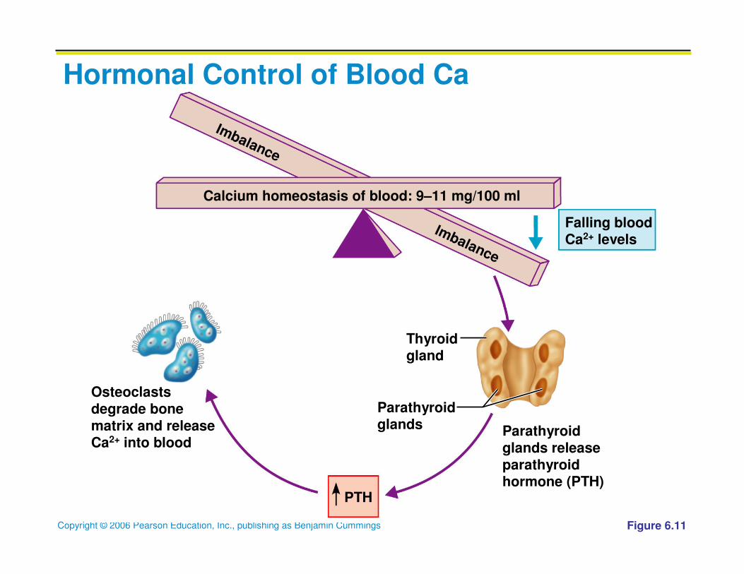
� Falling blood Ca2+ levels signal the parathyroid
glands to release parathyroid hormone (PTH)
� PTH signals osteoclasts to degrade bone matrix
and release Ca2+ into the blood
Copyright © 2006 Pearson Education, Inc., publishing as Benjamin Cummings
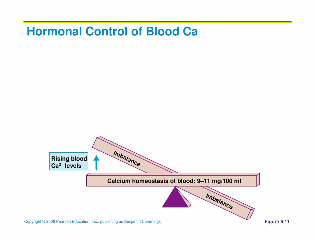
Hormonal Control of Blood Ca
Figure 6.11
Rising bloodCa2+ levels
Calcium homeostasis of blood: 9–11 mg/100 ml
Imbalance
Imbalance
Copyright © 2006 Pearson Education, Inc., publishing as Benjamin Cummings
Hormonal Control of Blood Ca
Figure 6.11
Thyroidgland
Rising bloodCa2+ levels
Calcium homeostasis of blood: 9–11 mg/100 ml
Imbalance
Imbalance
Copyright © 2006 Pearson Education, Inc., publishing as Benjamin Cummings
Hormonal Control of Blood Ca
Figure 6.11
Thyroidgland
Rising bloodCa2+ levels
Calcium homeostasis of blood: 9–11 mg/100 ml
Imbalance
Imbalance
Calcitoninsecreted
Copyright © 2006 Pearson Education, Inc., publishing as Benjamin Cummings
Hormonal Control of Blood Ca
Figure 6.11
Calcitoninstimulatescalcium saltdepositin bone
Thyroidgland
Rising bloodCa2+ levels
Calcium homeostasis of blood: 9–11 mg/100 ml
Imbalance
Imbalance
Calcitoninsecreted
Copyright © 2006 Pearson Education, Inc., publishing as Benjamin Cummings
Hormonal Control of Blood Ca
Figure 6.11
Calcitoninstimulatescalcium saltdepositin bone
Thyroidgland
Rising bloodCa2+ levels
Calcium homeostasis of blood: 9–11 mg/100 ml
Calcitoninsecreted
Copyright © 2006 Pearson Education, Inc., publishing as Benjamin Cummings
Hormonal Control of Blood Ca
Falling bloodCa2+ levels
Calcium homeostasis of blood: 9–11 mg/100 ml
Imbalance
Imbalance
Figure 6.11
Copyright © 2006 Pearson Education, Inc., publishing as Benjamin Cummings
Hormonal Control of Blood Ca
Parathyroidglands releaseparathyroidhormone (PTH)
Thyroidgland
Parathyroidglands
Falling bloodCa2+ levels
Calcium homeostasis of blood: 9–11 mg/100 ml
Imbalance
Imbalance
Figure 6.11
Copyright © 2006 Pearson Education, Inc., publishing as Benjamin Cummings
Hormonal Control of Blood Ca
Parathyroidglands releaseparathyroidhormone (PTH)
Thyroidgland
Parathyroidglands
Falling bloodCa2+ levels
Calcium homeostasis of blood: 9–11 mg/100 ml
PTH
Imbalance
Imbalance
Figure 6.11
Copyright © 2006 Pearson Education, Inc., publishing as Benjamin Cummings
Hormonal Control of Blood Ca
Parathyroidglands releaseparathyroidhormone (PTH)
Thyroidgland
Parathyroidglands
Osteoclastsdegrade bonematrix and releaseCa2+ into blood
Falling bloodCa2+ levels
Calcium homeostasis of blood: 9–11 mg/100 ml
PTH
Imbalance
Imbalance
Figure 6.11
Copyright © 2006 Pearson Education, Inc., publishing as Benjamin Cummings
Hormonal Control of Blood Ca

ERROR: undefined
OFFENDING COMMAND: f‘~
STACK:


![The Shoulder - madinaortho.com · [The Shoulder] Page | 11 Applied Anatomy BOONNEESS Sccaappuullaa Body is formed by intramembranous ossification Glenoid has 2 ossific centers](https://img.pdfslide.us/doc/110x75/5c07eea109d3f2a9648bd215/the-shoulder-the-shoulder-page-11-applied-anatomy-boonneess-sccaappuullaa.jpg)