Embed Size (px)
Citation preview
4CHAPTER
AGC kinases control phosphorylation and activation of
eukaryotic translation initiation factor 4B
Ankie G.M. van Gorp1, Arjan B. Brenkman2, Niels van den Broek2, Cornelis F. Calkhoven3, John W. Hershey4, Boudewijn M.T. Burgering2,
and Paul J. Coffer1
1Molecular Immunology Lab, Department of Immunology, University Medical Center Utrecht, Utrecht, The Netherlands
2Department of Physiological Chemistry, Centre for Biomedical Genetics, University Medical Center Utrecht, Utrecht, The Netherlands,
3Leibniz Institute for Age Research - Fritz Lipmann Institute, Beutenbergstr 11, D-07745 Jena, Germany4Department of Biological Chemistry, School of Medicine, University of California, Davis, CA, USA
Submitted for publication
Chapter 4 92
ABSTRACTEukaryotic translation initiation factor 4B (eIF4B) plays a critical role in stimulating the helicase activity of eIF4A to unwind inhibitory secondary structures in mRNAs and recruit the 40S ribosomal subunit to those mRNAs. eIF4B is a multiphosphorylated protein and phosphorylation can control its functional activity. In a screen to identify novel Protein Kinase B (PKB/c-akt) substrates, we identified eIF4B as a potential target. PKB was found to phosphorylate eIF4B on Ser422 in vitro and in vivo after mitogen-stimulation. Furthermore, we identified Ser406 as a novel mitogen-regulated phosphorylation site. Phosphorylation of Ser406 was found to be regulated by p90 ribosomal S6 kinase (RSK) in vivo. Utilising a translational control reporter system (TCRS), phosphorylation of both residues was found to be physiologically relevant in regulating the translational activity of eIF4B. These data provide novel insight into complex multi-kinase regulation of eIF4B phosphorylation and reveal an important mechanism by which PKB can regulate translation, potentially critical for the transforming capacity of this AGC kinase family member.
PKB phosphorylates and regulates eIF4B activity 93
InTRODUCTIOnRegulation of protein translation is crucial for the specific expression of proteins important for development, differentiation, cell growth and apoptosis (13,21). The ability of cells to regulate this process allows a rapid response to external stimuli without the necessity of mRNA synthesis, processing and transport. In transformed cells, components of the translation machinery are often deregulated or misexpressed and changes in the nucleolus; the suborganelle of the nucleus which functions as the centre of ribosome biogenesis, have long been recognized as a reliable marker of cellular transformation (7,29).Translational control mostly occurs at the level of initiation. The initiation phase of translation is regulated by a number of eukaryotic translation initiation factors (eIFs) (9,12). Initially, eIF4E binds to the cap structure at the 5’ end of the mRNA. eIF4E is part of a trimeric complex, termed eIF4F, together with scaffolding protein eIF4G and ATPase/RNA helicase eIF4A. eIF4A unwinds the secondary structure of the 5’UTR allowing the 40S ribosomal subunit to bind to the mRNA. The helicase activity of eIF4A is significantly increased by the co-factor eIF4B (18,35,36). eIF4B itself has three functional domains, namely two mRNA binding domains (22,25) and a DRYG domain necessary for dimerization and binding to eIF3 (24). The two RNA binding domains have distinct affinities for RNA, the argenine rich motif (ARM) binds mRNA with higher affinity and is essential for RNA helicase activity (22). The RNA recognition motif (RRM) binds with high affinity to 18S rRNA (23). Therefore, besides being a co-factor for eIF4A, eIF4B is thought to exhibit a bridge function between mRNA and rRNA (22).Translational control is intimately connected to the regulation of intracellular signal transduction pathways. Phosphorylation of initiation factors provides an important means to control the rate of mRNA binding (32). The phosphorylation state of eIF4E, eIF4G, eIF4B and eIF3 positively correlates with both translation and growth rates of the cell. Changes in phosphorylation, and thus translation, occur in response to a wide variety of extracellular signals including, viral infection, heat-shock and in response to cellular growth factors and cytokines (12,21). Global changes in protein synthesis after these events are relatively small but a subgroup of mRNAs exhibits a dramatic change in their rate of translation. Rajasekhar et al. recently demonstrated that upon Protein Kinase B (PKB/c-akt) and RAS signalling the profile of mRNA associated to polysomes was drastically altered, although the underlying mechanism remains unclear (31). Interestingly, these mRNAs mainly encoded proteins involved in the regulation of growth, transcription, cell-cell interactions and morphology. Thus, by controlling translation efficiency, general stimuli, such as growth factors and cytokines, can selectively induce or suppress the translation of specific set of genes and deregulation of these cellular mechanisms controlling translation can lead to cellular transformation (14). Mammalian target of rapamycin (mTOR) plays a major role in the regulation of global and specific mRNA translation. mTOR is activated by phosphatidylinositol
Chapter 4 94
3-kinase (PI3K) through PKB either by direct phosphorylation (26), or by phosphorylation of TSC2 which inactivates its GAP activity for the small G protein Rheb, a potent activator of mTOR (16). The best-studied downstream targets of mTOR activation are those involved in translation regulation, namely p70 S6 kinase (p70S6K) and eIF4E-binding proteins, (4E-BPs). p70S6K phosphorylates ribosomal protein S6, whose hyperphosphorylation status correlates with translation activity. The phosphorylation of the inhibitory 4E-BPs is required for their release of the proto-oncogene eIF4E resulting in increased cap-dependent translation (34,37). De-regulation of activation of the phosphatidylinositol 3-kinase (PI3K) pathway is found in a large variety of human cancers (20) and importantly, inhibition of translation by a specific mTOR inhibitor, rapamycin, can effectively block transformation initiated by perturbed PI3K signalling (10). This indicates that PI3K/PKB/mTOR mediated regulation of translational control is crucial for maintenance of neoplasia. eIF4B has long been known as a hyperphosphorylated protein (5), and eIF4B phosphorylation is responsive to extracellular stimuli including serum, insulin and phorbol esters (6). It had however remained elusive which kinase(s) are responsible for the phosphorylation of eIF4B, however, recently, two reports have been published concerning the regulation of phosphorylation a specific serine residue (Ser422). Raught et al. (33) implicated p70S6K as the specific Ser422 kinase but subsequently Shahbazian and co-workers (41) proposed that p70S6K and p90 S6 kinase (RSK) were both able to phosphorylate this residue. Both p70S6K and RSK are members of the AGC protein kinase family, which also contains PKB (30). This kinase family is defined by the high homology within their catalytic domains, resulting in similar substrate consensus sequences. The activity of these kinases, however, is differentially regulated, whereas PKB and p70S6K are components of the PI3K-mTOR pathway, RSK is activated by signalling through the small GTPase RAS. Recent evidence that long-term rapamycin treatment can inhibit PKB activity (39) made us re-examine the importance of mTOR signalling versus PKB signalling in the regulation of translation initiation. In this study, we show that PKB in vitro and in vivo can phosphorylate eIF4B within the RNA-binding domain at serine (Ser422). We demonstrate that PKB is the dominant AGC protein kinase family member phosphorylating Ser422 upon insulin stimulation in vivo. We also demonstrate regulation of a novel phosphorylation site (Ser406) and show that phosphorylation of this residue is regulated by RSK in vivo. Furthermore, we demonstrated that a non-phosphorylatable form of eIF4B enhances recognition of a uORF thus modulating translation. These data provide novel insight into the complex regulation of eIF4B phosphorylation in vivo. Furthermore, we demonstrate for the first time that eIF4B phosphorylation is a novel mechanism by which PKB can regulate protein translation and may be critical for the transforming potential of this AGC kinase family member.
PKB phosphorylates and regulates eIF4B activity 95
MATERIAL AnD METHODSCell cultureBa/F3 cells were cultured in RPMI 1640 medium with 8% Hyclone serum (Gibco, Paisley, UK) and recombinant mouse IL-3 produced in COS cells(4). For the generation of clonal Ba/F3 cells stably expressing myrPKB:ER*, the SRα-myrPKB:ER* construct was electroporated into Ba/F3 cells together with pSG5 conferring neomycin resistance and maintained in the presence of 1mg/ml G418 (Gibco, Paisley, UK) and IL-3. Clonal cell lines were generated by limited dilution. For cytokine withdrawal experiments, cells were washed twice with PBS and resuspended in AimV medium (Gibco, Paisley, UK). A14 cells and COS cells were cultured in Dulbecco’s modified Eagles Medium (Gibco, Paisley, UK) with 8% FCS (Gibco, Paisley, UK). A14 cells are NIH 3T3 derived cells that overexpress the insulin receptor (1). A14 cells were serum starved in DMEM supplemented with 0.1% FCS.
Antibodies and reagentsMonoclonal antibodies against phospho-PKB (Ser473) and the polyclonal antibodies against Phospho-eIF4B (Ser422) and Phospho (Ser/Thr) PKB substrate antibodies were from Cell Signaling Technologies (Hitchin, UK). Actin was from Santa Cruz Biotechnology Inc. (Santa Cruz, CA, USA). The phospho-Foxo3a (Thr32) and Phospho-Foxo3a (Ser253) antibodies were from Upstate Biotechnology Inc. (Lake Placid, NY, USA). Phospho-MAPK42/44(Thr202/Tyr204) and Phospho S6 (S235/S236) were from New England Biolabs (Hitchin, UK). The Anti-FLAG M2 monoclonal antibody peroxidase conjugate was purchased from Sigma (Seelze, Germany). 4-hydroxytamoxifen (4-OHT) and insulin were purchased from Sigma (Seelze, Germany). LY294002, U0126 and rapamycin were obtained from Biomol International LP (Hamburg, Germany)
Western blottingA14 cells were lysed in 1x sample buffer (60 mM Tris pH 6.8, 2% SDS, 10% glycerol, 2% β-mercaptoethanol and bromophenol blue) and boiled for 5 minutes. BaF3 cells were lysed in laemmli buffer (0.12M Tris HCL pH 6.8, 4% SDS, 20% Glycerol, 0.05 µg/µl bromophenol blue, and 35mM β-mercaptoethanol), boiled for 5 minutes and the protein concentration was determined. Equal amounts of sample were analyzed by SDS PAGE, electrophoretically transferred to PVDF membrane (Millipore, Bedford, MA) and probed with the respective antibodies. Immunocomplexes were detected using enhanced chemiluminescence (ECL, Amersham, Buckinghamshire, UK).
ImmunoprecipitationFor the immunoprecipitation assays either COS or A14 cells (9 cm dishes) were transfected with a total of 10 µg of plasmid DNA by the calcium phosphate or
Chapter 4 96
Polyethylene imine (PEI) precipitation method. The following morning the cells were washed with PBS and fresh medium was added to the cells. For serum starvation cells were again washed with PBS at the end of the day and DMEM containing 0.1% FCS was added to the cells. After another 24 hours of growth cells were stimulated as indicated and lysed in RIPA lysis buffer (20 mM Tris pH 7.8, 150 mM NaCl, 1% NP-40, 0.1% SDS, 0.5% sodiumdeoxycholin, 5mM EDTA, 1 mM Na3vo4, 10 µg/ml aprotinin, 10 µg/ml leupeptin, 1 mM PMSF). Lysates were centrifuged at maximum speed for 10 minutes to remove DNA and cellular debris. A part of the lysate was taken as a control for stimulations, 5x sample buffer was added to a final concentration of 1x (60 mM Tris pH 6.8, 2% SDS, 10% glycerol, 2% β-mercaptoethanol and bromophenol blue) and boiled for 5 minutes. The rest of the lysate was incubated for at least 2 hours with FLAG M2 agarose beads from Sigma (Seelze, Germany) at 4°C, subsequently beads were washed four times with RIPA lysis buffer and boiled in 1x sample buffer (60 mM Tris pH 6.8, 2% SDS, 10% glycerol, 2% β-mercaptoethanol and bromophenol blue).
Kinase assayAfter immunoprecipitation and washing, kinase buffer (20 mM HEPES pH 7.5, 5 mM MgCl2, 1 mM DTT, 2 mM ATP) and 200 ng of active PKBα (Upstate Biotechnology Inc., Lake Placid, NY, USA) was added to the FLAG M2 agarose beads and incubated at 37°C for 30 minutes. After incubation, 5x sample buffer was added to a final concentration of 1x (60 mM Tris pH 6.8, 2% SDS, 10% glycerol, 2% β-mercaptoethanol and bromophenol blue) and boiled for 5 minutes.
Tandem Mass spectrometryImmunoprecipitated eIF4B was digested with Trypsin (Roche) and enriched for phosphorylated peptides using a ~5mm length TiO2 microcolumn, packed in GE-Loader tip with a 3M Empore C8 plug from an extraction disc, essentially as described (17). Peptides were loaded onto this column in buffer A (80% acetonitrile, 0.1% trifluoric acid)/ 200g/l DHB(2,5-dihydroxibenzoic acid). Columns were washed once in bufferA/DHB followed by a wash in buffer A. The bound peptides were eluted with 20µl 1% ammonia in 5µl 10% Formic acid. Samples were directly subjected to nanoflow liquid (LC) chromatography (Agilent 1100 series) and concentrated on a C18 precolumn (100um ID, 2cm). Peptides were separated on an aquaTM C18 reversed phase column (kind gift of Prof. A. Heck, dimensions; 75µM ID, 20 cm) at a flow rate of 200nl/min with a 60 min. linear acetonitrile gradient from 0 to 90%. The LC system was directly coupled to a QTOF Micro tandem mass spectrometer (Micromass Waters, UK). A survey scan was performed from 400-1200 amu s-1 and precursor ions were sequenced in MS/MS mode at a threshold of 150 counts. Data were processed and subjected to database searches using MASCOT software (Matrixscience) against SWISSPROT and the NCBI non-redundant database, allowing for the detection of phosphorylation
PKB phosphorylates and regulates eIF4B activity 97
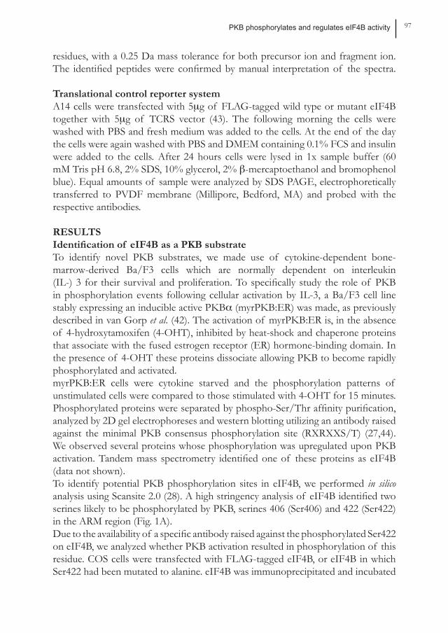
residues, with a 0.25 Da mass tolerance for both precursor ion and fragment ion. The identified peptides were confirmed by manual interpretation of the spectra.
Translational control reporter systemA14 cells were transfected with 5µg of FLAG-tagged wild type or mutant eIF4B together with 5µg of TCRS vector (43). The following morning the cells were washed with PBS and fresh medium was added to the cells. At the end of the day the cells were again washed with PBS and DMEM containing 0.1% FCS and insulin were added to the cells. After 24 hours cells were lysed in 1x sample buffer (60 mM Tris pH 6.8, 2% SDS, 10% glycerol, 2% β-mercaptoethanol and bromophenol blue). Equal amounts of sample were analyzed by SDS PAGE, electrophoretically transferred to PVDF membrane (Millipore, Bedford, MA) and probed with the respective antibodies.
RESULTSIdentification of eIF4B as a PKB substrateTo identify novel PKB substrates, we made use of cytokine-dependent bone-marrow-derived Ba/F3 cells which are normally dependent on interleukin (IL-) 3 for their survival and proliferation. To specifically study the role of PKB in phosphorylation events following cellular activation by IL-3, a Ba/F3 cell line stably expressing an inducible active PKBα (myrPKB:ER) was made, as previously described in van Gorp et al. (42). The activation of myrPKB:ER is, in the absence of 4-hydroxytamoxifen (4-OHT), inhibited by heat-shock and chaperone proteins that associate with the fused estrogen receptor (ER) hormone-binding domain. In the presence of 4-OHT these proteins dissociate allowing PKB to become rapidly phosphorylated and activated. myrPKB:ER cells were cytokine starved and the phosphorylation patterns of unstimulated cells were compared to those stimulated with 4-OHT for 15 minutes. Phosphorylated proteins were separated by phospho-Ser/Thr affinity purification, analyzed by 2D gel electrophoreses and western blotting utilizing an antibody raised against the minimal PKB consensus phosphorylation site (RXRXXS/T) (27,44). We observed several proteins whose phosphorylation was upregulated upon PKB activation. Tandem mass spectrometry identified one of these proteins as eIF4B (data not shown). To identify potential PKB phosphorylation sites in eIF4B, we performed in silico analysis using Scansite 2.0 (28). A high stringency analysis of eIF4B identified two serines likely to be phosphorylated by PKB, serines 406 (Ser406) and 422 (Ser422) in the ARM region (Fig. 1A). Due to the availability of a specific antibody raised against the phosphorylated Ser422 on eIF4B, we analyzed whether PKB activation resulted in phosphorylation of this residue. COS cells were transfected with FLAG-tagged eIF4B, or eIF4B in which Ser422 had been mutated to alanine. eIF4B was immunoprecipitated and incubated
Chapter 4 98
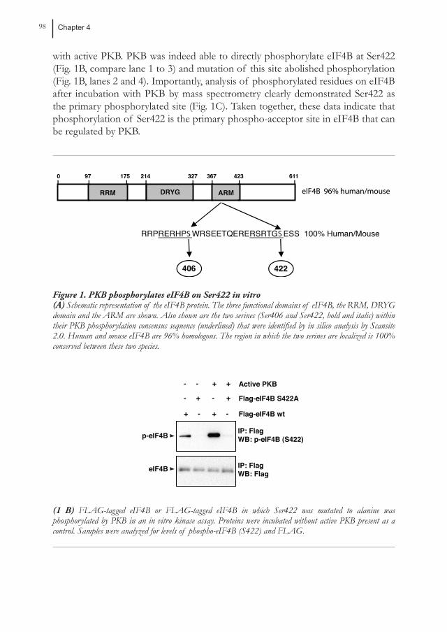
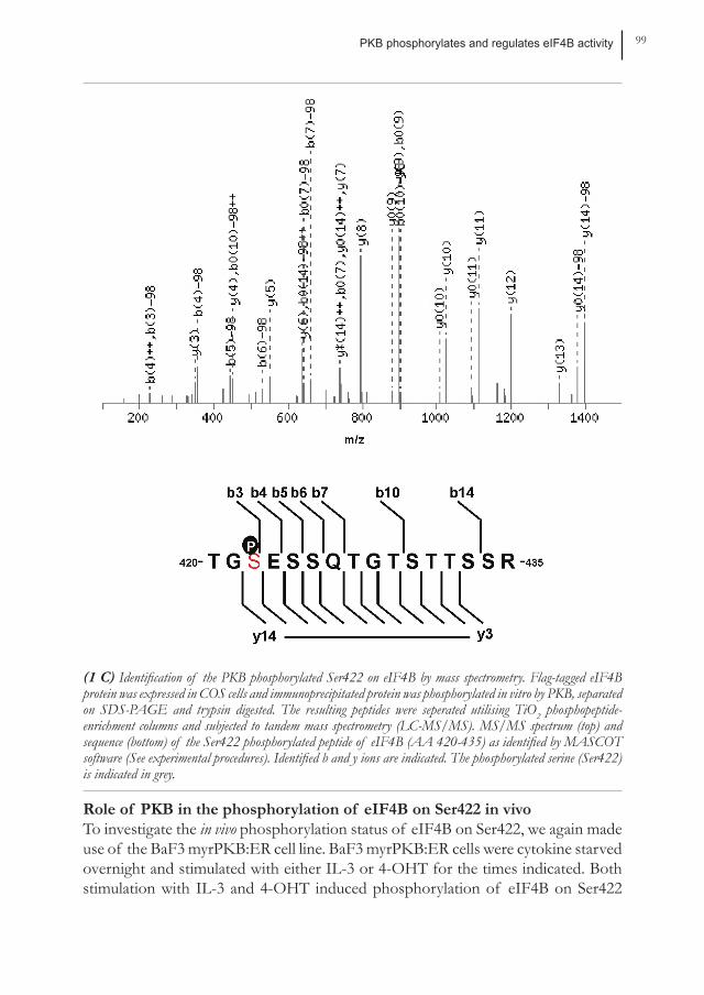
with active PKB. PKB was indeed able to directly phosphorylate eIF4B at Ser422 (Fig. 1B, compare lane 1 to 3) and mutation of this site abolished phosphorylation (Fig. 1B, lanes 2 and 4). Importantly, analysis of phosphorylated residues on eIF4B after incubation with PKB by mass spectrometry clearly demonstrated Ser422 as the primary phosphorylated site (Fig. 1C). Taken together, these data indicate that phosphorylation of Ser422 is the primary phospho-acceptor site in eIF4B that can be regulated by PKB.
eIF4B 96% human/mouseeIF4B 96% human/mouse
9797 175175 214214 327327 367367 423423 61161100
RRMRRM DRYGDRYG ARMARM
RRPRERHPS WRSEETQERERSRTGS ESS 100% Human/Mouse
406406 422422
Figure 1. PKB phosphorylates eIF4B on Ser422 in vitro (A) Schematic representation of the eIF4B protein. The three functional domains of eIF4B, the RRM, DRYG domain and the ARM are shown. Also shown are the two serines (Ser406 and Ser422, bold and italic) within their PKB phosphorylation consensus sequence (underlined) that were identified by in silico analysis by Scansite 2.0. Human and mouse eIF4B are 96% homologous. The region in which the two serines are localized is 100% conserved between these two species.
Flag-eIF4B wt
Flag-eIF4B S422A
+ - + -
- + - +
IP: FlagWB: p-eIF4B (S422)
IP: FlagWB: Flag
Active PKB- - + +
p-eIF4B
eIF4B
(1 B) FLAG-tagged eIF4B or FLAG-tagged eIF4B in which Ser422 was mutated to alanine was phosphorylated by PKB in an in vitro kinase assay. Proteins were incubated without active PKB present as a control. Samples were analyzed for levels of phospho-eIF4B (S422) and FLAG.
PKB phosphorylates and regulates eIF4B activity 99
(1 C) Identification of the PKB phosphorylated Ser422 on eIF4B by mass spectrometry. Flag-tagged eIF4B protein was expressed in COS cells and immunoprecipitated protein was phosphorylated in vitro by PKB, separated on SDS-PAGE and trypsin digested. The resulting peptides were seperated utilising TiO2 phosphopeptide-enrichment columns and subjected to tandem mass spectrometry (LC-MS/MS). MS/MS spectrum (top) and sequence (bottom) of the Ser422 phosphorylated peptide of eIF4B (AA 420-435) as identified by MASCOT software (See experimental procedures). Identified b and y ions are indicated. The phosphorylated serine (Ser422) is indicated in grey.
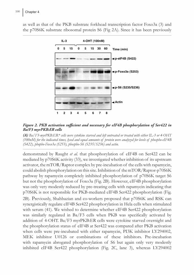
Role of PKB in the phosphorylation of eIF4B on Ser422 in vivoTo investigate the in vivo phosphorylation status of eIF4B on Ser422, we again made use of the BaF3 myrPKB:ER cell line. BaF3 myrPKB:ER cells were cytokine starved overnight and stimulated with either IL-3 or 4-OHT for the times indicated. Both stimulation with IL-3 and 4-OHT induced phosphorylation of eIF4B on Ser422
Chapter 4 100
as well as that of the PKB substrate forkhead transcription factor Foxo3a (3) and the p70S6K substrate ribosomal protein S6 (Fig 2A). Since it has been previously
0 5 15 0 5 15 30 60
IL-3
Time (min)
4-OHT (100nM)
p-eIF4B (S422)
p-Foxo3a (S253)
p-S6 (S235/S236)
Actin
1 2 3 4 5 6 7 8
Figure 2. PKB activation sufficient and necessary for eIF4B phosphorylation of Ser422 in Ba/F3 myrPKB:ER cells(A) Ba/F3-myrPKB:ER* cells were cytokine starved and left untreated or treated with either IL-3 or 4-OHT (100nM) for the indicated times, lysed and equal amounts of protein were analyzed for levels of phospho-eIF4B (S422), phopho-Foxo3a (S253), phospho-S6 (S235/S236) and actin.
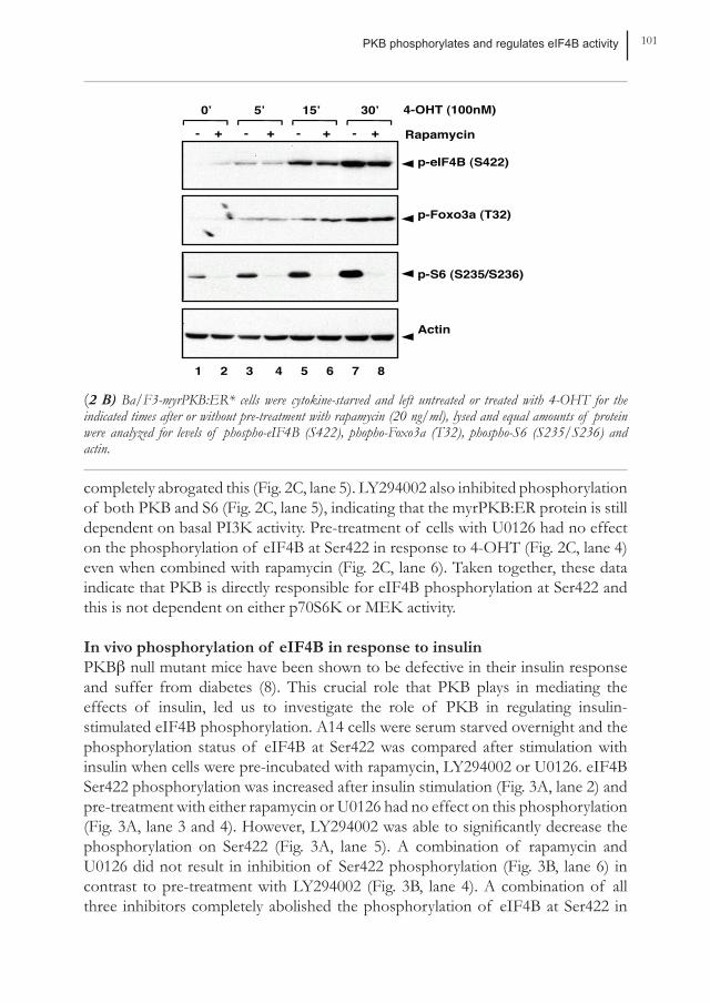
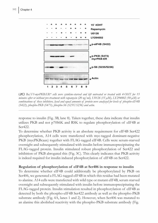
demonstrated by Raught et al. that phosphorylation of eIF4B on Ser422 can be mediated by p70S6K activity (33), we investigated whether inhibition of its upstream activator, the mTOR/Raptor complex by pre-incubation of the cells with rapamycin, could abolish phosphorylation on this site. Inhibition of the mTOR/Raptor-p70S6K pathway by rapamycin completely inhibited phosphorylation of p70S6K target S6 but not the phosphorylation of Foxo3a (Fig. 2B). However, eIF4B phosphorylation was only very modestly reduced by pre-treating cells with rapamycin indicating that p70S6K is not responsible for PKB-mediated eIF4B Ser422 phosphorylation (Fig. 2B). Previously, Shahbazian and co-workers proposed that p70S6K and RSK can synergistically regulate eIF4B Ser422 phosphorylation in Hela cells when stimulated with serum (41). We wished to determine whether eIF4B Ser422 phosphorylation was similarly regulated in Ba/F3 cells when PKB was specifically activated by addition of 4-OHT. Ba/F3 myrPKB:ER cells were cytokine starved overnight and the phosphorylation status of eIF4B at Ser422 was compared after PKB activation when cells were pre-incubated with either rapamycin, PI3K inhibitor LY294002, MEK inhibitor U0126 or combinations of these inhibitors. Pre-incubation with rapamycin abrogated phosphorylation of S6 but again only very modestly inhibited eIF4B Ser422 phosphorylation (Fig. 2C, lane 3), whereas LY294002
PKB phosphorylates and regulates eIF4B activity 101
Rapamycin- + - + - + - +
0’ 5’ 15’ 30’ 4-OHT (100nM)
p-eIF4B (S422)
p-Foxo3a (T32)
p-S6 (S235/S236)
Actin
1 2 3 4 5 6 7 8
(2 B) Ba/F3-myrPKB:ER* cells were cytokine-starved and left untreated or treated with 4-OHT for the indicated times after or without pre-treatment with rapamycin (20 ng/ml), lysed and equal amounts of protein were analyzed for levels of phospho-eIF4B (S422), phopho-Foxo3a (T32), phospho-S6 (S235/S236) and actin.
completely abrogated this (Fig. 2C, lane 5). LY294002 also inhibited phosphorylation of both PKB and S6 (Fig. 2C, lane 5), indicating that the myrPKB:ER protein is still dependent on basal PI3K activity. Pre-treatment of cells with U0126 had no effect on the phosphorylation of eIF4B at Ser422 in response to 4-OHT (Fig. 2C, lane 4) even when combined with rapamycin (Fig. 2C, lane 6). Taken together, these data indicate that PKB is directly responsible for eIF4B phosphorylation at Ser422 and this is not dependent on either p70S6K or MEK activity.
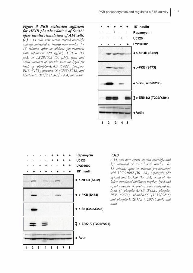
In vivo phosphorylation of eIF4B in response to insulinPkbβ null mutant mice have been shown to be defective in their insulin response and suffer from diabetes (8). This crucial role that PKB plays in mediating the effects of insulin, led us to investigate the role of PKB in regulating insulin-stimulated eIF4B phosphorylation. A14 cells were serum starved overnight and the phosphorylation status of eIF4B at Ser422 was compared after stimulation with insulin when cells were pre-incubated with rapamycin, LY294002 or U0126. eIF4B Ser422 phosphorylation was increased after insulin stimulation (Fig. 3A, lane 2) and pre-treatment with either rapamycin or U0126 had no effect on this phosphorylation (Fig. 3A, lane 3 and 4). However, LY294002 was able to significantly decrease the phosphorylation on Ser422 (Fig. 3A, lane 5). A combination of rapamycin and U0126 did not result in inhibition of Ser422 phosphorylation (Fig. 3B, lane 6) in contrast to pre-treatment with LY294002 (Fig. 3B, lane 4). A combination of all three inhibitors completely abolished the phosphorylation of eIF4B at Ser422 in
Chapter 4 102
- - - - + - + + +
15’ 4OHT
- - - + - + - + + Rapamycin
LY294002U0126
- - + - - + + - +- + + + + + + + +
p-S6 (S235/S236)
p-PKB (S473)myrPKB:ER
p-eIF4B (S422)
Actin
_
_
_
_
1 2 3 4 5 6 7 8 9
(2C) Ba/F3-myrPKB:ER* cells were cytokine-starved and left untreated or treated with 4-OHT for 15 minutes after or without pre-treatment with rapamycin (20 ng/ml), U0126 (15 µM), LY294002 (50 µM) or combinations of these inhibiters, lysed and equal amounts of protein were analyzed for levels of phospho-eIF4B (S422), phospho-PKB (S473), phospho-S6 (S235/S236) and actin.
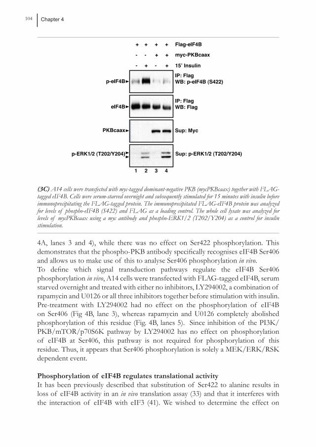
response to insulin (Fig. 3B, lane 8). Taken together, these data indicate that insulin utilizes PKB and not p70S6K and RSK to regulate phosphorylation of eIF4B at Ser422. To determine whether PKB activity is an absolute requirement for eIF4B Ser422 phosphorylation, A14 cells were transfected with myc-tagged dominant-negative PKB (mycPKBcaax) together with FLAG-tagged eIF4B. Cells were serum-starved overnight and subsequently stimulated with insulin before immunoprecipitating the FLAG-tagged protein. Insulin stimulated robust phosphorylation of Ser422 and inhibition of PKB abrogated this (Fig. 3C). This clearly indicates that PKB activity is indeed required for insulin induced phosphorylation of eIF4B on Ser422.
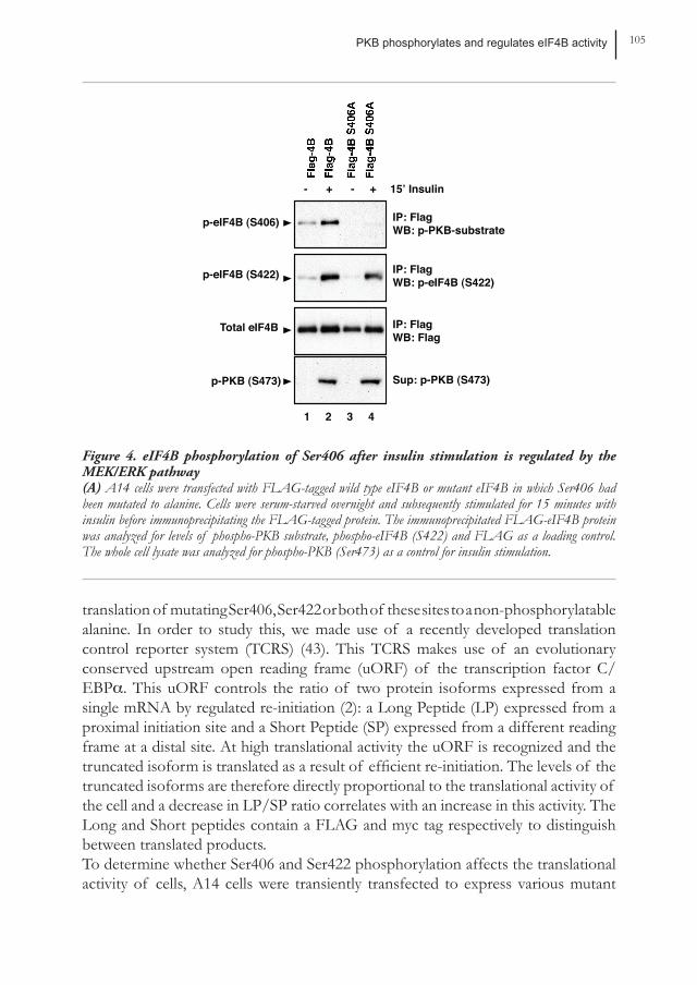
Regulation of phosphorylation of eIF4B at Ser406 in response to insulinTo determine whether eIF4B could additionally be phosphorylated by PKB on Ser406, we generated a FLAG-tagged eIF4B in which this residue had been mutated to alanine. A14 cells were transfected with wild type or mutant eIF4B, serum starved overnight and subsequently stimulated with insulin before immunoprecipitating the FLAG-tagged protein. Insulin stimulation resulted in phosphorylation of eIF4B as detected by both the phospho-eIF4B Ser422 antibody as well as the phospho-PKB substrate antibody (Fig. 4A, lanes 1 and 2). However, when Ser406 was mutated to an alanine this abolished reactivity with the phospho-PKB substrate antibody (Fig.
PKB phosphorylates and regulates eIF4B activity 103
15’ InsulinRapamycin
LY294002U0126
- - - - +
- - - + -
- - + - -- + + + +
p-S6 (S235/S236)
p-ERK1/2 (T202/Y204)
p-PKB (S473)
p-eIF4B (S422)
Actin
_
_
_
_
_
_
1 2 3 4 5
Figure 3 PKB activation sufficient for eIF4B phosphorylation of Ser422 after insulin stimulation of A14 cells. (A) A14 cells were serum starved overnight and left untreated or treated with insulin for 15 minutes after or without pre-treatment with rapamycin (20 ng/ml), U0126 (15 µM) or LY294002 (50 µM), lysed and equal amounts of protein were analyzed for levels of phospho-eIF4B (S422), phospho-PKB (S473), phospho-S6 (S235/S236) and phospho-ERK1/2 (T202/Y204) and actin.
15’ Insulin
Rapamycin
LY294002U0126
- + - + - + - +
- - + + - - + + - - - - + + + + - - - - + + + +
p-S6 (S235/S236)
p-ERK1/2 (T202/Y204)
p-PKB (S473)
p-eIF4B (S422)
Actin
_
_
_
_
_
_
1 2 3 4 5 6 7 8
(3B)A14 cells were serum starved overnight and left untreated or treated with insulin for 15 minutes after or without pre-treatment with LY294002 (50 µM), rapamycin (20 ng/ml) and U0126 (15 µM) or all of the before mentioned inhibitors together, lysed and equal amounts of protein were analyzed for levels of phospho-eIF4B (S422), phospho-PKB (S473), phospho-S6 (S235/S236) and phospho-ERK1/2 (T202/Y204) and actin.
Chapter 4 104
Sup: Myc
IP: FlagWB: Flag
IP: FlagWB: p-eIF4B (S422)
15’ Insulin- + - +
- - + +
+ + + +
myc-PKBcaax
Flag-eIF4B
p-eIF4B
eIF4B
PKBcaax
Sup: p-ERK1/2 (T202/Y204) p-ERK1/2 (T202/Y204)
1 2 3 4
(3C) A14 cells were transfected with myc-tagged dominant-negative PKB (mycPKBcaax) together with FLAG-tagged eIF4B. Cells were serum-starved overnight and subsequently stimulated for 15 minutes with insulin before immunoprecipitating the FLAG-tagged protein. The immunoprecipitated FLAG-eIF4B protein was analyzed for levels of phospho-eIF4B (S422) and FLAG as a loading control. The whole cell lysate was analyzed for levels of mycPKBcaax using a myc antibody and phospho-ERK1/2 (T202/Y204) as a control for insulin stimulation.
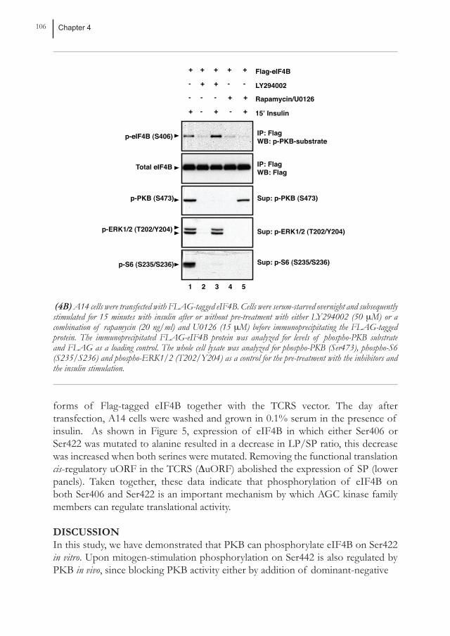
4A, lanes 3 and 4), while there was no effect on Ser422 phosphorylation. This demonstrates that the phospho-PKB antibody specifically recognises eIF4B Ser406 and allows us to make use of this to analyse Ser406 phosphorylation in vivo. To define which signal transduction pathways regulate the eIF4B Ser406 phosphorylation in vivo, A14 cells were transfected with FLAG-tagged eIF4B, serum starved overnight and treated with either no inhibitors, LY294002, a combination of rapamycin and U0126 or all three inhibitors together before stimulation with insulin. Pre-treatment with LY294002 had no effect on the phosphorylation of eIF4B on Ser406 (Fig 4B, lane 3), whereas rapamycin and U0126 completely abolished phosphorylation of this residue (Fig. 4B, lanes 5). Since inhibition of the PI3K/PKB/mTOR/p70S6K pathway by LY294002 has no effect on phosphorylation of eIF4B at Ser406, this pathway is not required for phosphorylation of this residue. Thus, it appears that Ser406 phosphorylation is solely a MEK/ERK/RSK dependent event.
Phosphorylation of eIF4B regulates translational activityIt has been previously described that substitution of Ser422 to alanine results in loss of eIF4B activity in an in vivo translation assay (33) and that it interferes with the interaction of eIF4B with eIF3 (41). We wished to determine the effect on
PKB phosphorylates and regulates eIF4B activity 105
Figure 4. eIF4B phosphorylation of Ser406 after insulin stimulation is regulated by the MEK/ERK pathway (A) A14 cells were transfected with FLAG-tagged wild type eIF4B or mutant eIF4B in which Ser406 had been mutated to alanine. Cells were serum-starved overnight and subsequently stimulated for 15 minutes with insulin before immunoprecipitating the FLAG-tagged protein. The immunoprecipitated FLAG-eIF4B protein was analyzed for levels of phospho-PKB substrate, phospho-eIF4B (S422) and FLAG as a loading control. The whole cell lysate was analyzed for phospho-PKB (Ser473) as a control for insulin stimulation.
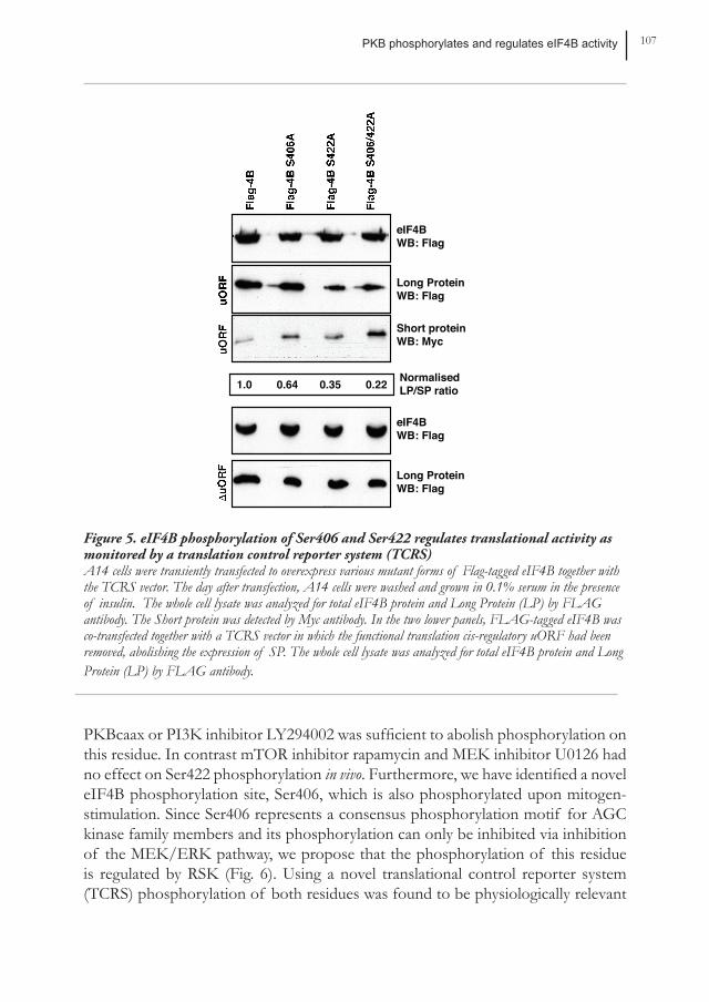
translation of mutating Ser406, Ser422 or both of these sites to a non-phosphorylatable alanine. In order to study this, we made use of a recently developed translation control reporter system (TCRS) (43). This TCRS makes use of an evolutionary conserved upstream open reading frame (uORF) of the transcription factor C/EbPα. This uORF controls the ratio of two protein isoforms expressed from a single mRNA by regulated re-initiation (2): a Long Peptide (LP) expressed from a proximal initiation site and a Short Peptide (SP) expressed from a different reading frame at a distal site. At high translational activity the uORF is recognized and the truncated isoform is translated as a result of efficient re-initiation. The levels of the truncated isoforms are therefore directly proportional to the translational activity of the cell and a decrease in LP/SP ratio correlates with an increase in this activity. The Long and Short peptides contain a FLAG and myc tag respectively to distinguish between translated products. To determine whether Ser406 and Ser422 phosphorylation affects the translational activity of cells, A14 cells were transiently transfected to express various mutant
IP: FlagWB: p-eIF4B (S422)
IP: FlagWB: Flag
IP: FlagWB: p-PKB-substrate
Sup: p-PKB (S473)
- + - + 15’ Insulin
p-eIF4B (S406)
p-eIF4B (S422)
Total eIF4B
p-PKB (S473)
1 2 3 4
Chapter 4 106
15’ Insulin+ - + - +
- - - + +
+ + + + +
Rapamycin/U0126
Flag-eIF4B- + + - - LY294002
IP: FlagWB: Flag
IP: FlagWB: p-PKB-substrate
Sup: p-PKB (S473)
Sup: p-ERK1/2 (T202/Y204)
Sup: p-S6 (S235/S236)
p-eIF4B (S406)
Total eIF4B
p-PKB (S473)
p-ERK1/2 (T202/Y204)
p-S6 (S235/S236)
1 2 3 4 5
(4B) A14 cells were transfected with FLAG-tagged eIF4B. Cells were serum-starved overnight and subsequently stimulated for 15 minutes with insulin after or without pre-treatment with either LY294002 (50 µM) or a combination of rapamycin (20 ng/ml) and U0126 (15 µM) before immunoprecipitating the FLAG-tagged protein. The immunoprecipitated FLAG-eIF4B protein was analyzed for levels of phospho-PKB substrate and FLAG as a loading control. The whole cell lysate was analyzed for phospho-PKB (Ser473), phospho-S6 (S235/S236) and phospho-ERK1/2 (T202/Y204) as a control for the pre-treatment with the inhibitors and the insulin stimulation.
forms of Flag-tagged eIF4B together with the TCRS vector. The day after transfection, A14 cells were washed and grown in 0.1% serum in the presence of insulin. As shown in Figure 5, expression of eIF4B in which either Ser406 or Ser422 was mutated to alanine resulted in a decrease in LP/SP ratio, this decrease was increased when both serines were mutated. Removing the functional translation cis-regulatory uORF in the TCRS (∆uORF) abolished the expression of SP (lower panels). Taken together, these data indicate that phosphorylation of eIF4B on both Ser406 and Ser422 is an important mechanism by which AGC kinase family members can regulate translational activity.
DISCUSSIOnIn this study, we have demonstrated that PKB can phosphorylate eIF4B on Ser422 in vitro. Upon mitogen-stimulation phosphorylation on Ser442 is also regulated by Pkb in vivo, since blocking PKB activity either by addition of dominant-negative
PKB phosphorylates and regulates eIF4B activity 107
Figure 5. eIF4B phosphorylation of Ser406 and Ser422 regulates translational activity as monitored by a translation control reporter system (TCRS) A14 cells were transiently transfected to overexpress various mutant forms of Flag-tagged eIF4B together with the TCRS vector. The day after transfection, A14 cells were washed and grown in 0.1% serum in the presence of insulin. The whole cell lysate was analyzed for total eIF4B protein and Long Protein (LP) by FLAG antibody. The Short protein was detected by Myc antibody. In the two lower panels, FLAG-tagged eIF4B was co-transfected together with a TCRS vector in which the functional translation cis-regulatory uORF had been removed, abolishing the expression of SP. The whole cell lysate was analyzed for total eIF4B protein and Long Protein (LP) by FLAG antibody.
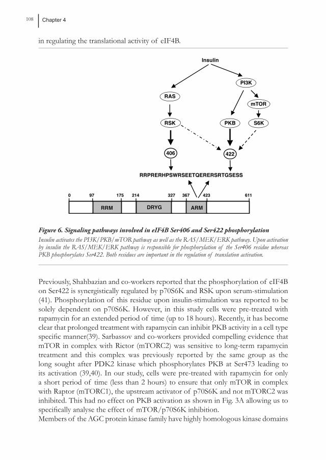
PKBcaax or PI3K inhibitor LY294002 was sufficient to abolish phosphorylation on this residue. In contrast mTOR inhibitor rapamycin and MEK inhibitor U0126 had no effect on Ser422 phosphorylation in vivo. Furthermore, we have identified a novel eIF4B phosphorylation site, Ser406, which is also phosphorylated upon mitogen-stimulation. Since Ser406 represents a consensus phosphorylation motif for AGC kinase family members and its phosphorylation can only be inhibited via inhibition of the MEK/ERK pathway, we propose that the phosphorylation of this residue is regulated by RSK (Fig. 6). Using a novel translational control reporter system (TCRS) phosphorylation of both residues was found to be physiologically relevant
Long ProteinWB: Flag
Short proteinWB: Myc
Long ProteinWB: Flag
1.0 0.64 0.35 0.22 NormalisedLP/SP ratio
eIF4BWB: Flag
eIF4BWB: Flag
Chapter 4 108
in regulating the translational activity of eIF4B.
Figure 6. Signaling pathways involved in eIF4B Ser406 and Ser422 phosphorylationInsulin activates the PI3K/PKB/mTOR pathway as well as the RAS/MEK/ERK pathway. Upon activation by insulin the RAS/MEK/ERK pathway is responsible for phosphorylation of the Ser406 residue whereas PKB phosphorylates Ser422. Both residues are important in the regulation of translation activation.
Previously, Shahbazian and co-workers reported that the phosphorylation of eIF4B on Ser422 is synergistically regulated by p70S6K and RSK upon serum-stimulation (41). Phosphorylation of this residue upon insulin-stimulation was reported to be solely dependent on p70S6K. However, in this study cells were pre-treated with rapamycin for an extended period of time (up to 18 hours). Recently, it has become clear that prolonged treatment with rapamycin can inhibit PKB activity in a cell type specific manner(39). Sarbassov and co-workers provided compelling evidence that mTOR in complex with Rictor (mTORC2) was sensitive to long-term rapamycin treatment and this complex was previously reported by the same group as the long sought after PDK2 kinase which phosphorylates PKB at Ser473 leading to its activation (39,40). In our study, cells were pre-treated with rapamycin for only a short period of time (less than 2 hours) to ensure that only mTOR in complex with Raptor (mTORC1), the upstream activator of p70S6K and not mTORC2 was inhibited. This had no effect on PKB activation as shown in Fig. 3A allowing us to specifically analyse the effect of mTOR/p70S6K inhibition. Members of the AGC protein kinase family have highly homologous kinase domains
RRPRERHPSWRSEETQERERSRTGSESSRRPRERHPSWRSEETQERERSRTGSESS
406406
PKBPKBRSKRSK S6KS6K
PI3KPI3K
RASRAS
Insulin
9797 175175 214214 327327 367367 423423 61161100
RRMRRM DRYGDRYG ARMARM
422422
mTORmTOR
PKB phosphorylates and regulates eIF4B activity 109
and similar substrate specificities, and can therefore be considered as potentially “promiscuous” when it comes to phosphorylation of target proteins. Care must therefore be taken in drawing conclusions from in vitro assays where, it is likely that various members of the AGC kinase family may phosphorylate substrates at the same site. In vivo, however, phosphorylation of substrates is likely to be a highly regulated process. In the case of eIF4B, three AGC kinase family members have now been shown to phosphorylate Ser422 in vitro, p70S6K, RSK and PKB respectively. Whereas insulin specifically utilizes PKB to phosphorylate this residue, serum may also utilize RSK to regulate Ser422 phosphorylation. Shahbazian and co-workers show after serum stimulation a temporal effect of MEK inhibitor U0126 and mTOR inhibitor rapamycin (41). U0126 effects the early phase of eIF4B phosphorylation whereas rapamycin effects the late phase. These data suggest that RSK is important for rapid regulation of eIF4B activity upon serum stimulation, and p70S6K (or PKB as we have indicated in the previous paragraph) is crucial for signal duration. In this study, we have shown that for insulin stimulation RSK does not play a role in Ser422 phosphorylation but this kinase is crucial in Ser406 phosphorylation. Therefore, it is safe to conclude that eIF4B phosphorylation and activation are regulated in a stimulus-dependent manner, and could be the reason why the Ser406 site was not identified by Raught and co-workers in their phosphomapping experiment after serum stimulation (33).While our data demonstrate that p70S6K is not responsible for insulin-mediated Ser406/422 phosphorylation, this does not discount a role for this AGC family member in eIF4B phosphorylation. It has been demonstrated that under conditions of amino acid refeeding p70S6K is activated while PKB remains inactive (11). Under these situations it is likely that p70S6K can phosphorylate eIF4B allowing for regulation of protein translation. Regulation of eIF4B phosphorylation may be a fundamental process in the regulation of protein translation in response to diverse extracellular stimuli. Our data suggests that utilizing various AGC kinase family members allows this mechanism of translational control to be regulated through distinct stimulus-specific intracellular signalling pathways. The effects of eIF4B phosphorylation on translation have, to some degree, been studied previously and eIF4B phosphorylation has been reported to correlate with high translational activity. In accordance with this, Holz et al. found that expression of a phosphomimetic form of eIF4B increased cap-dependent translation (15) and Shahbazian and co-workers found that phosphorylation of eIF4B on Ser422 stimulates its interaction with eIF3 (41). However, others observed an inhibition of translation upon eIF4B overexpression which was relieved when Ser422 was mutated into a non-phosphorylatable alanine (25,33). In this study, we also observe an increase in translational activity when Ser406 or/and Ser422 were mutated. It has been suggested that the relative amount of eIF4B expression is crucial for its effect on translation. Since eIF4B binds various members of the translation initiation complex including eIF3 and eIF4A, but also mRNA and 18S RNA, high expression
Chapter 4 110
levels could disrupt the correct stoichiometry of complex formation resulting in an inhibitory effect. Our data demonstrate that phosphorylation of eIF4B in the ARM region indeed regulates translational activity. Since activation of AGC protein kinase family members has an overall positive effect on translation, it is thus likely that this phosphorylation results in activation of eIF4B. In this study, we provide compelling evidence that phosphorylation of eIF4B can be regulated by both RAS and PKB signalling. Both pathways have been shown to be deregulated in a plethora of neoplasias. What role could eIF4B phosphorylation play in the process of transformation? eIF4B has been shown to play a critical role in stimulating the helicase activity of eIF4A to unwind inhibitory secondary structures in the 5’ untranslated region of mRNAs. These highly structured mRNAs are poorly translated when the translation initiation activity is decreased (19). Highly structured mRNAs often encode those proteins that are components of pathways critical to cell growth, such as growth factors, transcription factors, tyrosine kinases and receptors (38). Rajasekhar et al. recently demonstrated that upon PKB and RAS signalling the profile of mRNA associated to polysomes was drastically altered, these mRNAs mainly encoded for the proteins mentioned (31). Therefore activation of eIF4B by dysregulated RAS and PKB signalling may be critical in the induction of cellular transformation.Until recently, the effects PKB has on regulating translation were thought to be through increased mTOR activity. The inhibitory effects of rapamycin on PKB-induced transformation appeared to reveal the importance of mTOR as a downstream mediator of PKB signalling. However, the recent evidence that prolonged rapamycin treatment can itself inhibit PKB activation, re-emphasizes the importance of PKB itself as an oncogenic factor in regulating growth and proliferation. We suggest that oncogenic transformation as a result of uncontrolled PKB activity could be directly mediated by enhanced eIF4B activity, providing a novel rationale for the design of therapeutic strategies to inhibit tumour cell growth.
PKB phosphorylates and regulates eIF4B activity 111
REFEREnCE LIST
1. Burgering, B. M., R. H. Medema, J. A. Maassen, M. L. van de Wetering, A. J. van der Eb, F. McCormick, and J. L. Bos. 1991. Insulin stimulation of gene expression mediated by p21ras activation. EMBO J. 10:1103-1109.
2. Calkhoven, C. F., C. Muller, and A. Leutz. 2000. Translational control of C/EBPalpha and C/EBPbeta isoform expression. Genes Dev. 14:1920-1932.
3. Dijkers, P. F., K. U. Birkenkamp, E. W. Lam, n. S. Thomas, J. W. Lammers, L. Koenderman, and P. J. Coffer. 2002. FKHR-L1 can act as a critical effector of cell death induced by cytokine withdrawal: protein kinase B-enhanced cell survival through maintenance of mitochondrial integrity. J. Cell Biol. 156:531-542.
4. Dijkers, P. F., K. U. Birkenkamp, E. W. Lam, n. S. Thomas, J. W. Lammers, L. Koenderman, and P. J. Coffer. 2002. FKHR-L1 can act as a critical effector of cell death induced by cytokine withdrawal: protein kinase B-enhanced cell survival through maintenance of mitochondrial integrity. J. Cell Biol. 156:531-542.
5. Duncan, R. and J. W. Hershey. 1984. Heat shock-induced translational alterations in HeLa cells. Initiation factor modifications and the inhibition of translation. J. Biol. Chem. 259:11882-11889.
6. Duncan, R. and J. W. Hershey. 1985. Regulation of initiation factors during translational repression caused by serum depletion. Covalent modification. J. Biol. Chem. 260:5493-5497.
7. Gani, R. 1976. The nucleoli of cultured human lymphocytes. I. Nucleolar morphology in relation to transformation and the DNA cycle. Exp. Cell Res. 97:249-258.
8. Garofalo, R. S., S. J. Orena, K. Rafidi, A. J. Torchia, J. L. Stock, A. L. Hildebrandt, T. Coskran, S. C. Black, D. J. Brees, J. R. Wicks, J. D. Mcneish, and K. G. Coleman. 2003. Severe diabetes, age-dependent loss of adipose tissue, and mild growth deficiency in mice lacking Akt2/PKB beta. J. Clin. Invest 112:197-208.
9. Gingras, A. C., B. Raught, and n. Sonenberg. 1999. eIF4 initiation factors: effectors of mRNA recruitment to ribosomes and regulators of translation. Annu. Rev. Biochem 68:913-963.
10. Guertin, D. A. and D. M. Sabatini. 2005. An expanding role for mTOR in cancer. Trends Mol. Med. 11:353-361.
11. Hara, K., K. Yonezawa, Q. P. Weng, M. T. Kozlowski, C. Belham, and J. Avruch. 1998. Amino acid sufficiency and mTOR regulate p70 S6 kinase and eIF-4E BP1 through a common effector mechanism. J. Biol. Chem. 273:14484-14494.
12. Hershey, J. W. and W. C. Merrick. 2000. Pathway and mechanism of initiation of protein synthesis, p. 33-88. In N. Sonenberg, J. W. Hershey, and M. B. Mathews (ed.), Translational control of gene expression. Cold Spring Harbor Laboratory Press, Cold Spring Harbor, NY.
13. Holland, E. C., n. Sonenberg, P. P. Pandolfi, and G. Thomas. 2004. Signaling control of mRNA translation in cancer pathogenesis. Oncogene 23:3138-3144.
14. Holland, E. C., n. Sonenberg, P. P. Pandolfi, and G. Thomas. 2004. Signaling control of mRNA translation in cancer pathogenesis. Oncogene 23:3138-3144.
15. Holz, M. K., B. A. Ballif, S. P. Gygi, and J. Blenis. 2005. mTOR and S6K1 mediate assembly of the translation preinitiation complex through dynamic protein interchange and ordered phosphorylation events. Cell 123:569-580.
16. Inoki, K., Y. Li, T. Zhu, J. Wu, and K. L. Guan. 2002. TSC2 is phosphorylated and inhibited by Akt and suppresses mTOR signalling. Nat. Cell Biol. 4:648-657.
17. Larsen, M. R., T. E. Thingholm, O. n. Jensen, P. Roepstorff, and T. J. Jorgensen. 2005. Highly selective enrichment of phosphorylated peptides from peptide mixtures
Chapter 4 112
using titanium dioxide microcolumns. Mol. Cell Proteomics. 4:873-886. 18. Lawson, T. G., K. A. Lee, M. M. Maimone, R. D. Abramson, T. E. Dever, W.
C. Merrick, and R. E. Thach. 1989. Dissociation of double-stranded polynucleotide helical structures by eukaryotic initiation factors, as revealed by a novel assay. Biochemistry 28:4729-4734.
19. Lodish, H. F. 1976. Translational control of protein synthesis. Annu. Rev. Biochem 45:39-72.
20. Luo, J., B. D. Manning, and L. C. Cantley. 2003. Targeting the PI3K-Akt pathway in human cancer: rationale and promise. Cancer Cell 4:257-262.
21. Mamane, Y., E. Petroulakis, O. LeBacquer, and n. Sonenberg. 2006. mTOR, translation initiation and cancer. Oncogene 25:6416-6422.
22. Methot, n., A. Pause, J. W. Hershey, and n. Sonenberg. 1994. The translation initiation factor eIF-4B contains an RNA-binding region that is distinct and independent from its ribonucleoprotein consensus sequence. Mol. Cell Biol. 14:2307-2316.
23. Methot, n., G. Pickett, J. D. Keene, and n. Sonenberg. 1996. In vitro RNA selection identifies RNA ligands that specifically bind to eukaryotic translation initiation factor 4B: the role of the RNA remotif. RNA. 2:38-50.
24. Methot, n., M. S. Song, and n. Sonenberg. 1996. A region rich in aspartic acid, arginine, tyrosine, and glycine (DRYG) mediates eukaryotic initiation factor 4B (eIF4B) self-association and interaction with eIF3. Mol. Cell Biol. 16:5328-5334.
25. naranda, T., W. B. Strong, J. Menaya, B. J. Fabbri, and J. W. Hershey. 1994. Two structural domains of initiation factor eIF-4B are involved in binding to RNA. J. Biol. Chem. 269:14465-14472.
26. nave, B. T., M. Ouwens, D. J. Withers, D. R. Alessi, and P. R. Shepherd. 1999. Mammalian target of rapamycin is a direct target for protein kinase B: identification of a convergence point for opposing effects of insulin and amino-acid deficiency on protein translation. Biochem J. 344 Pt 2:427-431.
27. Obata, T., M. B. Yaffe, G. G. Leparc, E. T. Piro, H. Maegawa, A. Kashiwagi, R. Kikkawa, and L. C. Cantley. 2000. Peptide and protein library screening defines optimal substrate motifs for AKT/PKB. J. Biol. Chem. 275:36108-36115.
28. Obenauer, J. C., L. C. Cantley, and M. B. Yaffe. 2003. Scansite 2.0: Proteome-wide prediction of cell signaling interactions using short sequence motifs. Nucleic Acids Res. 31:3635-3641.
29. Pandolfi, P. P. 2004. Aberrant mRNA translation in cancer pathogenesis: an old concept revisited comes finally of age. Oncogene 23:3134-3137.
30. Parker, P. J. and S. J. Parkinson. 2001. AGC protein kinase phosphorylation and protein kinase C. Biochem Soc. Trans. 29:860-863.
31. Rajasekhar, V. K., A. Viale, n. D. Socci, M. Wiedmann, X. Hu, and E. C. Holland. 2003. Oncogenic Ras and Akt signaling contribute to glioblastoma formation by differential recruitment of existing mRNAs to polysomes. Mol. Cell 12:889-901.
32. Raught, B., A. C. Gingras, and n. Sonenberg. 2000. Regulation of ribosomal recruitment in eukaryotes, p. 245-295. In N. Sonenberg, J. W. Hershey, and M. B. Mathews (ed.), Translational control of gene expression. Cold Spring Harbor Laboratory Press, Cold Spring Harbor, NY.
33. Raught, B., F. Peiretti, A. C. Gingras, M. Livingstone, D. Shahbazian, G. L. Mayeur, R. D. Polakiewicz, n. Sonenberg, and J. W. Hershey. 2004. Phosphorylation of eucaryotic translation initiation factor 4B Ser422 is modulated by S6 kinases. EMBO J. 23:1761-1769.
34. Richter, J. D. and n. Sonenberg. 2005. Regulation of cap-dependent translation by eIF4E inhibitory proteins. Nature 433:477-480.
35. Rogers, G. W., Jr., n. J. Richter, W. F. Lima, and W. C. Merrick. 2001. Modulation
PKB phosphorylates and regulates eIF4B activity 113
of the helicase activity of eIF4A by eIF4B, eIF4H, and eIF4F. J. Biol. Chem. 276:30914-30922.
36. Rozen, F., I. Edery, K. Meerovitch, T. E. Dever, W. C. Merrick, and n. Sonenberg. 1990. Bidirectional RNA helicase activity of eucaryotic translation initiation factors 4A and 4F. Mol. Cell Biol. 10:1134-1144.
37. Ruggero, D., L. Montanaro, L. Ma, W. Xu, P. Londei, C. Cordon-Cardo, and P. P. Pandolfi. 2004. The translation factor eIF-4E promotes tumor formation and cooperates with c-Myc in lymphomagenesis. Nat. Med. 10:484-486.
38. Ruggero, D. and P. P. Pandolfi. 2003. Does the ribosome translate cancer? Nat. Rev. Cancer 3:179-192.
39. Sarbassov, D. D., S. M. Ali, S. Sengupta, J. H. Sheen, P. P. Hsu, A. F. Bagley, A. L. Markhard, and D. M. Sabatini. 2006. Prolonged rapamycin treatment inhibits mTORC2 assembly and Akt/PKB. Mol. Cell 22:159-168.
40. Sarbassov, D. D., D. A. Guertin, S. M. Ali, and D. M. Sabatini. 2005. Phosphorylation and regulation of Akt/PKB by the rictor-mTOR complex. Science 307:1098-1101.
41. Shahbazian, D., P. P. Roux, V. Mieulet, M. S. Cohen, B. Raught, J. Taunton, J. W. Hershey, J. Blenis, M. Pende, and n. Sonenberg. 2006. The mTOR/PI3K and MAPK pathways converge on eIF4B to control its phosphorylation and activity. EMBO J. 25:2781-2791.
42. van Gorp, A. G., K. M. Pomeranz, K. U. Birkenkamp, R. C. Hui, E. W. Lam, and P. J. Coffer. 2006. Chronic Protein Kinase B (PKB/c-akt) Activation Leads to Apoptosis Induced by Oxidative Stress-Mediated Foxo3a Transcriptional Up-regulation. Cancer Res. 66:10760-10769.
43. Wiesenthal, V., A. Leutz, and C. F. Calkhoven. 2006. A translation control reporter system (TCRS) for the analysis of translationally controlled processes in the vertebrate cell. Nucleic Acids Res. 34:e23.
44. Zhang, H., X. Zha, Y. Tan, P. V. Hornbeck, A. J. Mastrangelo, D. R. Alessi, R. D. Polakiewicz, and M. J. Comb. 2002. Phosphoprotein analysis using antibodies broadly reactive against phosphorylated motifs. J. Biol. Chem. 277:39379-39387.