Embed Size (px)
Citation preview
1
Prevalence and Characterization of Non-O157 Shiga Toxin Producing Escherichia coli 1
Isolated from Commercial Ground Beef in the United States† 2
3
Joseph M. Bosilevac1* and Mohammad Koohmaraie
2,3 4
5
USDA, ARS, U.S. Meat Animal Research Center, P. O. Box 166, State Spur 18D, Clay Center, 6
Nebraska 689331; IEH Laboratories and Consulting Group, 15300 Bothell Way NE, Lake 7
Forest Park, WA 98155,2 and College of Food and Agriculture, King Saud University, Riyadh, 8
Saudi Arabia3
9
10
11
Running title: Non-O157 STEC in U.S. Beef 12
Key words: Shiga toxin producing Escherichia coli, ground beef, beef carcasses 13
14
15
16
Corresponding author. Mailing address: USDA, ARS, U.S. Meat Animal Research Center, 17
P. O. Box 166, State Spur 18D, Clay Center, NE 68933. Phone: (402) 762-4225. Fax: 18
(402) 762-4149. E-mail: [email protected]. 19
20
† USDA is an equal opportunity provider and employer. Names are necessary to report factually 21
on available data; however, the USDA neither guarantees nor warrants the standard of the 22
product, and the use of the name by USDA implies no approval of the product to the exclusion of 23
others that may also be suitable. 24
Copyright © 2011, American Society for Microbiology and/or the Listed Authors/Institutions. All Rights Reserved.Appl. Environ. Microbiol. doi:10.1128/AEM.02833-10 AEM Accepts, published online ahead of print on 21 January 2011
on July 5, 2018 by guesthttp://aem
.asm.org/
Dow
nloaded from

2
Abstract 25
Escherichia coli O157:H7 is a Shiga toxin (stx) producing E. coli (STEC) that has been 26
classified as an adulterant in U.S. beef. However, numerous other non-O157 STEC are 27
associated with diseases of varying severity, and have become an increasing concern to the beef 28
industry, regulatory officials and the public. This study reports the prevalence and 29
characterization of non-O157 STEC in commercial ground beef (n=4,133) obtained from 30
numerous manufacturers across the U.S. for a period of 24 months. All samples were screened 31
by DNA amplification for the presence of Shiga toxin genes, which were present in 1006 32
(24.3%) of the samples. Then culture isolation of a STEC from all samples that contained stx1 33
and/or stx2 was attempted. Of the 1006 stx screened positive ground beef samples, 300 (7.3% of 34
the total 4,133) were confirmed to have at least one strain of STEC present by culture isolation. 35
In total 338 unique STEC isolates were recovered in the 300 samples that yielded a STEC. All 36
unique STEC isolates were serotyped, and were characterized for the presence of known 37
virulence factors. These included Shiga toxin subtypes, intimin subtypes, and accessory 38
virulence factors related to adherence (saa, iha, lifA), toxicity (cnf, subA, astA), iron acquisition 39
(chuA), the presence of the large 60MDa virulence plasmid (espP, etpD, toxB, katP, toxB) and a 40
pathogenicity molecular risk assessment (MRA, based on presence of various O-Island nle 41
genes). Results of this characterization identified ten STEC (0.24% of total 4,133) that may be 42
considered a significant food safety threat defined by the presence of eae, subA, and nle genes. 43
on July 5, 2018 by guesthttp://aem
.asm.org/
Dow
nloaded from
3
Introduction 44
More than 70 different serotypes of Shiga toxin–producing Escherichia coli (STEC) have 45
been described that cause disease in humans (13). Illnesses range from mild diarrhea to bloody 46
diarrhea to hemorrhagic colitis (HC) and hemolytic-uremic syndrome (HUS). E. coli O157:H7 47
is the STEC most often associated with the most severe forms of disease (13, 61, 70). However, 48
numerous non-O157 STEC have also been linked to illnesses and outbreaks of disease. Six O-49
groups (comprising thirteen serotypes) have been described by the CDC as the cause of 71% of 50
non-O157 STEC disease (13). These serotypes have been identified as: O26:H11 or NM, 51
O45:H2 or NM, O103:H2, H11, H25 or NM, O111:H8 or NM, O121:H19 or H7 and O145:NM. 52
The proportion of these non-O157 STEC serogroups breaks down as follows: O26 (22%), O111 53
(16%), O103 (12%), O121 (8%), O45 (7%), and O145 (5%). Although infrequently isolated and 54
reported, it is estimated that non-O157 STEC may cause diarrhea at frequencies similar to those 55
of other important enteric bacterial pathogens such as Salmonella and Shigella (63) while also 56
having infections resulting in HUS and outbreaks (13). The non-O157 STEC are likely under 57
counted however due to difficulties confirming in the lab (32). 58
Cattle are recognized as the reservoir of many STEC serotypes (47). In fact there are 59
ample data on the prevalence of non-O157 STEC from fed beef through processing (1) to 60
boneless beef trim destined for grinding (10) but there is a lack of information on the prevalence 61
of non-O157 STEC found in finished ground beef. The study of beef trim destined for grinding 62
showed that 10 to 30% of beef trim contained STEC (10), however, the results of blending the 63
various lean and fat materials during production of ground beef on the rates of STEC present in 64
the finished product are unknown. 65
A STEC seropathotype classification based upon the serotype, association with outbreaks, 66
on July 5, 2018 by guesthttp://aem
.asm.org/
Dow
nloaded from
4
HUS, and diarrhea has been proposed by Karmali et al (40). This classification system is a tool 67
to assess the clinical and public health risks associated with different non-O157 STEC strains. 68
The predominant EHEC serotype responsible for HC and HUS, O157:H7, is classified as group 69
A. The other non-O157 STEC serotypes recognized as belonging to the EHEC group 70
comprising the thirteen serotypes listed above, are considered group B. Group C in this 71
classification system is made up of strains referred to as atypical EHEC. The atypical EHEC are 72
less frequently involved in hemorrhagic diseases than typical EHEC, but are a frequent cause of 73
diarrhea. The principal serotypes identified in group C are O91:H21, O113:H21, O104:H21. 74
STEC serotypes not associated with HC, HUS or outbreaks are numerous and belong to groups 75
D and E. These two groups are delineated from one another by their source. Group D have been 76
isolated from humans while group E have only been isolated from animals (40). 77
Various factors and toxins contribute to the increasing virulence of STEC (19, 73). Strains 78
that produce Shiga toxin 2 (Stx2) and more specifically Shiga toxin 2 subtype c (Stx2c) have 79
been suggested to be more likely to cause HUS than are those that produce Shiga toxin 1 (stx1) 80
alone (9, 29). Other toxins purported to increase virulence in strains are subtilase (subA; 42, 55 81
76), lymphocyte activating factor (lifA also known as efa1; 43), cytotoxic necrotizing factor (cnf; 82
7), and heat stable toxin (astA; 79). The EHEC contain a large heterologous virulence plasmid, 83
referred to as the 60MDa virulence plasmid, that contains a number of different genes associated 84
with disease espP, katP, eptD, toxB and the EHEC hemolysin (ehx) (15, 16, 39, 51). Other 85
hemolysins such as ChuA and HylA have also been described to influence the iron acquisition 86
and virulence of STEC (33, 69, 77). Finally, there is a group of factors associated with 87
adherence that influences virulence. Intimin (eae) is contained in the locus of enterocyte 88
effacement (LEE) and along with the tir gene product is responsible for the intimate attachment 89
on July 5, 2018 by guesthttp://aem
.asm.org/
Dow
nloaded from

5
and classic pedestal formation of the characteristic adherence/effacing (A/E) lesion in HC and 90
HUS (47). In the absence of intimin, other factors such as saa and iha have been identified that 91
correlate with increased adherence (37, 56, 65). The interplay between these various genes is not 92
understood but each has been described to provide an additional trait that increases virulence in 93
STEC. 94
Finally, comparisons of the genomes of E. coli O157:H7 and E. coli K12 have identified 95
numerous islands of genetic material unique to each. The genomic regions specific to E. coli 96
O157:H7 have been termed O-islands. Studies of O-islands 36, 57, 71, and 122 have identified 97
genes associated with increased virulence (20, 40, 75). In fact a molecular risk assessment 98
system for STEC strains has been proposed based on the compliment of these four O-islands 99
(20). This project aims to determine the prevalence of non-O157 STEC in commercial ground 100
beef in the U.S., identify the serotypes of the STEC present and distinguish those that may be 101
pathogenic STEC (pSTEC). There are essentially two approaches to defining pSTEC, either 102
epidemiologically (identifying serogroups, particularly the “top six”) or seropathotypically 103
(based on molecular markers associated with pathogenicity). We chose the broader second 104
option which offers a much larger set of results and that identifies as many pSTEC as possible. 105
Narrowly focusing on only the described “top six” STEC poses the problem of identifing 106
numerous isolates of little pathogenic concern while missing others significant pSTEC, 107
especially since nearly one third of pSTEC are not within the “top six” serogroups. Our goal was 108
to not allow the impact of these other pSTEC to go unappreciated in our studies. 109
110
Materials and Methods 111
on July 5, 2018 by guesthttp://aem
.asm.org/
Dow
nloaded from

6
Design. Four-thousand one-hundred thirty-three identity-blinded ground beef samples 112
that had been collected previously were used (11). The samples were collected by 18 different 113
commercial ground beef producers from unique product lots produced on different days. No 114
more than one sample per day, per supplier, was used in this study. The 18 producers were 115
located in seven of the eight Beef Industry Food Safety Council (BIFSCo) regions of the U.S. 116
over a period of 24-months (11). The BIFSCo regions are: 1, northwest (WA, OR, ID); 2, west 117
(CA, NV); 3, southwest (AZ, NM, TX); 5, upper mid-west (NE, ND, SD, MN, WS); 6, Central 118
(IA, KS, MO); 7, southeast (OK, AR, LA, NC SC FL AL MS GA TN); and 8, northeast (IL, IN, 119
KT, MS, ME. MD, NJ, NY, NH, CN, RI, OH WV VA PN DL). Region 4 (MT, CO, WY, UT) 120
was not represented in the sample collection due to logistical problems. All samples were 121
finished ground beef that had been screened by the manufacturer for E. coli O157:H7 before 122
release. The presence of stx1, and stx2 genes was determined by PCR amplification, if Shiga 123
toxin genes were present, the samples were processed using a phenotypic screening assay to 124
isolate non-O157 STEC. STEC isolates were then characterized for serotype, the presence and 125
subtype of virulence genes, as well as other toxin, adherence and virulence plasmid associated 126
genes. Further, the presence of determinants within O-islands 36, 57, 71, and 122 were 127
determined as was the resistance to antibiotics of each isolate. 128
Screening for Shiga toxin genes. The ground beef samples (65g) were placed in 585 129
mL tryptic soy broth (TSB) and incubated at 42°C for 6 h then held for 6-12 h at 4°C as 130
previously described (11). Two 1 mL portions of each enrichment were archived as frozen 131
(-70°C) 30% glycerol stocks. Between 2 and 12 weeks after the date of freezing, 24 to 48 132
different glycerol stocks were thawed, vortexed to thoroughly mix, and maintained on ice. One 133
microliter was removed from each, and placed into a separate 25 µL multiplex PCR reaction that 134
on July 5, 2018 by guesthttp://aem
.asm.org/
Dow
nloaded from

7
detected stx1, stx2, eae, and ehx performed as previously described (52). Products of the PCR 135
amplifications were separated by electrophoresis, stained using ethidium bromide, then 136
photographed and interpreted for the presence of the 4 possible reaction products. Only 137
enrichments that had either stx1 and/or stx2 were considered positive for STEC and used in 138
prevalence calculations. 139
Isolation of non-O157 STEC. The samples enrichments determined by PCR to contain 140
either stx1 and/or stx2, were assayed by spiral plating on to plates of washed sheep blood agar 141
containing mitomycin (wSBAm; 64). Each enrichment was serial diluted to 1:500 and 1:5000 in 142
cold (4°C) buffered peptone water (BPW). Fifty microliters of each dilution was spiral plated 143
using an Autoplate 4000 spiral plater (Spiral Biotech, Norwood, MA) on to wSBAm plates. The 144
plates were incubated overnight at 37°C then viewed on a white light box (VeriQuest 100, 145
Photodyne Technologies, Inc., Northridge, CA) for colonies surrounded by zones of hemolysis. 146
The suspect hemolytic phenotype was a thin zone of 1 mm or less surrounding the colony (5). 147
However, many phenotypic variations of hemolysis not indicative of ehx were often present, in 148
these cases additional colonies representative of each variation were also picked for screening. 149
A minimum of 4 (if present) and a maximum of 6 colonies per sample were picked into 150
individual wells of 96-well screening plates containing 100 µL TSB per well. After suspect 151
colonies were picked, the wSBAm plates were placed at 4°C. The 96-well screening plates were 152
incubated overnight at 37°C, and screened by PCR as described above to identify stx-containing 153
isolates. If at least one suspect colony from a sample did not contain stx1 and/or stx2, the 154
wSBAm plates were removed from 4°C and subjected to another round of suspect colony 155
picking. The additional round of screening increased the number of isolate positive samples by 156
15 to 20%. All stx-containing isolates were checked for purity by streaking for isolation on 157
on July 5, 2018 by guesthttp://aem
.asm.org/
Dow
nloaded from
8
Sorbitol MacConkey Agar containing 5-bromo-4-chloro-3-indolyl-β-D-glucuronide (SMAC-158
BCIG; Oxoid-CM0981, Remel Inc., Lenexa KS). If more than one colony type was observed, 159
each was re-screened as described above until a pure culture resulted. All purified isolates of 160
STEC were transferred to tryptic soy agar (TSA) plates for characterization. 161
Characterization of STEC isolates. All stx-containing isolates were confirmed to be E. 162
coli by biochemical assays using Fluorocult LMX Broth (Merck KGaA, Darmstadt, Germany), 163
and SensiTiter Gram negative ID panels (Trek Diagnostic Systems, Inc.) or API 20E strips 164
(bioMerieux Inc., Hazelwood MO) all used according to their manufacturers recommendations. 165
Once an isolate was established as being an stx-containing E. coli, its serotype was determined 166
by molecular and serologic identification of O serogroup. PCR was used to molecularly identify 167
O groups: O26 (22), O45 (21), O55 (21), O103 (28), O111 (23), O113 (22), O117 (46), O121 168
(27), O126 (46), O145 (25) and O146 (46). A VTEC specific antisera set (Laboratorio de 169
Referencia de E. coli, Lugo Spain) was used to confirm the PCR results and identify O groups: 170
O1, O2, O4, O5, O6, O8, O9, O15, O16, O17, O20, O22, O25, O27, O32, O39, O41, O46, O48, 171
O64, O74, O75, O77, O81, O82, O84, O86, O88, O91, O98, O101, O104, O105, O109, O110, 172
O112, O116, O118, O119, O123, O128, O132, O136, O139, O141, O150, O153, O157, O162, 173
O163, O165, O166, O168, O171, O172, O174 (OX3), O176, O177, and O178 (6, 34). The H 174
group of each isolate was determined by molecular methods involving sequencing the fliC gene 175
(74) and comparison for like sequences in GenBank. This method allows the identification of 44 176
of the 53 different H types coded for by fliC. H-types H3, H17, H35, H36, H44, H47, H53, H54, 177
H55 that are encoded by flk, fll and flm were not determined by this method. In situations where 178
the fliC gene could not be resolved from one of the other H-type encoding genes, the fliC gene is 179
given and the possible other H-type is listed as a superscript. All other O and/or H types are 180
on July 5, 2018 by guesthttp://aem
.asm.org/
Dow
nloaded from
9
referred to as untypable, indicating that the serotype is outside the limits of the above mentioned 181
O and H typing methodologies. 182
Previously described PCR methods and primers were used with isolates found positive 183
for stx1 to determine the subtype of Shiga toxins 1, or 1c (80), and with isolates found positive 184
for stx2 to determine Shiga toxin subtypes 2 (referred to hereafter as 2a), 2c, 2d and 2e (4, 81). 185
Isolates found to be stx2 positive but non-typable using the PCR methods are referred to simply 186
as stx2 positive. If an isolate contained more than one type of stx2 allele, the subtypes are given 187
in a combined notation (ex: stx 2ac) but no attempts were made to quantitate the number of each 188
allele present. In a similar fashion, previously described methods were used with isolates found 189
positive for eae to identify intimin subtypes α1, α2, β1, β2, γ, δ, ε, θ, and ζ (6, 8). Since this 190
sub-typing is not inclusive of all defined subtypes, any isolate that was eae positive that did not 191
sub-type is referred to as untypable and presented simply as eae positive without the subtype 192
indicator. The presence of four genes associated with the large 60 MDa virulence plasmid, toxB, 193
espP, katP, and etpD, was determined by PCR using previously described methods (15, 16). 194
Additional PCR-based assays were used to identify toxin encoding genes (subA, 54; lifA, 43; cnf, 195
7; and astA, 78), adherence encoding genes (iha, 65 and saa, 53) and hemolysin genes (hylA, 44 196
and chuA, 35), in order to determine their distribution within the STEC isolates. Finally, the 197
genes described for molecular risk assessment (20) associated with O-islands 122 (genes nleB, 198
nleE, and ent) OI-57 (genes G2-3, G5-2 and G6-2), OI-36 (genes nleC, H1-1, and nleB2) and OI-199
71 (genes nleG, nleG9, nleF, H1-2, nleA and G2-1) were examined using the previously 200
described PCR amplifications (20). The serotype and the rates of possession of the various 201
pathogenicity related genes were combined to identify seropathotypes of the STEC isolates (40). 202
on July 5, 2018 by guesthttp://aem
.asm.org/
Dow
nloaded from

10
Statistical analysis. STEC isolates obtained by each study method were sorted 203
according to serotype and screening PCR positive reaction pattern (stx1, stx2, eae, and ehx). 204
Comparisons of percent isolation, and mean isolation rate were examined using a one-way 205
analysis of variance (ANOVA) and the Bonferroni multiple-comparison posttest. Comparisons 206
of median values of data sets were made using the Kruskal-Wallis test for nonparametric data 207
and Dunn’s multiple-comparison posttest. Comparisons of data sets with only two groups of 208
values were made using either a two-tailed unpaired t test or the Mann-Whitney U test for 209
nonparametric data. Interactions between study methods and isolate serotypes and/or screening 210
PCR positive reaction patterns were examined by ANOVA factorial analysis using Prism 5 211
(GraphPad Software, La Jolla, CA), and P < 0.05 were considered significantly different. The 212
DIFFER procedure of PEPI software (USD, Inc., Stone Mountain, GA) was used to calculate the 213
pair wise differences among each STEC serogroup and each screening PCR positive reaction 214
with significance defined at P < 0.05. 215
216
Results 217
The overall suggested prevalence of STEC in ground beef based on the presence of either 218
stx1 or stx2 was 24.3% (Table 1). The samples were screened in each calendar month and were 219
from numerous locations in 7 of the BIFSCo microbiological monitoring regions of the U.S. The 220
samples were submitted on a voluntary basis and as such, their spatial and temporal distribution 221
was non-uniform and, the monthly and regional analysis produced wide ranging results. The 222
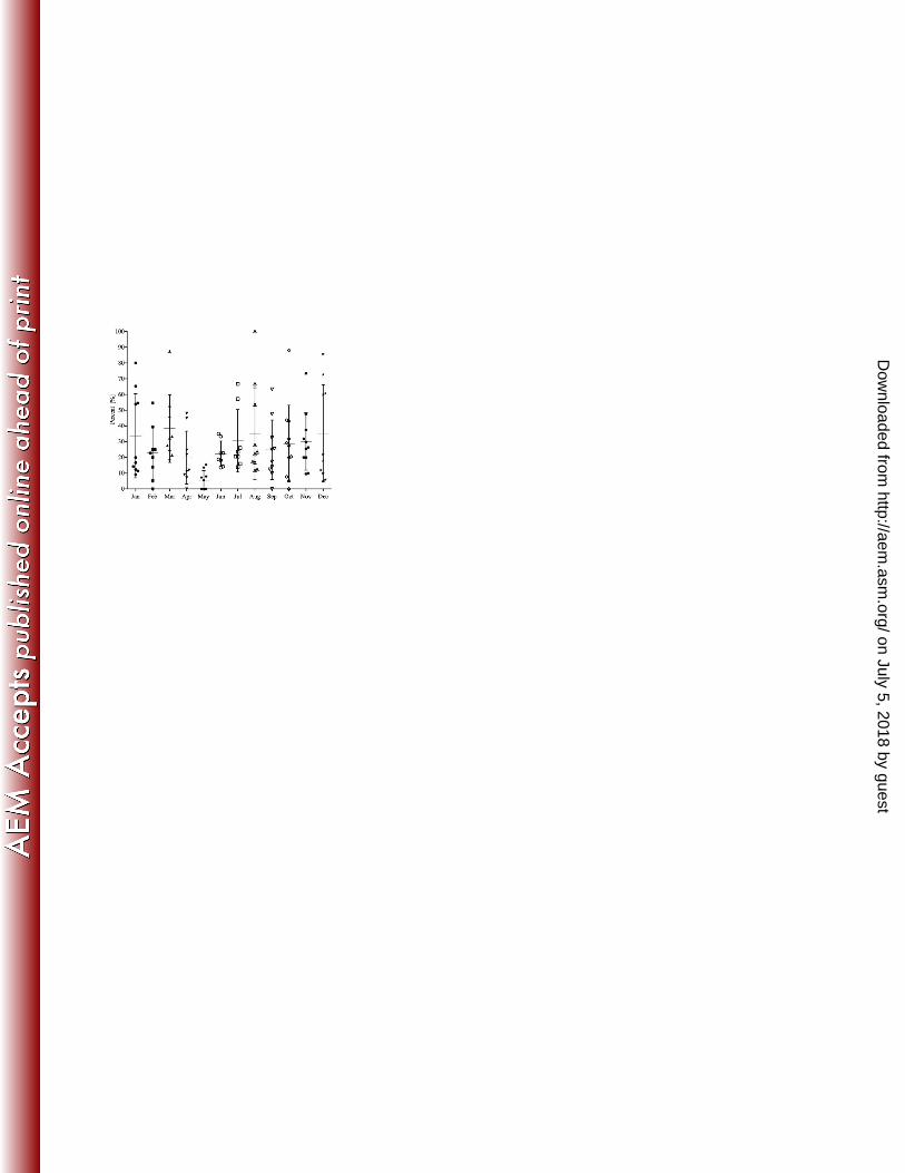
prevalence of stx in screened samples over time (Figure 1) showed the mean monthly prevalence 223
ranged from a low of 5.5% in May to a high of 38.4% in March. With few exceptions, 224
comparisons of monthly prevalence levels of stx in the screened samples were not different. 225
on July 5, 2018 by guesthttp://aem
.asm.org/
Dow
nloaded from

11
Examining the prevalence of stx in screened samples by region (Figure 2) showed the mean 226
prevalence across regions ranged from 13.1% to 39.4%, with regions 7 and 8 having stx 227
prevalence rates higher (P < 0.05) than regions 2 and 5. 228
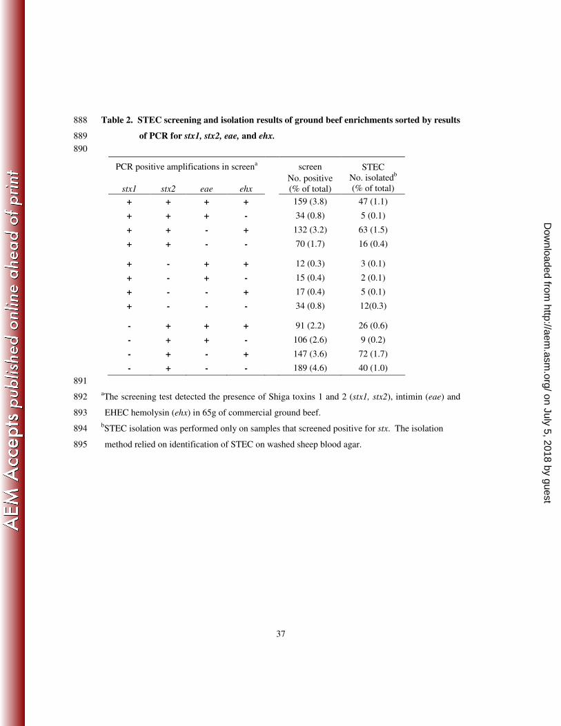
The screening test of the samples provided the presence of intimin (eae) and the EHEC 229
hemolysin (ehx) in addition to stx1 and stx2 (Table 2). The least common screening result (7.8% 230
samples) was for the presence of stx1 alone. Whereas just over half (53%) on the screened 231
positive samples had only stx2 present. Intimin was present with stx in 41.5% of screened 232
positive samples. Samples that screened positive for either stx1 or stx2 had similar proportions 233
of samples that were also positive for eae, 34.6 and 37.6% respectively. A larger proportion 234
(48.9%) of samples (P < 0.05) that were stx1 plus stx2 positive also contained eae. The presence 235
of ehx was observed in 55.5% of the screened samples with stx, but the proportion of samples 236
with ehx and with both stx1 and stx2 was significantly greater (P < 0.05) than in samples that had 237
only either stx1 or stx2 alone, 73.7% compared to 37.2 and 44.7% respectively. 238
All 1,006 samples that had screened positive for either stx1 and/or stx2 regardless of the 239
presence of eae or ehx were subjected to the STEC isolation and 338 unique isolates resulted 240
from 300 samples. Most samples yielded isolates of one serotype, but 32 positive samples 241
yielded isolates of two different serotypes and 3 samples yielded isolates of 3 different serotypes. 242
The rate of isolating a STEC in a positive screen sample was not different (P > 0.05) if the 243
sample had screened positive for stx1, stx2 or stx1 and stx2. Significantly fewer (P < 0.05) STEC 244
were isolated from samples that had screened positive for the presence of eae in addition to stx 245
(22%), compared to samples that did not screen positive for eae (35%). The isolation method 246
targeted the enterohemolytic phenotype associated with ehx on blood agar (5), therefore nearly 247
twice as many STEC (39% versus 20%) were isolated in samples that screened positive for ehx 248
on July 5, 2018 by guesthttp://aem
.asm.org/
Dow
nloaded from

12
even though colonies that demonstrated all types of hemolytic phenotypes were examined. The 249
largest proportion of samples to yield an isolate were those that screened positive for stx2 and 250
ehx, while the least likely were those that screened positive for stx2 and eae. 251
Numerous O-serogroups were present. The most frequently identified serogroups, in 252
order of frequency, were: O113, O8, O22, O117, O163, O174, O171, O116, and O20. These 253
nine serogroups represented 53% of all that were identified while at least 33 serogroups made up 254
the remaining 47%. The single most frequently identified serogroup was O113 which made up 255
11% of all the isolates. Fifty-six (16.7%) of the isolates were of an untypable O group, 256
indicating they were either outside the range of our typing system, rough, or serologically 257
untypable. The H typing protocol used identified 22 different H types in 319 isolates. Nineteen 258
isolates were H untypable, indicating that they may have been outside the scope of our typing 259
scheme, or were H- or non-motile. In total 99 different STEC serotypes were identified 260
(Table 3). The serotypes identified most often were O113:H21 (9.5%) followed by O8:H19 and 261
O117:H7 (4.4% and 4.7%). These serotypes were isolated from samples originating from five 262
(O8:H19) and six (O113:H21 and O117:H7) of the regions. 263
The Shiga toxin of each isolate was characterized by molecular subtyping (Table 4). 264
Shiga toxin 1 variants were present in 138 and shiga toxin 2 variants were present in 296 of the 265
338 isolates. Overall 15 different patterns of stx genes were observed. The most common stx 266
gene pattern was the sole presence of stx2a, which was the only stx gene present in 89 isolates 267
that were made up of 42 different serotypes. The most common serotypes in this group were 268
O8:H19, O22:H49, O113:H21 O163:H19 and Ount:H11, each identified 5 or more times. The 269
next most common stx subtype patterns present were the possession of stx1+stx2a and the 270
possession of stx2a+stx2c, both identified in 56 of the STEC isolates. Serotypes O8:H19, 271
on July 5, 2018 by guesthttp://aem
.asm.org/
Dow
nloaded from
13
O82:H8, and O88:H25 were the most commonly observed to possess stx1+stx2a, while O91:H21 272
and O113:H21 were the most common that possessed stx2+stx2c. The other frequently observed 273
stx gene patterns were the possession of either stx1 alone or stx2c alone, present in 41 and 39 of 274
the STEC isolates, respectively. 275
The most uncommon stx gene patterns present were those that included stx1c and stx2d. 276
Three stx1 genes were subtyped as stx1c. The stx1c containing STEC isolates were serotypes 277
O15:H27, O104:H7 and O150:H8. The first two of which also carried stx2c, while the last, 278
O150:H8, only possessed stx1c. Shiga toxin 2d was present in a total of 6 STEC isolates. It was 279
the only stx present in 3 of the isolates and present with stx2a and stx2c in 2 and 1 of the 280
remaining isolates respectively. Serotypes identified that possessed stx2d were O22:H8, 281
O112:H2, O171:H2 and Ount:H7. There were 17 STEC isolates that possessed stx2 as 282
determined by the screening PCR but that could not be subtyped. The non-subtypable stx2 were 283
therefore categorized as stx2x. There were 14 different serotypes in the stx2x group, 3 of which 284
were O113:Hunt. 285
Genes indicating the presence of the 60 MDa large virulence plasmid (ehxA, katP, espP, 286
and etpD) were present in 293 and absent in 45 of the STEC isolates. The most common of these 287
genes present were ehxA and espP in 266 and 246 of the isolates, respectively. The genes for 288
katP and etpD were rarely identified, and present only 5 and 4 times, respectively. Eight 289
different patterns in the genes of the large virulence plasmid were observed, ranging from the 290
presence of one to three of the genes. The most common patterns present were the combination 291
of ehxA+espP, in 218 isolates, and each of these markers alone, 41 isolates with ehxA, and 25 292
with espP. No isolates contained all four markers, as did the control E. coli O157:H7, but 3 293
isolates did possess 3 of the genes. A serotype O163:H19 contained the combination of 294
on July 5, 2018 by guesthttp://aem
.asm.org/
Dow
nloaded from
14
ehxA+katP+etpD, while isolates of serotypes O145:H28 and O26:H11 contained the combination 295
of ehxA+katP+espP. Twenty-three serotypes were identified among the STEC isolates that 296
lacked any markers for the large virulence plasmid. A third of the isolates that appeared to lack 297
the large virulence plasmid were serotypes O171:H2, O22:H8 and O121:H7. 298
The intimin gene was present in nine of the isolates (see Supplemental Table). Subtyping 299
of this gene showed that five variants could be identified. The intimin gene subtypes were 300
specific within certain serotypes. The β1-subtype was found in a serotype O26:H11; the γ-301
subtype was found in serotypes O145:H28 and O117:Hunt; the ε-subtype was present in 4 302
isolates of serotype O103:H2; while the ζ-subtype was present in serotype Ount:H25 and an 303
untypable intimin gene subtype was present in a serotype Ount:H8. These STEC were isolated 304
from samples originating from 4 different regions and between the months of October through 305
May. 306
The intimin gene-containing isolates and an additional isolate of serotype O26:H21 were 307
the only isolates to also possess any of the markers indicating the presence of virulence 308
associated O-islands, OI-122, OI-57, OI-36 and OI-71 (Table 5). Three genes were screened for 309
in OIs-122, 57 and 36, while six genes were targeted in OI-71. In this screening, between 2 and 310
10 of the OI-genes were identified indicating that 10 STEC isolates contained 2 to 4 of these O-311
islands. One isolate of serotype O26:H11, four isolates of serotype O103:H2 and one isolate of 312
serotype Ount:H8 had between 6 and 10 of these markers indicating the presence of all 4 O-313
islands. Only the O26:H11 STEC isolate had markers suggesting a complete OI-122 and OI-57 314
were present. Two of the O103:H2 isolates contained markers suggesting a complete OI-36 was 315
present. All other O-islands when present in any of the isolates were incomplete. Two isolates 316
did not contain any markers indicating the presence of OI-36 but did contain portions of OI-122, 317
on July 5, 2018 by guesthttp://aem
.asm.org/
Dow
nloaded from

15
OI-57 and OI-71. These were serotypes O145:H28 and O26:H11. Out of all the genetic markers 318
screened, nleB and nleE, in OI-122; nleB2 in OI-36; G2-3 in OI-57; and nleF in OI-71 were the 319
most frequently observed. Two markers in OI-71, H1-2 and G2-1 were not found in any of the 320
STEC isolates but only in the E. coli O157:H7 control. 321
The STEC isolates also were screened for the presence of additional putative virulence 322
factors: subA, lifA, cnf, and astA; adherence factors: iha and saa; and iron acquisition factors: 323
chuA and hlyA (Supplemental Table). The subA gene was present in 49% of the STEC isolates 324
and no isolate that contained subA contained eae. The presence of subA was observed in 52 325
different serotypes and in some cases the presence or absence of subA appeared to be a defining 326
characteristic of a serotype. For instance, 88% of STEC O8:H19, 97% of STEC O113:H21, 327
100% of STEC O116:H21, and 100% of STEC of serogroups O163 (H11, H19, and H46) 328
contained subA, while 100% of STECs O91:H21 and O22:H8 did not. 329
A small number of STEC isolates contained lifA, cnf or astA. Six isolates of serotypes 330
O26:H8, O26:H11 and O103:H21 contained lifA. Five of these six isolates also contained eae 331
and nle gene markers for OIs. Nine STEC isolates of 6 different serotypes contained cnf. Half 332
of this group were made up of serotypes O8:H16 and Ount:H21. STEC isolates that contained 333
cnf did not contain subA or astA and more commonly (7 of 9) only contained stx1. Seventeen 334
STEC isolates contained allele I and/or allele II of astA. Two of the astA containing STEC 335
possessed allele I, and were serotypes O145:H28 and Ount:H16, while fourteen STEC contained 336
allele I and were of 10 serotypes. One STEC contained both alleles of astA, and this strain was 337
the STEC Ount:H25 that contained eae-ζ. The group of STEC isolates containing astA 338
possessed varied types of stx genes. Another uncommon factor was chuA, which was present in 339
on July 5, 2018 by guesthttp://aem
.asm.org/
Dow
nloaded from

16
29 of the STEC isolates. STEC of 21 different serotypes contained chuA, and there was no 340
remarkable association with any of the other virulence factors examined. 341
A large portion of the STEC isolates contained iha (88%) and saa (73%). STEC that did 342
not contain iha were mostly non-remarkable, with the exception of STEC O103:H2 that all 343
contained eae and which lacked iha. The saa gene was always present in the absence of eae and 344
often found in isolates that also carried some portion of the large virulence plasmid (97%) as 345
compared to those that lacked the large virulence plasmid (29%). 346
347
Discussion 348
These studies were performed to determine the prevalence of STEC in commercial 349
ground beef in the U.S. and to attempt to determine what proportion of those may be significant 350
pathogens. Commercial ground beef was the selected product because of its wide distribution to 351
consumers and because it is formulated from varied beef source materials including finely 352
textured beef (FTB) and imported frozen lean boneless beef trim. Numerous providers supplied 353
samples from across the U.S. for 24 months to generate as extensive and as representative of a 354
sample pool as possible. 355
Screening samples for stx genes showed regional and seasonal differences in its 356
prevalence. However we must warn against attempts to over interpret this data. For instance, 357
although the prevalence in May was statistaically lower than prevalence in March and 358
November, the samples submitted for this month were more limited in number and locations.359
Isolates by region showed that the most common serotypes of STEC could be found in all 360
regions. The rate of STEC isolation by month in a region ranged as low as 10% in Regions 2 361
and 5 and as high as 60% in Region 7. This variability in the rates of STEC isolation may be due 362
on July 5, 2018 by guesthttp://aem
.asm.org/
Dow
nloaded from

17
to the load of STEC present in the samples. However methods to enumerate or measure the 363
concentration of STEC before enrichment are not feasible in a large scale study such as this. In 364
future experiments, direct measurements or a most probable number (MPN) methods should be 365
considered to determine if such information can provide a reliable estimate of STEC 366
concentration in a sample. 367
Screening for the presence of stx1 and stx2 by PCR may not have been the best method 368
for the initial determination of prevalence of STEC since other bacteria can carry the stx genes. 369
Our results were similar to those that reported 23% stx gene presence in samples of beef 370
collected from grocery stores in Seattle, Washington (62). In studies of minced meat and beef 371
samples from France, the prevalence was found to be 15% (58) in one study and only 11% in 372
another (60). Sixteen percent of ground beef samples examined in an Australian study (3) were 373
reported to contain stx genes. The studies listed for comparison used geographically limited 374
sampling areas. If limited to only one sample set from one supplier, we observed stx prevalence 375
rates that ranged from 0%-100%. The prevalence of STEC in ground beef samples appears to be 376
highly variable and the factors contributing to this are an area for continued study. 377
All samples that were screened positive for either stx1 and/or stx2 were taken forward for 378
the isolation of STEC. The isolation rates of STEC from eae- and ehx- positive samples varied 379
(Table 2). These values are higher than those previously reported by our group when an 380
alternative STEC isolation method (colony hybridization) was used (1). There are several 381
potential reasons not every PCR positive sample produced a STEC isolate. 1) other species of 382
bacteria besides E. coli can possess stx genes, therefore the positive PCR signal may not have 383
been in a STEC, 2) PCR can detect one copy of the stx genes, therefore the concentration of 384
STEC in the samples may have been too low to recover an isolate while still producing a positive 385
on July 5, 2018 by guesthttp://aem
.asm.org/
Dow
nloaded from

18
PCR signal, and 3) the isolation method relied upon hemolytic phenotypes and some STEC do 386
not posses either ehx or other hemolysins (13). 387
There are admitted limitations to the methods used in this study. The serotyping scheme 388
was limited to the 70 VTEC O-antisera available, and the H typing was based on sequencing of 389
only fliC. Therefore, O-untypable and H-untypable results were likely greater than would be 390
reported if alternate serotyping methods had been used. Our other characterization methods 391
relied on PCR amplification of genes and did not follow up PCR negative strains with a 392
secondary method such as hybridization to determine if a factor was indeed absent due to the 393
large number of STEC that were isolated for characterization. 394
The most frequent serotype identified in ground beef was O113:H21. This serotype was 395
isolated from multiple independent sources over multiple months of sample collection. STEC of 396
this serotype have been associated with a cluster of three HUS cases in Australia in 1998 (57), 397
but rarely implicated in human disease in the U.S. (13). Without a direct comparison beween our 398
O113:H21 isolates and the Australian strains it is difficult to determine why such a prevalent 399
STEC is not more frequently reported in the U.S. One isolate each of O145:H28 and O26:H11 400
was found in the ground beef samples. STEC of these serotypes are considered frequent and 401
significant sources of human disease (13) suggesting that those infections may not necessarily be 402
from beef. 403
The virulence of STEC is a multi-factorial trait that is not fully understood and the 404
genetic markers that define a pathogenic STEC (pSTEC) need to be determined. We attempted 405
to use known and suggested virulence factors to characterize and identify those STEC isolates 406
most likely to be pSTEC or defined as a seropathotype B or C (40). 407
on July 5, 2018 by guesthttp://aem
.asm.org/
Dow
nloaded from

19
The first trait examined was the stx subtype. A number of studies have documented that 408
Shiga toxin subtypes stx2a and stx2c are more often associated with HUS (17, 24) while stx2d 409
and stx2e are less often associated with HUS (36, 38). The subtyping method used was based on 410
PCR amplification of unique regions of the stx2 and stx1 (stx1c) genes. Although PCR-411
restriction fragment length polymorphism (PCR-RFLP) has often been described for subtyping 412
stx variants (4), we prefer the PCR amplification methods used herein because PCR-RFLP is 413
vulnerable to single-nucleotide changes and can be difficult to interpret if an isolate contains 414
more than one subtype of stx, as was often seen in the ground beef STEC isolates. 415
The most frequently observed stx2 variant in our isolates was stx2a seen in 222 of the 416
isolates, followed by stx2c in 130 isolates. These two stx were present together in 74 isolates of 417
numerous serotypes. The presence of multiple alleles of stx is not an uncommon feature of 418
STEC (59), but has been described to be significantly less frequent in non-O157 STEC compared 419
to O157:H7 (30, 24). The least common stx2 subtype was stx2d. The mucus-activatable stx, 420
Stx2d, has been associated with severe human disease and has been described to have limited 421
occurrence in cattle reservoirs (31, 66, 81). Its occurrence in isolates originating from cows and 422
beef was shown to be 6 of 153 isolates or 3.9% (81). Our study showed stx2d to be present at 423
half this rate, 1.8% of all STEC isolated. This is most likely due to the fact that all of our isolates 424
were from ground beef, whereas the previous report also included isolates from humans which 425
may have skewed the prevalence upward. Two of the stx2d STEC in our study were serogroup 426
O22, a serogroup that has previously been described to carry this stx variant (81). 427
Unlike stx2, stx1 has few easily identifiable subtypes. Subtype 1c has been described to 428
be commonly found among STEC isolated from sheep but seldom from cattle (12) and also 429
isolated from asymptomatic or mildly ill humans (49). There were only 3 stx1c containing 430
on July 5, 2018 by guesthttp://aem
.asm.org/
Dow
nloaded from

20
isolates found in our study (a low percentage of the total), and they were found in serotypes 431
O15:H27, O104:H7 and O150:H8. To the best of our knowledge, these serotypes have not 432
previously been reported as containing stx1c. 433
Most of the primary virulence determinants of STEC are chromosomally encoded. These 434
include the Shiga toxin variants and the LEE as well as other OIs. However, plasmids have been 435
described a playing role in the pathogenesis of STEC related disease (39). The 60 MDa plasmid, 436
also termed pO157 because it is found in nearly all clinical O157:H7 isolates from humans (45, 437
51), contains numerous genes associated with STEC pathogenesis. Reports correlate this large 438
virulence plasmid with hemolytic activity of ehx and adherence to intestinal epithelial cells 439
though other genes (67). STEC of different serotypes are known to harbor large plasmids and the 440
large plasmids of STEC are not uniform genetic elements but heterogeneous in both their gene 441
composition and arrangement (15, 39). However, their necessity to conferring virulence may be 442
arguable because the pO157 plasmid from sorbitol fermenting E. coli O157:H7 lacks katP, espP, 443
and toxB (14). The heterogeneous nature of the large virulence plasmid was born out by our 444
results. Our analysis of the large virulence plasmid in the STEC isolates from ground beef 445
showed there were at least eight variations present based on the presence of the five genetic 446
markers examined. None of the STEC that contained the large virulence plasmid possessed the 447
toxB gene, other reports have described toxB as the least frequent of these genes in non-O157 448
STEC (18). 449
Other toxins reported to contribute to the virulence of STEC and the severity of STEC 450
disease have been described. These include subA (54, 55), astA (26 41, 72, 79), and cnf (7). 451
SubA, is the prototype subtilase cytotoxin that was detected in a LEE-negative O113:H21 STEC 452
strain that was responsible for an 1998 outbreak of HUS in Australia (57). SubA is a highly 453
on July 5, 2018 by guesthttp://aem
.asm.org/
Dow
nloaded from

21
potent and lethal subtilase cytotoxin that is unrelated to any bacterial toxin. Khaiton (42) found a 454
significant number of subA positive STEC in North American STEC isolates, whereas Wolfson 455
(76) did not. Nearly one-half of our STEC isolates (49%) contained subA which is greater than 456
that reported by Khaiton (42) who found 6 of 24 (25%) in cattle isolates. Heat-stable enterotoxin 457
1 (astA) is a genetically distinct toxin structurally related to heat-stable enterotoxin I (ST I) of 458
enterotoxigenic E. coli (ETEC) (79). The astA gene was found in 31% (41) and 43% (26) of 459
STEC of swine origin and identified in serogroups O8:H-, O100:H
-, O121:H
- and H10, and 460
O159:H-. The prevalence of astA in ground beef STEC was infrequent, found in 17 isolates (5% 461
of STEC) which were of 13 different serotypes. The serotypes of STEC isolated from ground 462
beef containing astA were not the same as any of those described for swine. cnf is a pair of toxins 463
produced by uropathogenic E. coli strains that mediates its effects via the activation of small 464
GTP-binding proteins. We used a global primer set that did not distinguish between the two 465
(cnf-1 and cnf-2). Nine (2.7%) of the STEC isolates possessed cnf, this low rate of prevalence in 466
STEC is not uncommon (7, 50). 467
Several other factors have been described as adhesion factors in STEC that increase its 468
ability to adhere to hosts and establish an infection (68). These include ToxB (67), Saa (56), Iha 469
(65), and Efa1, also termed LifA (48). These adhesins are encoded either in the large plasmid 470
harbored by STEC strains or in chromosomal O islands (OIs). ToxB and Saa are plasmid 471
encoded, and an association between the presence of saa and enterohemolysin gene ehx has been 472
reported (56). Iha is encoded in OIs 43 and 48, while Efa1 is encoded in OI-122 (40). 473
Therefore, our observations that the occurrences of these genes correlates with the presence the 474
large virulence plasmid or with the presence of some of the OI genetic markers is not surprising. 475
on July 5, 2018 by guesthttp://aem
.asm.org/
Dow
nloaded from
22
In E. coli O157:H7 the gene chuA, codes for a 69-kDa outer membrane protein 476
responsible for heme uptake (69). ChuA, is thought to contribute to the colonization of human 477
hosts and to the pathogenicity of E. coli strains causing extra-intestinal infections. Hoffmann et 478
al (35) reported that 30% of E. coli strains isolated from the environment and about 70% of E. 479
coli strains isolated from human sources carried chuA. The chuA gene was present in 29 (8.6%) 480
of the STEC isolates in this study, and many were of serotypes associated with HUS or severe 481
human disease including an O145:H28 and an O103:H2. Therefore, chuA may serve as a useful 482
marker to identify pSTEC. 483
After applying the molecular risk assessment profile described by Coombes et al (20) 484
only 10 STEC isolates were identified that contained some genetic determinants indicating the 485
presence of at least a portion of OI-122, OI-57, OI-36 and/or OI-71 as well as other significant 486
virulence factors (Table 6). As expected isolates that were O26:H11, O103:H2 and O145:H28 487
were identified as possessing 4 to 10 of the genetic determinants and all 4 OI, except for the 488
O145 in which OI-36 was absent. Additionally this screening identified STEC O26:H21, 489
O117:Hunt, Ount:H8 and Ount:H25 that contained some of these genetic determinants as well. 490
Efforts to focus primarily on the six most frequent serogroups of STEC may not identify these 491
additional potentially pathogenic STEC. The analysis which assigned STEC isolates to 492
seropathotypes showed only 3% contained any portion of an O-island associated with disease, 493
suggesting they belong to seropathotype group B or C (severe disease potential). This implies 494
that 97% of STEC isolates from beef belong in the low incidence and no severe disease 495
seropathotype groups D and E. 496
The seropathotype groups B and C were found in all regions at least once. However, 497
these categories of isolates were not found in July, August, or September, which are the high-498
on July 5, 2018 by guesthttp://aem
.asm.org/
Dow
nloaded from

23
prevalence months for E. coli O157:H7. Since some non-O157 STEC have been described to 499
cross react with commonly used E. coli O157:H7 molecular detection tests (2), it is possible that 500
the industry practice of E. coli O157:H7 test-and-hold reduced the prevalence of these pathogens 501
during that time period as well. The most frequently isolated serotype of pSTEC was O103:H2. 502
This serotype was isolated from samples originating in 3 different regions (5, 7 and 8). Only one 503
of these O103:H2 isolates was positive for stx2c, all others contained only stx1. All contained 504
intimin of subtype epsilon. This is the most prevalent of the top six serogroups found in this 505
study. 506
Much attention is currently being placed on the development of detection tests for the 507
most common human disease related STEC. The most recently proposed methods released by 508
FSIS (71) state to screen enrichments for stx and eae. In this study, four of the STEC of the 509
greatest pathogenic potential were isolated from enrichments that had initially screened negative 510
for eae. Had a positive screen for eae been a selection criteria, one STEC O26:H11 and two 511
O103:H2 would have gone undetected. 512
Of the six most frequent STEC serogroups found in human disease, only isolates of 513
serogroups O26, O103, O121 and O145 were identified in ground beef. Further, a number of 514
these isolated STEC lacked virulence factors associated with severe disease. An additional 515
O26:H21 and O103:H21 were isolated that contained Shiga toxins but few of the other virulence 516
factors in our evaluation. Nine STEC O121s of four H-types (H7, H8, H16 and H19) were 517
isolated. Only two contained subA, and four possessed saa and genes of the large virulence 518
plasmid which is not enough to raise them to the category of a pSTEC or seropathotype B or C. 519
Narrowly focusing on only the described “top six” STEC (13) will identify numerous isolates of 520
little pathogenic concern while missing others that should not go unnoticed. 521
on July 5, 2018 by guesthttp://aem
.asm.org/
Dow
nloaded from

24
In conclusion, from 4,133 samples of U.S. commercial ground beef we have determined 522
that the prevalence of STEC may be as high as 24.3%, and was confirmed in 7.3% of the 523
samples. When the 338 resulting isolates were narrowed down to those most likely to be pSTEC 524
or in seropathotypes B or C, the prevalence rate was reduced to 0.24%. Based on the virulence 525
factor analysis and our best judgment of the data analysis, we believe screening ground beef 526
enrichments for some combination of stx1, stx2, ehx, eae, subA, chuA, nleB and nleF may be a 527
good approach to identifying samples that might harbor pSTEC. However, further work is 528
needed to validate this approach. 529
530
Acknowledgement 531
We gratefully thank the participating ground beef producers for their role in supplying, 532
collecting and submitting samples for this work. We also thank Dennis Johnson for assistance 533
organizing the receipt and maintenance of confidentiality of suppliers. We thank Greg Smith, 534
Sydney Bidleman, and Scott Schroetlin for technical support; Cheryl Yates for secretarial 535
support; and Dr. Terrance Arthur and Dr. Norasak Kalchayanand for scientific support. This 536
project was funded in part by The Beef Checkoff. 537
on July 5, 2018 by guesthttp://aem
.asm.org/
Dow
nloaded from

25
REFERENCES 538
1. Arthur, T. M., G. A. Barkocy-Gallagher, M. Rivera-Betancourt, and M. Koohmaraie. 2002. 539
Prevalence and characterization of non-O157 Shiga toxin-producing Escherichia coli on carcasses in 540
commercial beef cattle processing plants. Appl. Environ. Microbiol. 68:4847-4852. 541
542
2. Arthur, T. M., J. M. Bosilevac, X. Nou, and M. Koohmaraie. 2005. Evaluation of culture- and 543
PCR-based detection methods for Escherichia coli O157:H7 in inoculated ground beef. J. Food Prot. 544
68:1566-1574. 545
546
3. Barlow, R. S., K. S. Gobius, and P. M. Desmarchelier. 2006. Shiga toxin-producing Escherichia 547
coli in ground beef and lamb cuts: Results of a one-year study. Int. J. Food Microbiol. 111:1–5. 548
549
4. Bastian, S. N., I. Carle, and F. Grimont. 1998. Comparison of 14 PCR systems for the detection and 550
subtyping of stx genes in Shiga-toxin-producing Escherichia coli. Res. Microbiol. 149:457-472. 551
552
5. Bettelheim, K. A. 1995. Identification of enterohaemorrhagic Escherichia coli by means of their 553
production of enterohaemolysin. J. Appl. Bacteriol. 79:178-180. 554
555
6. Blanco, J. E., M. Blanco, J. Blanco, A. Mora, L. Balaguer, M. Mourino, A. Juarez, and W. H. 556
Jensen. 1996. O serogroups, biotypes and eae genes in Escherichia coli isolated from diarrheic and 557
healthy rabbits. J. Clin. Microbiol. 34:3101-3107. 558
559
7. Blanco, J., M. Blanco, M. P. Alonso, J. E. Blanco, J. I. Garabal, and E. A. Gonzalez. 1992. 560
Serogroups of Escherichia coli strains producing cytotoxic necrotizing factors CNF1 and CNF2. FEMS 561
Microbiol. Lett. 96:155-160. 562
563
8. Blanco, M., J. E. Blanco, A. Mora, G. Dahbi, M. P. Alonso, E. A. Gonzalez, M. I. Bernardez, and 564
J. Blanco. 2004. Serotypes, virulence genes, and intimin types of Shiga toxin (verotoxin)-producing 565
Escherichia coli isolates from cattle in Spain and identification of a new intimin variant gene (eae-xi). 566
J. Clin. Microbiol. 42:645-651. 567
568
9. Boerlin, P., S. A. McEwen, F. Boerlin-Petzold, J. B. Wilson, R. P. Johnson, and C. L. Gyles. 1999. 569
Associations between virulence factors of Shiga toxin-producing Escherichia coli and disease in 570
humans. J. Clin. Microbiol. 37:497–503. 571
on July 5, 2018 by guesthttp://aem
.asm.org/
Dow
nloaded from
26
572
10. Bosilevac, J. M., M. N. Guerini, D. M. Brichta-Harhay, T. M. Arthur, and M. Koohmaraie. 2007. 573
Microbiological characterization of imported and domestic boneless beef trim used for ground beef. J. 574
Food Prot. 70:440-449. 575
576
11. Bosilevac, J. M., M. N. Guerini, N. Kalchayanand, and M. Koohmaraie. 2009. Prevalence and 577
characterization of salmonellae in commercial ground beef in the United States. Appl. Environ. 578
Microbiol. 75:1892-1900. 579
580
12. Brett, K. N., V. Ramachandran, M. A. Hornitzky, K. A. Bettelheim, M. J. Walker, and S. P. 581
Djordjevic. 2003. stx1c Is the most common Shiga toxin 1 subtype among Shiga toxin-producing 582
Escherichia coli isolates from sheep but not among isolates from cattle. J. Clin. Microbiol. 41:926-936. 583
584
13. Brooks, J. T., E. G. Sowers, J. G. Wells, K. D. Greene, P. M. Griffin, R. M. Hoekstra, and N. A. 585
Strockbine. 2005. Non-O157 Shiga toxin–producing Escherichia coli infections in the United States, 586
1983–2002. J. Infect. Dis. 192:1422–1429. 587
588
14. Brunder, W., H. Karch, and H. Schmidt. 2006. Complete sequence of the large virulence plasmid 589
pSFO157 of the sorbitol-fermenting enterohemorrhagic Escherichia coli O157:H– strain 3072/96. Int. 590
J. Med. Microbiol. 296:467-474. 591
592
15. Brunder, W., H. Schmidt, M. Frosch, and H. Karch. 1999. The large plasmids of Shiga-toxin-593
producing Escherichia coli (STEC) are highly variable genetic elements. Microbiology 145:1005-594
1014. 595
596
16. Burland, V., Y. Shao, N. T. Perna, G. Plunkett, F. R. Blattner, and H. J. Sofia. 1998. The 597
complete DNA sequence and analysis of the large virulence plasmid of Escherichia coli O157:H7. 598
Nucl. Acids Res. 26:4196-4204. 599
600
17. Caprioli, A., I. Luzzi, A. Gianviti, H. Russmann, and H. Karch. 1995. Pheno-genotyping of 601
verotoxin 2 (VT2)-producing Escherichia coli causing haemorrhagic colitis and haemolytic uraemic 602
syndrome by direct analysis of patients’ stools. J. Med. Microbiol. 43:348-353. 603
604
on July 5, 2018 by guesthttp://aem
.asm.org/
Dow
nloaded from

27
18. Cergole-Novella, M. C., L. S. Nishimura, L. F. Dos Santos, K. Irino, T. M. Vaz, A. M. Bergamini, 605
and B. E. C. Guth. 2007. Distribution of virulence profiles related to new toxins and putative 606
adhesins in Shiga toxin-producing Escherichia coli isolated from diverse sources in Brazil. FEMS 607
Microbiol. Lett. 274:329–334. 608
609
19. Coburn, B. A., W. Deng, J. L. Puente, M. A. Karmali, and B. B. Finlay. 2006. Bacterial genetic 610
determinants of non-O157 STEC outbreaks and hemolytic-uremic syndrome after infection. J. Infect. 611
Dis. 194:819–827. 612
613
20. Coombes, B. K., M. E. Wickham, M. Mascarenhas, S. Gruenheid, B. Brett Finlay, and M. A. 614
Karmali. 2008. Molecular analysis as an aid to assess the public health risk of non-O157 Shiga toxin-615
producing Escherichia coli strains. Appl. Environ. Microbiol. 74:2153–2160. 616
617
21. DebRoy, C., P. M. Fratamico, E. Roberts, M. A. Davis, and Y. Liu. 2005. Development of PCR 618
assays targeting genes in O-antigen gene clusters for detection and identification of Escherichia coli 619
O45 and O55 serogroups. Appl. Environ. Microbiol. 71:4919-4924. 620
621
22. DebRoy, C., E. Roberts, J. Kundrat, M. A. Davis, C. E. Briggs, and P. M. Fratamico. 2004. 622
Detection of Escherichia coli Serogroups O26 and O113 by PCR Amplification of the wzx and wzy 623
Genes. Appl. Environ. Microbiol. 70:1830–1832. 624
625
23. Durso, L. M., J. L. Bono, and J. E. Keen. 2007. Molecular serotyping of Escherichia coli O111:H8. 626
J. Microbiol. Methods. 69:381-383. 627
628
24. Eklund, M., K. Leino, and A. Siitonen. 2002. Clinical Escherichia coli strains carrying stx genes: 629
stx variants and stx-positive virulence profiles. J. Clin. Microbiol. 40:4585-4593. 630
631
25. Feng, L., S. N. Senchenkova, J. Tao, A. S. Shashkov, B. Liu, S. D. Shevelev, P. R. Reeves, J. Xu, Y. 632
A. Knirel, and L. Wang. 2005. Structural and genetic characterization of enterohemorrhagic 633
Escherichia coli O145 O antigen and development of an O145 serogroup-specific PCR assay. J. 634
Bacteriol. 187:758–764. 635
636
on July 5, 2018 by guesthttp://aem
.asm.org/
Dow
nloaded from

28
26. Fratamico, P. M., A. A. Bhagwat, L. Injaian, and P. J. Fedorka-Cray. 2008. Characterization of 637
Shiga toxin-producing Escherichia coli strains isolated from swine feces. Foodborne Pathog. Dis. 638
5:827-838. 639
640
27. Fratamico, P. M., C. E. Briggs, D. Needle, C.-Y. Chen, and C. DebRoy. 2003. Sequence of the 641
Escherichia coli O121 O-antigen gene cluster and detection of enterohemorrhagic E. coli O121 by PCR 642
amplification of the wzx and wzy genes. J. Clin. Microbiol. 41: 3379–3383. 643
644
28. Fratamico, P. M., C. DebRoy, T. P. Strobaugh, Jr., and C.-Y. Chen. 2005. DNA sequence of the 645
Escherichia coli O103 O antigen gene cluster and detection of enterohemorrhagic E. coli O103 by PCR 646
amplification of the wzx and wzy genes. Can. J. Microbiol. 51:515-522. 647
648
29. Friedrich, A. W., M. Bielaszewska, W. L. Zhang, M. Pulz, T. Kuczius, A. Ammon, and H. Karch. 649
2002. Escherichia coli harboring Shiga toxin 2 gene variants: frequency and association with clinical 650
symptoms. J. Infect. Dis. 185:74–84. 651
652
30. Fürst, S., J. Scheef, M. Bielaszewska, H. Rüssmann, H. Schmidt, and H. Karch. 2000. 653
Identification and characterization of Escherichia coli strains of O157 and non-O157 serogroups 654
containing three distinct Shiga toxin genes. J. Med. Microbiol. 49:383-386. 655
656
31. Gobius, K. S., G. M. Higgs, and P. M. Desmarchelier. 2003. Presence of activatable Shiga toxin 657
genotype (stx2d) in Shiga-toxigenic Escherichia coli from livestock sources. J. Clin. Microbiol. 658
41:3777-3783. 659
660
32. Gould, H. 2009. Update on the epidemiology of STEC in the United States. Centers for Disease 661
Control and Prevention. Presented at the 2009 Annual Capital Area Food Protection Association ‘‘Non-662
O157 STEC: Waiting for the other shoe to drop,’’ Washington, DC, 15 September 2009. 663
664
33. Guerinot, M. L. 1994. Microbial iron transport. Annu. Rev. Microbiol. 48:743-772. 665
666
34. Guinee, P. P. M., C. M. Agterberg, and W. H. Jansen. 1972. Escherichia coli O antigen typing by 667
means of a mechanized microtechnique. Appl. Microbiol. 24:127-131. 668
669
on July 5, 2018 by guesthttp://aem
.asm.org/
Dow
nloaded from
29
35. Hoffmann, H., M. W. Hornef, S. Schubert, and A. Roggenkamp. 2001. Distribution of the outer 670
membrane haem receptor protein ChuA in environmental and human isolates of Escherichia coli. Int. J. 671
Med. Microbiol. 291:227-230. 672
673
36. Ito, H., A. Terai, H. Kurazono, Y. Takeda, and M. Nishibuchi. 1990. Cloning and nucleotide 674
sequencing of Vero toxin 2 variant genes from Escherichia coli O91:H21 isolated from a patient with 675
the hemolytic uremic syndrome. Microb. Pathog. 8:47-60. 676
677
37. Jenkins, C., N. T. Perry, T. Cheasty, D. J. Shaw, G. Frankel, G. Dougan, G. J. Gunn, H. R. Smith, 678
A. W. Paton, and J. C. Paton. 2003. Distribution of the saa gene in strains of Shiga toxin-producing 679
Escherichia coli of human and bovine origins. J. Clin. Microbiol. 41:1775-1778. 680
681
38. Jenkins, C., G. A. Willshaw, J. Evans, T. Cheasty, H. Chart, D. J. Shaw, G. Dougan, G. Frankel, 682
and H. R. Smith. 2003. Subtyping of virulence genes in verocytotoxin-producing Escherichia coli 683
(VTEC) other than serogroup O157 associated with disease in the United Kingdom. J. Med. Microbiol. 684
52:941-947. 685
686
39. Johnson, T. J., and L. K. Nolan. 2009. Pathogenomics of the virulence plasmids of Escherichia coli. 687
Microbiol. Mol. Biol. Rev. 73:750-774. 688
689
40. Karmali, M. A., M. Mascarenhas, S. Shen, K. Ziebell, S. Johnson, R. Reid-Smith, J. Isaac-Renton, 690
C. Clark, K. Rahn, and J. B. Kaper. 2003. Association of genomic O island 122 of Escherichia coli 691
EDL 933 with verocytotoxin-producing Escherichia coli seropathotypes that are linked to epidemic 692
and/or serious disease. J. Clin. Microbiol. 41:4930-4940. 693
694
41. Kaufmann, M., C. Zweifel, M. Blanco, J. E. Blanco, J. Blanco, L. Beutin, and R. Stephan. 2006. 695
Escherichia coli O157 and non-O157 Shiga toxin-producing Escherichia coli in fecal samples of 696
finished pigs at slaughter in Switzerland. J. Food Prot. 69:260-266. 697
698
42. Khaitan, A., D. M. Jandhyala, C. M. Thorpe, J. M. Ritchie, and A. W. Paton. 2007. The operon 699
encoding SubAB, a novel cytotoxin, is present in shiga toxin-producing Escherichia coli isolates from 700
the United States. J. Clin. Microbiol. 45:1374–1375. 701
702
on July 5, 2018 by guesthttp://aem
.asm.org/
Dow
nloaded from

30
43. Klapproth, J.-M. A. 2010. The role of lymphostatin/EHEC factor for adherence-1 in the pathogenesis 703
of gram negative infection. Toxins 2:954-962. 704
705
44. Lehmacher, A, H. Meier, S. Aleksic, and J. Bockemühl. 1998. Detection of hemolysin variants of 706
Shiga toxin-producing Escherichia coli by PCR and culture on vancomycin-cefixime-cefsulodin blood 707
agar. Appl. Environ. Microbiol. 64:2449-2453. 708
709
45. Levine, M. M., J. G. Xu, J. B. Kaper, H. Lior, V. Prado, B. Tall, J. Nataro, H. Karch, and K. 710
Wachsmuth. 1987. A DNA probe to identify enterohemorrhagic Escherichia coli of O157:H7 and 711
other serotypes that cause hemorrhagic colitis and hemolytic uremic syndrome. J. Infect. Dis. 156:175-712
182. 713
714
46. Liu, Y, C. DebRoy, and P. Fratamico. 2007. Sequencing and analysis of the Escherichia coli 715
serogroup O117, O126, and O146 O-antigen gene clusters and development of PCR assays targeting 716
serogroup O117-, O126-, and O146-specific DNA sequences. Mol. Cell Probes 21:295-302. 717
718
47. Nataro, J. P. and J. B. Kaper. 1998. Diarrheagenic Escherichia coli. Clinical Microbiol. Rev. 719
11:142-201. 720
721
48. Nicholls, L., T. H. Grant, and R. M. Robins-Browne. 2000. Identification of a novel genetic locus 722
that is required for in vitro adhesion of a clinical isolate of enterohaemorrhagic Escherichia coli to 723
epithelial cells. Mol. Microbiol. 35:275–288. 724
725
49. Ochoa T. J., and T. G. Cleary. 2003. Epidemiology and spectrum of disease of Escherichia coli 726
O157. Curr. Opin. Infect. Dis. 16:259-263. 727
728
50. Osek, J, P. Gallien, and D. Protz. 2000. Characterization of shiga toxin-producing Escherichia coli 729
strains isolated from calves in Poland. Comp. Immunol. Microbio. Infect. Dis. 23:267-276. 730
731
51. Ostroff, S. M., P. I. Tarr, M. A. Neill, J. H. Lewis, N. Hargrett-Bean, and J. M. Kobayashi. 1989. 732
Toxin genotypes and plasmid profiles as determinants of systemic sequelae in Escherichia coli 733
O157:H7 infections. J. Infect. Dis. 160:994-998. 734
735
on July 5, 2018 by guesthttp://aem
.asm.org/
Dow
nloaded from
31
52. Paton, A. W., and J. C. Paton. 1998. Detection and characterization of Shiga toxigenic Escherichia 736
coli by using multiplex PCR assays for stx1, stx2, eaeA, enterohemorrhagic E. coli hlyA, rfbO111, and 737
rfbO157. J. Clin. Microbiol. 36:598–602. 738
739
53. Paton, A.W., and J. C. Paton. 2002. Direct detection and characterization of Shiga toxigenic 740
Escherichia coli by multiplex PCR for stx1, stx2, eae, ehxA, and saa. Clin. Microbiol. 40:271–274. 741
742
54. Paton, A. W., and J. C. Paton. 2005. Multiplex PCR for direct detection of Shiga toxigenic 743
Escherichia coli strains producing the novel subtilase cytotoxin. J. Clin. Microbiol. 43:2944–2947. 744
745
55. Paton, A. W., P. Srimanote, U. M. Talbot, H. Wang, and J. C. Paton. 2004. A new family of 746
potent AB(5) cytotoxins produced by Shiga toxigenic Escherichia coli. J. Exp. Med. 200:35-46. 747
748
56. Paton, A. W., P. Srimanote, M. C. Woodrow, and J. C. Paton. 2001. Characterization of Saa, a 749
novel auto agglutinating adhesin produced by locus of enterocyte effacement-negative Shiga-toxigenic 750
Escherichia coli strains that are virulent for humans. Infect. Immun. 69:6999-7009. 751
752
57. Paton, A.W., M. C. Woodrow, R. M. Doyle, J. A. Lanser, and J. C. Paton. 1999. Molecular 753
characterization of a Shiga toxigenic Escherichia coli O113:H21 strain lacking eae responsible for a 754
cluster of cases of hemolytic-uremic syndrome. J. Clin. Microbiol. 37:3357-61. 755
756
58. Perelle, S., F. Dilasser, J. Grout, and P. Fach. 2006. Screening food raw materials for the presence 757
of the world’s most frequent clinical cases of Shiga toxin-encoding Escherichia coli O26, O103, O111, 758
O145, and O157. Int. J. Food Microbiol. 113:284–288 759
760
59. Persson, S., K. E. P. Olsen, S. Ethelberg, and F. Scheutz. 2007. Subtyping method for Escherichia 761
coli Shiga toxin (verocytotoxin) 2 variants and correlations to clinical manifestations. J. Clin. 762
Microbiol. 45:2020–2024. 763
764
60. Pradel, N., V. Livrelli, C. de Champs, J. B. Palcoux, A. Reynaud, F. Scheutz, J. Sirot, B. Joly, and 765
C. Forestier. 2000. Prevalence and characterization of Shiga toxin-producing Escherichia coli 766
isolated from cattle, food, and children during a one-year prospective study in France. J. Clin. 767
Microbiol. 38:1023–1031. 768
769
on July 5, 2018 by guesthttp://aem
.asm.org/
Dow
nloaded from

32
61. Rivero, M. A., J. A. Passucci, E. M. Rodriguez, and A. E. Parma. 2010. Role and clinical course of 770
verotoxigenic Escherichia coli infections in childhood acute diarrhoea in Argentina. J. Med. Microbiol. 771
59:345-352 772
773
62. Samadpour, M., J. E. Ongerth, J. Liston, N. Tran, D. Nguyen, T. S. Whittam, R. A. Wilson, and 774
P. I. Tarr. 1994. Occurrence of Shiga-like toxin-producing Escherichia coli in retail fresh seafood, 775
beef, lamb, pork, and poultry from grocery stores in Seattle, Washington. Appl. Environ. Microbiol. 776
60:1038–1040. 777
778
63. Slutsker L., A. A. Ries, K. D. Greene, J. G. Wells, L. Hutwagner, and P. M. Griffin. 1997. 779
Escherichia coli O157:H7 diarrhea in the United States: clinical and epidemiologic features. Ann. 780
Intern. Med. 126:505-513. 781
782
64. Sugiyama, K., K. Inoue, and R. Sakazaki. 2001. Mitomycin-supplemented washed blood agar for 783
the isolation of Shiga toxin-producing Escherichia coli other than O157:H7. Lett. Appl. Microbiol. 784
33:193-195. 785
786
65. Tarr, P. I., S. S. Bilge, J. C. Vary, Jr., S. Jelacic, R. L. Habeeb, T. R. Ward, M. R. Baylor, and T. 787
E. Besser. 2000. Iha: a novel Escherichia coli O157:H7 adherence-conferring molecule encoded on a 788
recently acquired chromosomal island of conserved structure. Infect. Immun. 68:1400-1407. 789
790
66. Tasara, T., M. Bielaszewska, S. Nitzsche, H. Karch, C. Zweifel, and R. Stephan. 2008. Activatable 791
Shiga toxin 2d (Stx2d) in STEC strains isolated from cattle and sheep at slaughter. Vet. Microbiol. 792
131:199-204. 793
794
67. Tatsuno, I., M. Horie, H. Abe, T. Miki, K. Makino, H. Shinagawa, H. Taniguchi, S. Kamiya, T. 795
Hayashi, and C. Sasakawa. 2001. toxB gene on pO157 of enterohemorrhagic Escherichia coli 796
O157:H7 is required for full epithelial cell adherence phenotype. Infect. Immun. 69:6660-6669. 797
798
68. Toma, C., E. Martínez Espinosa, T. Song, E. Miliwebsky, I. Chinen, S. Iyoda, M. Iwanaga, and 799
M. Rivas. 2004. Distribution of putative adhesins in different seropathotypes of Shiga toxin-producing 800
Escherichia coli. J. Clin. Microbiol. 42:4937-4946. 801
802
on July 5, 2018 by guesthttp://aem
.asm.org/
Dow
nloaded from

33
69. Torres, A. G., and S. M. Payne. 1997. Haem iron-transport system in enterohaemorrhagic 803
Escherichia coli O157:H7. Mol. Microbiol. 23:825-833. 804
805
70. Tozzi, A. E., A. Caprioli, F. Minelli, A. Gianvita, L. De Petris, and A. Edefonti. 2003. Shiga toxin-806
producing Escherichia coli infections associated with hemolytic uremic syndrome, Italy, 1988-2000. 807
Emerg. Infect. Dis. 9:106-108. 808
809
71. USDA Food Safety Inspection Service. 2010. Detection and Isolation of non-O157 Shiga-toxin 810
Producing Escherichia coli Strains (STEC) from Meat Products. Microbiological Laboratory 811
Guidebook version 5B.00. Available at www.fsis.usda.gov/PDF/Mlg_5B_00.pdf. Accessed Nov 30, 812
2010. 813
814
72. Vial, P. A., R. Robins-Browne, H. Lior, V. Prado, J. B. Kaper, J. P. Nataro, D. Maneval, A. 815
Elsayed, and M. M. Levine. 1988. Characterization of enteroadherent-aggregative Escherichia coli, a 816
putative agent of diarrheal disease. J. Infect. Dis. 158:70-79 817
818
73. Wang, G., C. G. Clark, and F. G. Rodgers. 2002. Detection in Escherichia coli of the genes 819
encoding the major virulence factors, the genes defining the O157:H7 serotype, and components of the 820
type 2 Shiga toxin family by multiplex PCR. J. Clin. Microbiol. 40:3613-3619. 821
822
74. Wang, L., D. Rothemund, H. Curd, and P. R. Reeves. 2003. Species-wide variation in the 823
Escherichia coli flagellin (H-antigen) gene. J. Bacteriol. 185:2936-2943. 824
825
75. Wickham, M. E., C. Lupp, M. Mascarenhas, A. Vazquez, B. K. Coombes, N. F. Brown, B. A. 826
Coburn, W. Deng, J. L. Puente, M. A. Karmali, and B. B. Finlay. 2006. Bacterial genetic 827
determinants of non-O157 STEC outbreaks and hemolytic-uremic syndrome after infection. J. Infect. 828
Dis. 194:819-827. 829
830
76. Wolfson, J. J., D. M. Jandhyala, L. A. Gorczyca, Z. Qadeer, C. M. Thorpe, S. D. Manning, J. 831
Hadler, and J. T. Rudrik. 2009. Prevalence of the operon encoding subtilase cytotoxin in non-O157 832
Shiga toxin-producing Escherichia coli isolated from humans in the United States. J. Clin. Microbiol. 833
47:3058-3059. 834
835
on July 5, 2018 by guesthttp://aem
.asm.org/
Dow
nloaded from

34
77. Wyckoff, E. E., D. Duncan, A. G. Torres, M. Mills, K. Maase, and S. M. Payne. 1998. Structure of 836
the Shigella dysenteriae haem transport locus and its phylogenetic distribution in enteric bacteria. Mol. 837
Microbiol. 28:1139-1152 838
839
78. Yamamoto, T., and P. Echeverria. 1996. Detection of the enteroaggregative Escherichia coli heat-840
stable enterotoxin 1 gene sequences in enterotoxigenic E. coli strains pathogenic for humans. Infect. 841
Immun. 64:1441–1445. 842
843
79. Yatsuyanagi, J., S. Saito, Y. Miyajima, K. Amano, and K. Enomoto. 2003. Characterization of 844
atypical enteropathogenic Escherichia coli strains harboring the astA gene that were associated with a 845
waterborne outbreak of diarrhea in Japan. J. Clin. Microbiol. 41:2033-2039. 846
847
80. Zhang, W., M. Bielaszewska, T. Kuczius, and H. Karch. 2002. Identification, characterization, and 848
distribution of a Shiga toxin 1 gene variant (stx(1c)) in Escherichia coli strains isolated from humans. 849
J. Clin. Microbiol. 40:1441-1446. 850
851
81. Zheng, J., S. Cui, L. D. Teel, S. Zhao, R. Singh, A. D. O'Brien, and J. Meng. 2008. Identification 852
and characterization of Shiga toxin type 2 variants in Escherichia coli isolates from animals, food, and 853
humans. Appl. Environ. Microbiol. 74:5645-5652. 854
on July 5, 2018 by guesthttp://aem
.asm.org/
Dow
nloaded from

35
Figure 1. Distribution of stx prevalence in commercial ground beef by month. Distribution 855
of the monthly prevalence of stx in commercial ground beef samples based on PCR screening for 856
the presence of stx1 and/or stx2 for a period of 24 months (n = 4,133). Mean (horizontal bars) 857
and standard deviation (vertical bars) for each month shown. Each point within a month 858
represents a regional prevalence value. Note that only some regions are represented twice in 859
each month. The STEC prevalence in May was significantly lower than during March and 860
November (P < 0.05). All other comparisons between months are not different (P > 0.05). 861
862
Figure 2. Distribution of stx prevalence in commercial ground beef by region. Distribution 863
of regional prevalence of STEC in commercial ground beef samples based on PCR screening for 864
the presence of stx1 and/or stx2 from seven of the eight BIFSCo microbiological monitoring 865
regions (n=4,133). The BIFSCo regions are: 1, northwest (WA, OR, ID); 2, west (CA, NV); 3, 866
southwest (AZ, NM, TX); 5, upper mid-west (NE, ND, SD, MN, WS); 6, Central (IA, KS, MO); 867
7, southeast (OK, AR, LA, NC SC FL AL MS GA TN); and 8, northeast (IL, IN, KT, MS, ME. 868
MD, NJ, NY, NH, CN, RI, OH WV VA PN DL). Region 4 (MT, CO, WY, UT) was not 869
represented in the sample collection due to logistical problems. Mean (horizontal bars) and 870
standard deviation (vertical bars) for each region shown. Each point within a region represents a 871
monthly prevalence value. Note that only some months are represented twice in each region. 872
The prevalence in samples from Regions 2 and 5 was less than that of Regions 7 and 8 873
(P < 0.05), all other comparisons between regions were not different (P > 0.05). 874
on July 5, 2018 by guesthttp://aem
.asm.org/
Dow
nloaded from

36
Table 1. Summary of non-O157 STEC screening, isolation and characterization from 875
commercial ground beef samples. 876
877
Total samples
in studya
Samples stx
positiveb
Samples
STEC
isolatedc
Samples
pathogenic
STEC isolatedd
number
(%)
4,133
(100%)
1,006
(24.3%)
300
(7.3%)
10
(0.24%)
878
aStudy samples were submitted from 18 commercial ground beef producers located in 7 of the 8 879
BIFSCo microbiological monitoring regions for a total of 24 months. 880
bThe determination of stx positive for a sample was based on PCR detection of either stx1 and/or 881
stx2. 882
cSTEC isolation was performed only on samples that screened positive for stx. The isolation 883
method relied on identification of STEC on washed sheep blood agar. 884
dThe determination of pathogenic STEC was based on an isolate containing intimin or subtilase, 885
and genetic markers indicating the presence of at least one virulence related O-island (OI-36, 886
OI-57, OI-71 or OI-122). 887 on July 5, 2018 by guesthttp://aem
.asm.org/
Dow
nloaded from
37
Table 2. STEC screening and isolation results of ground beef enrichments sorted by results 888
of PCR for stx1, stx2, eae, and ehx. 889
890
PCR positive amplifications in screena screen
stx1 stx2 eae ehx
No. positive
(% of total)
STEC
No. isolatedb
(% of total)
+ + + + 159 (3.8) 47 (1.1)
+ + + - 34 (0.8) 5 (0.1)
+ + - + 132 (3.2) 63 (1.5)
+ + - - 70 (1.7) 16 (0.4)
+ - + + 12 (0.3) 3 (0.1)
+ - + - 15 (0.4) 2 (0.1)
+ - - + 17 (0.4) 5 (0.1)
+ - - - 34 (0.8) 12(0.3)
- + + + 91 (2.2) 26 (0.6)
- + + - 106 (2.6) 9 (0.2)
- + - + 147 (3.6) 72 (1.7)
- + - - 189 (4.6) 40 (1.0)
891
aThe screening test detected the presence of Shiga toxins 1 and 2 (stx1, stx2), intimin (eae) and 892
EHEC hemolysin (ehx) in 65g of commercial ground beef. 893
bSTEC isolation was performed only on samples that screened positive for stx. The isolation 894
method relied on identification of STEC on washed sheep blood agar. 895
on July 5, 2018 by guesthttp://aem
.asm.org/
Dow
nloaded from
38
Table 3. Serotypesa of STEC isolated from commercial ground beef
b. 896
897
O1:H11 O22:H49 O91:H14 O113:Hunt O146:H21 Ount:H8
O1:H19 O22:Hunt O91:H21 O116:H21 O147:H21 Ount:H10
O5:H7 O26:H11 O101:H19 O116:Hunt O150:H8 Ount:H11
O5:H14 O26:H21 O103:H21 O117:H7 O157:H21 Ount:H14
O8:H8 O32:H40 O103:H2(35)
O117:H2 O163:H11 Ount:H16
O8:H16 O41:H11 O104:H7 O117:Hunt O163:H19 Ount:H18
O8:H19 O41:H25 O105:H18 O121:H7 O163:H46 Ount:H19
O8:H25 O41:H2(35)
O109:H5 O121:H8 O168:H8 Ount:H21
O8:H49 O48:H7 O109:H27 O121:H16 O171:H2(35)
Ount:H25
O15:H27 O74:H8 O109:H48 O132:H18 O172:H16 Ount:H38(44)
O17:H45 O74:H28 O110:Hunt O139:H7 O174:H7 Ount:H46
O20:H7 O74:H42 O112:H8 O139:H19 O174:H21 Ount:H49
O20:H19 O82:H8 O112:H19 O141:H8 O174:H28 Ount:Hunt
O20:Hunt O86:H8 O112:H45 O141:H49 O174:H2(35)
O22:H8 O88:H25 O112:H2(35)
O141:Hunt O174:Hunt
O22:H11 O88:Hunt O112:Hunt O145:H28 Ount:H2(35)
O22:H19 O91:H10 O113:H21 O146:H28 Ount:H7
898
aO groups: O26, O45, O55, O103, O111, O113, O117, O121, O126, O145 and O146 were 899
identified by PCR, while O groups: O1, O2, O4, O5, O6, O8, O9, O15, O16, O17, O20, O22, 900
O25, O27, O32, O39, O41, O46, O48, O64, O74, O75, O77, O81, O82, O84, O86, O88, O91, 901
O98, O101, O104, O105, O109, O110, O112, O116, O118, O119, O123, O128, O132, O136, 902
O139, O141, O150, O153, O157, O162, O163, O165, O166, O168, O171, O172, O174, O176, 903
O177, O178 and OX3 were identified by serology. H groups were determined by sequence 904
analysis of fliC to identify 44 of the 53 different H types. H-types H3, H17, H35, H36, H44, 905
H47, H53, H54, H55 were not determined by this method. In situations where a conclusive 906
serotype was not evident, the O and/or H type is referred to as untypable (unt). Then sequence 907
indicated more than one possible H type, fliC is given first, then alternate H-type is superscript. 908
bSerotypes of 338 STEC isolates from 300 samples. 909
on July 5, 2018 by guesthttp://aem
.asm.org/
Dow
nloaded from
39
Table 4. Summary of Shiga toxin sub-typing in isolates of STECa. 910
911
Shiga toxin subtype(s)
b
n 1 1c 2a 2c 2d 2xc
89 +
56 + +
56 + +
41 +
39 +
18 + + +
15 + +
10 +
7 + +
3 +
2 + +
1 +
1 + +
1 + +
1 + +
912
a338 STEC isolates from ground beef were subjected to stx subtyping using PCR based methods. 913
The number of isolates containing each pattern of stx subtypes is shown. Copy number of each 914
allele was not determined. 915
bstx1 was subtyped to stx1 and stx1c, stx2 was subtyped to stx2a stx2c, stx2d and stx2e. No stx2e 916
were identified. 917
c17 stx2 positive isolates were untypable by the methods used and were designated as stx2x. 918
on July 5, 2018 by guesthttp://aem
.asm.org/
Dow
nloaded from

40
Table 5. Distribution of genetic markers within virulence associated O-islands of 919
STEC isolatesa positive for one or more of the indicated genes. 920
921
a338 STEC isolates from ground beef were analyzed for presence of the four O-islands shown. 922
Any isolate containing a genetic marker indicating the presence of an OI are listed. 923
bWithin OI-71 six genes were targeted (nleG, nleG9, nleF, H1-2, nleA and G2-1), but H1-2 and 924
G2-1 were not detected in any isolate therefore these markers are not listed. 925
cAll other STEC isolates contained intimin (eae) whereas this isolate contained subtilase (subA) 926
OI-122 OI-57 OI-36 OI-71b
STEC nleB nleE ent G2-3 G5-2 G6-2 nleC H1-1 nleB2 nleG nleG9 nleF nleA
O26:H11 + + + + + + + + + +
O103:H2 + + + + + + + +
O103:H2 + + + + + + +
O103:H2 + + + + + + +
O103:H2 + + + + + +
O26:H21c + + + + + +
Ount:H8 + + + + + +
O145:H28 + + + +
O117:Hunt + +
Ount:H25 + +
on July 5, 2018 by guesthttp://aem
.asm.org/
Dow
nloaded from

41
Table 6. Summary of all potential pathogenic STEC of seropathotypes B or C isolated from 927
4,133 samples of ground beef. 928
929
930
931
932
933
934
935
936
937
938
939
940
aTen STEC isolates meet the criteria of being considered a pathogenic STEC. 941
bUntypable O group or H type indicated by “unt”. 942
cStx1 subtypes 1 or 1c indicated. 943
dStx2 subtypes 2a (a), 2c (c) or 2d (d) indicated. 944
eIntimin subtypes Beta-1 (B1), Epsilon (E), Gamma (G) or zeta (Z) indicated, “+” indicates 945
intimin positive but subtype not reactive with any of the preceding. 946
fSubA represents the subtilase cytotoxin. 947
gMolecular risk assessment (MRA) based on presence of O-islands 36, 57, 71 and 122, genetic 948
markers within those OI’s (up to 16 possible). Values indicate the number of OI or partial OI 949
present, and the number of genetic markers present for indicated IOs. 950
STEC isolate characterizationa MRA
g
serotypeb stx1
c stx2
d eae
e subA
f OI marker
1 O26:H11 1 - B1 - 4 10
2 O103:H2 1 - E - 4 8
3 O103:H2 1 c E - 4 7
4 O103:H2 1 - E - 4 6
5 O103:H2 1 - E - 4 7
6 O145:H28 1 - G - 3 4
7 O26:H21 - a - + 3 6
8 O117:Hunt 1 c G - 2 2
9 Ount:H8 1 a + - 4 6
10 Ount:H25 1 - Z - 2 2
on July 5, 2018 by guesthttp://aem
.asm.org/
Dow
nloaded from

![Downloaded from //aem.asm.org/content/aem/early/2015/10/19/AEM.02806-15.full.pdf · 49 high concentrations required (10% (v/v)), make it impractical for widespread use [5]. 50 .](https://img.pdfslide.us/doc/110x75/5e2ff5466d34a663a65c52ab/downloaded-from-aemasmorgcontentaemearly20151019aem02806-15fullpdf.jpg)
![Appl. Environ. Microbiol. doi:10.1128/AEM.03300-12 ...aem.asm.org/content/early/2012/12/14/AEM.03300-12.full.pdf · 22 KP: kurt.pfister@tropa.vetmed.uni-muenchen.de ... [15] was used](https://img.pdfslide.us/doc/110x75/5b1657b77f8b9a4f6d8b6b96/appl-environ-microbiol-doi101128aem03300-12-aemasmorgcontentearly20121214aem03300-12fullpdf.jpg)