Embed Size (px)
Citation preview

Advantages of a Pulsed CO 2 Laser inDirect Pulp Capping: A Long-Term
In Vivo StudyAndreas Moritz, MD, DMD,* Ulrich Schoop, MD, DMD, Kawe Goharkhay, MD,
and Wolfgang Sperr, MD, DMD
Department of Conservative Dentistry, School of Dentistry, University of Vienna,A-1090 Vienna, Austria
Background and Objective: A previous study [Moritz et al., ZStomatol 1996; 93:451–454] had shown that favourable results indirect pulp capping could be achieved using a continuous waveCO2 laser in addition to the conventional calcium hydroxidedressing technique. In this study, these results are compared tothose achieved using a CO2 laser working in superpulsed mode.Study Design/Materials and Methods: A total of 260 direct pulpcapping procedures were carried out; 130 were performed witha superpulsed CO2 laser, followed by a calcium hydroxide dress-ing, and 130 conventionally by applying only a calcium hydrox-ide preparation. Recall examinations were performed after 1week and monthly for 18 months after treatment. A final exami-nation was carried out 2 years after treatment. Thermal testswere used for vitality assessments and laser Doppler flowmetryfor direct measurement of pulpal blood flow.Results: In the group of pulps treated with the superpulsed CO2laser, the last recall examination at 2 years revealed that 93% ofthe teeth had remained vital. In the control group, the successrate was considerably lower (66.6%). Exposure site sizes andaverage patient age were nearly identical in both groups.Conclusion: The CO2 laser seems to be a valuable aid in directpulp capping; the efficiency of laser treatment can be increasedby using a pulsed CO2 laser. Lasers Surg. Med. 22:288–293,1998. © 1998 Wiley-Liss, Inc.
Key words: dentin; endodontics; teeth; vitality
INTRODUCTION
Direct pulp capping is considered a validtreatment method in today’s endodontics, becausesuccessful capping can preserve tooth vitality inan exposed pulp cavity. Calcium hydroxide prepa-rations are normally used as capping material.Several preparations are commercially available[3]. Due to superficial necrosis resulting from thehighly alkaline pH of the capping material andthe subsequent formation of fiber-rich scar tissue,normal pulp cells are transformed into secondaryodontoblasts and secondary dentin is formed [2].
The success rates reported in the literaturevary between 44% and 95% and depend on thesize of the exposed pulp area, the location of the
site to be capped, and the age of the patients[2,4–6].
Between 1985 and 1987, Melcer et al. [7–9]suggested that the CO2 laser be used for directpulp capping and stated that the advantages oflaser irradiation would be an improved restora-tion (scarring) of dentin tissue through formationof secondary dentin and, above all, sterilizationbecause of the thermal effects of laser treatment.In 1986, Melcer [8] even described successful pulp
*Correspondence to: Andreas Moritz, M.D., DMD, Departmentof Conservative Dentistry, School of Dentistry, University ofVienna, Wachringerstrasse 25a, A-1090 Vienna, Austria.
Accepted 5 February 1998
Lasers in Surgery and Medicine 22:288–293 (1998)
© 1998 Wiley-Liss, Inc.
restoration after direct capping of inflamed pulpswith laser.
Three years ago in our department, westarted cw CO2 laser-assisted direct pulp capping.First results were encouraging [1]. The next stepwas to introduce a superpulsed CO2 laser intoclinical treatment.
This study deals with CO2 laser-supporteddirect capping of clinically asymptomatic pulps.We used a cold test and laser Doppler flowmetryfor vitality assessment. This method is perfectlysuitable for measurements of pulpal blood flow asshown by Gazelius et al. in 1986 [11] and Wilder-Smith in 1988 [12].
MATERIALS AND METHODS
All test persons were recruited from the pa-tients of our department. They were randomlysubdivided into two groups (a laser group and acontrol group). In all patients, the pulps had beenaccidentally exposed during (mechanical) cavitypreparation because of deep decay. According tothermal vitality tests performed before treat-ment, all teeth had been vital and without anyclinical signs of pulpitis prior to treatment. Treat-ment by direct pulp capping was therefore indi-cated. In most cases, a rubber dam was appliedbefore the start of the preparation procedure. In afew cases it was applied later, when the exposureof the pulp was detected.
Our study included a total of 260 patients; allof which had given their written consent to par-ticipate in our study. In 130 patients, pulp cap-ping treatment was carried out with the super-pulsed CO2 laser, whereas the remaining 130 pa-tients were treated with a conventional cappingtechnique using Kerr Lifet dressing, which con-tains calcium hydroxide.
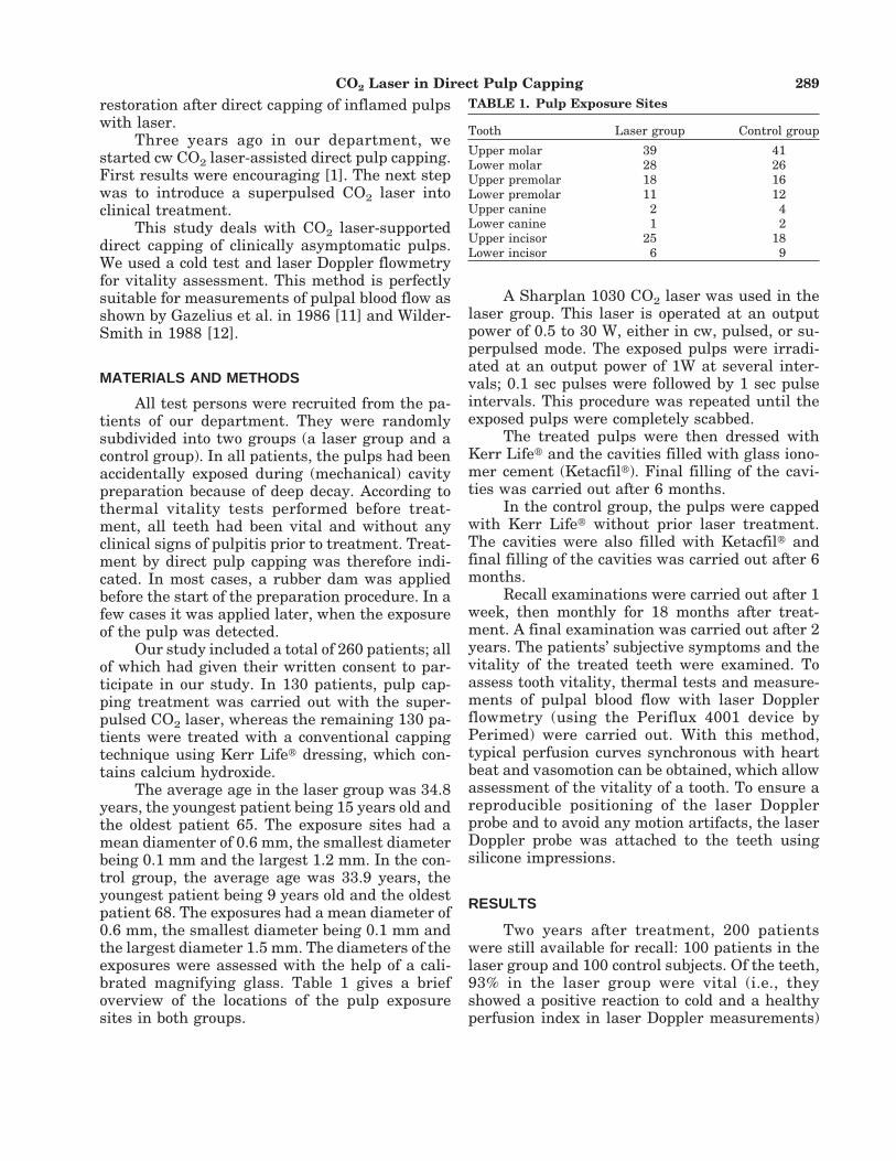
The average age in the laser group was 34.8years, the youngest patient being 15 years old andthe oldest patient 65. The exposure sites had amean diamenter of 0.6 mm, the smallest diameterbeing 0.1 mm and the largest 1.2 mm. In the con-trol group, the average age was 33.9 years, theyoungest patient being 9 years old and the oldestpatient 68. The exposures had a mean diameter of0.6 mm, the smallest diameter being 0.1 mm andthe largest diameter 1.5 mm. The diameters of theexposures were assessed with the help of a cali-brated magnifying glass. Table 1 gives a briefoverview of the locations of the pulp exposuresites in both groups.
A Sharplan 1030 CO2 laser was used in thelaser group. This laser is operated at an outputpower of 0.5 to 30 W, either in cw, pulsed, or su-perpulsed mode. The exposed pulps were irradi-ated at an output power of 1W at several inter-vals; 0.1 sec pulses were followed by 1 sec pulseintervals. This procedure was repeated until theexposed pulps were completely scabbed.
The treated pulps were then dressed withKerr Lifet and the cavities filled with glass iono-mer cement (Ketacfilt). Final filling of the cavi-ties was carried out after 6 months.
In the control group, the pulps were cappedwith Kerr Lifet without prior laser treatment.The cavities were also filled with Ketacfilt andfinal filling of the cavities was carried out after 6months.
Recall examinations were carried out after 1week, then monthly for 18 months after treat-ment. A final examination was carried out after 2years. The patients’ subjective symptoms and thevitality of the treated teeth were examined. Toassess tooth vitality, thermal tests and measure-ments of pulpal blood flow with laser Dopplerflowmetry (using the Periflux 4001 device byPerimed) were carried out. With this method,typical perfusion curves synchronous with heartbeat and vasomotion can be obtained, which allowassessment of the vitality of a tooth. To ensure areproducible positioning of the laser Dopplerprobe and to avoid any motion artifacts, the laserDoppler probe was attached to the teeth usingsilicone impressions.
RESULTS
Two years after treatment, 200 patientswere still available for recall: 100 patients in thelaser group and 100 control subjects. Of the teeth,93% in the laser group were vital (i.e., theyshowed a positive reaction to cold and a healthyperfusion index in laser Doppler measurements)
TABLE 1. Pulp Exposure Sites
Tooth Laser group Control group
Upper molar 39 41Lower molar 28 26Upper premolar 18 16Lower premolar 11 12Upper canine 2 4Lower canine 1 2Upper incisor 25 18Lower incisor 6 9
CO2 Laser in Direct Pulp Capping 289

and asymptomatic. Seven teeth were affected bypulpitis and required extirpation. In the controlgroup, 68 teeth remained vital, corresponding to asuccess rate of 68%.
The upper curve in the Figure 1 shows thatthe number of vital teeth in the superpulsed lasergroup dropped only slightly during the recall pe-riod, whereas in the control group, only 68 teethwere vital at the last recall examination. Thelower curve in Figure 1 shows the vitality devel-opment in the control group.
None of the groups showed a significant cor-relation between the size of the exposed pulp re-gion, the patients’ average age, type of the teethexamined (molar, incisor, etc.), and success oftreatment. Figures 2 and 3 illustrate the successof treatment in relation to the size of the exposedpulp region and the patients’ age, respectively.
All perfusion values obtained in the laserDoppler examination were consistent with the re-sults of the cold test. There were no discrepancieswith regard to pulpal blood flow and sensitivity.
DISCUSSION
As stated by Ketter [2] in 1978, the earlybeginnings of treatment of exposed pulps dateback to the year 1765. In 592 cases followed-upout of a total of 1,429 cases, Reuver [4], in 1992,found a success rate of 91% in the group of 10–20-year-old patients, which continuously droppedto 58% in the group of 70–80-year old patients. Ina type of opening, which the author described as“punctiform,” the success rate ranged ∼73%; whenan area of 1–9 mm2 was exposed, it was 61%. Atotal of 68% of the teeth were vital at the lastrecall examination. However, the timing of thelast recall examination varied between 4 monthsand 24 years in Reuver’s study.
A long-term study carried out by Marti in1979 [5] revealed that the healing success de-pends on the age of the patients. Marti [5] found asuccess rate of 84% in the group of 10–25-year-oldpatients, which dropped to only 44% in the groupof 46–70-year-old patients. This may be due to the
Fig. 1. Course of vitality.
290 Moritz et al.

fact that the pulps of younger subjects are richerin cells and have a higher ability to regenerate.
In 1979, Honegger et al. [6] reported a suc-cess rate of 83% in a total of 110 teeth subjected torecall examinations. Like Ketterl in 1987 [2],Schroeder [13] and Kopel [14] stated that directpulp cappying is indicated only in pulps that havenot been affected by inflammation a priori.
An obvious problem in an in vivo study in-volving such a large number of patients is theircontinuous availability for recall examinations. Infact, 200 out of 260 patients treated were stillavailable for recall after 24 months.
The success rate of 93% in the superpulsedlaser group is markedly higher than that obtainedin the control group and the success rates re-ported for conventional pulp capping techniquesin the literature [2,4–6]. It is even higher than thesuccess rate achieved using the cw CO2 laser [1].Similar to this study, the quoted authors workedon pulps that have been exposed during the re-moval of carious decay and were clinically asymp-tomatic.
None of the groups examined in this studyshowed a significant correlation between the suc-cess rate and the size of the exposed sites, or thepatient’s mean age.
Another interesting aspect seems to be thechronological course of vitality. The most notice-able loss of tooth vitality takes place within thefirst 4 months posttreatment. These 4 monthsseem to be the decisive period.
The most important effects of laser irradia-tion seem to be sterilization and scar formation[7–9] in the irradiated area due to thermal effects,which may help to preserve the pulp from bacte-rial invasion. In addition, laser irradiation shouldminimize the formation of a hematoma betweenthe pulp tissue and the calcium hydroxide dress-ing, allowing a close contact between the dressingand the exposed pulp. Another effect of lasertreatment may be the direct stimulation of dentinformation, as indicated by Paschoud and Holz in1988 [14]. However, in contrast to this study,these authors examined the effects of soft laserson dentin formation.
The long intervals (1 sec) between the pulses(0.1 sec), the relatively lower power setting (1 W)and the wavelength (10,6 mm), which is absorbedby ∼100 mm, is sufficient to avoid any thermaldamage to the pulp.
The reduced thermal strain on the pulp ex-erted by the superpulsed CO2 laser seems to beresponsible for the greater success rate in com-
Fig. 2. Tooth vitality in relation to the size of opening.
CO2 Laser in Direct Pulp Capping 291

parison with the cw CO2 laser. However, one ad-vantage of the cw CO2 laser used in the previousstudy (Lasersat by SATELEC) is its easy applica-tion. Since the actual laser is contained in thehandpiece, a bulky delivery system is avoided.
In the laser Doppler method, laser light inthe infrared range, which is delivered to the tis-sue to be examined, undergoes a Doppler fre-quency shift. A sensor records the wavelength ofthe light backscattered by moving particles(erythrocytes). Conclusions about blood flow inthe capillary region can be drawn by comparingthe wavelengths of the light transmitted to thetissue under study and the backscattered light.Because the measurements are not affected bythe function of the sensitive pulpal nerve-endings,possible false negative results of an indirect testcan be avoided, as shown by Gazelius in 1986 [10].
Vitality control with the laser Doppler tech-nique has shown to be very reliable, also in lux-ated and re-perfused teeth, as demonstrated byOlgart et al. in 1988 [15]. In our examinations, theDoppler vitality results were fully congruent withthe results of the thermal tests.
We believe that our results give clear proof ofthe favorable effects of laser application in direct
pulp capping, for which a pulsed laser should bepreferred.
REFERENCES
1. Moritz A, Schoop U, Ebrahim-Nahuray D, Sperr W. Vor-teile der der CO2-Laseranwendung bei der direkten Pul-penuberkappung mit Kalziumhydroxid-Eine Pilotstudie.Z Stomatol 1996; 939:451–454.
2. Ketterl W. Endodontie. Zahnerhaltung II, Bd. 3 Praxis d.Zahnheilkunde. Hrsg. New York: Verlag Urban undSchwarzenberg Wien 1987;3–81.
3. Foreman PC, Barnes IE. Review of calcium hydroxide.Int Endod J 1990; 23:283–297.
4. Reuver J. 592 Pulpauberkappungen in einer zahnarztli-chen Praxis-eine klinische Prufung (1966–1990). DtschZahnarztl Z 1992; 47:29–32.
5. Marti F. Direkte Pulpauberkappungen mit Calxyl. EineLangzeitstudie, Diss. Universitat Bern, 1979.
6. Honegger D, Holz J, Baume LJ. Controle clinique a longterme du coiffage pulpaire direct (realise par les etudi-ants de la SMD, Geneve). SSO Schweiz Monatsschr Zahn-heilkd 1979; 89:1020–1041.
7. Melcer J, Chaumette MT, Melcer F. Experimental re-search on the preparation of dentin-pulp tissue of teethexposed to CO2 laser beams in dogs and macaques (Ma-
Fig. 3. Tooth vitality in relation to patient age.
292 Moritz et al.

caca mulatta and Macaca fascicularis). C R Soc Biol(Paris) 1985: 179:577–585.
8. Melcer J. Latest treatment in dentistry by means of theCO2 laser beam. Lasers Surg Med 1986; 6:396–398.
9. Melcer J, Chaumette MT, Melcer F. Dental pulp ex-posed to the CO2 laser beam. Lasers Surg Med 1987;7:347–352.
10. Gazelius B, Olgart L, Edwall L. Non-invasive recording ofblood flow in human dental pulp. Enod Dent Traumatol1986; 2:219–221.
11. Wilder-Smith P. A new method for the non-invasive mea-surement of pulpal blood flow. Int Endod J 1988; 21:307–312.
12. Schroeder A. Endodontie-Ein Leitfaden fur Studium undPraxis. Quintessenz, Berlin, 1981;21–80.
13. Kopel HM. Considerations for the direct pulp cappingprocedure in primary teeth: A review of literature. ASDCJ Dent Child 1992; 59:141–149.
14. Paschoud Y, Holz J. Effect of the soft laser on the neofor-mation of a dentin bridge following direct pulp capping ofhuman teeth with calcium hydroxide. A histologicalstudy with the scanning electron microscope. SchweizMonatsschr Zahnmed 1988; 98:345–356.
15. Olgart L, Gazelius B, Lindh-Stromberg U. Laser Dopplerflowmetry in assessing vitality in luxated permanentteeth. Int Endod J 1988; 21:1–7.
CO2 Laser in Direct Pulp Capping 293