Embed Size (px)
Citation preview


Advances in Equine Laparoscopy
This book is accompanied by a companion website:
www.wiley.com/go/ragle
The website includes:
• Video clips demonstrating selected techniques
Advances in Equine LaparoscopyEdited by
Claude A. Ragle
A John Wiley & Sons, Inc., Publication
This edition first published 2012 © 2012 by John Wiley & Sons, Inc.
Wiley-Blackwell is an imprint of John Wiley & Sons, formed by the merger of Wiley’s global Scientific, Technical and Medical business with Blackwell Publishing.
Editorial offices: 2121 State Avenue, Ames, Iowa 50014-8300, USAThe Atrium, Southern Gate, Chichester, West Sussex, PO19 8SQ, UK9600 Garsington Road, Oxford, OX4 2DQ, UK
For details of our global editorial offices, for customer services and for information about how to apply for permission to reuse the copyright material in this book please see our website at www.wiley.com/wiley-blackwell.
Authorization to photocopy items for internal or personal use, or the internal or personal use of specific clients, is granted by Blackwell Publishing, provided that the base fee is paid directly to the Copyright Clearance Center, 222 Rosewood Drive, Danvers, MA 01923. For those organizations that have been granted a photocopy license by CCC, a separate system of payments has been arranged. The fee codes for users of the Transactional Reporting Service are ISBN-13: 978-0-4709-5877-3/2012.
Designations used by companies to distinguish their products are often claimed as trademarks. All brand names and product names used in this book are trade names, service marks, trademarks or registered trademarks of their respective owners. The publisher is not associated with any product or vendor mentioned in this book. This publication is designed to provide accurate and authoritative information in regard to the subject matter covered. It is sold on the understanding that the publisher is not engaged in rendering professional services. If professional advice or other expert assistance is required, the services of a competent professional should be sought.
Library of Congress Cataloging-in-Publication DataAdvances in equine laparoscopy / edited by Claude Ragle. p. ; cm. – (Advances in veterinary surgery) Includes bibliographical references and index. ISBN 978-0-470-95877-3 (hardcover : alk. paper) 1. Horses–Surgery. 2. Horses–Diseases–Diagnosis. 3. Veterinary laparoscopic surgery. I. Ragle, Claude. II. Series: Advances in veterinary surgery. [DNLM: 1. Horse Diseases–surgery. 2. Horses–surgery. 3. Laparoscopy–methods. 4. Laparoscopy–veterinary. SF 951] SF951.A38 2012 636.1’0897–dc23 2012014116
A catalogue record for this book is available from the British Library.
Wiley also publishes its books in a variety of electronic formats. Some content that appears in print may not be available in electronic books.
Set in 9.5/11.5 Pt Palatino by Toppan Best-set Premedia Limited
Cover design by Garth Stewart
DisclaimerThe publisher and the author make no representations or warranties with respect to the accuracy or completeness of the contents of this work and specifically disclaim all warranties, including without limitation warranties of fitness for a particular purpose. No warranty may be created or extended by sales or promotional materials. The advice and strategies contained herein may not be suitable for every situation. This work is sold with the understanding that the publisher is not engaged in rendering legal, accounting, or other professional services. If professional assistance is required, the services of a competent professional person should be sought. Neither the publisher nor the author shall be liable for damages arising herefrom. The fact that an organization or Website is referred to in this work as a citation and/or a potential source of further information does not mean that the author or the publisher endorses the information the organization or Website may provide or recommendations it may make. Further, readers should be aware that Internet Websites listed in this work may have changed or disappeared between when this work was written and when it is read.
1 2012
Dedication
This book is dedicated to my colleagues in laparoscopy who shared so generously to enrich our knowledge to care for the horse.

Contents
Contributors ix
Foreword xiJohn P. Walmsley
Foreword xvMark D. Markel, Chair of the Board of TrusteesACVS Foundation
Preface xvii
Acknowledgment xix
Section I Laparoscopic Skills and Instrumentation 3
1 FoundationsofLaparoscopy 5John P. Caron
2 FundamentalLaparoscopicSkills 13Claude A. Ragle and Boel A. Fransson
3 SuturingandKnot-TyingTechniques 21Fabrice Rossignol and Josef Boening
4 FundamentalsofEnergySources 35Scott E. Palmer
5 ReusableEquipment 41Christopher J. Chamness
6 DisposableEquipment 57John C. Huhn
Section II Laparoscopy in the Standing Horse 69
7 SedationandAnalgesiaintheStandingHorse 71Tamara Grubb
8 DiagnosticTechniques 83Dean A. Hendrickson
9 EvaluationofHorseswithSignsofAcuteandChronicAbdominalPain 93Andreas Klohnen
10 ClosureoftheNephrosplenicSpace 119Michael Roecken
11 Adhesiolysis 129Dean A. Hendrickson and Kayla Cochran
12 MesentericRentRepair 135Dean A. Hendrickson
13 Cryptorchidectomy 139Dwayne H. Rodgerson
14 PeritonealFlapHernioplastyTechniqueforPreventingtheRecurrenceofAcquiredStrangulatingInguinalHerniationintheStallion 149Hans Wilderjans
vii
viii Contents
15 InguinalHernioplastyUsingCyanoacrylate 161Fabrice Rossignol, Céline Mespoulhes-Rivière, and Josef Boening
16 IntersexGonadectomy 167Donna L. Shettko and Dean A. Hendrickson
17 BilateralOvariectomyintheMare 177Claude A. Ragle
18 OvariectomyfortheRemovalofLargePathologicOvariesinMares 189Hans Wilderjans
19 ImbricationoftheMesometriumtoRestoreNormal,HorizontalOrientationoftheUterusintheMare 203Palle Brink, Jim Schumacher, and John Schumacher
20 Nephrectomy 211Michael Roecken
21 RepairoftheRupturedEquineBladder 221Dean A. Hendrickson and Monika Lee
22 EquineThoracoscopy 229John Peroni
Section III Laparoscopy in the Recumbent Horse 239
23 GeneralAnesthesiaintheRecumbentHorse 241Tamara Grubb
24 Colopexy 253David G. Wilson
25 MeshIncisionalHernioplasty 257John P. Caron
26 Cryptorchidectomy 267John P. Caron
27 InguinalHernioplasty 277Fabrice Rossignol and Josef Boening
28 InguinalHerniorrhaphyintheFoal 287John P. Caron
29 OvariectomyintheMare 295Ted Fischer
30 OvariohysterectomyintheMare 301Claude A. Ragle
31 Laparoscopic-andEndoscopic-AssistedRemovalofCysticCalculi 311Michael Roecken
32 Splenectomy 323Ceri Sherlock and John Peroni
33 Hand-AssistedLaparoscopy 329Dwayne H. Rodgerson
Index 335
This book is accompanied by a companion website:
www.wiley.com/go/ragle
Thewebsiteincludes:
• Videoclipsdemonstratingselectedtechniques

Contributors
Josef Boening, DVM, DECVSTierarztliche Klinik, Telgte, Germany
Palle Brink, DVM, DECVSJägersro Equine ATG Clinic, Jagersro, Malmo, Sweden
John P. Caron, DVM, MS, DACVSCollege of Veterinary Medicine, Michigan State University, East Lansing, MI
Christopher J. ChamnessKarl Storz Endoscopy, Director of Global Business Development-Veterinary, Santa Barbara, CA
Kayla Cochran, DVMCollege of Veterinary Medicine, Colorado State University, Fort Collins, CO
Ted Fischer, DVM, DACVSChino Valley Equine Hospital, Chino Hills, CA
Boel A. Fransson, DVM, PhD, DACVSCollege of Veterinary Medicine, Washington State University, Pullman, WA
Tamara Grubb, DVM, MS, DACVACollege of Veterinary Medicine, Washington State University, Pullman, WA
Dean A. Hendrickson, DVM, MS, DACVSCollege of Veterinary Medicine, Colorado State University, Fort Collins, CO
John C. Huhn, DVM, MSVeterinary Medical Director, Covidien Animal Health, Salem, CT
Andreas Klohnen, DVM, DACVSChino Valley Equine Hospital, Chino Hills, CA
Monika Lee, BSCollege of Veterinary Medicine, Colorado State University, Fort Collins, CO
Céline Mespoulhes-Rivière, DVM, DECVSEcole Nationale Vétérinaire díAlfort, Maisons-Alfort Cedex, France
Scott E. Palmer, VMD, DABVPNew Jersey Equine Clinic, Millstone Twp, NJ
John Peroni, DVM, MS, DACVSCollege of Veterinary Medicine, University of Georgia, Athens, GA
Claude A. Ragle, DVM, DACVS, DABVP-EPCollege of Veterinary Medicine, Washington State University, Pullman, WA
ix

x Contributors
Michael Roecken, DVM, Priv.-Doz.Veterinary Clinic Starnberg, Starnberg, Germany; Department of Equine Surgery, School of Veterinary Medicine, Justus-Liebig-University, Giessen, Germany
Dwayne H. Rodgerson, DVM, MS, DACVSHagyard Equine Medical Institute, Lexington, KY
Fabrice Rossignol, DVM, DECVSClinique de Grosbois, Boissy Saint Leger, France
Jim Schumacher, DVM, MS, MVB, DACVSCollege of Veterinary Medicine, University of Tennessee, Knoxville, TN
John Schumacher, DVM, MS, DACVIM, ABVPCollege of Veterinary Medicine, Auburn University, Auburn, AL
Ceri Sherlock, DVM, DACVSCollege of Veterinary Medicine, University of Georgia, Athens, GA; School of Veterinary Medicine and Science, Sutton Bonington Campus, University of Nottingham, Sutton Bonington, Leicestershire, LE12 5RD, England
Donna L. Shettko, DVM, DACVSCollege of Veterinary Medicine, Western University of Health Sciences, Pomona, CA
Hans Wilderjans, DVM, DECVSDierenkliniek De Bosdreef, Moerbeke-Waas, B-9180, Belgium
David G. Wilson, DVM, DACVSWestern College of Veterinary Medicine, University of Saskatchewan, Sasketchewan, Canada
Foreword
Advances in Equine Laparoscopy is a textbook that will be welcomed by all equine laparoscopic sur-geons. Since the 1980s, when merely examining the equine abdomen endoscopically was an excit-ing revelation, some techniques such as laparo-scopic ovariectomy and cryptorchidectomy have developed and matured to become the treatment of choice for most surgeons. By 2012, however, a wide range of laparoscopic procedures have been reported and a publication conflating all this infor-mation is most appropriate.
In the human field, despite several reports of endoscopic procedures in the nineteenth century, the received birthplace of laparoscopy was in Dresden in 1901 when George Kelling examined a dog’s abdomen using a Nitze cystoscope; this was followed by endoscopic examinations of the human abdomen. As early as 1927, the first human laparoscopic textbook, Lehrbuch und Atlas der Laparo- und Thorakoskopie by Korbsch, was printed. Another significant early contributor was Heinz Kalk, who developed new lens systems in 1929 and published widely on liver and gall bladder disease. Progress predicated not only on the ambi-tion of surgeons but also on technological devel-opments. It was not until the late 1960s that the real expansion of laparoscopy began when Step-toe’s Laparoscopy for Gynaecology, describing lapar-oscopic sterilization techniques, instrumentation, and the use of electrocoagulation, was published.
In the 1970s, over 250,000 laparoscopic steriliza-tions were performed annually in the United States, but there was little formal training and complication rates were high. As a result, the American Association of Gynecologic Laparo-scopists was founded to inform surgeons and to monitor complications, and at about the same time, Chamberlain and Brown (1978) in Britain analyzed prospectively the complication rate in 50,000 laparoscopies. Gradually, an evaluation of what was going wrong and the introduction of credentialing programs has led to a reduction in morbidity and mortality in human laparoscopy.
There are other influences on the use of new techniques: One is the kudos of being able to perform them and another is public pressure for them to be performed, neither driven by the most important indication, viz, the outcomes of the pro-cedures. In human surgery, Cameron and Gadacz (1991) considered these influences a factor in the high incidence of bile duct injuries in laparoscopic cholecystectomies, which were 10 times that of open surgery. Nowadays, medical laparoscopy is under constant review and boasts many laparo-scopic procedures that have proven benefits over open surgery.
Laparoscopy has been a stimulating and chal-lenging addition to the veterinary surgeon’s rep-ertoire and as a veterinary discipline it is scarcely 25 years old. As with other fields of surgery, the
xi

xii Foreword
development of veterinary laparoscopy has fol-lowed that of human laparoscopy but has pro-gressed more slowly. The size of the horse’s abdomen and the weight of some abdominal organs have restricted the range of possible proce-dures. Like human surgeons in the early days, those of us performing equine laparoscopies in the late 1980s had little guidance and progress was slow, even though our arthroscopic hand–eye skills possibly gave us a better start than our human colleagues who at first only had training in general surgery. There are few studies analyz-ing morbidity and mortality associated with equine laparoscopy. Anecdotally, high complica-tion rates do not seem to have been the problem they were in human surgery: Perhaps they just have not been reported or maybe we have learned from the human experience. Either way, analysis of complication rates is called for, but at least formal laparoscopic tuition is now available and has become part of the residents’ training program so we can hope that many of the pitfalls will be avoided.
No doubt we also have been driven to perform laparoscopies for the benefit of our image as state-of-the-art surgeons, a fatal trap if our technique is insecure or if there are no benefits to the horse over open surgery. Client pressure can be difficult to ignore or assuage and can also lead us into per-forming procedures beyond our capabilities. For the benefit of our patients and ultimately their owners, we have a responsibility to evaluate our outcomes; comparative studies that will prove or disprove the benefits of procedures being per-formed laparoscopically are sorely needed. Once we have this information, we are in a stronger position to advise owners on the best course of action and we are unlikely to perform a laparos-copy because it is fun to do but has no benefit.
A great contribution to the discipline was made when in 2002 Ted Fischer’s landmark textbook, Equine Diagnostic and Surgical Laparoscopy, was published giving equine laparoscopists confidence to increase their repertoire and to consolidate their experience. This new text, Advances in Equine Laparoscopy, is the fruition of these experiences. Despite its title, the basics are not forgotten. The critical appraisal of training methods is a reminder that certain skills are difficult to acquire and there
are many ways to learn them. Laparoscopic skills are some of the most difficult to master in the field of surgery, and a basic competence in instrument and tissue handling in a 3-D cavity with 2-D vision is absolutely essential. Laparoscopic knots cannot be learnt during surgery nor can intracorporeal suturing and knot tying, two famously difficult techniques to master. However since they may be needed in a crisis, especially when hemorrhage is involved, they must be part of the surgeon’s armory before he or she embarks on laparoscopy in the clinical patient. This book offers the oppor-tunity for the inexperienced to discover what there is to learn and how to go about it, and for the experienced it is a reminder to maintain their skills. The thorough coverage of most procedures in common use today provides a basis and, one would hope, a stimulus for comparative studies between open surgery and laparoscopy. It is a reflection of the acceptance of equine laparoscopy that the evaluation of open techniques may have to be retrospective for some procedures. However, an objective appraisal is called for and where enough studies are published even a meta- analysis would be useful.
The time is opportune for a text that not only consolidates our knowledge of the bread-and-butter laparoscopic procedures, such as ovariec-tomy and cryptorchidectomy, but also offers detailed accounts of those that are proving their efficacy such as closure of the nephrosplenic space and herniorrhaphy. More advanced procedures including nephrectomy, splenectomy, cystotomy, and ovariohysterectomy are also covered. Written by surgeons with good experience of the tech-niques, this book will facilitate a widening of the laparoscopic repertoire and encourage experi-enced surgeons to master advanced techniques. Laparoscopy is an unforgiving activity that depends on good technique, and a study of this book will enable surgeons to avoid the mistakes of their predecessors, progress more safely, and benefit the horse and its owner. The skill, knowl-edge, and judgment of the surgeon are the factors on which the success of surgical procedures depends. Advances in Equine Laparoscopy will enhance the quality of all three and will make a significant contribution to the development of this discipline.

Foreword xiii
References
Cameron, J.L. & Gadacz, T.R. (1991) Laparoscopic chole-cystectomy (Editorial). Annals of Surgery, 213, 1–2.
Chamberlain, G. & Brown, J.C. (eds.) (1978) Gynaecologi-cal Laparoscopy: Report on the Confidential Enquiry into Gynaecological Laparoscopy. Royal College of Obstetri-cians and Gynaecologists, London.
John P. Walmsley, MA, VetMB, CertEO, DipECVS, HonFRCVS
The Liphook Equine HospitalForest Mere
LiphookHants GU30 7JG
UK

Foreword
The American College of Veterinary Surgeons (ACVS) Foundation is excited to present Advances in Equine Laparoscopy as the second book in the book series Advances in Veterinary Surgery. The ACVS Foundation is an independently chartered philanthropic organization devoted to advancing the charitable, educational, and scientific goals of the ACVS. Founded in 1965, the ACVS sets the standards for the specialty of veterinary surgery. The ACVS, which is approved by the American Veterinary Medical Association, administers the board certification process for diplomates in vet-erinary surgery and advances veterinary surgery and education. One of the principal goals of the ACVS Foundation is to foster the advancement of the art and science of veterinary surgery. The Foundation achieves these goals by supporting investigations in the diagnosis and treatment of surgical diseases; increasing educational opportu-nities for surgeons, surgical residents, and veteri-nary practitioners; improving surgical training of residents and veterinary students; and bettering animal patients’ care, treatment, and welfare. This collaboration with Wiley-Blackwell benefits all who are interested in veterinary surgery by pre-
senting the latest evidence-based information on a particular surgical topic.
This edition is an outstanding example of the promise of this series. Advances in Equine Laparos-copy is edited by Dr. Claude A. Ragle, a diplomate of the ACVS and a prominent equine surgeon. He has assembled the leaders in the field of equine laparoscopy presenting the important techniques and applications for this minimally invasive pro-cedure. As you read through this book, you will find the latest information on the foundations and fundamental skills required for laparoscopy, equipment, techniques for sedation and analgesia required for the use of laparoscopy in horses, and the techniques and applications for a wide range of laparoscopic and thoracoscopic procedures. The ACVS Foundation is proud to partner with Wiley-Blackwell on this important series and is proud to present this second book in the series Advances in Veterinary Surgery.
Mark D. MarkelChair of the Board of Trustees
ACVS Foundation
xv

Preface
Advances in Equine Laparoscopy (AEL) represents an important update in the practice of minimally invasive surgery (MIS) of the horse. It has been more than three decades since the publication of Animal Laparoscopy, the first veterinary laparos-copy book in which Dr. Witherspoon and col-leagues described the basic technique and instrumentation for the laparoscopy of the horse. Ten years separate this publication of AEL from Equine Diagnostic and Surgical Laparoscopy, the first book dedicated to the topic of laparoscopy of the horse. AEL builds upon the stellar contributions of these preceding books. It represents a collabora-tive gathering of current advances in techniques and instrumentation and is designed to augment and contrast existing texts. The book is organized such that the first several chapters review the fun-damental foundations of equine laparoscopy. The following chapters are clinical techniques and are divided into groups based on standing or recum-bent positioning of the horse and then on the body system being operated where applicable. A chapter reviewing sedation, analgesia, and/or anesthesia of the horse for laparoscopy precedes each of the
two groups of chapters that are based upon the horse’s position during the operation. Each opera-tive technique chapter follows a general frame-work to provide continuity and to ease locating of information by the reader. This scheme of organi-zation inherently leads to some overlap between related techniques but importantly provides the reader a broader perspective in how clinical experts may approach similar operative chal-lenges in another way. With few exceptions, every technique presented is a procedure that is a current standard of practice in laparoscopy. Not included in this edition of AEL are areas of MIS that are still in their infancy in equine surgery, such as Single Incision Laparoscopic Surgery (SILS), Natural Orifice Translumenal Endoscopic Surgery (NOTES) and Laparoscopic Robotic Surgery; it is anticipated that progress in these techniques in the horse may warrant inclusion in a future edition. This book will be of great interest to all that are interested in the field of MIS and provides an excellent platform for the future advancement of equine laparoscopy.
xvii

Acknowledgment
It is with great appreciation I recognize the ACVS Foundation’s Board of Trustees and Dr. Mark Markel (Chair) for creating the Advances in Veteri-nary Surgery series of books and for recognizing the significant progress of laparoscopy in equine surgery. This book would not have been possible without the expert guidance of Erica Judisch and Susan Engelken of Wiley-Blackwell. I also
acknowledge Rachael Lencioni, veterinary student extraordinaire, for her tireless efforts and organi-zational skills working on this book, and Dr. William Dernell, my department chair, for his encouragement. Finally, I would like to acknowl-edge my extended family and friends, particularly, Boel and my two wonderful daughters, Selma and Ella, for their unconditional love.
xix


Advances in Equine Laparoscopy

Section ILaparoscopic Skills and Instrumentation

5
Origins of laparoscopy
Since antiquity, health-care providers have striven to visually examine the internal anatomy of their patients. Indeed, rectal specula and natural light were in use in the era of Hippocrates of Cos (Rosin 1993). Ambient light or external light sources sup-plemented with diverse lenses or mirrors to examine the ears, nose, mouth, rectum, and vagina/uterus were used until the mid-nineteenth century. Among the first using artificial light was the lichtleiter or “light transmitter” invented by the German physician Philip Bozzini in 1804. The light source for the device was a wax candle, and its glow was reflected by a mirror into a speculum that Bozzini used for the examination of a variety of body orifices and the uterus (Figure 1.1). Unfor-tunately, as a result of contemptuous reviews of his invention by his contemporaries, the innova-tion went largely unrecognized by the medical community (Rathert et al. 1974). Perhaps inspired by the work of Bozzini, in 1853, Antonin Desormeaux developed an “endoscope” with a lamp fueled by a mixture of turpentine and alcohol (Kaiser & Corman 2001). The light from the flame
was concentrated and directed by a concave mirror to the area of interest and permitted evaluation of the cervix, uterus, and urinary bladder, although it lacked sufficient light for gastroscopy (Figure 1.2) (Rosin 1993; Lau et al. 1997). Unlike the reproach Bozzini’s efforts engendered, Desormeaux received an award from the Royal Academy of Medicine in Paris and went on to use his endo-scope for diagnostic and therapeutic procedures in his practice at the Necker Hospital in Paris during the 1860s (Kaiser & Corman 2001).
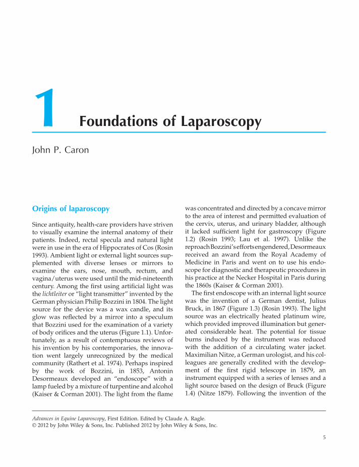
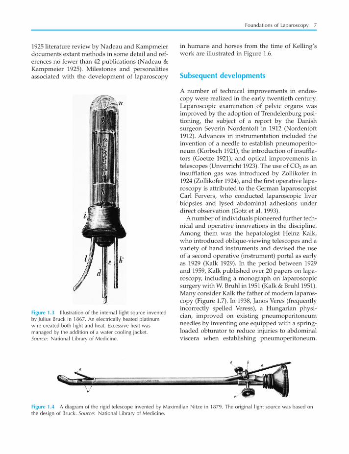
The first endoscope with an internal light source was the invention of a German dentist, Julius Bruck, in 1867 (Figure 1.3) (Rosin 1993). The light source was an electrically heated platinum wire, which provided improved illumination but gener-ated considerable heat. The potential for tissue burns induced by the instrument was reduced with the addition of a circulating water jacket. Maximilian Nitze, a German urologist, and his col-leagues are generally credited with the develop-ment of the first rigid telescope in 1879, an instrument equipped with a series of lenses and a light source based on the design of Bruck (Figure 1.4) (Nitze 1879). Following the invention of the
1 Foundations of Laparoscopy
John P. Caron
Advances in Equine Laparoscopy, First Edition. Edited by Claude A. Ragle.© 2012 by John Wiley & Sons, Inc. Published 2012 by John Wiley & Sons, Inc.

6 Advances in Equine Laparoscopy
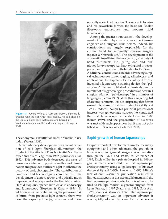
reflected into the speculum with a head mirror, a technique he called “ventroscopy” (Von Ott 1901). While his efforts presaged Natural Orifice Trans-lumenal Endoscopic Surgery (NOTES®) proce-dures, it is not regarded as laparoscopy in the conventional sense by many medical historians. Priority for “true” laparoscopic examination of the abdomen is afforded the German surgeon Georg Kelling, who used a Nitze-style cystoscope to examine the peritoneal cavity of dogs in 1901. Kelling created pneumoperitoneum with filtered air and called the technique “koelioskopie” (Figure 1.5) (Kelling 1902; Rosin 1993). Although Kelling later reported the use of laparoscopic methods in a series of human patients, Hans Christian Jaco-baeus, a Swedish surgeon, is credited with the first report of a relatively large series of “laparothora-koskopie” procedures in people, published in 1911 (Jacobaeus 1911). Unlike Kelling, Jacobaeus did not use abdominal insufflation; however, many of his patients had ascites, which afforded working space. As is typical in medicine, progress tends to occur in parallel; an American surgeon, Bertram Bernheim, also reported on two laparoscopically managed patients in 1911 (Bernheim 1911). Evi-dently, enthusiasm for the concept of laparoscopy spread rapidly in the ensuing years; a
incandescent bulb by Thomas A. Edison in 1880, it was incorporated into a telescope developed by the Glaswegian David Newman in 1883 (Dame-word 1992). Further developments in lenses and miniaturization of incandescent light bulbs fos-tered additional advances so that endoscopy of the upper airway, lower urogenital tract, esophagus, stomach, and anorectum began to be conducted with some frequency. Although the thoracic and peritoneal cavities were yet to be examined, the equipment and methodology to do so was largely in place (Lau et al. 1997).
In 1901, the abdominal cavity of a pregnant woman was examined by Dimitri von Ott, who employed a speculum introduced into the abdom-inal cavity via small colpotomy. External light was
Figure 1.1 Illustration of the lichtleiter of Philip Bozzini. The light from the candle is reflected by a mirror through an attached speculum (not shown) into the patient. Source: National Library of Medicine.
Figure 1.2 Diagram of Antonin Desormeaux’s endoscope of 1853. A lamp fueled by turpentine and alcohol provided the artificial light that was focused and directed through the speculum. Source: National Library of Medicine.
Foundations of Laparoscopy 7
in humans and horses from the time of Kelling’s work are illustrated in Figure 1.6.
Subsequent developments
A number of technical improvements in endos-copy were realized in the early twentieth century. Laparoscopic examination of pelvic organs was improved by the adoption of Trendelenburg posi-tioning, the subject of a report by the Danish surgeon Severin Nordentoft in 1912 (Nordentoft 1912). Advances in instrumentation included the invention of a needle to establish pneumoperito-neum (Korbsch 1921), the introduction of insuffla-tors (Goetze 1921), and optical improvements in telescopes (Unverricht 1923). The use of CO2 as an insufflation gas was introduced by Zollikofer in 1924 (Zollikofer 1924), and the first operative lapa-roscopy is attributed to the German laparoscopist Carl Fervers, who conducted laparoscopic liver biopsies and lysed abdominal adhesions under direct observation (Gotz et al. 1993).
A number of individuals pioneered further tech-nical and operative innovations in the discipline. Among them was the hepatologist Heinz Kalk, who introduced oblique-viewing telescopes and a variety of hand instruments and devised the use of a second operative (instrument) portal as early as 1929 (Kalk 1929). In the period between 1929 and 1959, Kalk published over 20 papers on lapa-roscopy, including a monograph on laparoscopic surgery with W. Bruhl in 1951 (Kalk & Bruhl 1951). Many consider Kalk the father of modern laparos-copy (Figure 1.7). In 1938, Janos Veres (frequently incorrectly spelled Veress), a Hungarian physi-cian, improved on existing pneumoperitoneum needles by inventing one equipped with a spring-loaded obturator to reduce injuries to abdominal viscera when establishing pneumoperitoneum.
1925 literature review by Nadeau and Kampmeier documents extant methods in some detail and ref-erences no fewer than 42 publications (Nadeau & Kampmeier 1925). Milestones and personalities associated with the development of laparoscopy
Figure 1.3 Illustration of the internal light source invented by Julius Bruck in 1867. An electrically heated platinum wire created both light and heat. Excessive heat was managed by the addition of a water cooling jacket. Source: National Library of Medicine.
Figure 1.4 A diagram of the rigid telescope invented by Maximilian Nitze in 1879. The original light source was based on the design of Bruck. Source: National Library of Medicine.
8 Advances in Equine Laparoscopy
optically correct field of view. The work of Hopkins and his coworkers formed the basis for flexible fiber-optic endoscopes and modern rigid laparoscopes.
Among the greatest innovators in the develop-ment of modern laparoscopy was the German engineer and surgeon Kurt Semm. Indeed, his contributions are largely responsible for the current trend for minimally invasive surgery (Spaner & Warnock 1997). The development of the automatic insufflator, the morcellator, a variety of hand instruments, the ligating loop, and tech-niques for extracorporeal knot tying and intracor-poreal suturing are all attributable to Dr. Semm. Additional contributions include advancing surgi-cal techniques for tumor staging, adhesiolysis, and applications for bipolar electrocautery. He also invented a laparoscopic training device, the “pel-vitrainer.” Semm published extensively and a number of his gynecologic procedures appear in a surgical atlas on “pelvicoscopy” in a number of languages (Semm 1992). With this staggering list of accomplishments, it is not surprising that Semm earned his share of habitual detractors (Litynski 1996a). Indeed, though his principal professional interest was gynecologic surgery, he conducted the first laparoscopic appendectomy in 1980 (Semm 1989), and the presentation of his work was met with such opposition that it was not pub-lished until 3 years later (Vilardell 2006).
Rapid growth of human laparoscopy
Despite important developments in electrocautery equipment and other advances, the growth of laparoscopy in general surgery was tentative through the 1960s and 1970s. In September of 1985, Erich Mühe, in a private hospital in Böblin-gen Germany, conducted the first laparoscopic cholecystectomy using a “galloscope” of his own design (Litynski 1996b; Lau et al. 1997). Mühe’s lack of enthusiasm for publication resulted in limited awareness of this accomplishment, and the first laparoscopic cholecystectomy is often attrib-uted to Phillipe Mouret, a general surgeon from Lyon, France, in 1987 (Nagy et al. 1992; Gotz et al. 1993; Spaner & Warnock 1997). The laparoscopic cholecystectomy was an important advance; it was rapidly adopted by a number of centers in
Figure 1.5 Georg Kelling, a German surgeon, is generally credited with the first “true” laparoscopy. He published on the use of a Nitze-style cystoscope and filtered air insufflation to examine the abdominal organs of dogs in 1901.
The eponymous insufflation needle remains in use today (Veress 1938).
A revolutionary development was the introduc-tion of cold light fiberglass illumination, the product of the efforts of French scientist Max Four-estier and his colleagues in 1952 (Fourestier et al. 1952). This advance both decreased the risks of burns associated with previous methods of illumi-nation and provided sufficient light to enhance the quality of endophotography. The contribution of Fourestier and his colleagues, combined with the development of a more robust and optically much improved rod lens system by the English physicist Harold Hopkins, opened new vistas in endoscopy and laparoscopy (Hopkins & Kapany 1954). In addition to virtually eliminating the pervasive risk of burns from previous light sources, there was now the capacity to enjoy a wider and more