Embed Size (px)
Citation preview

1
Advances in Endoscopy Update Newer Technologies in Adult GI
NASPGHAN 2013Clinical Session 2 • Endoscopy
October 11, 2013Chicago Hilton Downtown • Chicago, Illinois
John A. Martin, MD, FASGE
Associate Professor of Medicine and SurgeryDirector of Endoscopy
Northwestern University Feinberg School of Medicine • Chicago, Illinois
Disclosure
In the past 12 months, I have had no relevant financial relationships with the
manufacturers of any commercial products and/or providers of commercial services discussed in this CME activity
2
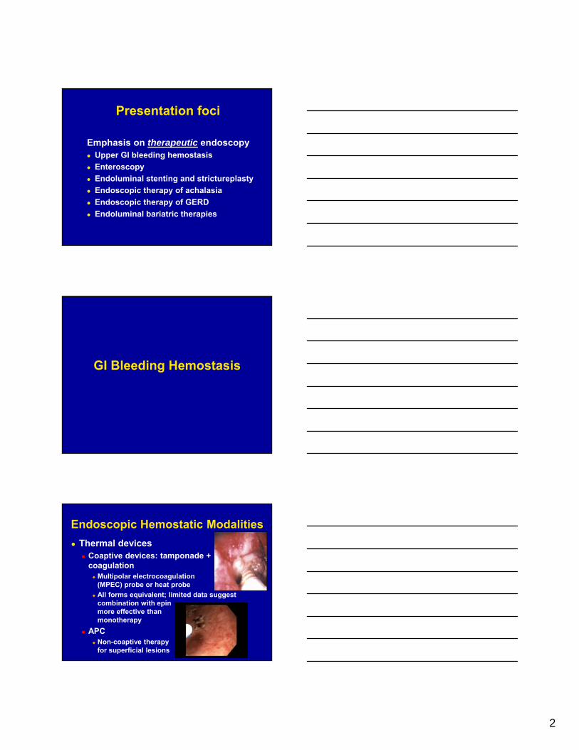
Presentation foci
Emphasis on therapeutic endoscopy Upper GI bleeding hemostasis
Enteroscopy
Endoluminal stenting and strictureplasty
Endoscopic therapy of achalasia
Endoscopic therapy of GERD
Endoluminal bariatric therapies
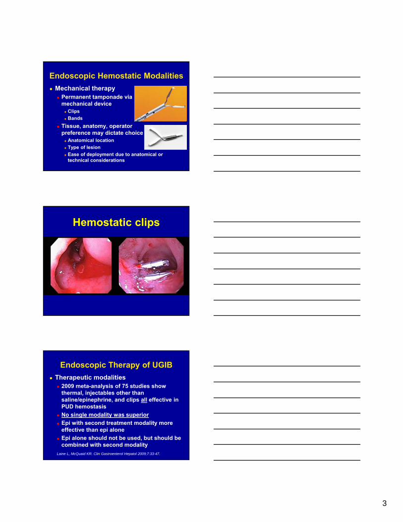
GI Bleeding Hemostasis
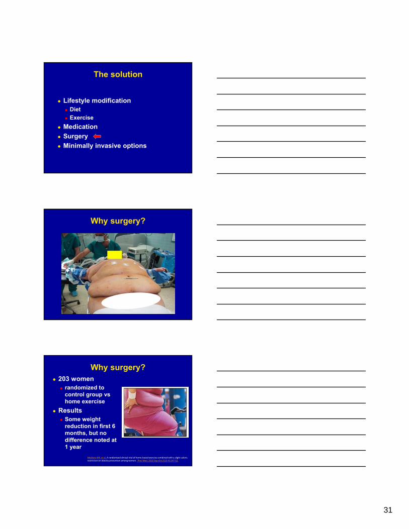
Thermal devices Coaptive devices: tamponade +
coagulation Multipolar electrocoagulation probe
(MPEC) probe or heat probe
All forms equivalent; limited data suggest combination with epin more effective than monotherapy
APC Non-coaptive therapy
for superficial lesions
Endoscopic Hemostatic Modalities
3
Mechanical therapy Permanent tamponade via
mechanical device Clips
Bands
Tissue, anatomy, operator preference may dictate choice Anatomical location
Type of lesion
Ease of deployment due to anatomical or technical considerations
Endoscopic Hemostatic Modalities
Hemostatic clips
Therapeutic modalities 2009 meta-analysis of 75 studies show
thermal, injectables other than saline/epinephrine, and clips all effective in PUD hemostasis
No single modality was superior
Epi with second treatment modality more effective than epi alone
Epi alone should not be used, but should be combined with second modality
Endoscopic Therapy of UGIB
Laine L, McQuaid KR. Clin Gastroenterol Hepatol 2009;7:33-47.

4
Combination Therapy vs. Hemostatic Clips Study
Prospective randomized controlled trial of acute non-variceal upper GI bleeding
All pts on high dose proton pump inhibitors
Primary Control Rebleeding Rate
Saltzman JR. Am J Gastroenterol 2005;100:1503
0
10
20
30
40
50
60
70
80
90
100
Hemoclips
Combination
0
10
20
30
Hemoclips
Combination
P=0.45 P=0.49
% %
Hemostatic Clips for Upper GI Bleed
Meta-analysis of 15 RCT’s of 1156 patients 390 clips alone
242 clips and injection
359 injection alone
165 thermocoagulation with or without injection
Hemoclips superior to injection therapy alone Definitive hemostasis 87% vs. 75%
Hemoclips comparable to thermal coagulation Definitive hemostasis 82% vs. 81%
Sung JJ. Gut 2007;56:1364
When to Use Hemostatic Clips
Ideal for hemoclips Lesion pliable
Lesion accessible
<2 mm vessel
<2 cm ulcer defect
Difficult for hemoclips Indurated or fibrotic base
Challenging locations Lesser curve stomach
Posterior wall stomach
Posterior duodenumVisible Vessel

5
No prospective trials comparing methods for acute UGIB due to vascular abnormalities Vascular ectasias
Dieulafoy lesions
GAVE
Endoscopic marking Consider tattooing difficult-to-locate lesions
Place clip whether endotherapy succeeds or fails to facilitate IR / surgical intervention
Upper GI Vascular Abnormalities
New hemostatic clips
A Peek at New Technologies in Hemostasis
Over-the-scope Clip
Kirschniak A. Gastrointest Endosc 2007;66:162

6
New hemostatic clips
A Peek at New Technologies in Hemostasis
New hemostatic clips
A Peek at New Technologies in Hemostasis
New hemostatic clips
A Peek at New Technologies in Hemostasis

7
New hemostatic clips
A Peek at New Technologies in Hemostasis
New hemostatic clips
A Peek at New Technologies in Hemostasis
Monopolar coagulation grasping forcep
A Peek at New Technologies in Hemostasis

8
Saltzman JR. Gastrointest Endosc 2010;72(4):796
Monopolar Cautery
Monopolar device Designed for endoscopic bleeding
Flat jaws for grasping
Rotational ability
Grounding pad required
Optimal settings (stomach) 50 Watts for 2 or 3 seconds
Role of monopolar cautery in the management of upper GI bleeding needs to be determined
Doppler probe
A Peek at New Technologies in Hemostasis
Doppler Ultrasound
Wong RC. Gastroenterology 2009;137:1897

9
Doppler Signal Before and After Endoscopic Therapy
Application of Doppler guided hemostasis has the potential to help reduce ulcer rebleeding
Jensen DM. DDW 2010
Hemostatic Nanopowder SprayMechanism of action:
Tamponade (rapid velocity application)
Dehydration of fluid within blood
Activation of clotting cascade
Activation of platelets
Aims: To assess the efficacy and safety of a novel hemostatic nanomaterial in short and long term hemostasis in a survival GI bleeding animal model
Conclusions: Endoscopic application of this nanopowder is safe and highly effective in achieving hemostasis in an anticoagulated severe GI bleeding animal model
Giday SA. Endoscopy 2011;43:296
(Forrest 1b = oozing)Sung JJY. Endoscopy
2011;43:291
Delivery catheter
Bleeding peptic ulcer
Human Hemostatic Spray Initial Trial

10
New hemostatic spray
A Peek at New Technologies in Hemostasis
New hemostatic spray
A Peek at New Technologies in Hemostasis
Hemospray Considerations
Effective only in actively oozing or spurting bleeding lesions
Does not require special expertise
Can be rapidly used if bleeding occurs after polypectomy or sphincterotomy
May be effective in difficult locations
Further clinical studies are needed

11
Consult new 2012 ASGE Guidelines at www.asge.org “The role of endoscopy in the management
of acute non-variceal upper GI bleeding” Gastrointest Endosc 2012;75:1132-1138. Management of PUD with adherent clot is
controversial
Injection, thermal, and mechanical therapies are all effective
Epinephrine alone should not be used in PUD bleeding, but should be combined with 2nd agent
Upper GI Bleeding 2012: Summary
Consult new 2012 ACG Guidelines at www.gi.org
Upper GI Bleeding 2012: Summary
Laine L, Jensen DM. Management of Patients with Ulcer Bleeding. ACG Practice Guidelines. Am J Gastroenterol 2012;107:345-360.
Consult new 2012 ACG Guidelines at www.gi.org
Upper GI Bleeding 2012: Summary
Laine L, Jensen DM. Management of Patients with Ulcer Bleeding. ACG Practice Guidelines. Am J Gastroenterol 2012;107:345-360.

12
Enteroscopy
Diagnostic and therapeutic options Colonoscopy with ileoscopy Video Capsule Endoscopy (VCE) Push Enteroscopy (with or without overtube) Balloon Enteroscopy (peroral or peranal) Intraoperative Enteroscopy (laparoscopic or
open) Rotational Enteroscopy *UGIS / SBFT (for evaluation of masses,
strictures) CT enterography / MR enterography Contrast angiography Tagged-RBC scan Meckel’s scan
Background
Deep enteroscopy: diagnostic and therapeutic

13
Background
Deep enteroscopy: diagnostic and therapeutic Balloon enteroscopy
Background
Deep enteroscopy: diagnostic and therapeutic
Background
Deep enteroscopy: diagnostic and therapeutic Balloon enteroscopy

14
Overtube Scope
Overtube Scope
40 cm
Overtube Scope
Overtube Scope
Reduction
Background
Courtesy Patrick Pfau, MD, Univ of Wisconsin.
Background
Deep enteroscopy: diagnostic and therapeutic Rotational enteroscopy
Performance characteristics
Deeper insertion = superior visualization compared to push enteroscopy
Total small intestinal examination in 12-25%; diagnostic yield 40%
Clinical yield for VCE and DBE equivalent: 60%
Kawamura T. GIE 2008. Pasha S. Clin Gastro Hep 2008.

15
Balloon enteroscopy caveats
It takes a long time… 120-200 minutes peroral or retrograde
Effortful May require anesthesia
(logistical issues, risk, cost)
Skill acquisition
Requisite expertise Diagnostic
Therapeutic
Balloon enteroscopy caveats
Surgical anatomical caveats: fixed bowel Peritoneal adhesions
Anatomotic strictures
Esophageal strictures
Balloon enteroscopy caveats
Surgical anatomical caveats: fixed bowel Roux-en-Y anatomy
Anastomoses– Ectatic anastomoses
– Hairpin turns
» Fixed
» Scope radius
» Scope stiffness
Peritoneal windows
Gastric looping– Hiatal hernia

16
Balloon enteroscopy caveats
Surgical anatomical caveats: fixed bowel Roux-en-Y anatomy
Anastomoses– Ectatic anastomoses
– Hairpin turns
» Fixed
» Scope radius
» Scope stiffness
Peritoneal windows
Gastric looping– Hiatal hernia
Choosing Your EquipmentWhat Gets Me Farther?
In randomized trials, double balloon and single balloon enteroscopy achieved comparable antegrade insertion distances1,2
In a single study, insertion depth with DBE was ~ 50 cm greater than SBE but this did not hold significance after comparisons
In a study comparing total enteroscopy (both antegrade and retrograde in same patients), total enteroscopy rate for SBE was 0% and 57.1% in DBE groups3
1Efthymiou M et al, GIE, 2012, 2 Domagk D et al, Endoscopy, 2011, 3 Takano N et al, GIE, 2011
Study Follow-up Duration Findings Rebleeding rate (%)
Double Balloon Enteroscopy
Gerson (2009) 30 monthsVascular lesions 45Normal DBE 42Overall 42
Shinozaki (2010) 29.7 monthsVascular lesions 60Normal DBE 37Overall 39
May (2011) 55 monthsVascular lesions 42Normal DBE N/AOverall N/A
Samaha (2012) 22.6 monthsVascular lesions 46Normal DBE N/AOverall N/A
Single Balloon Enteroscopy
Kushnir (2013) 23.9 monthsVascular lesions 48Normal SBE 56Overall 45
Kushnir VM, Dig Dis Sci, 2013
Enteroscopy for Small Bowel Bleeding Effective?

17
Deep enteroscopycomplications
Balloon enteroscopy Post-procedure distention/pain common
(> 20%)
Major complication rate 0.8 – 5 % Perforation 1-3%
Higher when intervention added
Rare pancreatitis
Mensink P. Endoscopy 2007. Kamal A. GIE 2008.
Deep enteroscopy: indications
Suspected Small Bowel Bleeding Obscure Occult
Obscure Overt
Detection or Resection of small bowel polyps/tumors
Suspected inflammatory bowel disease/enteropathy
Therapy of small bowel stricture
Altered anatomy ERCP
Clinical application
Capsule enteroscopy and balloon / rotational enteroscopy are complimentary
Per Dr. Rosh’s lecture Consider capsule first given non-
invasive, with lower complication risk and no sedation requirement
Consider going straight to rotational or balloon enteroscopy if suspicion for treatable lesion is high

18
Clinical application
Capsule enteroscopy and balloon / rotational enteroscopy are complimentary (continued) Positive capsule findings
Tissue acquisition
Treatment
Negative capsule findings …with persistent strong clinical suspicion
for intestinal pathology
Clinical application
Choice of deep enteroscopy platform is largely institution-dependent, and institutionally-driven Endoscope manufacturer holding
contract for unit
Availability of local operator experience and expertise
Applies to capsule as well as balloon / rotational enteroscopy
Clinical application
On the other hand… Choose capsule if
Purely diagnostic
Stricture unlikely or excluded
Radiologic studies are negative
Choose push enteroscopy with colonoscope if likely to be near ligament of Treitz or TI Easier, faster
Larger channel for aspiration, accessories
Dial-in stiffening feature, flushing pump capability
Consider quick repeat EGD first in appropriate cases, particularly if you didn’t perform the index EGD

19
Biliary Endoscopy
56
CCD-video choledochoscopy with NBI
CCD-video choledochoscopy
Image courtesy Professor Takao Itoi, MD, Tokyo Medical University
57
CCD-video choledochoscopy with NBI
Image courtesy Professor Takao Itoi, MD, Tokyo Medical University
CCD-video choledochoscopy

20
58
Direct-video choledochoscopy
Larghi and Waxman, GIE 2006;63:853.
Per-oral choledochoscopy (POCS)
59
CCD-video choledochoscopy with NBI
Image courtesy Irving Waxman, MD, University of Chicago
Per-oral choledochoscopy (POCS)
60
Deep-enteroscopic ERC
Altered-anatomy ERCP

21
61
Deep-enteroscopic ERC
Altered-anatomy ERCP
Luminal Stenting
Benign esophageal stricture management
Dilation Passage Balloon
Intralesional corticosteroid injection Strictureplasty
Needle-knife Endoscopic scissor Argon Plasma Coagulation (APC)
Stent therapy: long-term/continuous/gradual dilator Migration Chest pain Not durable

22
Treatments: Stents
Treatments: Stents
Increasing literature in benign disease, but all small series
*For SEMS (all): use in benign disease is off-label
No role for uncovered or partially-covered SEMS
Only fully-covered stents in benign indications FC-SEPS: FDA approved indication
**FC-SEMS: off-label use
Treatments: Stents
Stent therapy: concept in benign esophageal strictures Temporary, long-term/continuous/gradual dilator Stricture remodeling Initial enthusiasm was tempered by
Migration Chest pain Not durable AE fistulas (Rogart, et al., Endoscopy 2007)
Biodegradable stents Tissue ingrowth Potential for serial stenting without removal
Caveat: radiation and chemotx increase stent complications

23
Treatments: Stents
PC-SEMS: partially-covered metallic
FC-SEMS: fully-covered metallic
SEPS: fully-covered plastic
deWijkerslooth LRH, et al., Am J Gastroenterol 2011;106:2080.
Why we don’t use partially covered SEMS in benign disease
Why we don’t use partially covered SEMS in benign disease
Hirdes, et al., Endoscopy2011;43:156 4 patients PC-SEMS for
benign perforation or leak
Median dwell time 29 days
Endoscopic removal led to perforation in 4/4

24
Treatments: SEPS stentsStudy n Stricture
typeStent type
Duration stenting
Outcome Migrations Complic’s
Repici2004GIE
15 Mixed PolyflexSEPS
6 wks 80% dys-free at mean 22 mos
Migr 7%Complic 0
Evrard2004GIE
21 Mixed PolyflexSEPS
2d-56 wks 80% dys-free at median f/u21 mos
Migr 52%Airway compr 5%
Dua2008 AJG(prosp)
40 Mixed; most anast/corrosive/XRT
PolyflexSEPS
4 wks 40% dys-free at median 53 wk follow up
Migr 22%Death 1 bldFistula 1Perf 2
Oh2010DDS
13 Anast 11/13 PolyflexSEPS
6 wks 23% dysph-free @ µ 37 d, r 6-120 d
Migr 30%No major complic’s
Repici2010APT
130SysRvw
Mixed PolyflexSEPS
?; med f/u 13 mo
52% sympfree at med 13 mo f/u
Migr 24%Maj comp9%, dth 1%
Treatments: FC-SEMS stents
Study n Stricture type
Stent type
DurationStent/post
Outcome Migrations + Complications
Kim 2009 Eur Radiol
55PR
Corrosive 80%; else mixed
Tae-woong Niti-S
1 wk-6 mo/µ 38 mos
38% patency at 6 mos; 33% at 1 yr
Migr 25%Ovrgrth 31%
Senousy 2010 DDS
7RT
Mixed anast/pep/XRT/PDT
Alimaxx 4-84 d, µ 37 d/µ 172 d
“Clin impvmt dysphagia” 100%
Migr 39%Minor complic only
Eloubeidi 2011GIE
19PR
Mixed Alimaxx 6-300 d, 64±74d/24-360 d total f/u
30d median symptom -free post stent plcmt
Migr 37%No major compl
Hirdes 2012 GIE
15 Mixed Wallflex Med 109 d or to migr/ obstr/pain
100% dysph recur med15 d post-remvl
Migr 33%Asp pneum 7%Ovgrth 50%
New technology: biodegradable stent
Biodegradable esophageal stent: Ella-CS Uncovered stent 25mm dia, 60-135mm
length Polydioxanone Similar to polyester Degrades by hydrolysis Hydrolysis accelerated by
low pH Not removable Radial force begins to
deteriorate ~ 5 wks at pH 7 and 37°C in vitro 2/3 at 7 wks 50% at 9 wks Repici, et al., GIE 2010;72:927

25
Treatments: biodegradable stents
Study n Strictype
Stent type
Duration Outcome P Migrations + Complic’s
Repici2010GIE
21 MixedPeptic/caustic /anast
Ella-BD 53 wks medianfollow up
45% dys-free @ 53 wks f/u;med dysscore 3 to 1
<0.01 Migr 10%Bleeding 1/21
Van Boeckel2011 CGH
18 Mixed Ella-BD 166 days median follow up
33% dys-free @ 166 d f/u;med dysscore 3 to 0
<0.0001 Migr 22%Bleeding 1/18Obstr 2/18Ovrgrth 2/18
Canena2012 BMC Gastro
10 MixedPeptic/anast/XRT
Ella-BD 18.5 mo median follow up
30% dys-free @ median f/u18.5 mo (r 11-21 mo)
Migr 20%
Treatments: incisional therapyIncisional therapy
For anastomoticstrictures
Needle-knife incision Radial incision &
cutting Scissor incision
Beilstein GIE 2005
Hordijk GIE 2009
Treatments: incisional therapy
Needle-knife incisional strictureplasty Hordijk, et al. GIE 2009;70:849.
62 pts previously untreated anastomotic strictures
Randomized, controlled, prospective: 31:31 Savary:IS
Not blinded
Outcomes examined at 1, 3, 6 mos Mean dilations: 2.9 vs 3.3; P = 0.46
Success rate (% pts with ≤ 5 dilations / 6 mos): 80.6% vs 67.7%; P = 0.26

26
Treatments: incisional therapy
Endoscopic radial incision and cutting
Muto, et al. GIE 2012;75:965
Treatments: incisional therapy
Endoscopic radial incision and cutting Muto, et al. GIE 2012;75:965.
Non-randomized, retrospective
54 pts with refractory anastomotic strictures
Procedure time mean 14 min (r 5-40)
Outcome DS 0-1
– 6 mos: 63%
– 12 mos: 62%
Complications– Perforation 3.5%
The future More “beg-borrow-steal” Better, more durable biodegradable stents
Cardiac armamentaria
Stable, non-migrating, easily removable FC-SEMS designs
New knives ESD armamentaria
New scissors NOTES armamentaria: monopolar Made for tissue, not sutures
Better self-dilation methods Oral fluticasone ± other therapies
EoE armamentaria
Medication-eluting stents Cardiology/oncology armamentaria

27
Endoluminal AchalasiaTherapy
Pasricha P, Hawari R, Ahmed I , Chen J, Cotton P, Hawes R, Kalloo A, Kantsevoy S, Gostout CJ. Endoscopic Submucosal Esophageal Myotomy.
Endoscopy 2007;39:761-764, and DDW 2007, Washington, DC
Northwestern Interdisciplinary NOTES group

28
Endoluminal GERD Therapy
Roy-Shapira A. Endoscopy 2013 in press.
Roy-Shapira A. Endoscopy 2013 in press.

29
Daniloglu A, et al. Digestive Endoscopy 2013.
Daniloglu A, et al. Digestive Endoscopy 2013.
Bariatric Endoscopy

30
Source: Behavioral Risk Factor Surveillance System, CDC.
Obesity Trends* Among U.S. Adults: BFRSS, 2010(*BMI ≥30, or ~ 30 lbs. overweight for 5’ 4” person)
No Data <10% 10%–14% 15%–19% 20%–24% 25%–29% ≥30%
The problem
Source: Behavioral Risk Factor Surveillance System, CDC.
2000
Obesity Trends* Among U.S. AdultsBRFSS, 1990, 2000, 2010
(*BMI 30, or about 30 lbs. overweight for 5’4” person)
2010
1990
No Data <10% 10%–14% 15%–19% 20%–24% 25%–29% ≥30%
The problem
Obesity is now more prevalent world-wide than malnutrition from hunger
1.6 billion adults are overweight ≥ 400 million adults are obese
By 2015, 2.3 billion adults will be overweight > 700 million adults will be obese.
http://www.cdc.gov/obesity/data/trends.htmlWorld Health Organization, Obesity: preventing and managing the global epidemic: Report of a WHO consultation, WHO Technical Report Series 894, World Health Organization, Geneva, Switzerland (2000).
31
The solution
Lifestyle modification Diet
Exercise
Medication
Surgery
Minimally invasive options
Why surgery?
203 women randomized to
control group vs home exercise
Results Some weight
reduction in first 6 months, but no difference noted at 1 year
Mediano MF, et al. A randomized clinical trial of home‐based exercise combined with a slight caloric restriction on obesity prevention among women. Prev Med. 2010 Sep‐Oct;51(3‐4):247‐52.
Why surgery?

32
Sjostrom, et al. N Engl J Med 2007;357:741. Effects of Bariatric Surgery on Mortality in Swedish Obese Subjects.
Understanding bariatric surgical anatomy
Restrictive procedures Malabsorptive procedures Combination restrictive and
malabsorptive procedures
Restrictive Procedures
Gastric pouch
Mesh or silasticring/band
AdjustableLap band
Subcutaneousport
Illustration: John E. Pandolfino, MD
VBG Lap Band

33
Malabsorptive Procedures
BPD BPD + Duodenal Switch
Illustrations: John E. Pandolfino, MD
Roux-en-Y Gastric Bypass: restrictive and malabsorptive
Illustration: John E. Pandolfino, MD
Jejunojejunostomy
Anastomosis
Gastric Pouch
RemnantStomach
Safe and effective Rapid weight loss
Improved comorbidities
Durable results
Upsides of bariatric surgery
Illustrations: John Pandolfino, MD
34
The only durably effective therapy for severe obesity is currently surgery
Significantly reduces the risk of mortalityassociated with obesity
Upsides of bariatric surgery
Illustrations: John Pandolfino, MD
M. Magnusson, et al. Five‐year results of laparoscopic vertical banded gastroplasty in the treatment of massive obesity, Obes Surg 12 (2002), pp. 826–830.
If surgery is so effective, why deliver bariatric interventions
endoluminally?
Postoperative Complications
Mortality 1%
Anastomotic Leak 1.5%
Pulmonary Embolism 2%
Acute Gastric Distention rare
Pneumonia 1.9%
Wound Infection 6%
Stomal Stricture 3 – 20 % Stomal Ulceration 3 – 20 %
Marginal ulcer (J) Stomal ulcer (GP)
Staple line disruption 1% Internal Hernia rare Incisional Hernia 15% Fistula rare
Perioperative mortality of bariatric surgery is less than 1% but morbidity can be substantial:
Early (within 30 days) Late

35
Anastomotic Complications: where do they occur?
Pouch Stomal ulcer
Anastomosis Marginal ulcer Anastomotic stricture
Remnant stomach PUD
Duodenum PUD
Roux anastomosis Bleeding Stricture Ulceration
Illustrator: John E. Pandolfino, MD
ASGE Clinical Practice Guideline
Anderson, MA, et al. Gastrointest Endosc 2008;68:1.
Access at: www.asge.org
AGA Guidelines & Technical Review
Coming soon:
AGA Management of Post-bariatric Surgery Complications Guidelines
and Technical Review

36
Downsides of bariatric surgery
Complications Surgical / technical
Anastomotic– Ulcers– Strictures– Bleeding– Retained foreign material
Non-anastomotic– Staple-line disruptions– Leaks and fistulas– Non-anastomotic ulcers
Parietal– Wound infections– Hernias
Removing Foreign MaterialRemoving retained sutures: more than meets the eye
1 2
3 4
YES!
NO!
Removing Foreign Material
Removing retained sutures: what to do
1 2
3 4

37
Downsides of bariatric surgery
Complications Functional
Motility abnormalities
Dumping
SIBO
Nutritional Vitamins
Minerals
Trace elements
Secondary hyperpara-thyroidism: bone disease
Loss of endoscopic access to biliary tree in high-risk population
Downsides of bariatric surgery
Complications Functional
Motility abnormalities
Dumping
SIBO
Nutritional Vitamins
Minerals
Trace elements
Secondary hyperpara-thyroidism: bone disease
Loss of endoscopic access to biliary tree in high-risk population
Laparoscopic Adjustable Gastric Band

38
Gastric Banding Complications
Food impaction / pouch outlet obstruction
Band displacement / slippage
Band erosion
Gastric pouch dilatation
Esophageal dilatation
Gastric Banding Complications
Band erosion (partially migrated)
Video courtesy Prof. Raul Monserrat, Caracas, Venezuela
Sleeve Gastrectomy Complications

39
Sleeve Gastrectomy Complications
Sleeve Gastrectomy Complications
Sleeve Gastrectomy Complications

40
Downsides of bariatric surgery
Cost
Limited access
Irreversibility
Potential advantages of endoluminal bariatrics
No anastomosis
Non-resective
Some completely reversible
Potentially less expensive No OR time
Recover in endoscopy unit
Outpatient basis
Less invasive third option between medication / lifestyle and surgery
Potential advantages of endoluminal bariatrics
Thompson CC. Endoscopic Therapy of Obesity: a new paradigm in bariatric care. Gastrointestinal Endoscopy2010;72:505-507.

41
The role of the gastroenterologist
Moreno, et al., Endoscopy 2008;40:406.
Now: managing complications Robust impact for endoscopy
Increasing need
Role in bariatric surgery revision under active study
The future: endoluminal bariatric interventions? No FDA-approved, presently
marketed, dedicated devices in US
Restrictive, space- occupying, diversion devices in various stages of development
Gersin, et al., SurgInnovation 2007;14:275.
Endoluminal bariatrics: today’s paradigms
Restrictive
Malabsorptive
Diverting
Endoluminal bariatrics: today’s paradigms
Restrictive Volume-occupying devices
Intragastric balloons Restrictive procedures
Transoral gastroplasty Endoluminal vertical gastroplasty TERIS
Malabsorptive Duodeno-jejunal sleeve Gastro-duodeno-jejunal sleeve
(requires laparoscopic assistance) Diverting
Aspiration system

42
Full disclosure: ENDOLUMINAL BARIATRIC THERAPIES
NONE OF THE DEVICES BEING DISCUSSED TODAY ARE APPROVED BY THE US FDA FOR THE ENDOLUMINAL
TREATMENT OF OBESITY IN THE UNITED STATES
Intragastric balloons
Historical precedent: Garren-Edwards Gastric Bubble (GEGB), b. 1985; d. 1988
From Velchik, et al. J. Nucl Med 1989;30:692.
Intragastric balloons
Presently available balloons (not in US) BioEnterics Intragastric
Balloon (BIB) (Inamed-US)
Heliosphere BAG (Helioscopie-France)
Endogast (Combined endoscopic-surgical insertion; Districlass-France)
Images: Kahtani K, 2008; Trande P, 2008.

43
BIB Complications
Meta-analysis: 20 studies; 4240 pts Mortality 0.07%: 3 patients
2 gastric perforation in post-Nissen patients
1 aspiration during BIB insertion
Gastric perforation 0.21%: 9 patients 5 / 9 had prior gastric surgery
Bowel obstruction requiring endoscopy, surgery, or both for removal 0.17%: 7 pts
Gastroduodenal ulcers 0.4 %
Esophagitis 18.2%
Dumonceau. Obes Surg 2008;18:1611.
Intragastric balloon: synopsis
Effective in promoting short-term weight loss in ~2/3 patients: mean weight loss 17.8 kg
Safe if contraindications observed
Significant improvement in comorbidities in the short-term
No data regarding durable weight loss ≥ 2 yrs after BIB removal, or predictive factors for long-term success
May have role in pts with BMI 30.0-39.9 kg/m2 who have failed other weight loss approaches
May have role in superobese patients in preparation for and facilitating bariatric surgery
Dumonceau. Obes Surg 2008;18:1611.
Transoral gastroplasty (TOGa)
Endoluminal gastric stapling Transoral Gastroplasty (TOGa, Satiety,
Inc., Palo Alto, CA, USA)
Vertical line of titanium staples from Hisparallel to lesser curve
Direct visualization
Tubularization of proximal stomach
Adjustable and revisable
Outpatient procedure
Moreno, et al., Endoscopy 2008;40:406.

44
Transoral gastroplasty (TOGa)
Endoluminal gastric stapling Two components
TOGa sleeve stapler– 54 Fr, 8.6 mm scope through dedicated channel
– Anterior and posterior walls into 2 vacuum pods
– Stapler closed and fired
– 3 rows of 11 Ti transmural staples
– 1 cm prox to Z-line extending 4.5 cm distally, parallel to lesser curvature; can be extended
Transoral gastroplasty (TOGa)
Endoluminal gastric stapling Two components
TOGa sleeve stapler
Transoral gastroplasty (TOGa)
Endoluminal gastric stapling Two components
TOGa sleeve stapler
TOGa restrictor– 45 Fr
– Delivered alongsidescope
– Staples “pleats” at distal end of sleeve to restrict outflow

45
Sleeve stapler inserted, positioned along lesser curvature.
Transoral gastroplasty (TOGa)
Sail, wire deployed to spread tissue and keep separated.
Transoral gastroplasty (TOGa)
Vacuum applied to collect anterior and posterior wall tissue.
Transoral gastroplasty (TOGa)

46
Stapler jaws closed and fired.
Transoral gastroplasty (TOGa)
Stapled sleeve: repeat to lengthen (2 segments shown)
Transoral gastroplasty (TOGa)
Restrictor inserted into sleeve; scope alongside.
Transoral gastroplasty (TOGa)

47
Restrictions in place, retroflexion view.
Transoral gastroplasty (TOGa)
Deviere, et al. Surg Endosc 2008;22:589.
TOGa in action: Sreeni Jonnalagadda, MD, Christopher Eagon, MD, Washington University in St. Louis
Transoral gastroplasty (TOGa)
1 day post-TOGa
Transoral gastroplasty (TOGa)

48
Intact sleeve, 3 months
Transoral gastroplasty (TOGa)
Intact sleeve, 3 months
Transoral gastroplasty (TOGa)
Transoral gastroplasty (TOGa)
Pilot study (Deviere 2008, Moreno 2008) Initial 21 patients treated with original
version of device
6 month results published Deviere, et al, Surg Endosc 2008;22:589
Original protocol followed patients 6 mos
Extended protocol now reporting 12 month data (n=20)
Phase II now n=141

49
Transoral gastroplasty (TOGa)
Pilot study (Deviere 2008, Moreno 2008) 21 patients treated Feb-May 2006
Procedure time 2 hr 11 min
Anesthesia time 3 hr 8 min
Technical results 18 full double sleeves
1 single sleeve
2 partial second sleeves
Staple line gaps (mean 2.4 cm) in 13 / 21 pts at 6 mos
% excess weight loss and % excess BMI loss
Transoral gastroplasty (TOGa)
BMI decrease at 3, 6, 12 mos; p<0.0001 at 6 mos
Transoral gastroplasty (TOGa)

50
12 mo follow up data phases I and II: Moreno, et al
Transoral gastroplasty (TOGa)
0%
10%
20%
30%
40%
50%
60%
70%
% E
xce
ss W
eig
ht L
oss
Follow-up Point
%EWL - Belgian Patients
1 & 2 Sleeve Patients
n=38
n=6n=6
n=18
n=35
24 mo follow up data; n=38 at study inception
Transoral gastroplasty (TOGa)
Transoral gastroplasty (TOGa)
Current US, IDE-approved, multi-center study for FDA approval Randomized, blinded, sham-controlled
N=303 (273 US, 30 international)
9 US centers, 1 in Belgium
2:1 randomization (TOGA:sham)
1-year blinded period, crossover is allowed thereafter
Primary endpoint: difference in %EWL between arms
Other endpoints: comorbidity improvement, BMI change, QOL scores

51
Nitinol anchor with barbs and retrieval drawstring attached to impermeable fluoropolymer liner 2 feet long
Duodenojejunal bypass sleeve (DJBS)
Duodenojejunal bypass sleeve (DJBS)
Gersin, et al., Surg Innovation 2007;14:275.
Duodenojejunal bypass sleeve (DJBS)
Gersin, et al., SurgInnovation 2007;14:275.

52
First human study Rodriguez-Gunert, Surg Obes Rel Dis
2008;4:55.
n=12, prospective, open-label, single-center
Endoscopic / fluoroscopic deployment under GA
Diet: liquid > puree > solid over 4 weeks
Device removed after 12 weeks
71 adverse events: mainly abdo pain/N/V, but 1 oropharyngeal and 1 esoph tear
Duodenojejunal bypass sleeve (DJBS)
First human study 2 explanted day 9 due to abdominal pain
Mean EWL 23.6% (12.5-41.5)
Mean total weight loss 10.2 kg (6.1-16.6)
Average BMI 43 kg/m2 → 38.7 kg/m2
Mean BMI decrease 3.8 kg/m2
All patients reported greater satiety, decreased food intake
3/4 pts with type 2 DM resolved, 2/4 pts with HTN improved, 2/3 pts with hyperlipidemia improved
Duodenojejunal bypass sleeve (DJBS)
First randomized, controlled study Tarnoff, et al. Surg Endosc 2009; 23:650.
Open-label, randomized, controlled trial DJBS vs low-calorie diet
12 wks, 25 study pts / 14 controls
Mean BMI 42 study group / 40 in controls
4 had type 2 DM
Primary endpoint: difference in % EWL
Secondary endpoints Reduction HbA1C of 0.5% or off DM medication
Percentage with > 10% EWL
Duodenojejunal bypass sleeve (DJBS)

53
First randomized, controlled study 80% maintained DJBS for 12 weeks
without adverse events
UGI bleeding in 3 patients: mean 13.8 days, no transfusions
Anchor migration: 1 patient
Sleeve obstruction: 1 patient
Mean excess weight loss at 12 weeks 22% device patients
5% control group
p = 0.02
Duodenojejunal bypass sleeve (DJBS)
Duodenojejunal bypass sleeve (DJBS)
p = 0.02
Duodenojejunal bypass sleeve (DJBS)
Type 2 DM outcomes

54
Gastroduodenojejunal bypass sleeve (ValenTx)
Combined endoluminal-laparoscopic
DDW 2010 AGA Research Forum De Jong, Mathus-Vliegen, Verlaan,
Eshuis, Veldhuyzen, Fockens, Amsterdam
Overtube placed
5 transmural plications stapled near EGJ
5 silicone anchors placed through plications
Restrictive silicone device with 10 mm orifice attached to anchors
Transoral Endoscopic Restrictive Implant System (TERIS)
Transoral Endoscopic Restrictive Implant System (TERIS)
De Jong, et al, DDW 2010

55
Transoral Endoscopic Restrictive Implant System (TERIS)
De Jong, et al, DDW 2010
Transoral Endoscopic Restrictive Implant System (TERIS)
De Jong, et al, DDW 2010
De Jong 2010: TERIS pilot study 13 patients, median BMI 42.1 kg/m2
Median procedure time 142 min (93-184)
Pneumoperitoneum in 2 (1 deflated percutaneously, other self-resolved)
Gastric perforation pt #7 (to surgery)
Stapling device subsequently redesigned, CO2 insufflation used → no complications since
Followed for 6 months
Transoral Endoscopic Restrictive Implant System (TERIS)

56
De Jong 2010: TERIS No side-effects at 6 months
Median EWL 37.6% (9-56) at 6 months
Median BMI decreased from 42.1 to 35.8 kg/m2 (30-47)
Authors conclusions Weight loss was excellent
Results comparable to LAGB
De Jong, et al. Gastrointestinal Endoscopy 2010;72:497-504.
Transoral Endoscopic Restrictive Implant System (TERIS)
Currently in clinical trials Endoscopically placed implant very
similar to PEG tube
Aspiration takes place 20 min after meal
Patient connects tube to companion valve device which allows passive drainage of gastric contents with water lavage
1/3 – 1/2 of stomach contents removed
AspireAssist Aspiration Therapy System
AspireAssist Aspiration Therapy System

57
AspireAssist Aspiration Therapy System
Endoscopic technologies are delivering more and more formerly surgical therapies endoluminally
Traditional endoscopic therapies and algorithms are being refined actively
Results from longer-term, large, randomized, prospective, studies are needed and eagerly anticipated
Regulatory approval, comparative cost, and reimbursement remain major hurdles in delivery of these therapies
Conclusion