Embed Size (px)
Citation preview
2version 3.0 2018
Adult Advanced Life Support
CONTENTSObjectives ................................................................................................................................... 7
Introduction ................................................................................................................................. 8
The aims of Adult Advanced Life Support ............................................................................. 8
Cardiopulmonary arrest (CPR) ............................................................................................. 8
The deteriorating patient ......................................................................................................11
Conclusion .......................................................................................................................... 16
ALS ............................................................................................................................................ 17
The ALS flowchart ............................................................................................................... 17
Sequence when responding to an arrest ........................................................................... 18
Responsibilities of team members ...................................................................................... 22
Being organised and prepared ............................................................................................ 24
Conclusion .......................................................................................................................... 28
Rhythm Recognition ................................................................................................................ 29
Rhythms are a picture ......................................................................................................... 29
Types of rhythms ................................................................................................................. 35
Conclusion .......................................................................................................................... 36
Defibrillation .............................................................................................................................. 37
What is defibrillation ............................................................................................................ 37
Energy delivery ................................................................................................................... 40
Sequence for defibrillation .................................................................................................. 42
Conclusion .......................................................................................................................... 46
Advanced Airway Management ............................................................................................... 47
Recognising an airway obstruction .................................................................................... 47
Airway adjuncts ................................................................................................................... 48
Advanced airway devices ................................................................................................... 50
3version 3.0 2018
Adult Advanced Life Support
Conclusion .......................................................................................................................... 55
Drugs in Resuscitation ............................................................................................................ 56
The action of first line drugs ................................................................................................ 56
Adminstration ...................................................................................................................... 57
Conclusion .......................................................................................................................... 58
Cardioversion and Pacing ....................................................................................................... 59
Synchronised cardioversion ................................................................................................ 59
Temporary external transcutaneous cardiac pacing ........................................................... 61
Conclusion .......................................................................................................................... 64
Special Circumstances ............................................................................................................ 65
Anaphylaxis ......................................................................................................................... 65
Pregnancy ........................................................................................................................... 66
Hypothermia ........................................................................................................................ 66
Electrocution ....................................................................................................................... 67
Drug poisoning/toxicology ................................................................................................... 67
Near drowning ..................................................................................................................... 68
Conclusion .......................................................................................................................... 69
Post-resuscitation Care ........................................................................................................... 70
Aims of therapy ................................................................................................................... 70
Once the patient has been successfully resuscitated ......................................................... 71
Conclusion .......................................................................................................................... 75
Appendix 1 - Rhythms .............................................................................................................. 76
Sinus ................................................................................................................................... 78
Sinus Bradycardia ............................................................................................................... 79
Sinus Tachycardia ............................................................................................................... 80
Ventricular Fibrillation .......................................................................................................... 81
Pulseless Ventricular Tachycardia ...................................................................................... 83
4version 3.0 2018
Adult Advanced Life Support
Asystole .............................................................................................................................. 85
Pulseless Electrical Activity (PEA) ...................................................................................... 87
Ventricular Tachycardia with a pulse ................................................................................... 88
Torsades de Pointes ........................................................................................................... 90
Supraventricular tachycardia .............................................................................................. 92
Third Degree Block ............................................................................................................. 94
Idioventricular (Slow) .......................................................................................................... 96
Accelerated idioventricular (fast) ......................................................................................... 98
Atrial fibrillation .................................................................................................................... 99
Atrial flutter ........................................................................................................................ 100
Agonal ............................................................................................................................... 102
Appendix 2 - Resuscitation Drugs ........................................................................................ 106
First line drugs .................................................................................................................. 107
Second line and/or peri-arrest drugs ................................................................................. 109
Appendix 3 - Quiz Answers ....................................................................................................119
References .............................................................................................................................. 123
5version 3.0 2018
Adult Advanced Life Support
WELCOMEWelcome to the Ramsay Health Care Adult Advanced Life Support program.
The Australian Heart Foundation cites cardiovascular disease as accounting for 43,477 deaths in Australia in 2017, including almost 20,000 deaths from ischaemic heart disease (ABS cat. no. 3303.0), more than any other single cause of death for both men and women. It also states that of Australians aged 55-64 years of age, 8.8% are reported living with heart, stroke or vascular disease, and that prevalence increases with age to 39% for those Australians aged 85 years and over.
The aim of this program is to provide the knowledge and skills required to recognise and manage the patient with a life threatening event, in line with current guidelines from the Australian Resuscitation Council. The ANZCOR guidelines are available for free access at www.resus.org.au
About this course
This is not an ARC recognised/endorsed course. Ramsay hospitals accept the level of training provided according to the standards of the face to face and assessment components provided by each hospital. The Adult ALS program has been made possible with the support and critical contribution of ALS educators from around the country.
Prerequisites
Prior to enrollment in this course you must be deemed competent in Basic Life Support within the past 12 months
Structure
This program is divided into two undertakings:
1. The online module or equivalent document (this document)
2. Face-to-face training and practical assessment (according to site preferences e.g. case study or simulation)
Assessment requirements
Completion of the online module and quiz (in print or interactive format) is not equivalent to an ALS qualification.
It is a requirement of this program that you complete the online learning and achieve 100% in the accompanying assessment before you can be enrolled in the face-to-face session.
At successful completion of the module and assessment, you will be instructed how to proceed with enrolment in the face to face session.
6version 3.0 2018
Adult Advanced Life Support
Duration
It is recommended that you spend a minimum of 4 hours on the learning contained within this document. In order to register your completion of this document, you are required to log in to your eLearning and complete the online assessment. This assessment is accessible on Ramsay machines, personal computers, and iPads and tablets.
Additional resources
You will need your Athens login to access the recommended basic Anatomy and Physiology text:
• Essentials of Anatomy and Physiology – 6th Ed. (2011), Chapter 12: The Heart.
Note: You need to log in to Athens via the Ramsay Library website in order to access this resource. If you do not already have an Athens login (this is NOT your eLearning password) you should register at ramsaylibrary.com.au
Version Date Rationale2.1 20 April 2017 Added Athens login link to Rhythm Recognition
chapter introduction3 20 November 2018 Updates based on changes in ARC Guidelines
Document Version Control
7version 3.0 2018
Adult Advanced Life Support
OBJECTIVES
At the end of this learning, you should be able to:
• Describe the concept of the Chain of Survival• Discuss the importance of early recognition of potential cardiac arrests• Describe the ethical and legal requirements of CPR• Follow the Advanced Life Support algorithm• Identify cardiac rhythms requiring defibrillation• Describe the principles and application of defibrillation• Discuss the choice and application of airway adjuncts• Describe your role in assisting with advanced airway management• Describe how to assist with a tracheal intubation• Identify medication and doses used in resuscitation• Describe the principles and application of synchronised cardioversion• Describe the principles and application of non-invasive cardiac pacing• Discuss the requirements of post resuscitation care
8version 3.0 2018
Adult Advanced Life Support
THE AIMS OF ADULT ADVANCED LIFE SUPPORT
When a patient suffers a cardiac arrest in a hospital their chances of survival should be optimal. Advanced Life Support (ALS) aims to increase these chances through uniform procedure and early recognition practices.
Advanced Life Support is defined by the Australian Resuscitation Council as:
“Basic Life Support with the addition of invasive techniques e.g. defibrillation, advanced airway management, intravenous access and drug therapy.”
The role of the nurse in ALS
The role of the nurse has expanded to require the individual nurse to have a sound knowledge of and be able to demonstrate skills in both the care of and management of the patient during a life threatening emergency.
There is increased emphasis for the nurse regarding non clinical skills such as leadership, teamwork and communication to improve CPR performance and patient outcomes. Your role will not only be to undertake the clinical aspects of resuscitation but to act as role model and mentor to junior staff during the arrest situation.
Nurses with ALS training, following the algorithm and working with the team may show improved outcomes. However, “available research suggests that ALS knowledge and skills decay by 6 months to 1 year after training, and that skills decay faster than knowledge” (C.W.Yang et al, 2012). Regular education and assessment programs (3 – 4 times a year) based on scenarios allow the nurse to reflect and update their practical skills in a non-threatening environment, providing an opportunity to have hands on practice with the equipment.
The nurse involved in Advanced Life Support should be aware of and act within their scope of nursing practice.
CARDIOPULMONARY ARREST
Cardiopulmonary arrest, also known as Cardiac arrest, is defined by the abrupt cessation of the heart’s ability to contract effectively, resulting in loss of cardiac output and eventual death. In many hospitalised patients, cardiopulmonary arrest is neither a sudden nor an unpredictable event.
Cardiopulmonary arrest is generally triggered by failure in either the respiratory system or the cardiovascular system.
IntroductionIn this section, we’ll look at the aims of Advanced Life Support and the role of the nurse, as well as the what, why, how and when of cardiac arrest and resuscitation
9version 3.0 2018
Adult Advanced Life Support
The respiratory system
Arrest due to airway problems may be caused by (but is not limited to):
• CNS depression• Bodily fluids• Inflammation • Foreign body• Infection• Laryngo or bronchospasm
Any condition that prevents the respiratory tract’s ability to function normally will have a profound negative impact on the patient’s respirations and oxygen delivery to the tissues. Arrest due to breathing problems may be caused by (but not limited to):
• CNS depression• Muscle weakness• Exhaustion• Acute episodes of respiratory conditions• Lung disorders e.g. pneumothorax
The Cardiovascular System
Circulation problems may be a primary cardiac condition caused by:
• ischaemia or infarction• arrhythmia or cardiac failure due to
hypertrophy• tamponade myocarditis/cardiac tamponade
Secondary cardiac problems may be as a result of:
• respiratory obstruction• asphyxia• tension pneumothorax• severe blood loss• hypoxaemia• hypothermia • septic shock
Signs
The cardiac arrests with the worst outcomes are usually neither sudden nor unpredictable. In hospital, cardiopulmonary arrest usually presents as a final step in a sequence of progressive deterioration of the presenting illness, involving hypoxia and hypotension.
Regular and complete sets of core physiological observations are vital to aid the detection of the deteriorating patient. Often patients will not display noticeable physical signs that they are deteriorating, particularly if they are relatively young and fit with efficient compensatory mechanisms.
10version 3.0 2018
Adult Advanced Life Support
Young patients are able to compensate for disordered physiology efficiently and when their compensation mechanisms fail (e.g. they become hypotensive), it is an ominous sign and an indication of significant deterioration.
Signs of deterioration will include one or more of the following elements:
• Respiratory Rate• Heart Rate• Blood Pressure• Temperature• Glascow Coma Score - level of consciousness• Pain
The Chain of Survival
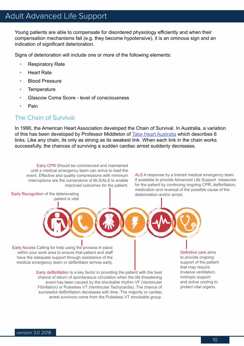
In 1990, the American Heart Association developed the Chain of Survival. In Australia, a variation of this has been developed by Professor Middleton of Take Heart Australia which describes 6 links. Like any chain, its only as strong as its weakest link. When each link in the chain works successfully, the chances of surviving a sudden cardiac arrest suddenly decreases.
Early Access Calling for help using the process in place within your work area to ensure that patient and staff have the adequate support through assistance of the medical emergency team or defibrillator arrives early.
Early Recognition of the deteriorating patient is vital
Early CPR Should be commenced and maintained until a medical emergency team can arrive to lead the
event. Effective and quality compressions with minimum interruptions are the cornerstone of BLS/ALS to enable
improved outcomes for the patient.
ALS A response by a trained medical emergency team if available to provide Advanced Life Support measures for the patient by continuing ongoing CPR, defibrillation, medication and reversal of the possible cause of the deterioration and/or arrest.
Definitive care aims to provide ongoing support of the patient that may require invasive ventilation, inotropic support and active cooling to protect vital organs.
Early defibrillation Is a key factor in providing the patient with the best chance of return of spontaneous circulation when the life threatening
event has been caused by the shockable rhythm VF (Ventricular Fibrillation) or Pulseless VT (Ventricular Tachycardia). The chance of successful defibrillation decreases with time. The majority or cardiac
arrest survivors come from the Pulseless VT shockable group.
11version 3.0 2018
Adult Advanced Life Support
THE DETERIORATING PATIENT
As you have already learned, recognising a deteriorating patient early on is the first critical step in the Chain of Survival. Your hospital or work area will already have systems in place to ensure that you are educated in the recognition of a deteriorating patient and have clear guidance on when to call for help.
The approach to all critically ill patients is the same. Let’s now look at the underlying principles.
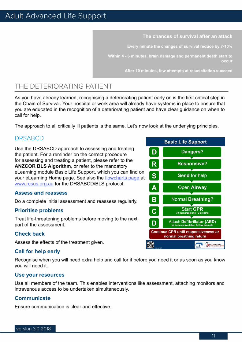
DRSABCD
Use the DRSABCD approach to assessing and treating the patient. For a reminder on the correct procedure for assessing and treating a patient, please refer to the ANZCOR BLS Algorithm, or refer to the mandatory eLearning module Basic Life Support, which you can find on your eLearning Home page. See also the flowcharts page at www.resus.org.au for the DRSABCD/BLS protocol.
Assess and reassessDo a complete initial assessment and reassess regularly.
Prioritise problemsTreat life-threatening problems before moving to the next part of the assessment.
Check backAssess the effects of the treatment given.
Call for help earlyRecognise when you will need extra help and call for it before you need it or as soon as you know you will need it.
Use your resourcesUse all members of the team. This enables interventions like assessment, attaching monitors and intravenous access to be undertaken simultaneously.
CommunicateEnsure communication is clear and effective.
The chances of survival after an attack
Every minute the changes of survival reduce by 7-10%
Within 4 - 6 minutes, brain damage and permanent death start to occur
After 10 minutes, few attempts at resuscitation succeed
12version 3.0 2018
Adult Advanced Life Support
Keep it simpleThe aim of initial treatment is to keep the patient alive, and achieve some clinical improvement. This will buy time for further treatment.
Effective Basic Life support is an essential component of Advanced Life SupportChest compressionsAll rescuers should perform chest compressions for all those who are unresponsive and not breathing normally. ANZCOR suggests that those who are trained and willing to give breaths do so for all persons in cardiac arrest. If rescuers do continuous chest compressions they should be at a rate of approximately 100 – 120/min (ANZCOR 2016 Guideline 8 - Cardiopulmonary Resuscitation)
Minimise interruptions to chest compressions CPR should not be interrupted to check for response or breathing. ANZCOR places a high priority on minimising interruptions for chest compressions. We seek to achieve this overall objective by balancing it with the practicalities of delivering 2 effective breaths between cycles of 30 chest compressions to the patient without an advanced airway. (ANZCOR 2016 Guideline 8 - Cardiopulmonary Resuscitation)
Medical Emergency Teams
Medical Emergency Teams (MET) are an example of a rapid response system and have been introduced in many hospitals to identify, review and treat patients at risk during the early phase of their deterioration. The MET not only responds to patients in cardiopulmonary arrest, but also to those with acute signs of deterioration.
Calling the team is reliant upon standardised criteria where staff are prompted to summon the team, as in the escalation processes outlined in Track and Trigger charts. Once arrived, the MET team will assess and treat the patient as required with the explicit aim of preventing further deterioration. A guide to standard criteria may include some or all of the following determinants:
• Threatened airway• All respiratory arrests• Respiratory rate <4/min or >35/min (Code Blue criteria <8/min)• Systolic BP <90 mmHg• Pulse rate >140/min or <40/min (Code Blue criteria >140/min)• Fall in GCS of >2 points or unresponsive on AVPU• Seizures• Fall in urine output <0.5mls/kg/hr• Oxygen Saturation <90% (Code Blue criteria )• All cardiac arrests (no pulse/cardiac output)• Any patient you are seriously concerned about that does not fit into the above criteria
13version 3.0 2018
Adult Advanced Life Support
Safety considerations
As a member of the ALS team, environmental awareness and safety should be paramount. It is important to don Personal Protective Equipment (PPE) and ensure the surrounds are safe for all team members prior to initiating any intervention.
Considerations may include:
• Environment free from potential hazards and risks – initial assessment and actioned appropriately
• All interventions are practiced according to National Health and Safety guidelines • Existing Multi-resistant Organisms (MRO) – PPE requirements• Cytotoxic precautions - Cytotoxic PPE requirements• Medical Radiology Imaging and exposure – Lead garments and PPE• Cardiac Catheter Labs and radiation exposure – Lead garments and PPE
Please refer to your facility guidelines for specific PPE requirements.
Ethical considerations
Law requires a collaborative approach between the health professional and patient and/or substitutive decision-maker about providing, withholding or withdrawing any life-sustaining medical measures. Please be aware that according to your Duty of Care, you are required to render whatever assistance that lies within your scope of practice (ANZCOR 2015 Guideline 10.5 - Legal and Ethical Issues related to Resuscitation).
ConsentCompetent adults have the right to refuse treatment when it is life-saving, even if it is not in their best interest. This right may be undermined if the person does not fully comprehend and understand their options regarding the situation.
Where active treatment is no longer appropriate, this should be explained to patients and/their substitutive decision maker.
In an emergency situation, reasonable efforts should be made to obtain consent, however there may be need for an immediate decision to be made about maintaining the life and health of a patient without proper consent.
When not to resuscitateFor out-of-hospital cardiac arrests little may be known about the previous medical condition of the patient and the basic rule is to start CPR except when there are reliable criteria or certain death.
For cardiac arrests that occur in hospital, a patient’s medical condition may have been defined as inappropriate to receive resuscitation if the patient is found to be pulseless.
Withholding CPR may then be appropriate if the prognosis is very poor, if the patient’s condition would render the attempt futile, or if the patient has made an informed judgement that he or she did not wish CPR to be attempted.
For more information, check your hospital for calling criteria and emergency response procedures
14version 3.0 2018
Adult Advanced Life Support
NFR ordersAn NFR (Not For Resuscitation) order means that in the event of a sudden deterioration in the patient’s condition, or if that patient is found to be in a state of cardio/respiratory arrest, the emergency response team within the hospital will not be activated. This order comes from the patient or the patient’s family and must be clearly documented for each individual. NFR does not mean not for escalation. It may still be appropriate to escalate to a MET (medical emergency) just not for CPR / defibrillation.
It is important to understand that a decision ‘not to resuscitate’ does not mean that the standard of general medical and nursing care is reduced in any way. It also does not mean that active or supportive treatment will be withdrawn, such as chemotherapy, rehydration, antibiotics and most importantly pain relief.
Advanced Health DirectiveThe Advanced Health Directive is a statement which can be made by any competent adult over the age of 18 years of age. This form makes provision for directives concerning what medical treatment the patient wants or does not want. It can also provide further instructions on life sustaining measures, what the person is willing to accept and what they do not want done.
The life sustaining measures include CPR or any measures to keep the heart working, including direction in mechanical ventilation, artificial feeding, use of blood products and/or hydration.
This form can be revised at any time or even revoked provided the adult is mentally competent.
The Advanced Health Directive does not constitute a NFR order. National Clinical Governance policies are available on the National Clinical Governance Unit intranet site under Polices > General. Please refer to your hospital’s Not for Resuscitation policy.
Legal requirements
Legally, healthcare staff are required to commence CPR promptly, to administer it effectively, and to also know when such measures are contraindicated and when they should cease.
Failure to startThe consequences of failing to respond to a cardiac arrest may be significant and life threatening. The reasons for such failure may be complex but are generally as a result of the following:
• Failure to recognise that a cardiac arrest has occurred• Feelings of inadequacy by the rescuers• Anxieties in connection with real or imagined risks as a result of undertaking resuscitation.
Failure to recognise that a patient is in need of CPR is a failure of training. Certainly this may be excusable in the case of the lay public but for nursing and medical staff who owe a duty of care to the patient, it could be regarded as negligent.
Refer to your hospital’s Not for Cardiopulmonary Resuscitation policy. The primary response, however, must always be that resuscitation will be attempted
in the event of a cardiac arrest unless there are specific NFR orders. In the absence of specific instructions, patients suffering cardiopulmonary arrest will
be assumed to require full CPR.
15version 3.0 2018
Adult Advanced Life Support
When to stopIn hospital, it is the responsibility of the leader of the resuscitation team to make the decision to stop CPR. The legal obligation rests with the most senior medically qualified doctor present.
Time should not be the major factor in the decision whether or not to continue. The size of the pupils or their lack of response to light are not reliable guides to the activity of the central nervous system or the likelihood of recovery. Factors influencing the decision to terminate resuscitation efforts include clinical history and prognosis; the arrest rhythm that the patient is in; and cardiovascular unresponsiveness to defibrillation, inotropic and stimulant drugs in the presence of effective oxygenation and basic life support.
When it is appropriate, the decision to discontinue advanced cardiac life support should be made by the doctor in charge, but all others involved in the attempt should be consulted, including the patient’s consultant.
DocumentationA ‘real time’ record, such as a form called the ‘Cardiopulmonary Resuscitation Record’, should be completed during the resuscitation and filed in the patient’s notes (a summary of the ‘code’ may be printable from your defibrillator, and this may be useful for real time record keeping and accurate documentation).
In addition to the real time record the team leader is to document events and outcomes in the patients records or nominate a medical team member to undertake this role.
Any medications ordered and administered during the resuscitation should be documented and signed for according to your local policy. The staff member responsible for the patient before the emergency should document the events leading up to making the emergency call and use the ISOBAR handover tool (or equivalent) if the patient requires transfer to a higher level of care.
Accurate and detailed descriptions of the resuscitation attempt may be invaluable if litigation is ever sought. Documentation should not be left for a junior staff member as the accurate recording of events is important to the legal processes.
Standard of careA nurse or doctor who accepts a ‘duty of care’ for a patient in need of CPR is expected to provide a reasonable standard of treatment.
A claim of inexperience or lack of training will not be successful as defence in an allegation of negligence if a practitioner has been called upon only to work within the limits of their own expected competence. It is important that practitioners should work within their Scope of Practice.
16version 2.0 2016
Advanced Life Support
16version 3.0 2018
Adult Advanced Life SupportQuizLet’s take a moment now to check what you’ve learned. This is not the final quiz, just practice to help you test your knowledge so far. You’ll find the answers in Appendix 3.
1. What is the primary aim of the Chain of Survival?
a. To give you a simple means to remember the order of response when someone goes into cardiac arrest
b. To highlight the relationship between early detection and defibrillation and the rates of survival of cardiac arrests
c. To remind you to use the DRSABCD approachd. To help you recognise a cardiac arrest
2. Every minute, the chances of survival after an arrest decreases by:
a. 3-5%b. 5-7%c. 7-10%d. 10-15%
3. What are the ethical considerations that need to be adhered to as the ALS trained nurse in an emergency?
a. NFR, DNR, current medical situation, Advanced Health Directiveb. Consultants orders, available equipment, family wishes, time of code, NFRc. Advanced Health Directive, NFR, Medical situation, Consultants ordersd. Consent, NFR, Medical condition, Advanced Health Directive
4. The legal requirements which have to be considered in a cardiac arrest situation are:
a. Effective cardiac compressions, documentation, cessation of CPR, Standard of careb. Medical prognosis, documentation, cessation of CPR, Standard of carec. Failure to start, documentation, cessation of CPR, Standard of cared. Standard of care, documentation, justice, empathy
CONCLUSION
In this chapter, we’ve looked at the aims of Advance Life Support, what a cardiac arrest is and how the Chain of Survival can increase the chances of recovery. We’ve also looked at the role of the nurse as well as the ethical and legal implications involved in resuscitation attempts.
17version 3.0 2018
Adult Advanced Life Support
THE ALS FLOWCHART
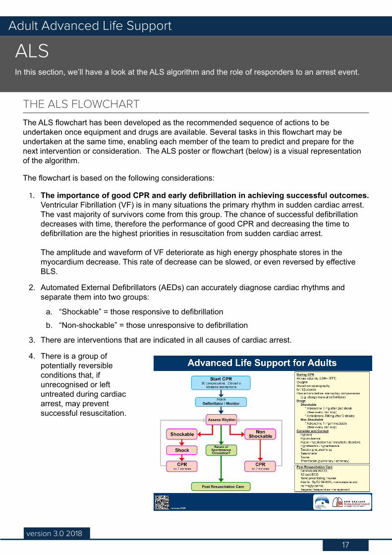
The ALS flowchart has been developed as the recommended sequence of actions to be undertaken once equipment and drugs are available. Several tasks in this flowchart may be undertaken at the same time, enabling each member of the team to predict and prepare for the next intervention or consideration. The ALS poster or flowchart (below) is a visual representation of the algorithm.
The flowchart is based on the following considerations:
1. The importance of good CPR and early defibrillation in achieving successful outcomes. Ventricular Fibrillation (VF) is in many situations the primary rhythm in sudden cardiac arrest. The vast majority of survivors come from this group. The chance of successful defibrillation decreases with time, therefore the performance of good CPR and decreasing the time to defibrillation are the highest priorities in resuscitation from sudden cardiac arrest. The amplitude and waveform of VF deteriorate as high energy phosphate stores in the myocardium decrease. This rate of decrease can be slowed, or even reversed by effective BLS.
2. Automated External Defibrillators (AEDs) can accurately diagnose cardiac rhythms and separate them into two groups:
a. “Shockable” = those responsive to defibrillationb. “Non-shockable” = those unresponsive to defibrillation
3. There are interventions that are indicated in all causes of cardiac arrest.
4. There is a group of potentially reversible conditions that, if unrecognised or left untreated during cardiac arrest, may prevent successful resuscitation.
ALSIn this section, we’ll have a look at the ALS algorithm and the role of responders to an arrest event.
18version 3.0 2018
Adult Advanced Life Support
SEQUENCE WHEN RESPONDING TO AN ARREST
Please note that some of the details contained in the following process descriptions may be different depending on your site.
1. The first responder
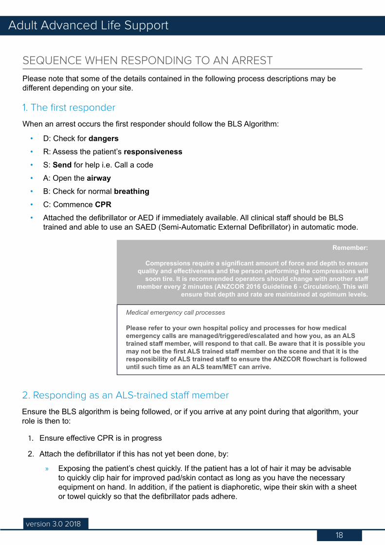
When an arrest occurs the first responder should follow the BLS Algorithm:
• D: Check for dangers• R: Assess the patient’s responsiveness• S: Send for help i.e. Call a code• A: Open the airway• B: Check for normal breathing• C: Commence CPR• Attached the defibrillator or AED if immediately available. All clinical staff should be BLS
trained and able to use an SAED (Semi-Automatic External Defibrillator) in automatic mode.
Remember:
Compressions require a significant amount of force and depth to ensure quality and effectiveness and the person performing the compressions will
soon tire. It is recommended operators should change with another staff member every 2 minutes (ANZCOR 2016 Guideline 6 - Circulation). This will
ensure that depth and rate are maintained at optimum levels.
Medical emergency call processes
Please refer to your own hospital policy and processes for how medical emergency calls are managed/triggered/escalated and how you, as an ALS trained staff member, will respond to that call. Be aware that it is possible you may not be the first ALS trained staff member on the scene and that it is the responsibility of ALS trained staff to ensure the ANZCOR flowchart is followed until such time as an ALS team/MET can arrive.
2. Responding as an ALS-trained staff member
Ensure the BLS algorithm is being followed, or if you arrive at any point during that algorithm, your role is then to:
1. Ensure effective CPR is in progress
2. Attach the defibrillator if this has not yet been done, by:
» Exposing the patient’s chest quickly. If the patient has a lot of hair it may be advisable to quickly clip hair for improved pad/skin contact as long as you have the necessary equipment on hand. In addition, if the patient is diaphoretic, wipe their skin with a sheet or towel quickly so that the defibrillator pads adhere.
19version 3.0 2018
Adult Advanced Life Support
» Attaching the pads. We’ll look at where the pads should go a little later. » Turning the defibrillator on ensuring it is in manual mode (depending on the type of
defibrillator you have at your site some may default to automatic mode). We’ll learn more about this operation in a later chapter. Check the default function of the defibrillator used in your area.
3. If an ALS/Medical Emergency team has not yet arrived, continue CPR and proceed along the ALS flowchart, directing any other staff present to undertake tasks if they haven’t yet been taken care of, e.g. ventilation, suction (if necessary), clearing the space around the patient, recording the event on the Resuscitation record, drawing up drugs, obtaining IV access and swapping with the person doing compressions.
Working with MET
MET and the on-call medical officer responding to a code will usually each take responsibility for particular tasks, for example gaining IV access and maintaining the airway. Please refer to
your local policies for the roles of the emergency team.
Your job is to coordinate with these responders while simultaneously coordinating others.
3. Prepare for defibrillation
As an ALS provider, once you have a defibrillator connected, proceed according to the ALS flowchart. Operating a defibrillator will be covered in the next chapter. For now, let’s focus on the flow of events as outlined in the ALS flowchart.
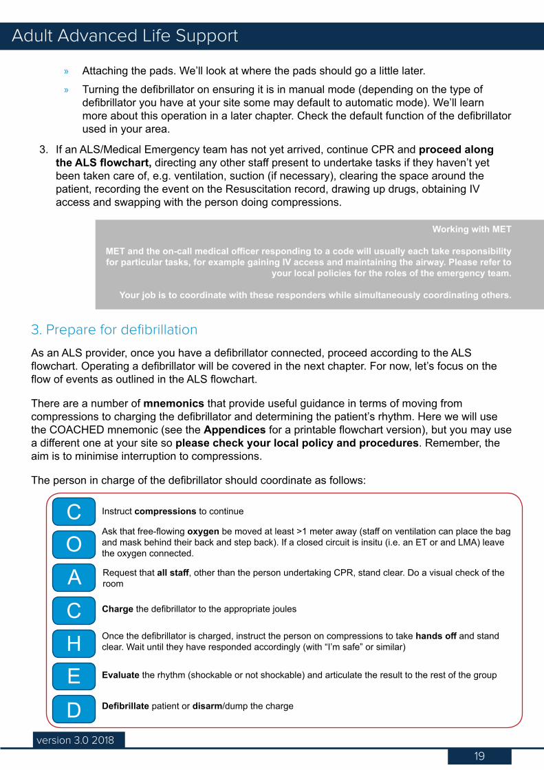
There are a number of mnemonics that provide useful guidance in terms of moving from compressions to charging the defibrillator and determining the patient’s rhythm. Here we will use the COACHED mnemonic (see the Appendices for a printable flowchart version), but you may use a different one at your site so please check your local policy and procedures. Remember, the aim is to minimise interruption to compressions.
The person in charge of the defibrillator should coordinate as follows:
COACHED
Instruct compressions to continue
Ask that free-flowing oxygen be moved at least >1 meter away (staff on ventilation can place the bag and mask behind their back and step back). If a closed circuit is insitu (i.e. an ET or and LMA) leave the oxygen connected.
Request that all staff, other than the person undertaking CPR, stand clear. Do a visual check of the room
Charge the defibrillator to the appropriate joules
Once the defibrillator is charged, instruct the person on compressions to take hands off and stand clear. Wait until they have responded accordingly (with “I’m safe” or similar)
Evaluate the rhythm (shockable or not shockable) and articulate the result to the rest of the group
Defibrillate patient or disarm/dump the charge
20version 3.0 2018
Adult Advanced Life Support
A note about charging safety:
The ANZCOR 2016 Guidelines (11.4 Electrical Therapy for Adult Advanced Life Support) state that biphasic defibrillators with self-adhesive defibrillation pads are
safe for compressions while charging, and recommend that compressions continue while charging to minimise interruptions to compressions and increase likelihood
of shock success, however you should refer to your hospital’s policy for any ‘hands off’ charging procedures.
4. Continue CPRNON-SHOCKABLE RHYTHM (Non-VF/VT, Asystole/PEA)
• Asystole is characterised by the absence of any cardiac electrical activity.• Pulseless Electrical Activity (PEA) (sometimes referred to as Electromechanical Dissociation
[EMD]) is the presence of a coordinated electrical rhythm without a detectable cardiac output. Please see the section Rhythm Recognition, and Appendix 1: Rhythms for more information.
• The prognosis in this group of cardiac rhythms or asystole is much less favourable than with VF/VT.
• During CPR advanced life support interventions are applied and potential causes of arrest sought.
• Defibrillation is not indicated and the emphasis is on CPR and other ALS interventions (e.g. intravenous access, consideration of advanced airway, drugs and pacing).
If the rhythm was not shockable and the defibrillator has been disarmed, immediately asses for return of spontaneous circulation. If there is no pulse or the rhythm is asystole/PEA, continue with CPR and administer (or administer under the direction of the emergency medical team) 1mg of Adrenaline immediately (ANZCOR 2018 Guideline 11.2 - Protocals for Adult Advanced Life Support).
SHOCKABLE RHYTHM• Ventricular fibrillation is asynchronous chaotic ventricular activity that produces no cardiac
output.• Pulseless ventricular tachycardia is a wide complex regular tachycardia associated with
no clinically detectable cardiac output. Please see the section Rhythm Recognition, and Appendix 1: Rhythms for more information.
• A defibrillator shock should be administered according to the algorithm.• Administer a single shock and immediately resume CPR for 2 minutes after delivery of shock.
Do not delay recommencing CPR to assess the rhythm.
Energy levels
• Monophasic: the energy level for adults should be set at maximum (usually 360 Joules) for all shocks.
• Biphasic waveforms: the default initial energy level for adults should be set at 200J. Other energy levels may be used providing there is relevant clinical data for a specific defibrillator that suggests that an alternative energy level provides adequate shock success (eg. usually greater than 90%).
21version 3.0 2018
Adult Advanced Life Support
ANZCOR suggests that if the first shock is not successful and the defibrillator is capable of delivering shocks of higher energy, it is reasonable to increase the energy to the maximum available for subsequent shocks (CoSTR 2015 weak recommendation, very low quality evidence).
Immediate CPR
Interruptions to CPR decrease the chance of survival from cardiac arrest. While defibrillation is of paramount importance for VF/VT, a period of well performed CPR immediately after each shock can help maintain myocardial and cerebral viability, and improves the likelihood of subsequent shock success.
CPR should be recommenced immediately after the shock has been delivered. DO NOT PAUSE COMPRESSIONS TO CHECK THE RHYTHM. The heart is slightly stunned when first defibrillated and the chance the patient will rearrest due to low cardiac output is higher until a resonable blood pressure is achieved with compressions.
Remember, the CPR ratio is 30:2, with 2 breaths at the completion of each 30 compressions (without a secured airway). It should take 2 minutes to complete 5 cycles of 30:2.
While CPR is underway, towards the end of the 2 minute cycle and if your hospital policy permits, charge the defibrillator again. Once charged, follow a mnemonic like COACHED again in order to check the patient’s rhythm with minimal interruptions to compressions.
• If it becomes necessary to shock a second time, you should administer (or will be directed to administer) 1mg of Adrenaline after the 2nd shock. This dose should then be administered every second cycle.
• If it becomes necessary to shock a third time, proceed as above (CPR/charge > pause > check rhythm > shock) and administer 300mg of Amioderone after the third shock.
• If the rhythm is not shockable, disarm the defibrillator, continue CPR, and return to the Assess rhythm point in the algorithm. Follow the steps again.
Simultaneously while CPR continues, you and the team should be ascertaining the cause of the arrest by considering the 4Hs and 4Ts and treating appropriately.
5. Return of spontaneous circulation
Has spontaneous circulation and normal breathing returned? If it has, proceed to post resuscitation care.
If circulation has not returned, CPR should be continued until the patient is spontaneously breathing. The patient should be reassessed every 2 minutes in regards to response to treatment (drug administration, compressions or a shock).
• If the rhythm is shockable: deliver another shock as per the previous step• If the rhythm is non-shockable: disarm the defibrillator and continue CPR as per the
previous step
22version 3.0 2018
Adult Advanced Life Support
6. Post-resuscitation care
Once spontaneous circulation has returned, you should:
• Re-evaluate ABCDE• Re-evaluate oxygenation and ventilation – aim for 94 -98% saturation/ normocapnia /
normoglycaemia• Treat any precipitating causes - 4Hs and 4Ts• Provide haemodynamic support through IV fluid administration• Record a 12 lead ECG for analysis• Escalate level of care and arrange safe transfer• Initiate targeted temperature management (TTM) if appropriate
RESPONSIBILITIES OF TEAM MEMBERS
As the ALS trained staff member on scene, and until a medical officer has arrived, it is your role to ensure the ALS flowchart is followed, and coordinate or ‘run’ the code. This means that you need to maintain an awareness of the overall situation – who is where and doing what, and whether the actions being taken are effective and following the ALS algorithm – regardless of what else you are doing, and be able to coordinate everyone’s efforts. Refer to Human Factors training for more information about this. The Human Factors program is available in your eLearning.
You will need to use your own judgement on what is needed, but be aware of everyone in the room. Is the CPR person tiring? Has the person on ventilation responded appropriately to the defibrillation call to stand clear? Who is investigating the cause of the arrest (4Hs & 4Ts)? Have the recommended drugs been administered? These are only some of the considerations you will have to take into account.
Above and beyond this, these are (commonly) the roles during a code:
• The team leader (responsible for allocating roles and running the Code)• The person managing the patient’s airway (once the medical officer has arrived at the code,
this is often their role)• The person operating the defibrillator• The person performing compressions (2 to 3 people may need to rotate through this role as
a responder who is starting to tire may not be able to maintain good quality compressions)• The person preparing access and administering any necessary drugs (this is often the
members of the MET)• The person recording the event (i.e. the scribe)
Let’s now look at how these teams appear and function at different types of hospitals. Remember, this is only a stereotypical view and teams may take different forms or function in slightly different ways at your site.
23version 3.0 2018
Adult Advanced Life Support
ALS trained, small hospitalYou are an ALS trained staff member at a small hospital. With your training, you are rostered on as part of a Medical Emergency Team on a regular basis.
In the event of a cardiac emergency, you will be called to respond with the rest of your team and the on-call medical officer. Your role can be any one of the following:
• The person ‘running’ the code (coordinating the resuscitation efforts)
• The person managing the patient’s airway
• The person operating the defibrillator
• The person performing compressions• The person preparing access and
administering any necessary drugs • The person recording the event (i.e.
the scribe)
ALS trained, large hospialYou are an ALS trained staff member at a medium to large hospital. With your training, you are the nominated ALS person on your ward. You may or may not be the only ALS person on your ward.
In the event of a cardiac emergency you may be the first or second person on the scene. Your role is to coordinate other staff members present in resuscitation efforts until the medical officer and/or MET arrive. Depending on the number of ward staff at the code, your role may also include one of the following:
• The person ‘running’ the code (coordinating the resuscitation efforts)
• The person managing the patient’s airway
• The person operating the defibrillator
• The person performing compressions• The person preparing access and
administering any necessary drugs • The person recording the event (i.e. the
scribe)
• The person managing the patient’s airway
• The person operating the defibrillator• The person performing compressions
• The person preparing access and administering any necessary drugs
• The person recording the event (i.e. the scribe)
Once MET or a medical officer has arrived, your role may be to continue coordinating efforts in conjunction with the MET and MO, or to act under direction from the MO.
ICU, CCU, ED/EUYou are a staff member working in ICU, CCU or Emergency. You are likely to already have ALS
certification and may have dealt with a number of cardiac arrests. You may be rostered on to a Medical Emergency Team regularly.
In the event of a cardiac emergency, you will be called to respond with the rest of your team and the on-call medical officer. Your role can be any one of the following:
24version 3.0 2018
Adult Advanced Life Support
BEING ORGANISED AND PREPARED
A code never runs according to plan. You must be organised and prepared to the best of your ability even while adjusting to a changing situation. While CPR is underway, you will have a moment to focus on the kinds of things that you and your team will need to consider.
During CPR you may have to be prepared to:
• Utilise available airway adjuncts• Deliver oxygen• Use waveform capnography• Prepare or adjust IV/IO access• Administer drugs• Send to lab any blood and other tests• Liaise with other medical staff• Communicate with the medical officer and MET
Remember
You must plan your actions before interrupting compressions (e.g. charge manual defibrillator as in
stated in the ALS algorithm).
25version 3.0 2018
Adult Advanced Life Support
The 4Hs and 4Ts
We have discussed previously some common reasons why a cardiac arrest may occur. Cardiac arrest maybe the result of potentially reversible causes requiring a specific treatment. During CPR you will need to consider whether you are dealing with these causes – referred to as the 4Hs and 4Ts - and correct them.
Hypoxia• Cause: Low oxygen levels in the blood• Considerations: Prior to the arrest what was the respiratory pattern, rate, ABGs, O2
saturation?• Treatment: Airway management and appropriate interventions need to be considered to
reverse the hypoxia. For example manual ventilation using the bag and mask, LMA or intubation
HypovolaemiaHypovolaemia may be significant in arrests associated with trauma, GI Bleed, dehydration, fluids shifts (burns), anaphylaxis, post-operative sepsis, surgery, and post-delivery complications/miscarriage.
• Cause: Low amount of circulating blood, either absolutely due to blood loss or relatively due to vasodilation
• Considerations: Look for signs of bleeding, severe dehydration (e.g. diarrhoea and vomiting). Compensatory systemic release of catecholamines promotes peripheral vasoconstriction, increased cardiac contractility and tachycardia. A low diastolic BP suggests arterial vasodilation (as in anaphylaxis or sepsis) (ANZCOR 2016 Guideline 9.2.7 – First Aid Management of Anaphylaxis)
• Treatment: Control bleeding and deliver appropriate fluid replacement e.g. crystalloid and blood products.
Hyper/hypokalaemia/metabolic disordersMetabolic abnormalities may be suggested by the patient’s underlying condition (e.g. renal failure), tests taken during the resuscitation, or clues given in the ECG.
• Cause: Electrolyte imbalance, for example disturbances in the level of potassium/calcium/ magnesium in the blood. Also check patient history for issues e.g. hypoglycaemia
• Considerations: Look for conditions such as renal failure, diabetes, dehydration and check patient medical history.
• Treatment: Aim of treatment is to reverse the disorder: » Hypokalaemia - 5mmol bolus KCL IV » Hyperkalaemia - to lower the potassium use shifting agents (glucose 25g + 10 units
of short acting insulin) and consider Sodium Bicarbonate. Cardio protective measures include 5 -10ml of 10% calcium chloride IV or 10ml of 10% calcium gluconate as per your hospital policy to stabilise cell membranes (this does not reduce serum potassium levels)
» Hypoglycaemia - 10-50% glucose IV » Hypomagnesia - 5mmol bolus of magnesium IV
26version 3.0 2018
Adult Advanced Life Support
Hypothermia/hyperthermiaHypothermia is associated with drowning incidents and drug overdose. Hyperthermia is associated with dehydration, heart disease, fever, heat stroke, or drug use.
• Cause: Dramatic increase or decrease in body temperate • Considerations: Look for signs of severely high or low body temperature such as skin
colour, cracked or blue lips• Treatment: The patient should be actively warmed or cooled. Heat the patient using a
space blanket if hypothermia. Cool with fluids, ice packs in armpits and behind the neck for hyperthermia. Specific modifications may be needed in cardiac arrest (refer to the Special Circumstances chapter)
Tension pneumothoraxTension pneumothorax results from the progressive build-up of air within the pleural space, usually due to a lung laceration which allows air to escape the pleural space but not return.
• Cause: A tear in the lung leading to lung collapse and twisting of the large blood vessels, including the insertion of invasive devices into the chest cavity
• Considerations: Observe and assess for tracheal deviation, unilateral air entry, and/or unequal rise of the chest when ventilating. Also look for bruising around the rib area and consider a thoracic assessment for broken ribs
• Treatment: Emergency chest decompression of the affected side by a skilled operator. Insert a 14 gauge (or larger) needle /cannulae, into the 2nd intercostal space (midclavicular line). Follow this with a chest drain when appropriate.
Tamponade• Cause: Fluid or blood in the pericardium, compressing the heart and preventing its ability
to contract, due to thoracic trauma, recent cardiac surgery, insertion of central lines, recent angiography, recent MI, recent pacemaker, mediastinal radiation therapy, known pericardial effusion, renal failure, or pericarditis (ARC ALS 2 3rd Australian edition 2016)
• Considerations: Assess history for blunt trauma/stab wounds or for rapidly decreasing blood pressure or complications from cardiac surgery/interventional procedures i.e. PPM insertion
• Treatment: Undertake cardiac ultrasound. Pericardial tap (pericardiocentesis) or emergency open heart surgery may be required
Toxins/poisons/drugsToxic reaction is either a result of accidental or deliberate overdose.
• Cause: Dependent on the toxin• Considerations: Look for symptoms of anaphylactic shock (rapidly decreasing vital signs) or
opiate induced respiratory arrest• Treatment: Maintain basic and advanced life support and try to ascertain the source of the
reaction and reverse it (administer antagonist)
27version 3.0 2018
Adult Advanced Life Support
Thrombosis (pulmonary/coronary)Thrombosis is the blockage of blood vessels to the lungs or heart by a blood clot or other material.
• Cause: Post-surgical complications, Myocardial Infarction (MI), cardiac arrhythmias, blood disorders
• Considerations: Patient history, e.g. chest pain, kidney failure (low molecular heparin), signs of a stroke (level of consciousness) or signs of transient ischemic attack (TIA)
• Treatment: Thrombolytic therapy if there is a high likelihood of Pulmonary Embolus. MI is treated by primary angioplasty. Increased risk of severe bleeding needs to be considered particularly post-surgery / trauma / head injury
ANZCOR 2018 Guideline. 11.2 - Protocols for Adult Advanced Life Support
28version 2.0 2016
Advanced Life Support
28version 3.0 2018
Adult Advanced Life SupportQuizLet’s take a moment now to check what you’ve learned. This is not the final quiz, just practice to help you test your knowledge so far. You’ll find the answers in Appendix 3.
1. What is the correct response to recognition of a non-shockable rhythm?
a. Charge the defibrillator in case the rhythm changesb. Ventilate the patientc. Continue good quality CPR and reassess the rhythm after 2 minutesd. Shock the patient then continue CPR
2. You are the ALS trained nurse responding to a call to resuscitate a patient on the surgical ward. Ward staff found the patient collapsed, commenced CPR and attached the defibrillator. What is your first priority?
a. Check the patient’s airwayb. Defibrillate the patientc. Obtain IV accessd. Assess the effectiveness of the CPR being performed while planning for rhythm check/
defibrillation
3. Failure to perform a visual sweep of the room and call “stand clear” (or similar) before pressing the Shock button constitutes a potential hazard to other staff members – True or False?
4. You should never charge the defibrillator pads on the patient’s chest wall as this may cause accidental discharge – True or False?
CONCLUSION
In this chapter, we’ve looked at the ALS algorithm and the roles of the ALS nurse during a cardiac arrest code. We’ve also looked at the 4Hs and 4Ts which may be causes of cardiac arrest.
29version 3.0 2018
Adult Advanced Life Support
Rhythm RecognitionIn this section, we’ll have a close look at the ECG and the components of a waveform, as well as what constitutes a normal rhythm and how to recognise and interpret the abnormal.
Cardiac Anatomy
The following content assumes that you understand the myocardial functions of the heart. If you need to brush up on your cardiac anatomy and physiology, now’s the time to do so. You can use whatever
text you have at hand, or follow the link to our recommended online resource, Anatomy and Physiology Essentials.
You will need your Athens login. See the RHC Library at http://ramsaylibrary.com.au for registration.
RHYTHMS ARE A PICTURE
It is important to be able to recognise the cardiac rhythms that may compromise cardiac output, precede cardiac arrest or complicate the recovery period after successful resuscitation.
Even if a definite ECG diagnosis cannot be made, it is important to be able to recognise that an arrhythmia (an abnormality in the rhythm) is present so that you can assess the effects on cardiac output and therefore act accordingly.
The electrocardiograph
Understanding of the basic principles of electrocardiograph (otherwise known as the ECG) recording measurements and how this relates to the action of the heart will help you in understanding how to read a rhythm strip.
About the ECGThe ECG is a diagnostic tool that reflects cardiac electrical activity. It is a record of the magnitude and direction of the electrical current which is generated by depolarisation (contraction) and repolarisation (relaxation) of the atria and ventricles.
• Rhythm strips – provide information relating to the rhythm and the rate.• 12 lead ECG – provides information about rate, rhythm, impulse conduction, electrical axis,
hypertrophy, ischemia and infarction.
It’s important to remember that the ECG will only show you what is occurring electrically within the heart, not how well it is pumping.
The measurement of the squares in an ECG strip is relevant to the ECG rhythm in that each part of the ECG has an allocated time frame for each conduction, so knowing the measurement will enhance your ability to interpret the rate and rhythm.
30version 3.0 2018
Adult Advanced Life Support
Reading a rhythm strip
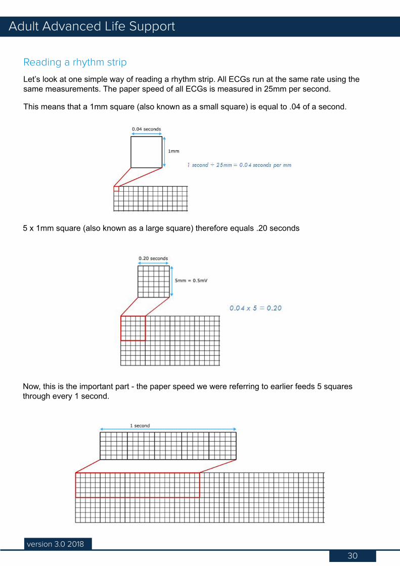
Let’s look at one simple way of reading a rhythm strip. All ECGs run at the same rate using the same measurements. The paper speed of all ECGs is measured in 25mm per second.
This means that a 1mm square (also known as a small square) is equal to .04 of a second.
5 x 1mm square (also known as a large square) therefore equals .20 seconds
Now, this is the important part - the paper speed we were referring to earlier feeds 5 squares through every 1 second.
31version 3.0 2018
Adult Advanced Life Support
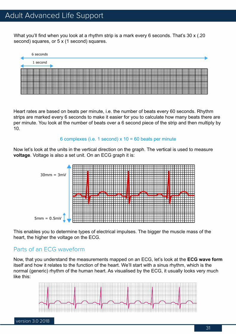
What you’ll find when you look at a rhythm strip is a mark every 6 seconds. That’s 30 x (.20 second) squares, or 5 x (1 second) squares.
Heart rates are based on beats per minute, i.e. the number of beats every 60 seconds. Rhythm strips are marked every 6 seconds to make it easier for you to calculate how many beats there are per minute. You look at the number of beats over a 6 second piece of the strip and then multiply by 10.
6 complexes (i.e. 1 second) x 10 = 60 beats per minute
Now let’s look at the units in the vertical direction on the graph. The vertical is used to measure voltage. Voltage is also a set unit. On an ECG graph it is:
This enables you to determine types of electrical impulses. The bigger the muscle mass of the heart, the higher the voltage on the ECG.
Parts of an ECG waveform
Now, that you understand the measurements mapped on an ECG, let’s look at the ECG wave form itself and how it relates to the function of the heart. We’ll start with a sinus rhythm, which is the normal (generic) rhythm of the human heart. As visualised by the ECG, it usually looks very much like this:
32version 3.0 2018
Adult Advanced Life Support
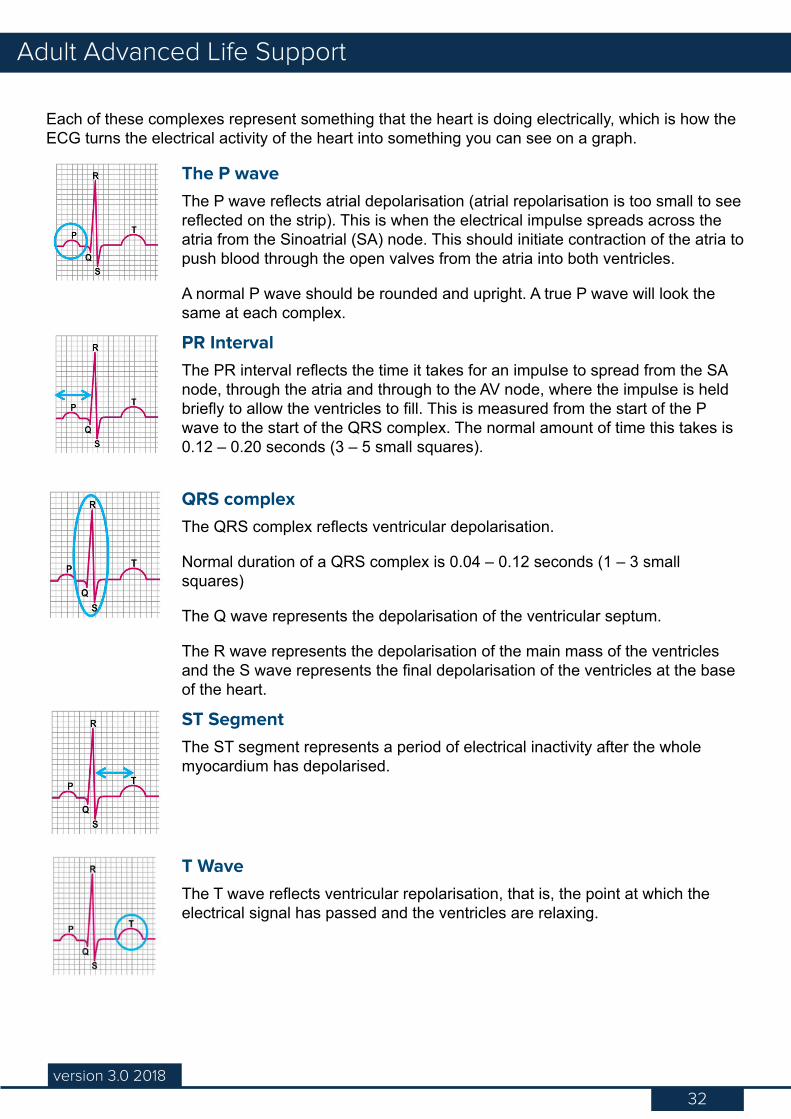
Each of these complexes represent something that the heart is doing electrically, which is how the ECG turns the electrical activity of the heart into something you can see on a graph.
The P waveThe P wave reflects atrial depolarisation (atrial repolarisation is too small to see reflected on the strip). This is when the electrical impulse spreads across the atria from the Sinoatrial (SA) node. This should initiate contraction of the atria to push blood through the open valves from the atria into both ventricles.
A normal P wave should be rounded and upright. A true P wave will look the same at each complex.
PR IntervalThe PR interval reflects the time it takes for an impulse to spread from the SA node, through the atria and through to the AV node, where the impulse is held briefly to allow the ventricles to fill. This is measured from the start of the P wave to the start of the QRS complex. The normal amount of time this takes is 0.12 – 0.20 seconds (3 – 5 small squares).
QRS complexThe QRS complex reflects ventricular depolarisation.
Normal duration of a QRS complex is 0.04 – 0.12 seconds (1 – 3 small squares)
The Q wave represents the depolarisation of the ventricular septum.
The R wave represents the depolarisation of the main mass of the ventricles and the S wave represents the final depolarisation of the ventricles at the base of the heart.
ST SegmentThe ST segment represents a period of electrical inactivity after the whole myocardium has depolarised.
T WaveThe T wave reflects ventricular repolarisation, that is, the point at which the electrical signal has passed and the ventricles are relaxing.
33version 3.0 2018
Adult Advanced Life Support
QT IntervalThe QT interval is the distance from the beginning of the QRS to the end of the T wave that represents the depolarisation and repolarisation of the ventricles. Calculating the measurement is dependent upon heart rate and the length of the QT interval. This is calculated by the ECG machine.
RR IntervalThe RR interval represents the distance between 2 consecutive R waves.
Analysing rhythms
Now let’s learn how to analyse what we’re seeing on an ECG. Cardiac rhythm is identified through the combination of several factors, some of which we have already touched upon.
Regularity and rate
Regularity refers to the predictability of the pattern. A regular pattern, where all complexes are occurring as expected, indicates that the rhythm is normal. An irregular pattern, or one where only certain complexes are occurring indicates that the rhythm is abnormal and that there is a conduction issue somewhere within the heart (for reasons yet to be determined).
Rate similarly gives you an indication of ‘what the heart is doing’. Since we know what constitutes a normal rate under general conditions, we can determine what issues potentially exist based on how much faster or slower the patient’s rate is compared to the normal.
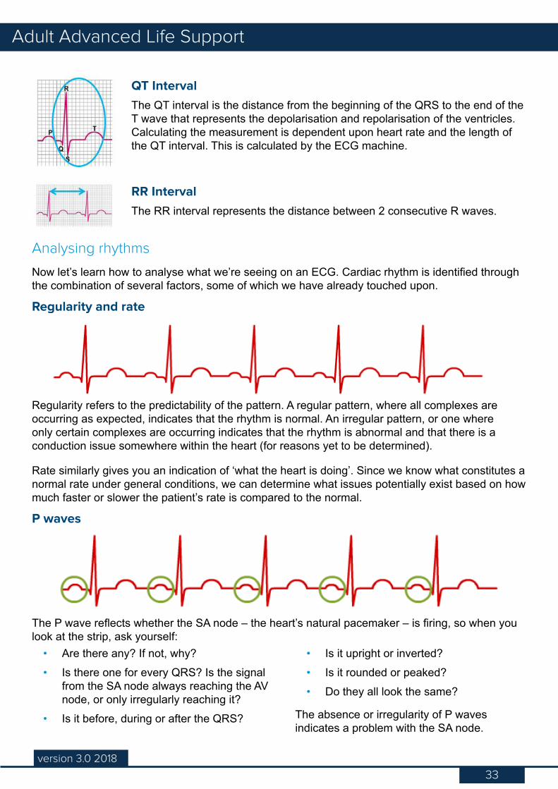
P waves
The P wave reflects whether the SA node – the heart’s natural pacemaker – is firing, so when you look at the strip, ask yourself:
• Are there any? If not, why?• Is there one for every QRS? Is the signal
from the SA node always reaching the AV node, or only irregularly reaching it?
• Is it before, during or after the QRS?
• Is it upright or inverted?• Is it rounded or peaked?• Do they all look the same?
The absence or irregularity of P waves indicates a problem with the SA node.
34version 3.0 2018
Adult Advanced Life Support
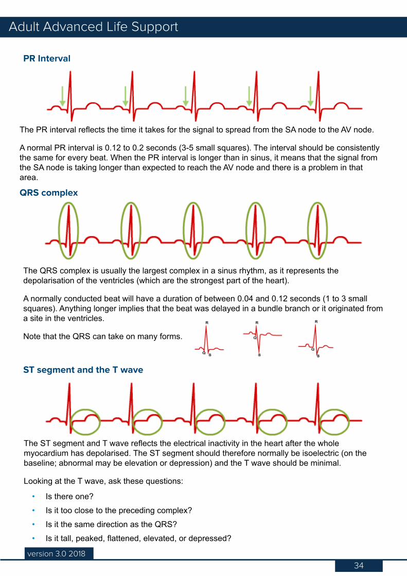
PR Interval
The PR interval reflects the time it takes for the signal to spread from the SA node to the AV node.
A normal PR interval is 0.12 to 0.2 seconds (3-5 small squares). The interval should be consistently the same for every beat. When the PR interval is longer than in sinus, it means that the signal from the SA node is taking longer than expected to reach the AV node and there is a problem in that area.
QRS complex
The QRS complex is usually the largest complex in a sinus rhythm, as it represents the depolarisation of the ventricles (which are the strongest part of the heart).
A normally conducted beat will have a duration of between 0.04 and 0.12 seconds (1 to 3 small squares). Anything longer implies that the beat was delayed in a bundle branch or it originated from a site in the ventricles.
Note that the QRS can take on many forms.
ST segment and the T wave
The ST segment and T wave reflects the electrical inactivity in the heart after the whole myocardium has depolarised. The ST segment should therefore normally be isoelectric (on the baseline; abnormal may be elevation or depression) and the T wave should be minimal.
Looking at the T wave, ask these questions:
• Is there one?• Is it too close to the preceding complex?• Is it the same direction as the QRS?• Is it tall, peaked, flattened, elevated, or depressed?
35version 3.0 2018
Adult Advanced Life Support
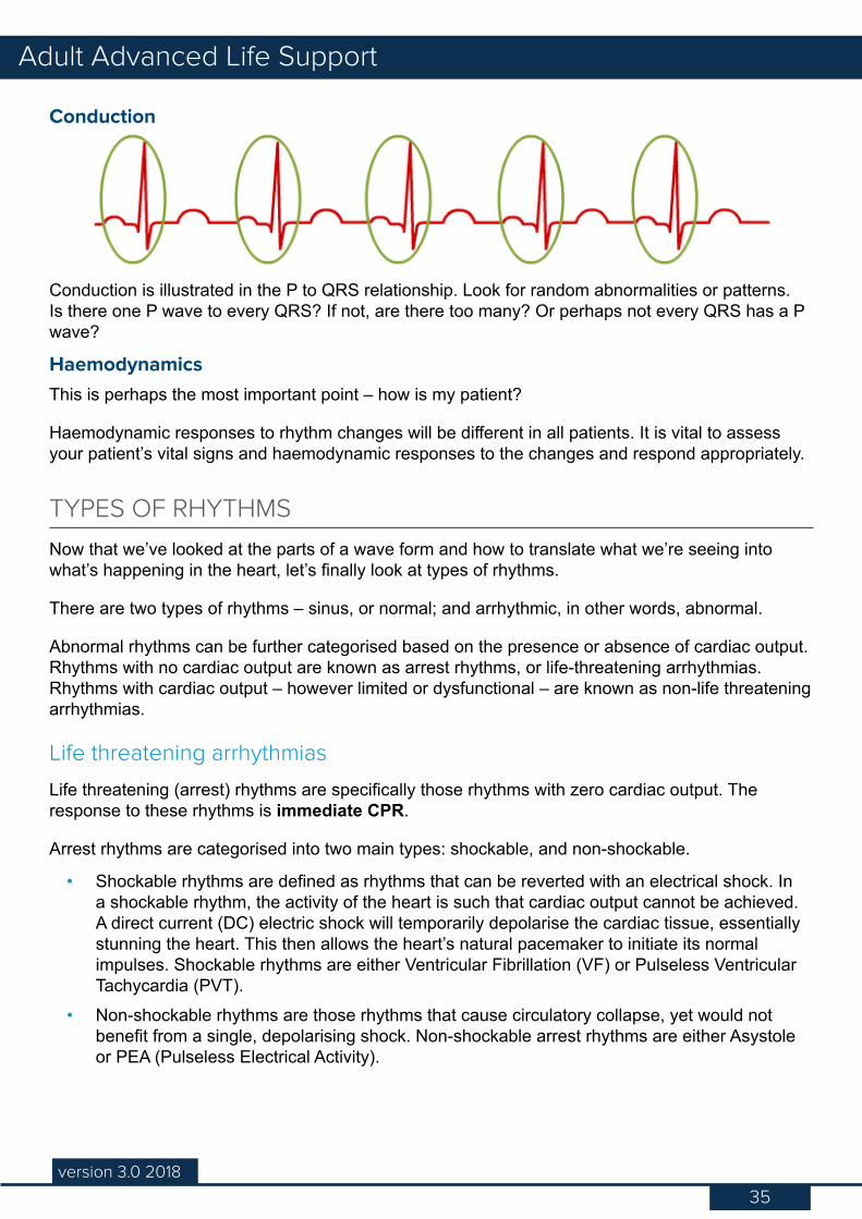
Conduction
Conduction is illustrated in the P to QRS relationship. Look for random abnormalities or patterns. Is there one P wave to every QRS? If not, are there too many? Or perhaps not every QRS has a P wave?
HaemodynamicsThis is perhaps the most important point – how is my patient?
Haemodynamic responses to rhythm changes will be different in all patients. It is vital to assess your patient’s vital signs and haemodynamic responses to the changes and respond appropriately.
TYPES OF RHYTHMS
Now that we’ve looked at the parts of a wave form and how to translate what we’re seeing into what’s happening in the heart, let’s finally look at types of rhythms.
There are two types of rhythms – sinus, or normal; and arrhythmic, in other words, abnormal.
Abnormal rhythms can be further categorised based on the presence or absence of cardiac output. Rhythms with no cardiac output are known as arrest rhythms, or life-threatening arrhythmias. Rhythms with cardiac output – however limited or dysfunctional – are known as non-life threatening arrhythmias.
Life threatening arrhythmias
Life threatening (arrest) rhythms are specifically those rhythms with zero cardiac output. The response to these rhythms is immediate CPR.
Arrest rhythms are categorised into two main types: shockable, and non-shockable.
• Shockable rhythms are defined as rhythms that can be reverted with an electrical shock. In a shockable rhythm, the activity of the heart is such that cardiac output cannot be achieved. A direct current (DC) electric shock will temporarily depolarise the cardiac tissue, essentially stunning the heart. This then allows the heart’s natural pacemaker to initiate its normal impulses. Shockable rhythms are either Ventricular Fibrillation (VF) or Pulseless Ventricular Tachycardia (PVT).
• Non-shockable rhythms are those rhythms that cause circulatory collapse, yet would not benefit from a single, depolarising shock. Non-shockable arrest rhythms are either Asystole or PEA (Pulseless Electrical Activity).
36version 3.0 2018
Adult Advanced Life Support
Non-life threatening arrhythmias
Non-life threatening rhythms are rhythms which may potentially cause decreased cardiac output. These rhythms can be serious, and may lead to a cardiac arrest if untreated. In these cases cardiac output will mean that the patient’s haemodynamic status is potentially compromised, requiring immediate recognition of the rhythm and prompt treatment to prevent poor outcomes.
CONCLUSION
In this chapter, we’ve examined the ECG waveform and looked at how what you’re seeing on a strip relates to the conduction in the heart.
37version 3.0 2018
Adult Advanced Life Support
WHAT IS DEFIBRILLATION
Defibrillation is the passage of electrical energy to the heart to simultaneously depolarise enough myocardial cells to produce repolarisation and enable the natural pacemaker tissue to resume control.
The Australian Resuscitation Council has determined that resuscitation outcomes show improvement when a defibrillator is attached to the patient as soon as it is available and shockable rhythms are immediately identified and shocked (ANZCOR 2016 Guideline 11.4 – Electrical Therapy for Adult Advanced Life Support).
The importance of good CPR and early defibrillation in achieving successful outcomes. Ventricular Fibrillation (VF) is in many situations the primary rhythm in sudden cardiac arrest. The vast majority of survivors come from this group. The chance of successful defibrillation decreases with time. Therefore the performance of good CPR and decreasing the time to defibrillation are the highest priorities in resuscitation from sudden cardiac arrest.
The amplitude and waveform of VF deteriorate as high energy phosphate stores in the myocardium decrease. This rate of decrease can be slowed, or even reversed by effective BLS.
DefibrillationIn this section, we’ll look at defibrillation. We’ll also examine the factors that determine successful and safe defibrillation.
Maintaining competency
Code of Practice for Registered Nurses states that it is the responsibility of the Registered Nurse to maintain competency in any
procedures they undertake as part of their nursing practice.
With regard to defibrillation, nurses are evaluated in their competency against the defibrillation policy in the department in which they work.
These may be set against the standards set through the ARC.
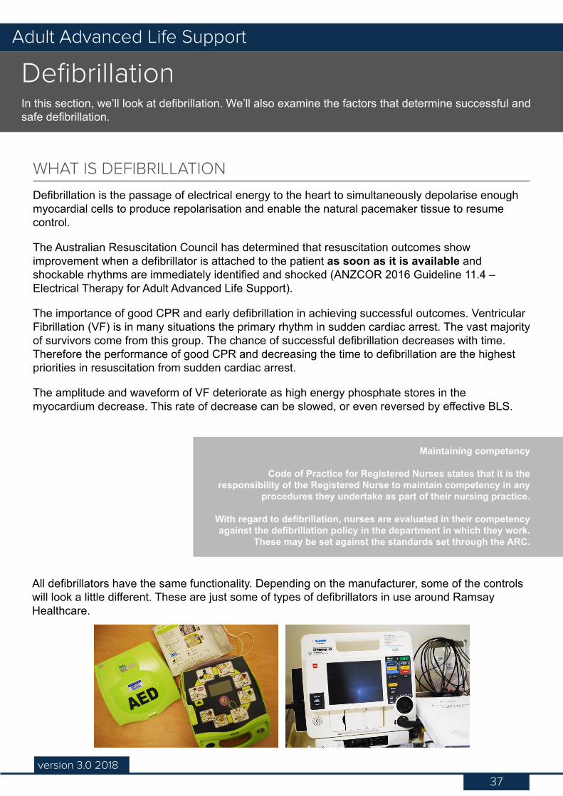
All defibrillators have the same functionality. Depending on the manufacturer, some of the controls will look a little different. These are just some of types of defibrillators in use around Ramsay Healthcare.
38version 3.0 2018
Adult Advanced Life Support
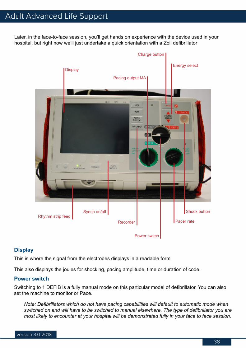
Later, in the face-to-face session, you’ll get hands on experience with the device used in your hospital, but right now we’ll just undertake a quick orientation with a Zoll defibrillator
Display
Power switch
Synch on/off
Recorder Pacer rateRhythm strip feed
Charge button
Pacing output MA
Energy select
Shock button
DisplayThis is where the signal from the electrodes displays in a readable form.
This also displays the joules for shocking, pacing amplitude, time or duration of code.
Power switchSwitching to 1 DEFIB is a fully manual mode on this particular model of defibrillator. You can also set the machine to monitor or Pace.
Note: Defibrillators which do not have pacing capabilities will default to automatic mode when switched on and will have to be switched to manual elsewhere. The type of defibrillator you are most likely to encounter at your hospital will be demonstrated fully in your face to face session.
39version 3.0 2018
Adult Advanced Life Support
Energy SelectThis controls the joules that the defibrillator will deliver and is used to increase/lower the joules or dissipate the charge.
Charge buttonThis button will charge up the machine in preparation to deliver a shock.
Defibrillators produce a direct current (DC) discharge which passes between two electrodes.
• Monophasic defibrillators deliver a single burst of energy from one pad to another. The energy on these defibrillators is set to 360J.
• Bi-phasic defibrillators send electricity from one pad to the other and then reverse direction. This compensates for transthoracic impedance (which we’ll look at shortly) and therefore uses less energy. The energy these defibrillators are capable of delivering is adjustable but the ARC states that the default should be set to 200J unless otherwise recommended by the manufacturer. If there is any doubt as to what the machine default is, it should be set to 200J.
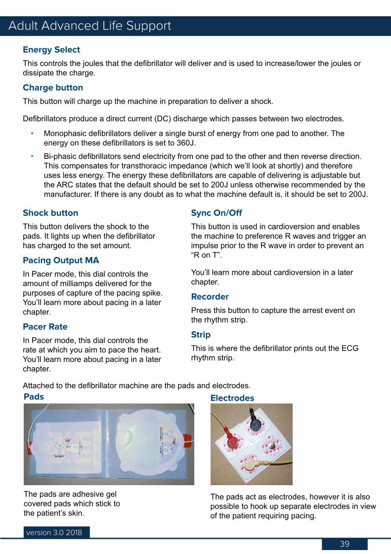
Pads Electrodes
Shock buttonThis button delivers the shock to the pads. It lights up when the defibrillator has charged to the set amount.
Pacing Output MAIn Pacer mode, this dial controls the amount of milliamps delivered for the purposes of capture of the pacing spike. You’ll learn more about pacing in a later chapter.
Pacer RateIn Pacer mode, this dial controls the rate at which you aim to pace the heart. You’ll learn more about pacing in a later chapter.
Sync On/OffThis button is used in cardioversion and enables the machine to preference R waves and trigger an impulse prior to the R wave in order to prevent an “R on T”.
You’ll learn more about cardioversion in a later chapter.
RecorderPress this button to capture the arrest event on the rhythm strip.
StripThis is where the defibrillator prints out the ECG rhythm strip.
Attached to the defibrillator machine are the pads and electrodes.
The pads are adhesive gel covered pads which stick to the patient’s skin.
The pads act as electrodes, however it is also possible to hook up separate electrodes in view of the patient requiring pacing.
40version 3.0 2018
Adult Advanced Life Support
ENERGY DELIVERY
Delivery of a single shock is now the standard. There is no evidence to suggest that a single or stacked regime of shock delivery improves outcome. There is evidence to show that effective compressions and oxygenation during arrest improves outcome, hence the emphasis on effective CPR.
Single shock
For biphasic defibrillators, a single shock of 200 joules is delivered.
All shocks for monophasic defibrillators, or where you are unsure of the recommended setting for a specific device, should be 360 joules.
If the first shock is not successful and the defibrillator is capable of delivering a higher energy shock, it is reasonable to increase the energy to maximum joules available for subsequent
shocks.
ANZCOR 2016 Guidelines section 11.4 - Electrical Therapy for Adult Advanced Life Support
Stacked shock
Stacked shocks are shocks that are delivered one after another. If the first shock does not have any notable effect, another shock should be delivered within 20 seconds (ANZCOR 2017 Guidelines Section 11.1 - Introduction to and Principles of In-hospital Resuscitation). The time required for rhythm recognition and recharging the defibrillator is short (<10 seconds), and up to three shocks are counted as the first shock in the ALS algorithm sequence.
The only situation where stacked shocks are now recommended is where:
1. The patient is previously well perfused and oxygenated;
2. The ECG is witnessed to change into a shockable rhythm;
3. The patient is non-responsive; and
4. A manual defibrillator (not a semi-automatic one) is already connected and ready for use with qualified personnel to operate (for example, in a Cath Lab when the patient is haemodynamically stable prior to the event, or with a patient who is immediately post-cardiac surgery or has previously had a witnessed monitored arrest).
In this specific situation, a delivery of three stacked shocks may be given, followed immediately by two minutes of CPR. The rationale for this is that successful reversion of the rhythm is proportional to speed of defibrillation and length of time the patient has been in the shockable rhythm. If a rhythm appears normally associated with an output after, for example, the second stacked shock, there is no need to deliver the third shock. Subsequent shock delivery will be single.
41version 3.0 2018
Adult Advanced Life Support
Successful defibrillation
You will normally recognise successful defibrillation by a change in the patient’s ECG rhythm.
Successful defibrillation depends on many patient and operational factors. These include:
• High quality CPR• Pre-shock pause before defibrillation and post-shock resumption of chest compressions <5
seconds• Pre-existing condition of the patient• The duration of pre-shock ventricular fibrillation and CPR (i.e. “down time”)• The functional state of the myocardium• Acid/base balance• Level of hypoxia and• Antiarrhythmic drugs
The likelihood of successful defibrillation may be improved by the administration of certain drugs such as Adrenaline. Time is the most important factor affecting defibrillation success. The aim is to have the shortest possible pause between when compressions cease and delivery of shock – under 5 seconds. Minimal interruption supports improved outcomes. (ANZCOR 2016 Guideline 11.4 – Electrical Therapy for Adult Advanced Life Support)
Transthoracic impedance
Transthoracic impedance is the resistance to current by the body. Skin, fat, muscle, bone and internal organs all contribute to the magnitude of the current that actually reaches the heart. Transthoracic impedance can be influenced by the following factors:
Body sizeThe range of weight in an adult’s body size does not greatly influence the energy requirements. However, the time that the energy takes to transmit from the sternal to apex pads may be slightly impeded.
Also consider breast size (male or female) in terms of positioning the pads. Pads placed directly on a large breast will affect transmission of energy. Ensure pads are not placed over breast tissue.
Body hairIt’s often difficult to obtain good electrode to skin contact in patients with hairy chests. This increases impedance, reduces defibrillation efficacy and may cause burns to the patient. If clippers are available immediately, use them to remove the hair from the area where the electrodes are to be placed. However, defibrillation should not be delayed if clippers are not immediately at hand.
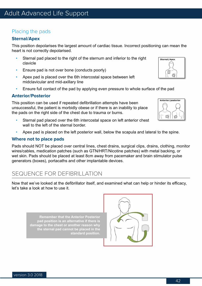
42version 3.0 2018
Adult Advanced Life Support
Placing the padsSternal/ApexThis position depolarises the largest amount of cardiac tissue. Incorrect positioning can mean the heart is not correctly depolarised.
• Sternal pad placed to the right of the sternum and inferior to the right clavicle
• Ensure pad is not over bone (conducts poorly)• Apex pad is placed over the 6th intercostal space between left
midclavicular and mid-axillary line• Ensure full contact of the pad by applying even pressure to whole surface of the pad
Anterior/PosteriorThis position can be used if repeated defibrillation attempts have been unsuccessful, the patient is morbidly obese or if there is an inability to place the pads on the right side of the chest due to trauma or burns.
• Sternal pad placed over the 6th intercostal space on left anterior chest wall to the left of the sternal border.
• Apex pad is placed on the left posterior wall, below the scapula and lateral to the spine.
Where not to place padsPads should NOT be placed over central lines, chest drains, surgical clips, drains, clothing, monitor wires/cables, medication patches (such as GTN/HRT/Nicotine patches) with metal backing, or wet skin. Pads should be placed at least 8cm away from pacemaker and brain stimulator pulse generators (boxes), portacaths and other implantable devices.
SEQUENCE FOR DEFIBRILLATION
Now that we’ve looked at the defibrillator itself, and examined what can help or hinder its efficacy, let’s take a look at how to use it.
Remember that the Anterior Posterior pad position is an alternative if there is
damage to the chest or another reason why the sternal pad cannot be placed in the
standard position.
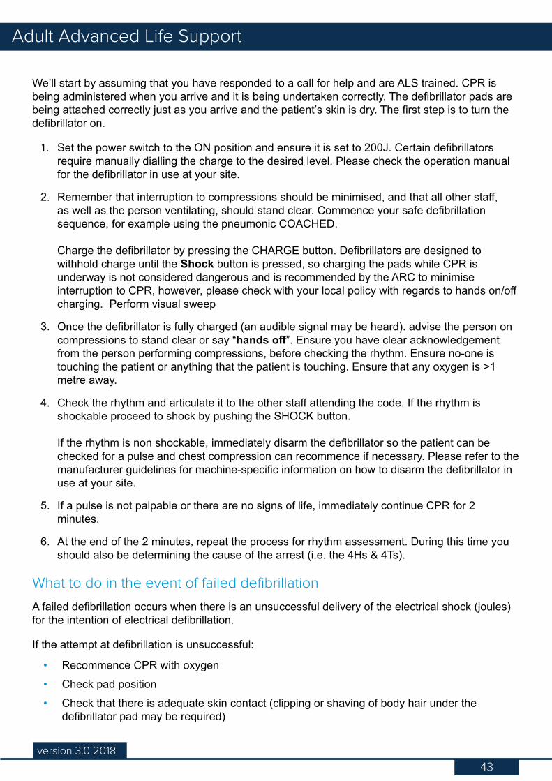
43version 3.0 2018
Adult Advanced Life Support
We’ll start by assuming that you have responded to a call for help and are ALS trained. CPR is being administered when you arrive and it is being undertaken correctly. The defibrillator pads are being attached correctly just as you arrive and the patient’s skin is dry. The first step is to turn the defibrillator on.
1. Set the power switch to the ON position and ensure it is set to 200J. Certain defibrillators require manually dialling the charge to the desired level. Please check the operation manual for the defibrillator in use at your site.
2. Remember that interruption to compressions should be minimised, and that all other staff, as well as the person ventilating, should stand clear. Commence your safe defibrillation sequence, for example using the pneumonic COACHED. Charge the defibrillator by pressing the CHARGE button. Defibrillators are designed to withhold charge until the Shock button is pressed, so charging the pads while CPR is underway is not considered dangerous and is recommended by the ARC to minimise interruption to CPR, however, please check with your local policy with regards to hands on/off charging. Perform visual sweep
3. Once the defibrillator is fully charged (an audible signal may be heard). advise the person on compressions to stand clear or say “hands off”. Ensure you have clear acknowledgement from the person performing compressions, before checking the rhythm. Ensure no-one is touching the patient or anything that the patient is touching. Ensure that any oxygen is >1 metre away.
4. Check the rhythm and articulate it to the other staff attending the code. If the rhythm is shockable proceed to shock by pushing the SHOCK button. If the rhythm is non shockable, immediately disarm the defibrillator so the patient can be checked for a pulse and chest compression can recommence if necessary. Please refer to the manufacturer guidelines for machine-specific information on how to disarm the defibrillator in use at your site.
5. If a pulse is not palpable or there are no signs of life, immediately continue CPR for 2 minutes.
6. At the end of the 2 minutes, repeat the process for rhythm assessment. During this time you should also be determining the cause of the arrest (i.e. the 4Hs & 4Ts).
What to do in the event of failed defibrillation
A failed defibrillation occurs when there is an unsuccessful delivery of the electrical shock (joules) for the intention of electrical defibrillation.
If the attempt at defibrillation is unsuccessful:
• Recommence CPR with oxygen• Check pad position• Check that there is adequate skin contact (clipping or shaving of body hair under the
defibrillator pad may be required)
44version 3.0 2018
Adult Advanced Life Support
• Consider changing the defibrillator pads (pads could be expired)• Consider anterior-posterior placement so that the maximum amount of current traverses the
myocardium
Failed defibrillation can fall into one of three categories:
Patient failurePatient failure can be due to:
• Prolonged downtime before resuscitation is commenced• Severe acid-base imbalance, myocardial hypoxia or severe electrolyte imbalance• Terminal condition pre-arrest• Severe drug toxicity, such as digoxin
Machine failureMachine failure can be due to:
• A malfunction of the defibrillator• Loss of battery charge during repeated defibrillation attempts. Always plug the defibrillator in
to the mains if repeated shocks are required.• The synch button is on/active.
Operator failureOperator failure can be due to:
• The defibrillator not being turned on• The defibrillator not being charged• Joules not being selected or the incorrect joules being selected• The pads are not connected to the defibrillator
Process and documentation
The following forms of documentation are critical to both the ongoing care of the patient and your hospital’s requirements for due process. Please check with your hospital policy for documentation requirements.
Resuscitation record The resuscitation record must be filled out by a member of the team during the event if possible.
This includes time of collapse, initial rhythm, number of shocks delivered, number of joules given at each shock, drugs used and in what amounts, patient response to resuscitation efforts and the duration of those efforts. If there were any failed attempts in interventions, as well as the rationale behind the decisions made during resuscitation – e.g. two failed attempts at intubation due to difficult or swollen airway therefore progressed to tracheotomy – then this also needs to be included in the resuscitation record.
45version 3.0 2018
Adult Advanced Life Support
The times on the record should match the time stamp on the defibrillator (be aware that some older model devices do not have a time visible on the screen which will make it difficult to ensure this correlation. Check with your local policy/procedure as to how to manage this if you have an older device at your site). If the time on the strip is different to the manual records, there can be legal implications. Also on the record must be the names of those staff members who attended the code. A record sheet is attached to the resuscitation trolley. Part of the post-event checklist is to restock this sheet along with any other items used from the trolley. Please refer to your hospital policy on correct documentation.
Post-event print outOne of the most important pieces of documentation in a cardiac event is a printout of the rhythm strip post-event.
If you are the person recording the arrest event on the resuscitation sheet it is also your responsibility to obtain a print out of the rhythm strip at the end of the code. This must be included with the resuscitation record in the patient’s file.
Post event countThe post-arrest count is conducted to correlate physically what has been used with the resuscitation record, most importantly the drugs given, duration of the code, who attended the code, shocks delivered, what equipment was used and what the next steps are. This is to both confirm the notes on the record and to prepare for restocking.
Resuscitation trolleyThe team leader or NUM will have allocated a staff member from the ward to restock the resuscitation trolley after an arrest. It is also checked and restocked (if necessary) on a daily basis as per hospital policy.
Team debriefPost-resuscitation it is important that the team members have time to reflect and debrief on the events, in addition to be able to discuss outcomes, processes, identify safety issues and opportunities for improvement.
FamilyEnsure family have been informed and relevant information material provided.
46version 2.0 2016
Advanced Life Support
46version 3.0 2018
Adult Advanced Life SupportQuizLet’s take a moment now to check what you’ve learned. This is not the final quiz, just practice to help you test your knowledge so far. You’ll find the answers in Appendix 3.
1. One of the conditions where it is recommended to deliver stacked shocks to a patient is:
a. Only when a previously well oxygenated, well perfused, witnessed and monitored patient develops a pulseless, shockable rhythm
b. Only when a cardiac patient is witnessed to collapse in their bathroomc. Only when you are called to an arrestd. Only when a telemetry patient is seen to have collapsed
2. Time is a critical factor that affects defibrillation success – True or False?
3. Which of the following is NOT a factor that influences transthoracic impedance?
a. Pad contact and body hairb. The patient’s chest wall sizec. Obesityd. Correct pad placement
CONCLUSION
In this chapter, we’ve looked in detail at the defibrillation process and how to operate a defibrillator. This should assist your learning when it comes time to practice these actions in the face-to-face session.
47version 3.0 2018
Adult Advanced Life Support
Early recognition and prompt treatment of the airway during an emergency is paramount in preventing secondary hypoxic damage. When unconscious, all the muscles relax, therefore the unprotected airway may easily be blocked by the tongue falling against the back wall of the throat. In the unconscious patient the care of the airway will take precedence over any injury.
RECOGNISING AN AIRWAY OBSTRUCTION
Airway obstruction in a cardiac arrest patient may be the result of the loss of consciousness, or the cause of the arrest. In both cases, it is vital to clear the obstruction. Airway obstruction may be partial or complete. In the conscious patient, the signs and symptoms of obstruction will be dependent upon severity. Partial obstruction may present as:
• Anxiety• Laboured, noisy breathing• Coughing• Air can still be felt escaping from the mouth
Complete obstruction may present with:
• Respiratory effort without breath sounds or air escaping from the mouth
Common causesUpper Airway
• Tongue• Blood, vomit, secretions• Foreign material• Oedema from burns or anaphylaxis• Infection• Trauma• Laryngeal spasm
Lower Airway • Secretions, blood• Pulmonary aspiration• Bronchospasm, asthma• Oedema (mucosal or pulmonary)
Advanced Airway ManagementIn this section, we’ll examine issues surrounding airway management during cardiac arrest. Choice of adjunct is dependent upon the training and experience of the provider and the equipment available.
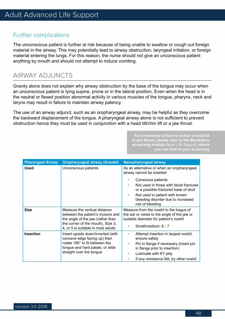
48version 3.0 2018
Adult Advanced Life Support
Further complications
The unconscious patient is further at risk because of being unable to swallow or cough out foreign material in the airway. This may potentially lead to airway obstruction, laryngeal irritation, or foreign material entering the lungs. For this reason, the nurse should not give an unconscious patient anything by mouth and should not attempt to induce vomiting.
AIRWAY ADJUNCTS
Gravity alone does not explain why airway obstruction by the base of the tongue may occur when an unconscious patient is lying supine, prone or in the lateral position. Even when the head is in the neutral or flexed position abnormal activity in various muscles of the tongue, pharynx, neck and larynx may result in failure to maintain airway patency.
The use of an airway adjunct, such as an oropharyngeal airway, may be helpful as they overcome the backward displacement of the tongue. A pharyngeal airway alone is not sufficient to prevent obstruction hence they must be used in conjunction with a head tilt/chin lift or a jaw thrust.
Pharyngeal Airway Oropharyngeal airway (Guedel) Nasopharyngeal airwayUsed Unconscious patients As an alternative or when an oropharyngeal
airway cannot be inserted
• Conscious patients• Not used in those with facial fractures
or a possible fractured base of skull• Not used in patient with known
bleeding disorder due to increased risk of bleeding
Size Measure the vertical distance between the patient’s incisors and the angle of the jaw (rather than the corner of the mouth). Size 3, 4, or 5 is suitable in most adults
Measure from the nostril to the tragus of the ear or nares to the angle of the jaw or suitable diameter for patient’s nostril
• Small/medium: 6 - 7
Insertion Insert upside down/inverted (with concave edge facing up) then rotate 180° to fit between the tongue and hard palate, or slide straight over the tongue
• Attempt insertion in largest nostril, ensure safety
• Pin in flange if necessary (insert pin in flange prior to insertion)
• Lubricate with KY jelly• If any resistance felt, try other nostril
For a reminder of how to action a head tilt or jaw thrust, please refer to the Mandatory eLearning module Basic Life Support, which
you can find in your eLearning
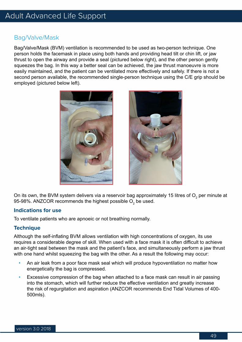
49version 3.0 2018
Adult Advanced Life Support
Bag/Valve/Mask
Bag/Valve/Mask (BVM) ventilation is recommended to be used as two-person technique. One person holds the facemask in place using both hands and providing head tilt or chin lift, or jaw thrust to open the airway and provide a seal (pictured below right), and the other person gently squeezes the bag. In this way a better seal can be achieved, the jaw thrust manoeuvre is more easily maintained, and the patient can be ventilated more effectively and safely. If there is not a second person available, the recommended single-person technique using the C/E grip should be employed (pictured below left).
On its own, the BVM system delivers via a reservoir bag approximately 15 litres of O2 per minute at 95-98%. ANZCOR recommends the highest possible O2 be used.
Indications for useTo ventilate patients who are apnoeic or not breathing normally.
TechniqueAlthough the self-inflating BVM allows ventilation with high concentrations of oxygen, its use requires a considerable degree of skill. When used with a face mask it is often difficult to achieve an air-tight seal between the mask and the patient’s face, and simultaneously perform a jaw thrust with one hand whilst squeezing the bag with the other. As a result the following may occur:
• An air leak from a poor face mask seal which will produce hypoventilation no matter how energetically the bag is compressed.
• Excessive compression of the bag when attached to a face mask can result in air passing into the stomach, which will further reduce the effective ventilation and greatly increase the risk of regurgitation and aspiration (ANZCOR recommends End Tidal Volumes of 400-500mls).
50version 3.0 2018
Adult Advanced Life Support
ADVANCED AIRWAY DEVICES
Following placement of an advanced airway device (by experienced staff) chest compression can continue without pausing to deliver ventilations. Ventilate the lungs at a rate of 6 to 10 ventilations per minute avoid hyperventilation/excessive ventilation aim to provide one breath after each 15 chest compressions.
Supraglottic Airway devices (LMA, Laryngeal Tube, Oesophageal-Tracheal Combitute) are generally considered easier to insert and can be inserted without interrupting chest compressions, and their use has been increasing in cardiac arrest situations.
LMA
The laryngeal mask airway (LMA) is a common piece of airway management equipment used throughout Ramsay hospitals. However, education and training in their use and insertion is required before you can be deemed competent to insert in the patient (please be aware that different brands of LMAs will require slightly different insertion techniques e.g. Laryngeal tube, i-Gel, Combitube).
The LMA consists of a wide bore tube with an elliptical inflated cuff designed to oppose the laryngeal opening. Although primarily for use in anaesthetised patients breathing spontaneously, it can also be used for positive pressure ventilation, which has been demonstrated to be more efficient and easier than with a bag/valve/mask.
Most LMAs are now single use. Drugs should not be administered via the LMA.
Advantages• Rapid establishment of a patent airway• Administration of 100% oxygen, can be inserted whilst CPR is in progress (minimising
interruption to compressions)• Effective ventilation with one person• Non-traumatic, does not require vigorous movements to align the head and neck• Successful insertion by personnel with minimum training time• Possible to minimise gastric inflation provided tidal volumes do not generate high inflation
pressures during intermittent positive pressure ventilation (> 20 cmH20) through the LMA.• Provided there are no leaks, compression can continue without a pause (breaths can be
delivered at a ratio of 15:1 or 10 per minute)
Disadvantages• Cannot administer medications via LMA• Short term airway• Aspiration of gastric contents is possible
Indications for useIndications for use in hospital patients requiring resuscitation:
• Cardiac arrest situations
51version 3.0 2018
Adult Advanced Life Support
• Profoundly unconscious patient with absent glossopharyngeal and laryngeal reflexes at risk of airway obstruction and who may need artificial ventilation when tracheal intubation is precluded by lack of available expertise or equipment
• Known or unexpectedly difficult intubation.
Essential equipmentIntubation equipment must be pre-prepared and ready to use:
• LMA sizes 3, 4 & 5. Backup/restock LMAs should be immediately available.• 50/30ml syringe (luerlock connection)• Gloves and other PPE• BVM/Air Viva • Oxygen-Powered Resuscitators (Mapleson Circuit)• Stethoscope• End tidal CO2 monitoring device• Lubrication• Bite block (rolled gauze or oropharyngeal airway)• Tape/ties to secure tube• Suction
TechniqueRefer to your hospital policy for the type of LMA used in your hospital/MET. Different types of LMA have different insertion techniques.
VentilationVentilation can be commenced using any standard self-inflating bag in conjunction with high flow oxygen. Inflation should be of adequate volume to allow the chest wall to rise. A small leak may be heard, however if it is too large then the LMA should be removed and a larger one inserted.
The inflation pressure should be kept to a minimum. The aim is to allow chest wall to rise whilst preventing potential gastric inflation or regurgitation.
RemovalEnsure that suction equipment is available prior to removal
Only remove the LMA if:
a. the patient demonstrates a return of airway protective reflexes;b. the patient requires ongoing ventilation with an endotracheal tube (ETT) and the
attendant is skilled and competent in intubation; orc. the resuscitation attempt is abandoned (and not a Coroners case)
If the patient is obviously regaining consciousness (i.e. after the patient can open their mouth on command) then the tape and bite block may be removed.
52version 3.0 2018
Adult Advanced Life Support
If the tube is to be removed prior to intubation then:
• Ensure suction equipment is available prior to removal of any airway adjunct• Remove on exhalation
The cuff may remain inflated during removal. The rationale for this is that it will help draw out secretions sat on top of the cuff rather than allowing those secretions to aspirate when the cuff is deflated.
Tracheal intubation
Effective chest compressions deliver at maximum 30% of cerebral blood perfusion, therefore in order to maximise cerebral oxygenation in a cardiac arrest the aim should be to deliver the highest oxygen concentration effectively. CPR must be maintained and if endotracheal intubation is attempted, laryngoscopy should be performed during chest compressions and attempts at intubation should not interrupt compressions for more than 5 seconds (ANZCOR 2016 Guideline 11.6 - Equipment and Techniques in Adult Advanced Life Support).
For this reason tracheal intubation is the gold-standard in airway management as oxygen concentrations of 100% can be achieved through a protected airway. It is indicated when personnel who are able to carry out the procedure with a reasonable level of skill and confidence is present.
However, to avoid substantial interruptions in chest compressions providers may defer attempts at intubation until return of spontaneous circulation if IV access is established and good bag mask technique is in use. If tracheal intubation is attempted ongoing CPR must be maintained and attempts at intubation should not interrupt cardiac compressions for more than 5 seconds (ANZCOR 2016 Guideline 11.6 - Equipment and Techniques in Adult Advanced Life Support).
Advantages• Isolation of the airway/maintenance of patent airway• Allows suction/clearance of trachea• Avoidance of gastric dilatation• Prevents aspiration• Facilitates intermittent positive pressure ventilation• Delivery of high concentrations of oxygen• Administration of first line drugs• Allows uninterrupted chest compressions (ratio 15:1 including no pause) and approximately
8-10 breaths per minute
Disadvantages• Training and experience essential, high failure rate if operator is inexperienced• Aggravation of airway obstruction (e.g. epiglottis)• Local injury (teeth, tongue, palate etc)• Potential to exacerbate cervical cord injury• Time without chest compressions (multiple attempts)• Incorrect placement i.e. oesophageal/right main bronchus
53version 3.0 2018
Adult Advanced Life Support
Complications - during• Trauma• Vagal stimulus Bradycardia• Hypoxia from misplaced tube• Laryngospasm• Substantial interruption to chest compressions
Complications whilst in place • Blockage• Dislodgment• Laryngeal/ Tracheal strictures• Mechanical ventilation complications
Complications post extubation• Aspiration• Obstruction• Laryngospasm• Stridor• Respiratory failure
Essential equipmentPrior to intubation, the Medical Emergency Team must have the intubation tray ready (or direct another staff member to do so).
• Laryngoscope: curved Macintosh blade - size 3 small adults – females - size 4 larger adults - males
• Check light source: ensure bulb is secure and working (single use, disposable Laryngoscope may be used), and if applicable, have spare bulbs and batteries available, charge batteries, and if applicable check fibre optic blade
• Tracheal tubes: female size 7.0 - 8.0 mm male size 8.0 - 9.0 mm• Syringe: for cuff inflation (10ml)• Stethoscope: to confirm correct position of tube in trachea• Tapes/ties: to secure tracheal tube• Suction: Yankeur sucker and flexible catheters• Lubricant jelly: water soluble KY jelly• Magill’s forceps: to remove objects from the oropharynx• Introducers: either soft bougies or semi-rigid stylets, intubation catheters• PPE• ETCO2 detection device (i.e. Easycap), ETCO2 monitoring device• Gastric tube (naso/orogastric), Nasal Gastric Tube
54version 3.0 2018
Adult Advanced Life Support
Waveform and colourmetric capnography
Waveform capnography measures end tidal CO2 in a non-invasive way. It is an effective method of monitoring CO2 during an arrest and is recommended to confirm the position of the ET tube. It may also be beneficial in providing feedback on the effectiveness of chest compressions and potential patient outcomes. A lack of CO2 return may be due to lack of cardiac output to the lungs.
Disposable one-use only colourmetric CO2 detectors are effective and available for use in emergency intubation where waveform capnography is not readily available on all arrest trolleys to confirm correct tube placement.
Cricoid pressure
Cricoid pressure helps prevent regurgitation of gastric contents and consequent pulmonary aspiration or to assist in viewing the vocal cords. It is not required on all patients. Please check with the MO managing the airway as to whether cricoid pressure is required.
During intubation a trained assistant may carry out this manoeuvre if requested by the MO by placing the thumb and forefinger on the cricoid cartilage (located below the thyroid cartilage). The complete cricoid ring is forced backwards occluding the oesophagus against the body of the cervical vertebrae.
Pressure is maintained until the operator instructs its release, after the tube has been placed through the vocal chords, the cuff has been inflated and tube position confirmed. Although effective at preventing passive regurgitation, cricoid pressure must be released when active vomiting occurs as it may lead to oesophageal rupture.
Cricothyroidotomy
A Cricothyroidotomy is where a surgical airway is created by inserting a tube into the trachea. It is an emergency airway undertaken by medical staff that has a limited life span. It is not a long-term option. It is used when ventilation and intubation have both failed, when there is a supraglottic obstruction or when there is laryngeal or palate trauma where the upper airway is completely obstructed.
Needle cricothyroidectomy is a procedure that only allows oxygenation of the patient; carbon dioxide is not eliminated and therefore its usefulness is limited to about 30 minutes, until intubation, a tracheostomy or surgical cricothyroidectomy can be performed to maintain oxygen and ventilation long term.
Complications include malpositioning of the catheter causing surgical emphysema, haemorrhage and oesophageal perforation.
Only the doctor running the code will perform this procedure.
55version 2.0 2016
Advanced Life Support
55version 3.0 2018
Adult Advanced Life SupportQuizLet’s take a moment now to check what you’ve learned. This is not the final quiz, just practice to help you test your knowledge so far. You’ll find the answers in Appendix 3.
1. The size of the ET tube that you would expect to use for an average female during an arrest is:
a. 4.0 – 5.0b. 7.0 – 8.0c. 6.0 – 7.0d. 9.0 – 9.5
2. The signs that an endotracheal intubation has been unsuccessful are:
a. A visible rise and fall of the chest wallb. Increase in oxygen saturationc. Absence of audible breath sounds through auscultationd. Absence of expiratory wheeze
3. Which of the following statements regarding intubation during a cardiac arrest is false?
a. Endotracheal intubation can be performed by ALS trained nursesb. Endotracheal intubation helps to prevent aspiration of stomach contentsc. The endotracheal tube provides a route for the administration of drugs if IV is absentd. Endotracheal intubation improves lung inflation and allows administration of 100%
oxygen
CONCLUSION
In this chapter we’ve looked at airway management during a cardiac arrest.
56version 3.0 2018
Adult Advanced Life Support
The use of medications during cardiac arrest have been shown to be beneficial in certain situations, improving patient outcomes and survival. Priorities are defibrillation, external cardiac compression, and oxygenation together with ventilation.
ALS providers should have a good understanding of the first line drugs used during a cardiac arrest as well as their action. First line drugs are Adrenaline and Amiodarone.
THE ACTION OF FIRST LINE DRUGS
The Sympathetic Nervous System (SNS) works when the body is stressed and reveals itself in the fight or flight response, as opposed to the Parasympathetic Nervous System (PNS) which is responsible for the body’s rest and digest function. The SNS has connections between the brain and the Central Nervous System, including the heart, lungs, and blood vessels. The cell membranes of the target organs have an alpha or a beta response. Adrenaline as the primary drug in cardiac arrest is used for its alpha and beta effects.
Alpha response
Alpha response occurs in the blood vessels, producing vasoconstriction, which leads to an increase in systemic arterial blood pressure.
Beta response
Beta response occurs at three different levels.
Beta 1 - Heart:• Increases heart rate (chronotropic) by stimulating the SA node to fire faster. This is a positive
chronotopic effect.• Improves conductivity (dromotropic) through the conduction pathways, such as the AV node
and the bundle branches.• Increases contraction force (inotropic) by stimulating the myocardium. This is a positive
inotropic effect. However, it does also increase myocardical oxygen demand.
Beta 2 – Respiration:The beta 2 effect is dilation of the bronchioles in the lungs, and a decrease in the Systemic Vascular Resistance (SVR) in the blood vessels. This decreases the afterload of the heart, making it easier for the heart to pump blood out, and decreases myocardial oxygen demand.
Drugs in ResuscitationIn this section we’ll look at the mode of administration of drugs used in resuscitation, as well as the action of first line medications
A NOTE ABOUT NURSE INITIATED DRUGS
This section does not change your Scope of Practice, and not all sites are the same concerning nurse-initiated drugs. Please check your local policy for guidance on what
drugs you may and may not administer without direct medical order.
57version 3.0 2018
Adult Advanced Life Support
CatecholaminesSome of the sympathetic fibres pass to the adrenal medulla, where they release catecholamines into the bloodstream. They work on the target organs through the same mechanism as the sympathetic pathway.
Naturally occurring catecholamines are Adrenaline, Noradrenaline, and Dopamine. Drugs that have the same actions as these catecholamines are called sympathomimetics.
Amiodarone is a membrane stabilising anti-arrhythmic which increase refractory period in the cardiac cycle, thereby slowing conduction at the AV node. It also has mild negative inotropic effect (alpha blocking) which causes peripheral vasodilation.
ADMINSTRATION
The route for drug administration during a cardiac arrest will depend upon available access. If central venous access is already established, this is the preferred route. If central venous is not available, ideally, the peripheral intravenous route should be used due to the insertion of a central venous catheter interrupting CPR and being associated with several potential complications.
If IV access fails the intraosseous route should be considered.
Intravenous
IV drug administration is the preferred route, via a large peripheral vein. This is a fast, reliable method for drug administration, the taking of bloods and/or fluid administration during a cardiac arrest.
If there are no visible peripheral veins, the external jugular vein may be considered. This vein is often prominent during an arrest. Lower limb veins should be avoided due to impairment of venous return below the diaphragm during cardiac arrest.
Intravenous drug administration must be followed by a fluid flush of (normal saline) at least 20-30mls and external cardiac compression. In addition, elevating the limb may speed up delivery to the central circulation.
Intraosseous
If IV access cannot be established, intraosseous (IO) access is safe and effective for fluid resuscitation and drug delivery in all age groups.
The method of insertion will be dependent upon available equipment with the most common sites for insertion being the proximal humerus, proximal tibia and distal tibia. It can be maintained for 24–48 hours, after which another route of access should be obtained. Intraosseous access has roughly the same absorption rate as IV access, and allows for blood sampling and fluid resuscitation as well as administration of high-volume drugs and fluids.
Contra indication to IO includes infection above or below the insertion site or trauma to a limb above or below the insertion site.
58version 3.0 2018
Adult Advanced Life Support
CONCLUSION
In this chapter we’ve learned about the mode of administration of drugs used in resuscitation and an overview of the action of the first line medications.
About the Endotrachael route
The use of the endotracheal tube to administer medications is of limited benefit and its use is not currently promoted. However, the following points using this method of administration may be found in the ARC guidelines (11.5: 2016).
If access cannot be gained the priorities are defibrillation, oxygenation and ventilation together with external cardiac compressions and this method of drug administration considered as a last resort.
If IV or (IO) access cannot be attained and an endotracheal tube is present, endotracheal administration of some medications is possible; although the absorption is variable and plasma concentrations are substantially lower than those achieved when the same drug is given by the intravenous route (increase in dose 3-10 times may be required).
Dilution with water instead of 0.9% normal saline may achieve better drug absorption. Adrenaline, lignocaine and atropine are the only medications which may be instilled via the endotracheal route, other cardiac arrest drugs should NOT be given endotracheally as they may cause mucosal and alveolar damage.
To administer ETT drugs clear the ET tube with suctioning and remove the catheter, then directly administer the drug via the syringe down the ET tube. Follow this by delivering 2-5 ventilations to disperse the drug into the alveoli.
For a list of rhythms management and drug dose and administration, please see Appendix 1.
For a complete list drugs, their alpha and beta responses, indications, effects, doses and dilutions, please see
Appendix 2.
59version 3.0 2018
Adult Advanced Life Support
Not all abnormal cardiac rhythms require full defibrillation. Many rhythms, as you have learned in the chapter on Rhythm Recognition, are not immediately life threatening nor will they benefit from defibrillation. If your patient is suffering from an arrhythmia, either fast or slow, that presents as an adverse sign of deterioration (for example low blood pressure, periods of syncope, heart failure or ongoing chest pain), techniques such as synchronised cardioversion and cardiac pacing may be used.
We’ll look at each of these in detail now.
SYNCHRONISED CARDIOVERSION
In synchronised cardioversion, the SA node is “reset”. Cardioversion stuns excitable cells in the atria (as in atrial fibrillation) that prevent the SA node from functioning normally rather than stopping it altogether. To achieve this, a lower setting than that used for DC defibrillation is applied.
Cardioversion is used mainly for atrial arrhythmias associated with haemodynamic compromise or other adverse signs. However it may also be undertaken as an elective procedure for patients in long term AF, for example, cardioversion is undertaken in a controlled environment. The patient is prepared and sedated for the procedure.
IndicationsCardioversion can be used for:
• Atrial fibrillation• Atrial flutter• Atrial tachycardia• Supraventricular tachycardia (SVT)
It can also be used in VT where the patient is conscious and has a pulse, and drug therapy has been ineffective or the patient is compromised.
Energy selectionLower joules are required than when defibrillating the heart in an arrest situation. The aim is to shock the SA node, not stop it altogether.
The following joule selection may be used (determined by the medical team) for each rhythm:
• Atrial Flutter and Atrial Tachycardia usually start at 50-100 joules• Atrial Fibrillation starts at 100 joules• SVT is commenced at 100 joules
The joules used can then be gradually increased depending on patient response. This should be undertaken as per the Medical Officer’s orders.
Cardioversion and PacingIn this chapter we’ll examine other ways in which an electrical impulse can be used to change the action of the pacemaker of the heart.
60version 3.0 2018
Adult Advanced Life Support
Pre-careThe patient should be fasted prior to the procedure and a 12 lead ECG should be obtained, as well as an IV cannula inserted. You should also have gained formal patient consent.
Pre-procedure requirements:
• Results for U&Es, INR (if previously on Warfarin)• Drugs for sedation – for example, Propofol, Midazolam, Fentanyl• Resuscitation and suction equipment
Shave the patient’s chest if required and apply the pads. A different placement may be used for atrial rhythms at the discretion of the cardiology team. Preoxygenate the patient during sedation.
Procedure for cardioversion
Please note, this process will vary according to the machine you are using.
1. Press the Synch button on defibrillator.
2. Ensure each complex is highlighted as per the defibrillator instructions with either a dot or triangle above the R wave. There must be 100% recognition. Failure of loss of R wave recognition may result in the patient converting to a life threatening arrhythmia.
3. Press the Energy Select button and select the energy level required as per the MO orders (the default setting will be for defibrillation).
4. Charge the defibrillator
5. After following safety checks that all staff are standing clear, press and hold the Shock button until the shock is delivered. The shock will only be delivered on the R wave of the complex, therefore there may be a delay between pressing the button and the shock being delivered.
6. Observe for return of normal rhythm.
If further shocks are required most defibrillators will require the synch button to be reset. This is a safety feature in case a DC shock is required.
Post reversion
Once the rhythm has reverted, maintain the patient’s airway until they are self-ventilating.
Remove adhesive pads whilst patient unconscious and stay with the patient until they have regained consciousness.
Continue to monitor them via ECG and perform a 12 lead ECG, making frequent haemodynamic observations and ensure you document events as per hospital policy.
61version 3.0 2018
Adult Advanced Life Support
TEMPORARY EXTERNAL TRANSCUTANEOUS CARDIAC PACING
Please note, this process will vary according to the machine you are using.
External transcutaneous cardiac pacing is the process by which the patient’s cardiac rhythm is augmented to increase the heart rate and therefore their cardiac output.
To initiate pacing, the patient must be monitored utilising the ECG leads on the defibrillator in addition to the pads so that it may sense and respond to the patient’s intrinsic rhythm on a continuous, uninterrupted basis. On many machines the pads are not able to read the rhythm and pace at the same time through one set of pads, therefore the defibrillator ECG leads must be connected to the patient.
The defibrillator is able to act as a pacemaker to provide an artificial electrical stimulus to the myocardium in order to initiate depolarisation of the atria, ventricle, or both. Pacing is the process of initiating an electrical stimulus to the cardiac muscle and aims to achieve both electrical and mechanical capture.
Capture
Capture – occurs both electrically and mechanically.
• Electrical capture is when an adequate electrical current has been delivered to the heart to produce depolarisation of the ventricles. This is evidenced by a spike on the ECG which is immediately followed by a wide QRS complex followed by a tall, broad T wave.
• Mechanical capture involves depolarisation of the ventricles and an adequate cardiac output (i.e. a palpable pulse). Patients who are being paced should have mechanical capture confirmed by palpating for the presence of a femoral pulse.
If there is a failure to capture:
• Increase the current• Reposition the electrodes on the patient’s chest• Consider a severe metabolic acidosis, hypoxia or drug toxicity
Sensing
This is when the pacemaker is sensing the patient’s intrinsic cardiac rhythm when in the demand mode. The pacemaker can sense the atria or ventricles or both. The external pacemaker senses the patient’s QRS complex (ventricular depolarisation), not the P wave. A mark appears on the QRS complex when sensing occurs.
When there is failure to sense during pacing:
• Increase the ECG size so the pacer can detect the patient’s QRS complex• Select a different ECG lead• Reposition the electrodes on the patient’s chest• If your site has access to a pacemaker technician, contact them immediately
62version 3.0 2018
Adult Advanced Life Support
Non-demand/fixed pacing
The pacemaker delivers a pacing stimulus at a set rate completely independent of the patient’s intrinsic heart activity. It can also be call Asynchronous Pacing. An indication for fixed mode maybe during transport. Movement of the patient trolley during transport may cause sensing issues (over-sensing). However, in general, this mode is rarely used, as competition between paced beats and the patient’s intrinsic rhythm can lead to ventricular arrhythmias (Phillips Healthcare, 2013).
Demand pacing
The pacemaker senses the patient’s intrinsic cardiac activity, and delivers pacing stimuli to capture only when the patient’s intrinsic rhythm falls below the set pacing rate or during prolonged pauses in the patient’s rhythm. It can also be called Synchronous Pacing. This is the more commonly used pacing mode.
Indications
External cardiac pacing is indicated for patients with symptomatic bradycardia and conduction blocks such as complete heart block.
BradycardiaPacing is indicated in slow rhythms if the patient is haemodynamically compromised (SBP < 90mmHg) and has not responded to drug therapy, such as atropine or isoprenaline. In this situation pacing aims to increase the patient’s heart rate and cardiac output.
Heart blockPacing is usually the treatment of choice in 3rd degree heart block, possibly in conjunction with or whilst awaiting additional medications to improve rate and contractility. Transcutaneous pacing may sometimes be indicated in other conduction blocks, depending on the degree of haemodynamic compromise.
Advantages and disadvantages
Transcutaneous pacing in a cardiac arrest situation has both its advantages and disadvantages depending on the use of the pacing process and the current situation.
AdvantagesThere are numerous advantages to pacing. It is easy and quick to prepare the patient for the procedure, and requires less training than internal pacing modes. There is also a lower risk to the patient.
DisadvantagesPacing can be quite uncomfortable for the conscious patient. Cutaneous nerve stimulation can result in a tingling, stinging, pinching, or burning sensation. There can also be skeletal muscle contraction that can cause tapping, twitching, or thudding sensations for the patient.
Skeletal muscle contractions can occur even with low energy levels, and patients often have trouble tolerating currents above 50 milliamps. As capture threshold often occurs above this level, analgesia and sedation should be considered for the conscious patient but only after adequate cardiac output has been produced.
63version 3.0 2018
Adult Advanced Life Support
Despite the discomfort for the patient, the administration of analgesia and sedation to a patient who does not have an adequate cardiac output may result in a haemodynamic state that precipitates a cardiac arrest.
Sequence for non-invasive cardiac pacing
Please note that this sequence is again based on the Zoll defibrillator and that the machine in use at your site may differ. Please check your manufacturer’s instructions.
1. Connect the defibrillator/pacing pads and ECG leads and confirm the presence of a rhythm. Check the pad expiry dates as all pads are pre-gelled and may dry out over time.
2. Record a rhythm strip.
3. Ensure the defibrillator pads are attached to the cable and place in the appropriate positions on the chest. If there is time, shave hair from the site to improve conduction. Have the leads pointing to the outside of the patient.
4. Turn on the Pacemaker.
5. Unless there is a default heart rate, select the required Heart rate. This will be determined by the treating medical officer who will request an appropriate pacing rate as per the situation.
6. With the milliamps (or current, as terminology may differ) set at zero, start increasing the milliamps until electrical capture is noted (where the artificial ‘spike’ triggers a response in the myocardium). Check for a femoral pulse to confirm cardiac output and mechanical capture.
7. Once mechanical capture has been confirmed, increase the milliamps by 10% if tolerated by patient as a margin of safety or as per your hospital policy.
8. Ensure patient comfort (e.g. administer analgesia or sedation).
9. Remember to record all observations including pacing mode, heart rate, threshold for mechanical capture and set milliamps.
64version 2.0 2016
Advanced Life Support
64version 3.0 2018
Adult Advanced Life SupportQuizLet’s take a moment now to check what you’ve learned. This is not the final quiz, just practice to help you test your knowledge so far. You’ll find the answers in Appendix 3.
1. Synchronised counter shock is delivered:
a. On the Q of the QRS complexb. On the P wave of the ECGc. On the R wave of the ECGd. On the T wave of the ECG
2. Which of the following arrhythmia is an indication for the delivery of synchronised cardioversion?
a. Atrial fibrillationb. Ventricular fibrillationc. Pulseless ventricular tachycardiad. Ventricular ectopic beat
3. To initiate pacing mode the patient must be connected to the defibrillator ECG leads and pads. True or False?
CONCLUSION
In this chapter, we’ve looked at ways to electrically enhance a slow heart rate and revert a tachyarrythmia to support the patient and prevent further deterioration.
There are additional options relating to the administration of medications which may also revert/enhance tachy and brady arrhythmias. Refer to your hospital policy’s and practice for chemical therapies.
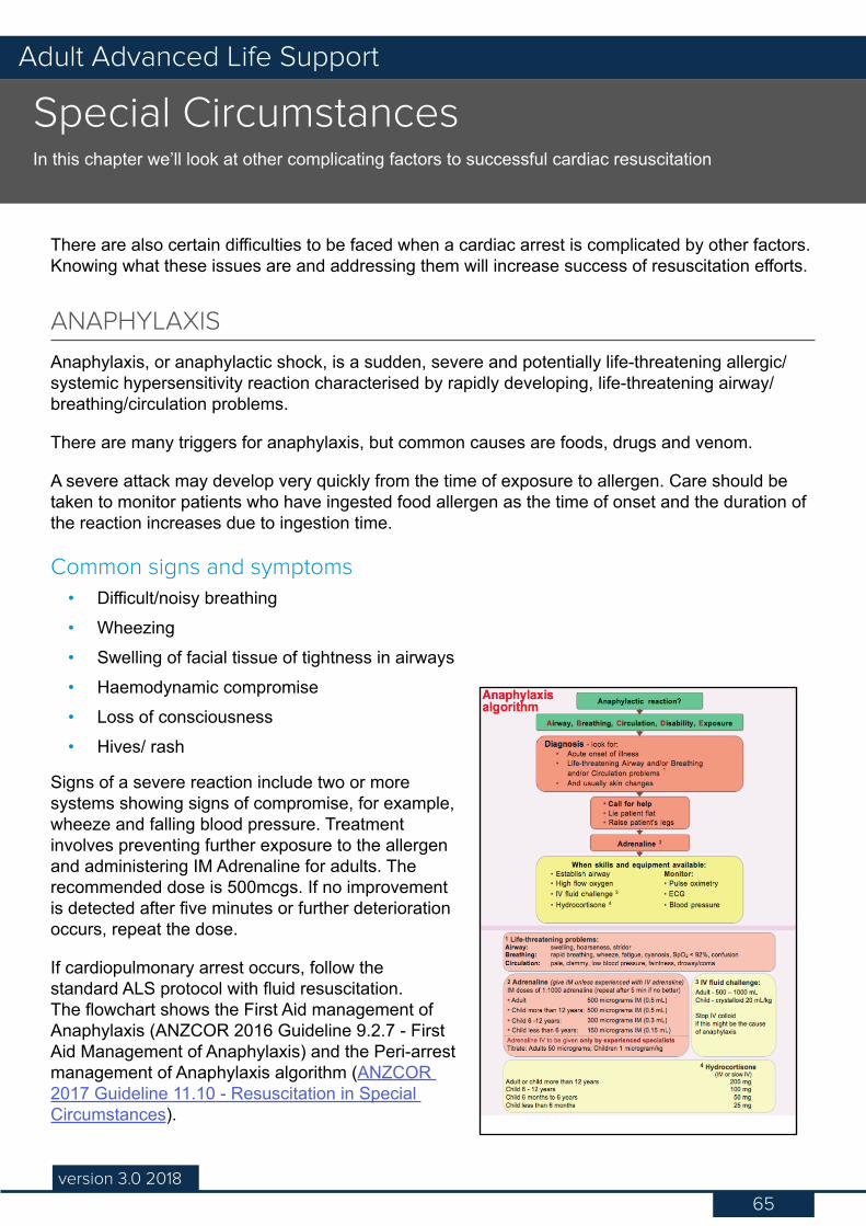
65version 3.0 2018
Adult Advanced Life Support
There are also certain difficulties to be faced when a cardiac arrest is complicated by other factors. Knowing what these issues are and addressing them will increase success of resuscitation efforts.
ANAPHYLAXIS
Anaphylaxis, or anaphylactic shock, is a sudden, severe and potentially life-threatening allergic/systemic hypersensitivity reaction characterised by rapidly developing, life-threatening airway/breathing/circulation problems.
There are many triggers for anaphylaxis, but common causes are foods, drugs and venom.
A severe attack may develop very quickly from the time of exposure to allergen. Care should be taken to monitor patients who have ingested food allergen as the time of onset and the duration of the reaction increases due to ingestion time.
Common signs and symptoms• Difficult/noisy breathing• Wheezing• Swelling of facial tissue of tightness in airways• Haemodynamic compromise• Loss of consciousness• Hives/ rash
Signs of a severe reaction include two or more systems showing signs of compromise, for example, wheeze and falling blood pressure. Treatment involves preventing further exposure to the allergen and administering IM Adrenaline for adults. The recommended dose is 500mcgs. If no improvement is detected after five minutes or further deterioration occurs, repeat the dose.
If cardiopulmonary arrest occurs, follow the standard ALS protocol with fluid resuscitation. The flowchart shows the First Aid management of Anaphylaxis (ANZCOR 2016 Guideline 9.2.7 - First Aid Management of Anaphylaxis) and the Peri-arrest management of Anaphylaxis algorithm (ANZCOR 2017 Guideline 11.10 - Resuscitation in Special Circumstances).
Special CircumstancesIn this chapter we’ll look at other complicating factors to successful cardiac resuscitation
66version 3.0 2018
Adult Advanced Life Support
PREGNANCY
In the obviously pregnant woman, the pregnant uterus causes pressure on the major abdominal vessels when she lies flat, reducing venous return to the heart.
Maternal cardiac arrest can occur as the result of haemorrhage, amniotic or pulmonary embolism, eclampsia or placental abruption, as well as the same causes of cardiac arrest as females of the same age group. The physiological changes as a result of pregnancy, such as increased blood volume, cardiac output, oxygen consumption and reduced lung volumes, can complicate the basic ABC management. For example, intubation is more difficult and gastric reflux is more likely.
The principles of basic and advanced life support apply to the pregnant patient and the emphasis is on the mother, as the foetus depends on her for survival. Without oxygen, the foetus will die and this means oxygen and adequate circulation needs to be provided.
The main points when resuscitating a pregnant woman include:
• Summon expert help immediately• Left lateral tilt: Manually displace pregnant uterus to the left to alleviate compression of
the major blood vessels. The angle of the tilt to the left still needs to allow for effective compressions, i.e. shoulders flat, pelvis tilted to the left. A gravid uterus will occlude the Vena Cava and the Aorta if the woman is left on her back. This can in itself produce ‘Supine Hypotension Syndrome’ as the pressure of the uterus on the major abdominal vessels can precipitate a cardiac arrest. The pregnant woman should be positioned on her back with her shoulders flat and sufficient padding under the right buttock to give an obvious pelvic tilt to the left.
• Aggressive airway management and early intubation: The foetus needs oxygen and gastric emptying is poor in the pregnant woman with a subsequent risk of aspiration.
Further procedures: If the arrest is due to a ruptured uterus or eclampsia, or if initial resuscitation efforts fail, then a peri-mortem emergency caesarean section is the only chance for delivery of the infant and must take place within 5 minutes after the mother’s cardiac arrest. Advanced life support must continue during and after surgery.
HYPOTHERMIA
Partly a protective mechanism, hypothermia will significantly reduce the basal metabolic rate for every degree below 36°C. This therefore reduces oxygen requirements and will slow the development of acidosis associated with anaerobic metabolism secondary to hypoxia. However, hypothermia is also a barrier to effective resuscitation because it produces arrhythmias and increases transthoracic impedance in defibrillation. The stiffness created by hypothermia in the chest cavity will impede effective chest compressions and conduction of electrical impulse from defibrillation.
To this end, it may be necessary to make modifications to ALS for the hypothermic patient:
• It is reasonable to withhold drugs until the core temperature >30°C – the intervals then should be doubled until >35°C.
• If VF persists after 3 shocks – delay further attempts until the body temperature is 28 – 30°C
67version 3.0 2018
Adult Advanced Life Support
Use all available resources e.g. active external methods like warm air, space blankets, warm blankets or a ‘bear hugger’ for extra corporeal rewarming (cardio-pulmonary bybass/VA -ECMO); and minimally invasive methods like warmed fluids. Patients should be rewarmed 1 to 1.5°C/hr to avoid acute acidosis and other systemic complications resulting in ‘rewarming shock’.
ELECTROCUTION
Electricity will travel the path of least resistance, with resistance lowest in moist areas. Serious internal damage occurs when the electricity is converted to heat, which occurs at the areas of highest resistance, such as bone.
Low voltage• Significant contact wounds and possible cardiac arrest, but little deep tissue damage.• Commonly household current, which is Alternating Current (AC)• Alternating Current (AC) can lead to cardiac arrhythmias, especially Asystole, VF and
hypoxia.
High voltage
Injury is caused by flash burns and Direct Current (DC) transmission.
• DC transmission will result in both full thickness cutaneous injury and deep tissue damage at both entry and exit points. Deep muscle damage can occur along the path of the current and may lead to Rhabdomyolosis with subsequent Acute Tubular Necrosis and renal failure.
• Overall damage is related to the combined effects of current, resistance and voltage.
Assessment and Management of Electrical Injuries
Ensure any power source is turned off and that it is safe to approach the victim. The priorities of assessment are ABC and an ECG.
Avoiding hypoxia will improve the outcome. Defibrillation is the choice for VF arrest although efficacy may be affected by the damage done to the heart from the electrical injury. With all electrical burns you need to look for entrance and exit sites, as they can give an indication of the pathway of the injury. The exit site is usually larger than the entry wound.
DRUG POISONING/TOXICOLOGY
The main consideration is for cardiotoxic poisons that inhibit conduction. Patients who experience a cardiac arrest following poisoning by a substance that slows conduction are very difficult to successfully resuscitate.
The focus should be on avoiding the cardiac arrest by neutralising or eliminating the poison.
68version 3.0 2018
Adult Advanced Life Support
NEAR DROWNING
Initial immersion will elicit the diving reflex, which lowers the heart rate and respiratory rate; followed by apnoea, producing hypoxia, tachycardia and subsequent acidosis. This will eventually lead to bradycardia, arrhythmias and cardiac arrest.
The main problems with near drowning are hypoxia, hypothermia and acidosis, all three reducing the effectiveness of resuscitation attempts.
Therefore, early airway management with intubation and ventilation is essential. Positive End Expiratory Pressure (PEEP), which is the maintenance of alveolar pressure (keeping the alveoli open), is often used to improve the relationship of ventilation to perfusion and therefore gas exchange.
Assessing temperature is a critical factor in treating the hypothermia that frequently accompanies immersion injuries.
69version 2.0 2016
Advanced Life Support
69version 3.0 2018
Adult Advanced Life SupportQuizLet’s take a moment now to check what you’ve learned. This is not the final quiz, just practice to help you test your knowledge so far. You’ll find the answers in Appendix 3.
1. The common signs of an anaphylaxis are:
a. Difficult/noisy breathing, hives/rash, loss of consciousness, muscle twitchingb. Hives/rash, welts and redness, loss of consciousness, thirstc. Difficult/noisy breathing, hives/rash, wheezing, loss of consciousnessd. Difficult/noisy breathing, wheezing, thirst, headaches
CONCLUSION
In this chapter, we’ve looked at the special circumstances relevant to cardiac arrest, as well as how to treat these complicating factors.
70version 3.0 2018
Adult Advanced Life Support
The aim of resuscitation is to ultimately have a patient with:
• A normal conscious level• No neurological deficit• A stable cardiac rhythm• Adequate oxygen perfusion
However, the return of spontaneous circulation does not mark the achievement of these aims; more likely it marks the start of a long and difficult post-resuscitation phase.
AIMS OF THERAPY
The aims of therapy after initial resuscitation are to:
• Determine and treat the cause of the cardiac arrest and prevent reoccurrence• Continue respiratory support• Maintain cerebral perfusion• Treat and prevent cardiac arrhythmias• Prevent secondary damage
This requires a range of assessments and observations to be undertaken following the cardiac arrest. Let’s have a look now at an idealised timeline for events during a resuscitation attempt in action.
While resuscitation is taking place
While resuscitation is taking place, if the patient has been critically ill, it is possible that there may be family members present. Unless they are disrupting resuscitation efforts, they should be permitted to stay if they so desire, as this has been shown to be beneficial in their adjusting to loss.
If a family member requests to stay in the room during resuscitation, a member of staff who can adequately support them should be allocated to care for them. This member of staff needs to be confident that they can explain what is occurring in a language the relatives will be able to assimilate at this stressful time.
If the resuscitation is unsuccessful, the relatives should be allowed to be with the patient in private as soon as is able.
Post-resuscitation CareIn this chapter we’ll look at considerations immediately following successful resuscitation
71version 3.0 2018
Adult Advanced Life Support
During a sudden, unexpected arrest
If an arrest is sudden and unexpected and relatives need contacting by phone, it is important that this call is made by someone who has knowledge of the family dynamics, so that the appropriate information can be communicated, whilst trying to minimise panic.
Normally this call will be made by the doctor in charge of the code, or the Nurse Unit Manager of the ward. The person who makes that call should try to be available to receive those relatives when they do arrive at the unit/ward.
Interpreter services
Consider that in certain circumstances you may need to contact an interpreter if the patient’s family do not speak English or if they are culturally and linguistically diverse. This is an important consideration as there can be culturally protocols that must be adhered to at the end of life.
ONCE THE PATIENT HAS BEEN SUCCESSFULLY RESUSCITATED
Once the patient has been successfully resuscitated, ventilation must be continued until the patient is awake, alert and cooperative. The patient must be able to protect his or her own airway and have a stable cardiovascular system before extubation. It is imperative that a full assessment of your patient occurs post-resuscitation, reviewing airway, breathing, circulation, disability and exposure to ensure optimum patient outcomes are achieved.
While in this stage, the medical officer running the code with the ALS nurse will check:
• Respiratory system: » Position and security of the ET tube if required » Assess for trauma to the chest wall/pneumothorax
• Cardiovascular system: » Listen for heart sounds, palpate major pulses » Look for grossly distended neck veins » Blood pressure
• Central Nervous system: » Perform an initial neurological assessment as a base line only » Assess pupillary size and reaction, limb movement and abnormal posturing
• Abdomen: » Examine for distension » Insert a urinary catheter and nasogastric tube
72version 3.0 2018
Adult Advanced Life Support
Patient stabilisation is achieved
It is imperative to restore the patient’s usual blood pressure or at least a systolic pressure greater than 100mmHg. Avoid hypoxia through use of 100% oxygen during cardiac arrest, and avoid hyperoxia - it is reasonable to titrate inspired oxygen to maintain SpO2 between 94 – 98%. In addition, maintain PaCO2 within normal range.
If the blood pressure falls, a vasopressor may be given by small increments (0.1mg) or an infusion until fluid status and the need for volume expansion can be assessed. As soon as possible all vasoactive drugs should be given by central access or dedicated central line lumen.
Hypo/hyperglycaemia and electrolyte disorders such as hypo/hypernatraemia may produce continuing cerebral damage. Blood glucose level, biochemical screen, arterial blood gases and a 12 lead ECG should be performed to guide further management. Monitor blood glucose regularly and treat any hyperglycaemia (>10mmol/L) and avoid hypoglycaemia
If ventricular fibrillation has been successfully reverted the anti-arrhythmic drug that was used may be continued as an infusion under medical instruction.
There is no evidence that corticosteroids are beneficial. Anti-epileptic medication may be required if seizures occur. Treat seizures if they occur and start maintenance therapy.
Targeted Temperature Management (TTM) describes the method and treatment of inducing a mild hypothermia in patients post cardiac arrest who remain unresponsive. TTM been shown to improve neurological outcomes in this cohort of patients. (ANZCOR 2016 Guideline 11.8 - Targeted Temperature Management (TTM) after Cardiac Arrest)
If TTM is considered, then the method of cooling will be dependent upon equipment available. Cooling methods include, ice packs, cool fluids, cooling blankets or Extra Corporeal Membrane Oxygenation (ECMO) for example. The important factor is that a constant temperature be maintained between 32 – 36°C, for a period of 12 – 24 hours (check hospital policy).
Additional considerations include avoiding hyperthermia and preventing the patient from shivering which will increase metabolic demands.
Patient transfer is then determined
Only once the patient is stable should the decision to transfer be made. This decision should come from the leader of the arrest situation and the receiving team. However, that being said, all post arrest patients who are intubated and medically stable are transported to ICU or CCU as it is assumed that they will require ventilation and further critical care treatment.
Having made the decision to transfer, continuous monitoring facilities need to be attached to the patient. All lines, drains, catheters and tubing should be secured.
Staff attending to the patient during transfer should be satisfied before they leave, that they have all equipment and skills necessary to deal with any complication or re-arrest during the interdepartmental/hospital trip.
73version 3.0 2018
Adult Advanced Life Support
Staff are debriefed
A debrief after the event is important for staff to examine what happened, how they felt about it and accept loss in the event of a death. Usually this debrief is conducted by the NUM or team leader, and includes all the staff on the ward who were present at the arrest, including ALS trained staff. The MET team responders will likely have their own internal debrief elsewhere.
The debrief is not for criticism of performance but for positive feedback. The opportunity for discussion about how and why events occurred should be included. Lastly, issues that may have emerged for areas of educational activity or equipment availability and system/procedure changes can be identified after debrief.
Often in the case of patients who have been on the ward for a while, staff might be interested in the patient’s post arrest outcomes. Usually the NUM will obtain this information and disseminate it to the interested staff members.
Sometimes arrest situations can be both stressful and traumatic. If you find that you are troubled as a result of your invovlement in an arrest situation you may benefit from talking with someone external to the event. If this is the case you can contact Ramsay’s EAS provider Benestar on 1300 360 364.
Keep in mind
Separate to the debrief, the NUM or team leader will open discussion with the attending staff members to determine prior causes and issues leading up to the arrest and how effective treatment and resuscitation efforts were. In this session the team will examine:
• The patient’s past medical history• The patient’s current drug therapy• Relevant events preceding the cardiac arrest• Possibility of drug overdose/anaphylactic reaction• Hypoglycaemia• Neurological event such as a stroke
74version 2.0 2016
Advanced Life Support
74version 3.0 2018
Adult Advanced Life SupportQuizLet’s take a moment now to check what you’ve learned. This is not the final quiz, just practice to help you test your knowledge so far. You’ll find the answers in Appendix 3.
1. Once a successful resuscitation has been achieved which of the following post resuscitation checks must be undertaken immediately?
a. Ensure that the patient is transferred to the ICU immediatelyb. Clean the room and put all the furniture back, as well as check the resuscitation trolley
and restock whatever was usedc. Check that all staff are handling the event well and discuss how the code response wentd. Fully assess patient, ensure airway patency is maintained and aim to achieve normative
blood pressure
2. Which of the following is important during the immediate post resuscitation analysis? (note: there is more than one correct answer)
a. Patient’s current drug therapyb. Possibility of drug overdose/anaphylactic reactionc. Patient’s past medical historyd. Relevant medical events leading up to the cardiac arrest
75version 3.0 2018
Adult Advanced Life Support
The purpose of this analysis is to ensure that no aspect of nursing care was deficient.
Congratulations. You have reached the end of this component of the ALS programme. You should now understand the concept of the Chain of Survival and understand the importance of early recognition of potential cardiac arrests.
You should also be able to:
• Describe the ethical and legal requirements of CPR• Follow the Advanced Life Support algorithm• Identify cardiac rhythms requiring defibrillation• Describe the principles and application of defibrillation• Discuss the determination and application of airway adjuncts• Describe your role in assisting in advanced airway management• Describe how to assist with a tracheal intubation• Identify medication and doses used in resuscitation• Describe the principles and application of synchronised cardioversion• Describe the principles and application of non-invasive cardiac pacing• Discuss the requirements of post resuscitation care
Keep in mind that no two codes will run the same. The conditions that led to the arrest are different in every patient. Resuscitation is one part routine and two parts adapting to changes.
Conclusion
Remember:
• Good CPR technique and early defibrillation increases chances of survival
• It is vital to know your rhythms and be able to recognise them immediately in order to act quickly
• It is vital to know when drugs can be administered and where they are required in the resuscitation process
• If you understand each person’s role in responding to a code and take a proactive and communicative approach to your response you will help enable the code to proceed smoothly.
76version 3.0 2018
Adult Advanced Life Support
This resource lists the common rhythms you may see on an ECG.
Sinus ................................................................................................................................... 75
Sinus Bradycardia ............................................................................................................... 76
Sinus Tachycardia ............................................................................................................... 77
Ventricular Fibrillation .......................................................................................................... 78
Pulseless Ventricular Tachycardia ...................................................................................... 80
Asystole .............................................................................................................................. 82
Pulseless Electrical Activity (PEA) ...................................................................................... 84
Ventricular Tachycardia with a pulse ................................................................................... 85
Torsades de Pointes ........................................................................................................... 87
Supraventricular Tachycardia .............................................................................................. 89
Third Degree Block ............................................................................................................. 91
Idioventricular (Slow) .......................................................................................................... 93
Accelerated Idioventricular (fast) ........................................................................................ 95
Atrial Fibrillation .................................................................................................................. 96
Atrial Flutter ......................................................................................................................... 97
Agonal ................................................................................................................................. 99
Appendix 1 - Rhythms
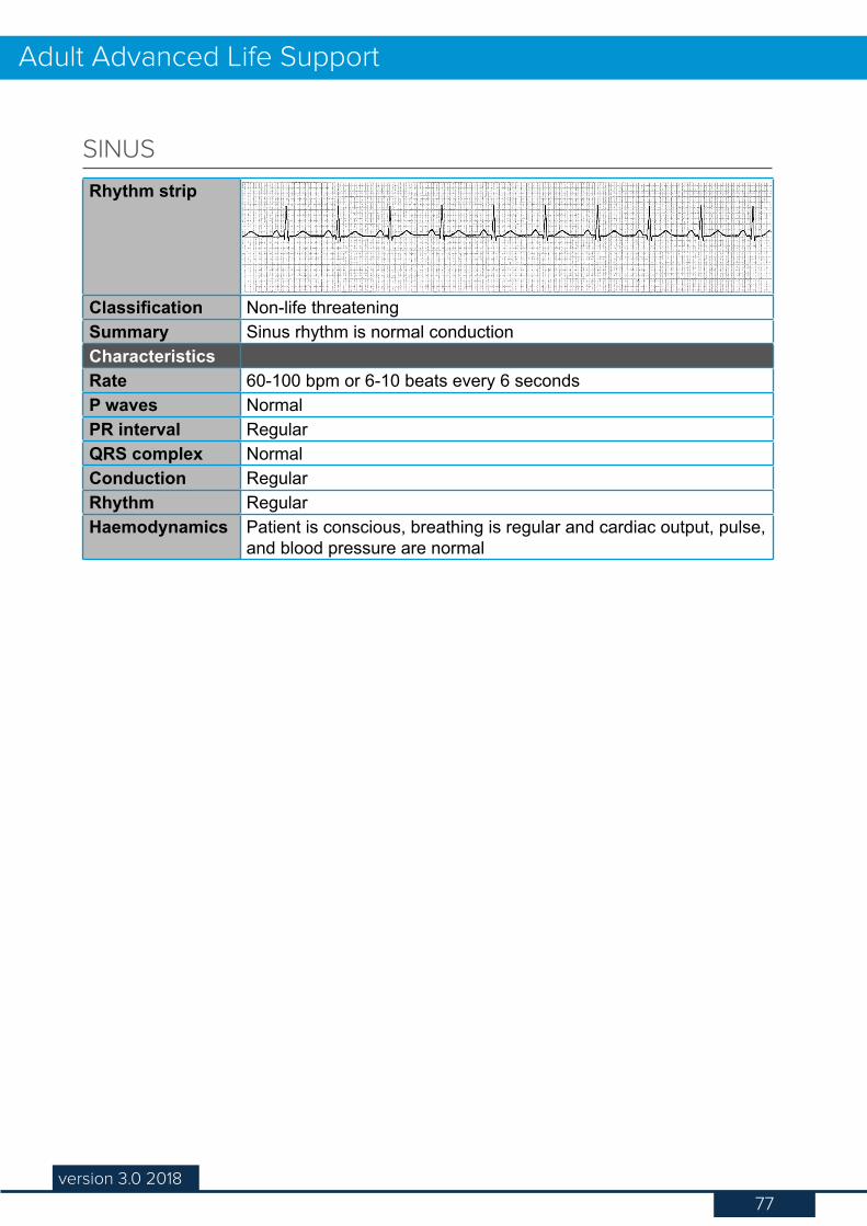
77version 3.0 2018
Adult Advanced Life Support
Rhythm strip
Classification Non-life threateningSummary Sinus rhythm is normal conductionCharacteristicsRate 60-100 bpm or 6-10 beats every 6 secondsP waves NormalPR interval RegularQRS complex NormalConduction RegularRhythm RegularHaemodynamics Patient is conscious, breathing is regular and cardiac output, pulse,
and blood pressure are normal
SINUS
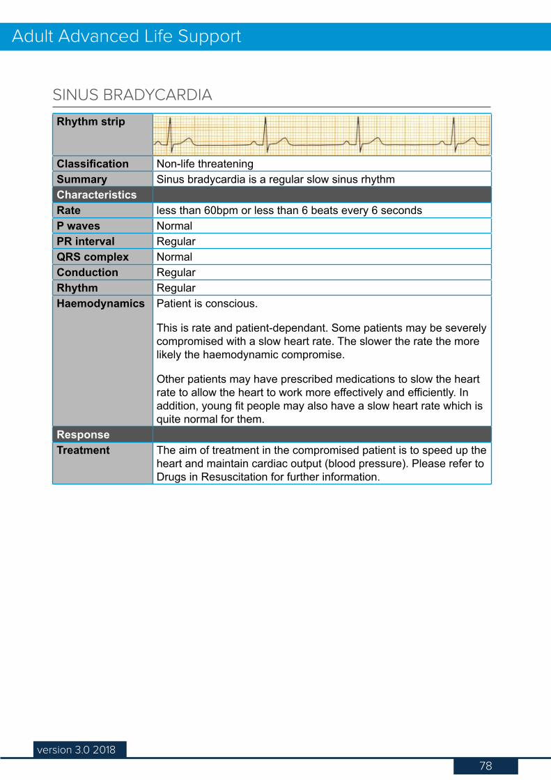
78version 3.0 2018
Adult Advanced Life Support
Rhythm strip
Classification Non-life threateningSummary Sinus bradycardia is a regular slow sinus rhythmCharacteristicsRate less than 60bpm or less than 6 beats every 6 secondsP waves NormalPR interval RegularQRS complex NormalConduction RegularRhythm RegularHaemodynamics Patient is conscious.
This is rate and patient-dependant. Some patients may be severely compromised with a slow heart rate. The slower the rate the more likely the haemodynamic compromise.
Other patients may have prescribed medications to slow the heart rate to allow the heart to work more effectively and efficiently. In addition, young fit people may also have a slow heart rate which is quite normal for them.
ResponseTreatment The aim of treatment in the compromised patient is to speed up the
heart and maintain cardiac output (blood pressure). Please refer to Drugs in Resuscitation for further information.
SINUS BRADYCARDIA
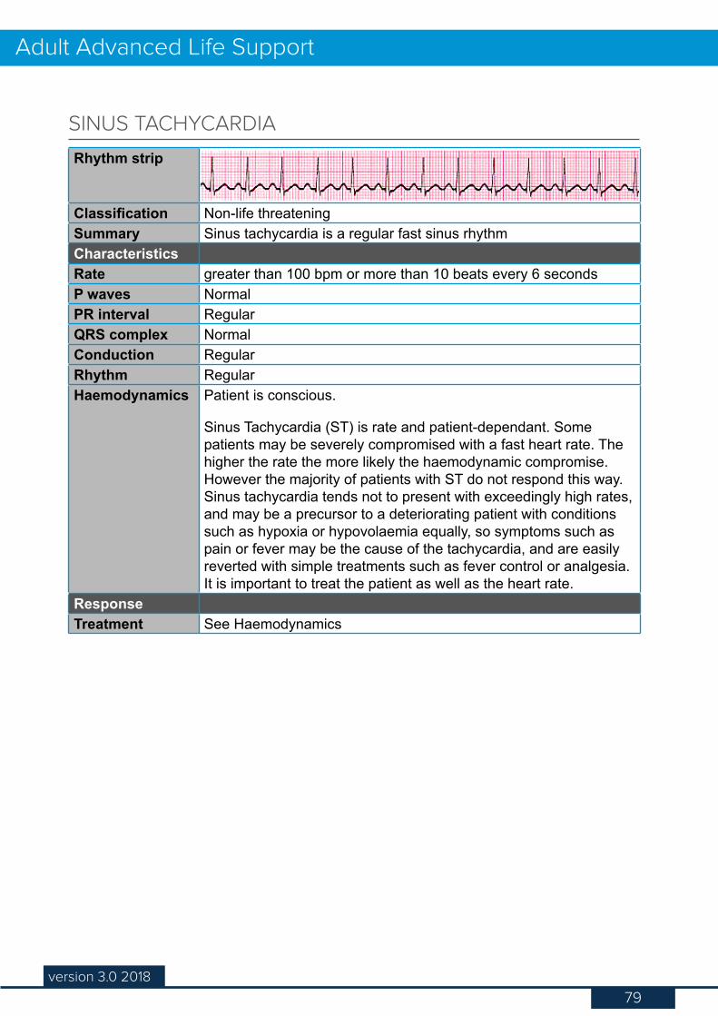
79version 3.0 2018
Adult Advanced Life Support
Rhythm strip
Classification Non-life threateningSummary Sinus tachycardia is a regular fast sinus rhythmCharacteristicsRate greater than 100 bpm or more than 10 beats every 6 secondsP waves NormalPR interval RegularQRS complex NormalConduction RegularRhythm RegularHaemodynamics Patient is conscious.
Sinus Tachycardia (ST) is rate and patient-dependant. Some patients may be severely compromised with a fast heart rate. The higher the rate the more likely the haemodynamic compromise. However the majority of patients with ST do not respond this way. Sinus tachycardia tends not to present with exceedingly high rates, and may be a precursor to a deteriorating patient with conditions such as hypoxia or hypovolaemia equally, so symptoms such as pain or fever may be the cause of the tachycardia, and are easily reverted with simple treatments such as fever control or analgesia. It is important to treat the patient as well as the heart rate.
ResponseTreatment See Haemodynamics
SINUS TACHYCARDIA
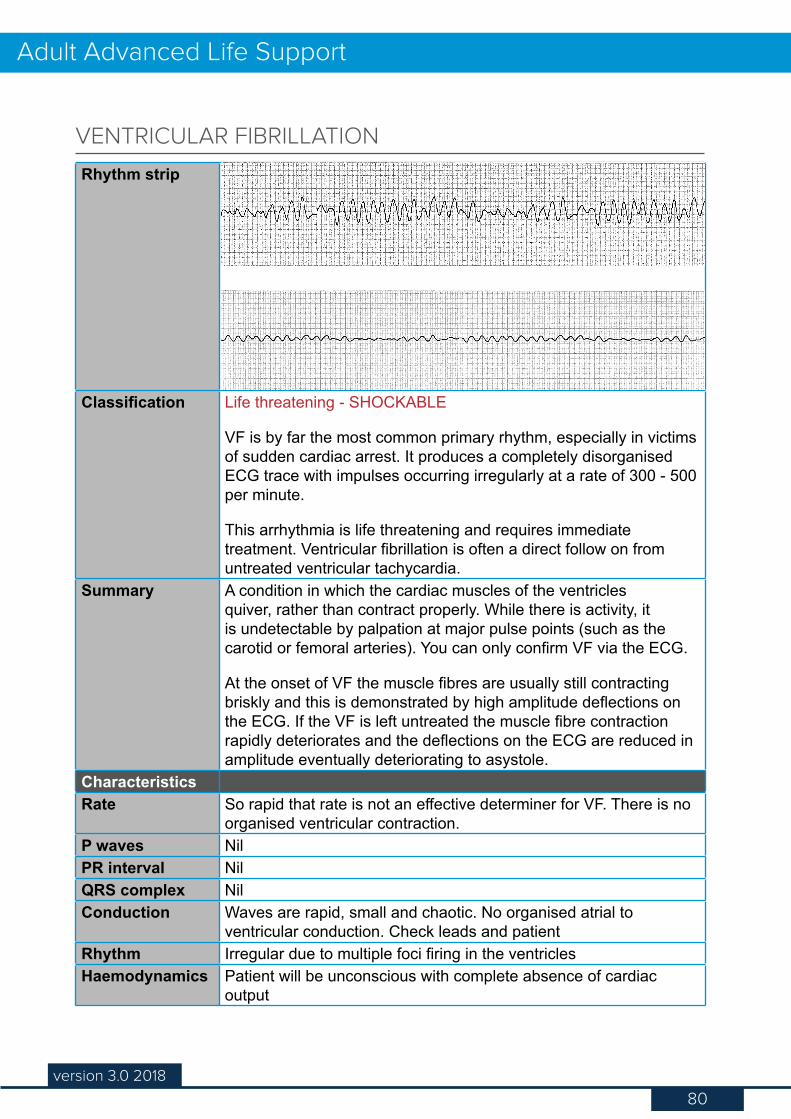
80version 3.0 2018
Adult Advanced Life Support
Rhythm strip
Classification Life threatening - SHOCKABLE
VF is by far the most common primary rhythm, especially in victims of sudden cardiac arrest. It produces a completely disorganised ECG trace with impulses occurring irregularly at a rate of 300 - 500 per minute.
This arrhythmia is life threatening and requires immediate treatment. Ventricular fibrillation is often a direct follow on from untreated ventricular tachycardia.
Summary A condition in which the cardiac muscles of the ventricles quiver, rather than contract properly. While there is activity, it is undetectable by palpation at major pulse points (such as the carotid or femoral arteries). You can only confirm VF via the ECG.
At the onset of VF the muscle fibres are usually still contracting briskly and this is demonstrated by high amplitude deflections on the ECG. If the VF is left untreated the muscle fibre contraction rapidly deteriorates and the deflections on the ECG are reduced in amplitude eventually deteriorating to asystole.
CharacteristicsRate So rapid that rate is not an effective determiner for VF. There is no
organised ventricular contraction.P waves NilPR interval NilQRS complex NilConduction Waves are rapid, small and chaotic. No organised atrial to
ventricular conduction. Check leads and patientRhythm Irregular due to multiple foci firing in the ventriclesHaemodynamics Patient will be unconscious with complete absence of cardiac
output
VENTRICULAR FIBRILLATION
81version 3.0 2018
Adult Advanced Life Support
ResponseNursing Ventricular Fibrillation always causes pulselessness and
unconsciousness. Commence CPR, administer O2 and prepare for defibrillation.
Treatment Defibrillation with 200J biphasic shock. CPRRecommended drugs
Adrenaline 1mg IV after the 2nd shock, repeated every 2nd cycle. Amiodarone 300mg IV after the 3rd shock and a second dose of 150mg may be given after the 5th shock.
Possible causes • Myocardial Infarction or Ischaemia• Acid base imbalances• Electric shock• Severe hypothermia• Electrolyte imbalances
Management The major determinant of survival of patients with VF or VT is the speed of effective defibrillation.
When a defibrillator is not readily available, the major determinant is adequate myocardial reperfusion and coronary perfusion pressure (determined by the CPR technique and peripheral vasoconstriction). To date, in the absence of or while awaiting a defibrillator, nothing has been shown to be more effective than good compressions.
Mortality Chance of successful defibrillation decreases with time. Amplitude and waveform of VF deteriorate as high energy phosphate stores in the myocardium decrease. This rate of decease of myocardial energy reserves can be slowed, but not halted, by the provision of effective CPR.
A defibrillatory shock should be attempted every 2 minutes until VF no longer exists. Reversible causes such as hypoxia, hyperkalaemia/hypokalaemia, hypothermia, acid/base disorders must be identified and treated.
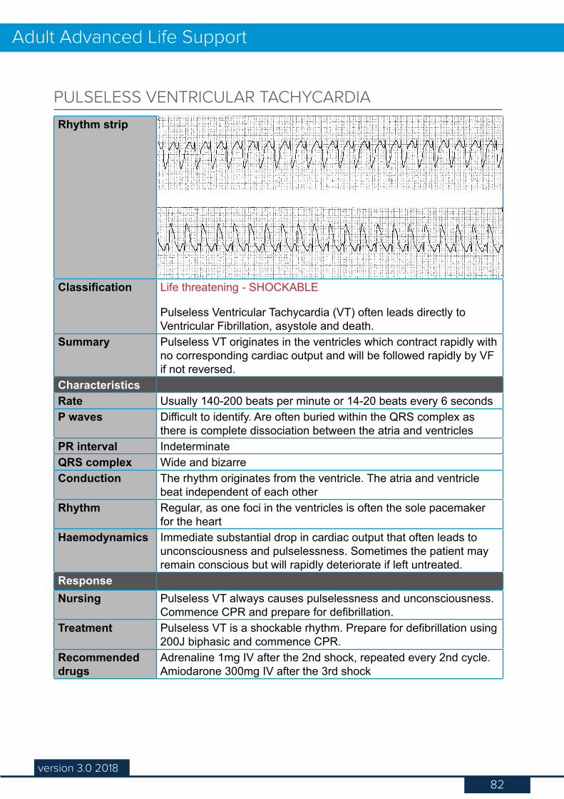
82version 3.0 2018
Adult Advanced Life Support
Rhythm strip
Classification Life threatening - SHOCKABLE
Pulseless Ventricular Tachycardia (VT) often leads directly to Ventricular Fibrillation, asystole and death.
Summary Pulseless VT originates in the ventricles which contract rapidly with no corresponding cardiac output and will be followed rapidly by VF if not reversed.
CharacteristicsRate Usually 140-200 beats per minute or 14-20 beats every 6 secondsP waves Difficult to identify. Are often buried within the QRS complex as
there is complete dissociation between the atria and ventriclesPR interval IndeterminateQRS complex Wide and bizarreConduction The rhythm originates from the ventricle. The atria and ventricle
beat independent of each otherRhythm Regular, as one foci in the ventricles is often the sole pacemaker
for the heartHaemodynamics Immediate substantial drop in cardiac output that often leads to
unconsciousness and pulselessness. Sometimes the patient may remain conscious but will rapidly deteriorate if left untreated.
ResponseNursing Pulseless VT always causes pulselessness and unconsciousness.
Commence CPR and prepare for defibrillation.Treatment Pulseless VT is a shockable rhythm. Prepare for defibrillation using
200J biphasic and commence CPR.Recommended drugs
Adrenaline 1mg IV after the 2nd shock, repeated every 2nd cycle. Amiodarone 300mg IV after the 3rd shock
PULSELESS VENTRICULAR TACHYCARDIA
83version 3.0 2018
Adult Advanced Life Support
Possible causes • Myocardial Infarction or Ischaemia• Cardiomyopathy• Electrolyte imbalances (Hypokalaemia)• Drug toxicity from digoxin
Management The major determinant of survival of patients with Pulseless VT is the speed of effective defibrillation.
When a defibrillator is not readily available, the major determinant is adequate myocardial reperfusion and coronary perfusion pressure (determined by the CPR technique and peripheral vasoconstriction). To date, in the absence of or while awaiting a defibrillator, nothing has been shown to be more effective than good compressions.
Mortality Chance of successful defibrillation decreases with time. Amplitude and waveform of VF deteriorate as high energy phosphate stores in the myocardium decrease. This rate of decrease of myocardial energy reserves can be slowed, but not halted, by the provision of effective CPR.
A defibrillatory shock should be attempted every 2 minutes until VF/VT no longer exists. Reversible causes such as hypoxia, hyperkalaemia/hypokalaemia, hypothermia, acid/base disorders must be identified and treated.
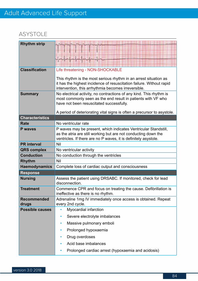
84version 3.0 2018
Adult Advanced Life Support
Rhythm strip
Classification Life threatening - NON-SHOCKABLE
This rhythm is the most serious rhythm in an arrest situation as it has the highest incidence of resuscitation failure. Without rapid intervention, this arrhythmia becomes irreversible.
Summary No electrical activity, no contractions of any kind. This rhythm is most commonly seen as the end result in patients with VF who have not been resuscitated successfully.
A period of deteriorating vital signs is often a precursor to asystole. CharacteristicsRate No ventricular rateP waves P waves may be present, which indicates Ventricular Standstill,
as the atria are still working but are not conducting down the ventricles. If there are no P waves, it is definitely asystole.
PR interval NilQRS complex No ventricular activityConduction No conduction through the ventriclesRhythm NilHaemodynamics Complete loss of cardiac output and consciousnessResponseNursing Assess the patient using DRSABC. If monitored, check for lead
disconnection.Treatment Commence CPR and focus on treating the cause. Defibrillation is
ineffective as there is no rhythm.Recommended drugs
Adrenaline 1mg IV immediately once access is obtained. Repeat every 2nd cycle.
Possible causes • Myocardial infarction• Severe electrolyte imbalances• Massive pulmonary emboli• Prolonged hypoxaemia• Drug overdoses• Acid base imbalances • Prolonged cardiac arrest (hypoxaemia and acidosis)
ASYSTOLE
85version 3.0 2018
Adult Advanced Life Support
Management An important consideration is the possibility of mistaken diagnosis. The presence of fine ventricular fibrillation may not be recognised for a number of reasons including equipment failure, excessive artefact, lead disconnection or an incorrect gain setting.
Make every effort to interpret or improve the quality of the recording of the rhythm. Be familiar with your equipment and how it represents a lead disconnection, for example, does the trace become a broken line?
If there is any uncertainty as to the rhythm, recommence CPR for 2 minutes to provide further effective compressions, then reassess. Compressions may coarsen a very fine VF line, making it more recognisable.
Mortality Recovery of patients who have primary cardiac disease that leads to asystole is very unlikely to occur after 15 minutes of unsuccessful CPR.
Important exceptions are cases of hypothermia, near drowning, or poisoning, all of which should be remedied or excluded before resuscitation attempts are abandoned.
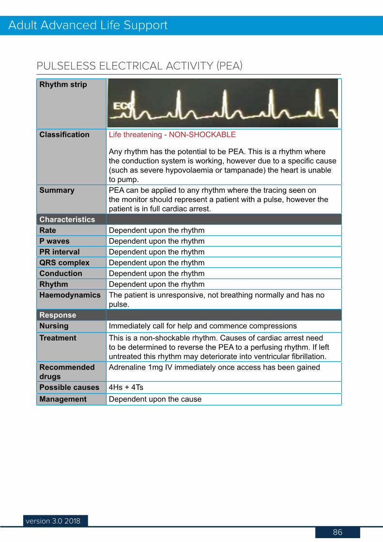
86version 3.0 2018
Adult Advanced Life Support
Rhythm strip
Classification Life threatening - NON-SHOCKABLE
Any rhythm has the potential to be PEA. This is a rhythm where the conduction system is working, however due to a specific cause (such as severe hypovolaemia or tampanade) the heart is unable to pump.
Summary PEA can be applied to any rhythm where the tracing seen on the monitor should represent a patient with a pulse, however the patient is in full cardiac arrest.
CharacteristicsRate Dependent upon the rhythmP waves Dependent upon the rhythmPR interval Dependent upon the rhythmQRS complex Dependent upon the rhythmConduction Dependent upon the rhythmRhythm Dependent upon the rhythmHaemodynamics The patient is unresponsive, not breathing normally and has no
pulse.ResponseNursing Immediately call for help and commence compressionsTreatment This is a non-shockable rhythm. Causes of cardiac arrest need
to be determined to reverse the PEA to a perfusing rhythm. If left untreated this rhythm may deteriorate into ventricular fibrillation.
Recommended drugs
Adrenaline 1mg IV immediately once access has been gained
Possible causes 4Hs + 4TsManagement Dependent upon the cause
PULSELESS ELECTRICAL ACTIVITY (PEA)
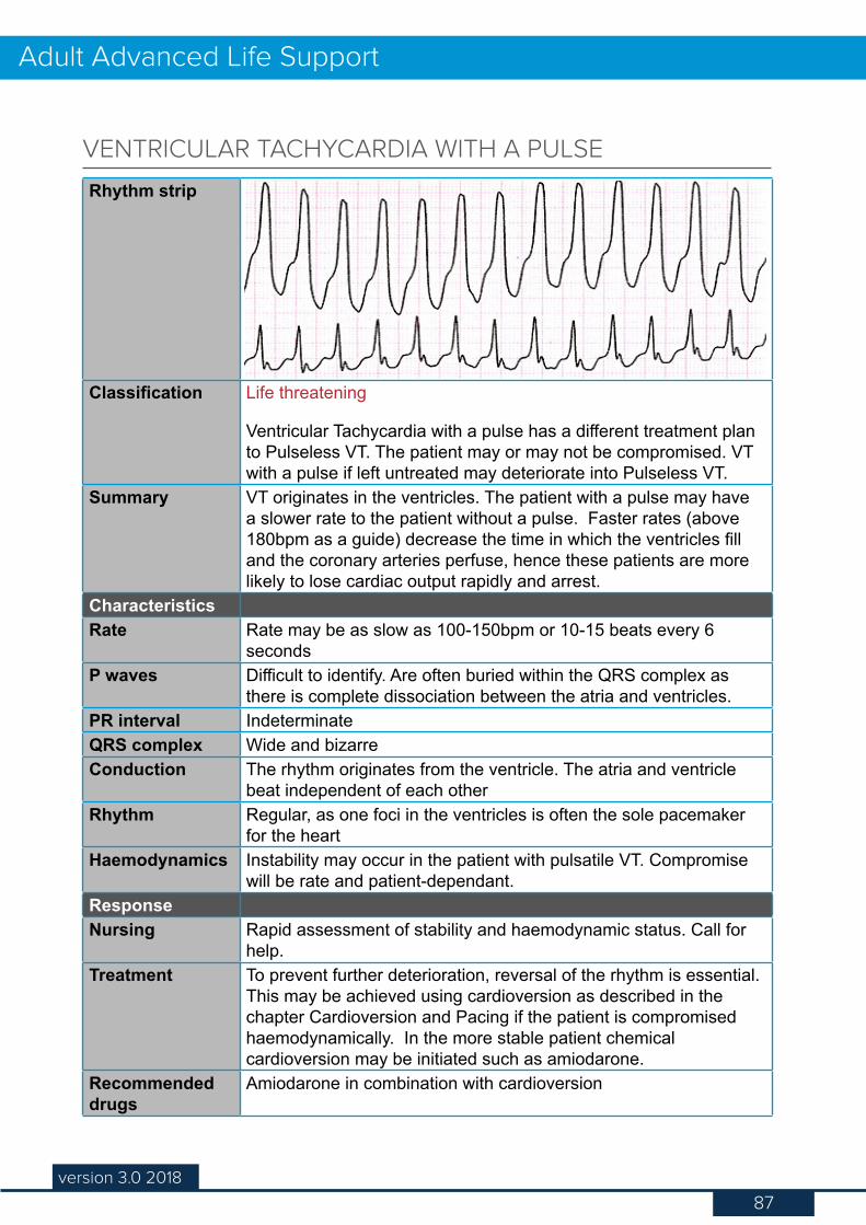
87version 3.0 2018
Adult Advanced Life Support
Rhythm strip
Classification Life threatening
Ventricular Tachycardia with a pulse has a different treatment plan to Pulseless VT. The patient may or may not be compromised. VT with a pulse if left untreated may deteriorate into Pulseless VT.
Summary VT originates in the ventricles. The patient with a pulse may have a slower rate to the patient without a pulse. Faster rates (above 180bpm as a guide) decrease the time in which the ventricles fill and the coronary arteries perfuse, hence these patients are more likely to lose cardiac output rapidly and arrest.
CharacteristicsRate Rate may be as slow as 100-150bpm or 10-15 beats every 6
secondsP waves Difficult to identify. Are often buried within the QRS complex as
there is complete dissociation between the atria and ventricles.PR interval IndeterminateQRS complex Wide and bizarreConduction The rhythm originates from the ventricle. The atria and ventricle
beat independent of each otherRhythm Regular, as one foci in the ventricles is often the sole pacemaker
for the heartHaemodynamics Instability may occur in the patient with pulsatile VT. Compromise
will be rate and patient-dependant.ResponseNursing Rapid assessment of stability and haemodynamic status. Call for
help.Treatment To prevent further deterioration, reversal of the rhythm is essential.
This may be achieved using cardioversion as described in the chapter Cardioversion and Pacing if the patient is compromised haemodynamically. In the more stable patient chemical cardioversion may be initiated such as amiodarone.
Recommended drugs
Amiodarone in combination with cardioversion
VENTRICULAR TACHYCARDIA WITH A PULSE
88version 3.0 2018
Adult Advanced Life Support
Possible causes • Myocardial Infarction or Ischaemia• Cardiomyopathy• Electrolyte imbalances (Hypokalaemia)• Drug toxicity from digoxin
Management Cardiac monitoring may occur for at least 24 hours to observe patient does not revert back into VT. Cardiology review and a treatment plan. Patient will require investigation into cause of VT and may require a defibrillator pacemaker implant is recurrent episodes occur.
Mortality Good outcomes if treated early.
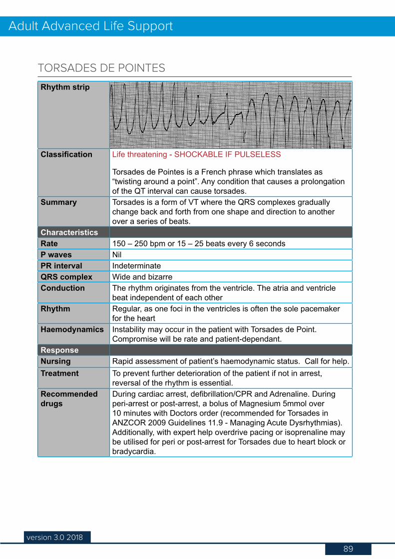
89version 3.0 2018
Adult Advanced Life Support
Rhythm strip
Classification Life threatening - SHOCKABLE IF PULSELESS
Torsades de Pointes is a French phrase which translates as “twisting around a point”. Any condition that causes a prolongation of the QT interval can cause torsades.
Summary Torsades is a form of VT where the QRS complexes gradually change back and forth from one shape and direction to another over a series of beats.
CharacteristicsRate 150 – 250 bpm or 15 – 25 beats every 6 secondsP waves NilPR interval IndeterminateQRS complex Wide and bizarreConduction The rhythm originates from the ventricle. The atria and ventricle
beat independent of each otherRhythm Regular, as one foci in the ventricles is often the sole pacemaker
for the heartHaemodynamics Instability may occur in the patient with Torsades de Point.
Compromise will be rate and patient-dependant.ResponseNursing Rapid assessment of patient’s haemodynamic status. Call for help.Treatment To prevent further deterioration of the patient if not in arrest,
reversal of the rhythm is essential. Recommended drugs
During cardiac arrest, defibrillation/CPR and Adrenaline. During peri-arrest or post-arrest, a bolus of Magnesium 5mmol over 10 minutes with Doctors order (recommended for Torsades in ANZCOR 2009 Guidelines 11.9 - Managing Acute Dysrhythmias). Additionally, with expert help overdrive pacing or isoprenaline may be utilised for peri or post-arrest for Torsades due to heart block or bradycardia.
TORSADES DE POINTES
90version 3.0 2018
Adult Advanced Life Support
Possible causes • Prolonged QT time• Many drugs may cause this arrhythmia• Electrolyte imbalance• Genetic predisposition
Management Call for help. If pulseless, immediate defibrillation, CPR and adrenaline to perfuse the brain. Lignocaine and other antiarrhythmics are avoided as they may prolong the QT interval and worsen the situation. Aim of therapy is to decrease the QT interval and so magnesium sulphate and isoprenaline can be used. Transcutaneous overdrive pacing may also be used.
Mortality Rapid reversal and correction of underlying cause improves outcomes
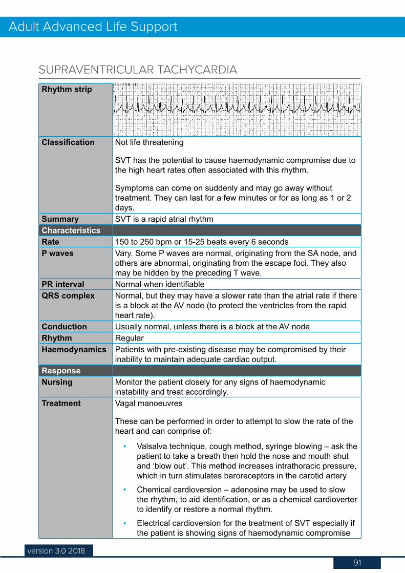
91version 3.0 2018
Adult Advanced Life Support
Rhythm strip
Classification Not life threatening
SVT has the potential to cause haemodynamic compromise due to the high heart rates often associated with this rhythm.
Symptoms can come on suddenly and may go away without treatment. They can last for a few minutes or for as long as 1 or 2 days.
Summary SVT is a rapid atrial rhythmCharacteristicsRate 150 to 250 bpm or 15-25 beats every 6 secondsP waves Vary. Some P waves are normal, originating from the SA node, and
others are abnormal, originating from the escape foci. They also may be hidden by the preceding T wave.
PR interval Normal when identifiableQRS complex Normal, but they may have a slower rate than the atrial rate if there
is a block at the AV node (to protect the ventricles from the rapid heart rate).
Conduction Usually normal, unless there is a block at the AV nodeRhythm RegularHaemodynamics Patients with pre-existing disease may be compromised by their
inability to maintain adequate cardiac output.ResponseNursing Monitor the patient closely for any signs of haemodynamic
instability and treat accordingly.Treatment Vagal manoeuvres
These can be performed in order to attempt to slow the rate of the heart and can comprise of:
• Valsalva technique, cough method, syringe blowing – ask the patient to take a breath then hold the nose and mouth shut and ‘blow out’. This method increases intrathoracic pressure, which in turn stimulates baroreceptors in the carotid artery
• Chemical cardioversion – adenosine may be used to slow the rhythm, to aid identification, or as a chemical cardioverter to identify or restore a normal rhythm.
• Electrical cardioversion for the treatment of SVT especially if the patient is showing signs of haemodynamic compromise
SUPRAVENTRICULAR TACHYCARDIA
92version 3.0 2018
Adult Advanced Life Support
Recommended drugs
Chemical cardioversion – adenosine may be used to slow the rhythm, to aid identification, or as a chemical cardioverter to identify or restore normal rhythm. Please refer to Drugs in Resuscitation for further information.
Possible causes Can be due to a rapid firing ectopic focus within the atria or a re-entry circuit that lets the impulse travel rapidly within the atria.
Management Investigations to find cause. Medications may be prescribedMortality Good outcomes with treatment
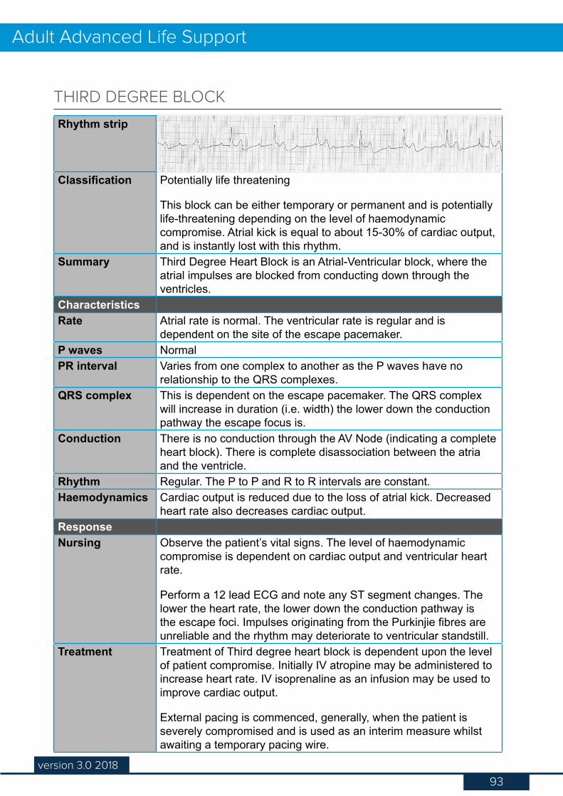
93version 3.0 2018
Adult Advanced Life Support
Rhythm strip
Classification Potentially life threatening
This block can be either temporary or permanent and is potentially life-threatening depending on the level of haemodynamic compromise. Atrial kick is equal to about 15-30% of cardiac output, and is instantly lost with this rhythm.
Summary Third Degree Heart Block is an Atrial-Ventricular block, where the atrial impulses are blocked from conducting down through the ventricles.
CharacteristicsRate Atrial rate is normal. The ventricular rate is regular and is
dependent on the site of the escape pacemaker.P waves NormalPR interval Varies from one complex to another as the P waves have no
relationship to the QRS complexes.QRS complex This is dependent on the escape pacemaker. The QRS complex
will increase in duration (i.e. width) the lower down the conduction pathway the escape focus is.
Conduction There is no conduction through the AV Node (indicating a complete heart block). There is complete disassociation between the atria and the ventricle.
Rhythm Regular. The P to P and R to R intervals are constant.Haemodynamics Cardiac output is reduced due to the loss of atrial kick. Decreased
heart rate also decreases cardiac output.ResponseNursing Observe the patient’s vital signs. The level of haemodynamic
compromise is dependent on cardiac output and ventricular heart rate.
Perform a 12 lead ECG and note any ST segment changes. The lower the heart rate, the lower down the conduction pathway is the escape foci. Impulses originating from the Purkinjie fibres are unreliable and the rhythm may deteriorate to ventricular standstill.
Treatment Treatment of Third degree heart block is dependent upon the level of patient compromise. Initially IV atropine may be administered to increase heart rate. IV isoprenaline as an infusion may be used to improve cardiac output.
External pacing is commenced, generally, when the patient is severely compromised and is used as an interim measure whilst awaiting a temporary pacing wire.
THIRD DEGREE BLOCK
94version 3.0 2018
Adult Advanced Life Support
Recommended drugs
Atropine 600mcg is given as a starting dose. Rate response to the first dose will determine subsequent delivery and amount. Isoprenaline may be also administered on medical instruction. See Appendix 2.
Possible causes • Coronary artery disease• Anterior or inferior wall myocardial infarction• Degenerative changes in the heart• Digoxin toxicity• Lignocaine or amiodarone infusion• Surgical injury
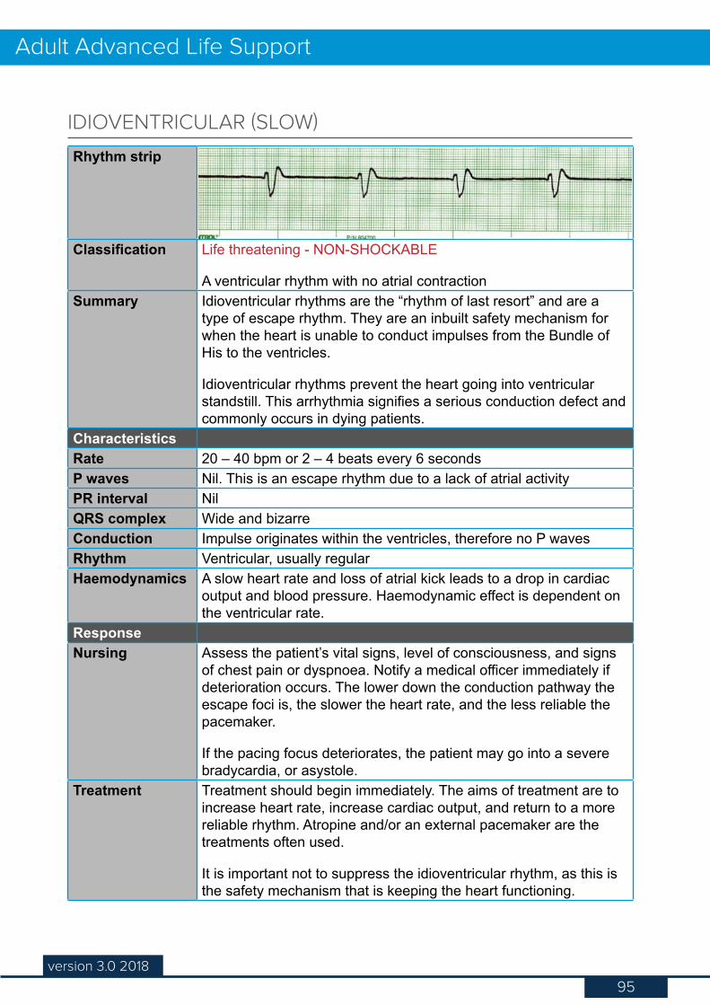
95version 3.0 2018
Adult Advanced Life Support
Rhythm strip
Classification Life threatening - NON-SHOCKABLE
A ventricular rhythm with no atrial contractionSummary Idioventricular rhythms are the “rhythm of last resort” and are a
type of escape rhythm. They are an inbuilt safety mechanism for when the heart is unable to conduct impulses from the Bundle of His to the ventricles.
Idioventricular rhythms prevent the heart going into ventricular standstill. This arrhythmia signifies a serious conduction defect and commonly occurs in dying patients.
CharacteristicsRate 20 – 40 bpm or 2 – 4 beats every 6 secondsP waves Nil. This is an escape rhythm due to a lack of atrial activityPR interval NilQRS complex Wide and bizarreConduction Impulse originates within the ventricles, therefore no P wavesRhythm Ventricular, usually regularHaemodynamics A slow heart rate and loss of atrial kick leads to a drop in cardiac
output and blood pressure. Haemodynamic effect is dependent on the ventricular rate.
ResponseNursing Assess the patient’s vital signs, level of consciousness, and signs
of chest pain or dyspnoea. Notify a medical officer immediately if deterioration occurs. The lower down the conduction pathway the escape foci is, the slower the heart rate, and the less reliable the pacemaker.
If the pacing focus deteriorates, the patient may go into a severe bradycardia, or asystole.
Treatment Treatment should begin immediately. The aims of treatment are to increase heart rate, increase cardiac output, and return to a more reliable rhythm. Atropine and/or an external pacemaker are the treatments often used.
It is important not to suppress the idioventricular rhythm, as this is the safety mechanism that is keeping the heart functioning.
IDIOVENTRICULAR (SLOW)
96version 3.0 2018
Adult Advanced Life Support
Recommended drugs
Isoprenaline may be also administered (see Appendix 2). Never treat with Lignocaine as this will suppress this rhythm, and ventricular standstill may occur.
Possible causes Idioventricular rhythms can accompany 3rd degree heart block, and be caused by:
• Myocardial Ischaemia• Metabolic imbalances• Pacemaker failure
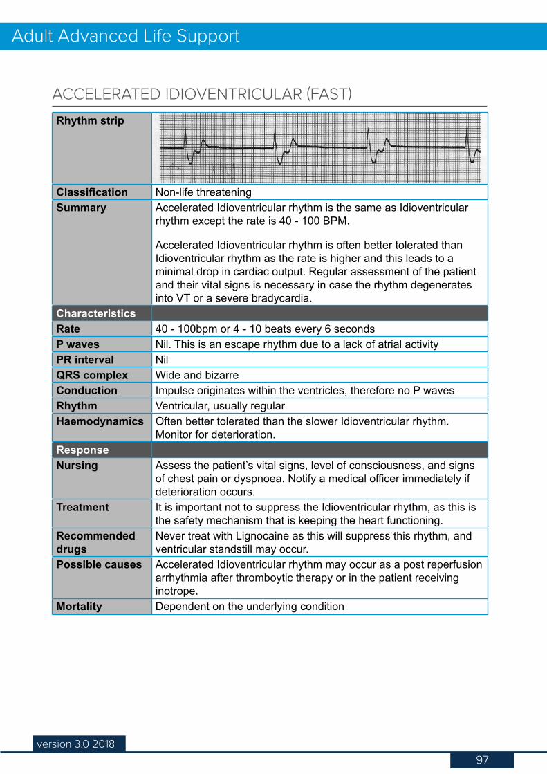
97version 3.0 2018
Adult Advanced Life Support
Rhythm strip
Classification Non-life threateningSummary Accelerated Idioventricular rhythm is the same as Idioventricular
rhythm except the rate is 40 - 100 BPM.
Accelerated Idioventricular rhythm is often better tolerated than Idioventricular rhythm as the rate is higher and this leads to a minimal drop in cardiac output. Regular assessment of the patient and their vital signs is necessary in case the rhythm degenerates into VT or a severe bradycardia.
CharacteristicsRate 40 - 100bpm or 4 - 10 beats every 6 secondsP waves Nil. This is an escape rhythm due to a lack of atrial activityPR interval NilQRS complex Wide and bizarreConduction Impulse originates within the ventricles, therefore no P wavesRhythm Ventricular, usually regularHaemodynamics Often better tolerated than the slower Idioventricular rhythm.
Monitor for deterioration.ResponseNursing Assess the patient’s vital signs, level of consciousness, and signs
of chest pain or dyspnoea. Notify a medical officer immediately if deterioration occurs.
Treatment It is important not to suppress the Idioventricular rhythm, as this is the safety mechanism that is keeping the heart functioning.
Recommended drugs
Never treat with Lignocaine as this will suppress this rhythm, and ventricular standstill may occur.
Possible causes Accelerated Idioventricular rhythm may occur as a post reperfusion arrhythmia after thromboytic therapy or in the patient receiving inotrope.
Mortality Dependent on the underlying condition
ACCELERATED IDIOVENTRICULAR (FAST)
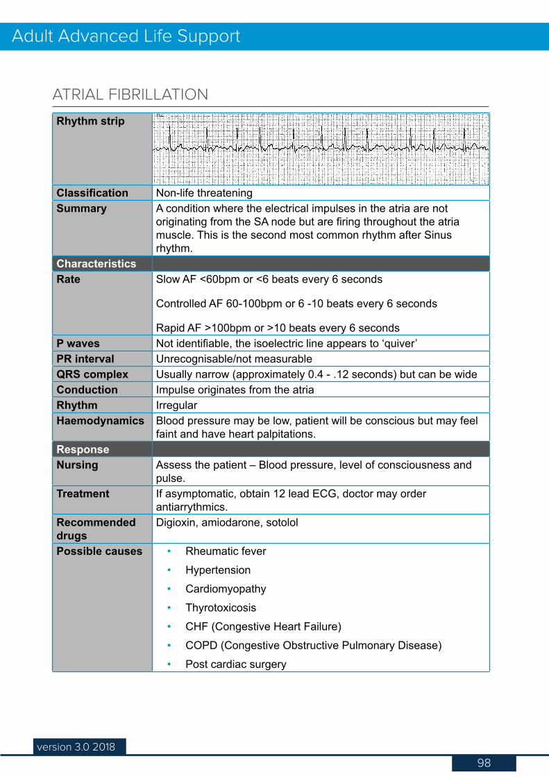
98version 3.0 2018
Adult Advanced Life Support
Rhythm strip
Classification Non-life threateningSummary A condition where the electrical impulses in the atria are not
originating from the SA node but are firing throughout the atria muscle. This is the second most common rhythm after Sinus rhythm.
CharacteristicsRate Slow AF <60bpm or <6 beats every 6 seconds
Controlled AF 60-100bpm or 6 -10 beats every 6 seconds
Rapid AF >100bpm or >10 beats every 6 secondsP waves Not identifiable, the isoelectric line appears to ‘quiver’PR interval Unrecognisable/not measurableQRS complex Usually narrow (approximately 0.4 - .12 seconds) but can be wideConduction Impulse originates from the atriaRhythm IrregularHaemodynamics Blood pressure may be low, patient will be conscious but may feel
faint and have heart palpitations.ResponseNursing Assess the patient – Blood pressure, level of consciousness and
pulse.Treatment If asymptomatic, obtain 12 lead ECG, doctor may order
antiarrythmics.Recommended drugs
Digioxin, amiodarone, sotolol
Possible causes • Rheumatic fever• Hypertension• Cardiomyopathy• Thyrotoxicosis• CHF (Congestive Heart Failure)• COPD (Congestive Obstructive Pulmonary Disease)• Post cardiac surgery
ATRIAL FIBRILLATION
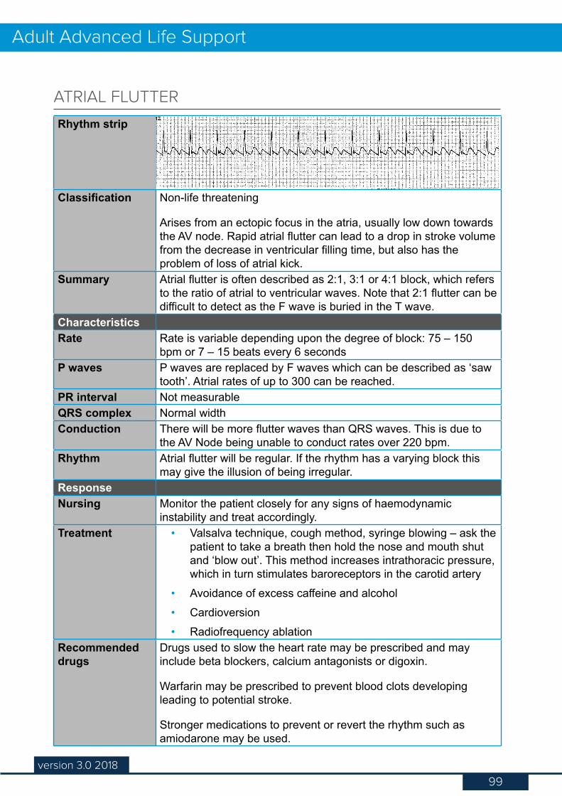
99version 3.0 2018
Adult Advanced Life Support
Rhythm strip
Classification Non-life threatening
Arises from an ectopic focus in the atria, usually low down towards the AV node. Rapid atrial flutter can lead to a drop in stroke volume from the decrease in ventricular filling time, but also has the problem of loss of atrial kick.
Summary Atrial flutter is often described as 2:1, 3:1 or 4:1 block, which refers to the ratio of atrial to ventricular waves. Note that 2:1 flutter can be difficult to detect as the F wave is buried in the T wave.
CharacteristicsRate Rate is variable depending upon the degree of block: 75 – 150
bpm or 7 – 15 beats every 6 secondsP waves P waves are replaced by F waves which can be described as ‘saw
tooth’. Atrial rates of up to 300 can be reached.PR interval Not measurableQRS complex Normal widthConduction There will be more flutter waves than QRS waves. This is due to
the AV Node being unable to conduct rates over 220 bpm.Rhythm Atrial flutter will be regular. If the rhythm has a varying block this
may give the illusion of being irregular.ResponseNursing Monitor the patient closely for any signs of haemodynamic
instability and treat accordingly.Treatment • Valsalva technique, cough method, syringe blowing – ask the
patient to take a breath then hold the nose and mouth shut and ‘blow out’. This method increases intrathoracic pressure, which in turn stimulates baroreceptors in the carotid artery
• Avoidance of excess caffeine and alcohol• Cardioversion• Radiofrequency ablation
Recommended drugs
Drugs used to slow the heart rate may be prescribed and may include beta blockers, calcium antagonists or digoxin.
Warfarin may be prescribed to prevent blood clots developing leading to potential stroke.
Stronger medications to prevent or revert the rhythm such as amiodarone may be used.
ATRIAL FLUTTER
100version 3.0 2018
Adult Advanced Life Support
Possible causes • Rheumatic heart disease• Overactive thyroid• Hypertension• Coronary heart disease• Cor pulmonale• Pericarditis• Hypoxia• Digoxin toxicity
Mortality Haemodynamic status will be monitored in addition to further investigation. An overactive thyroid can precipitate Atrial Flutter therefore a thyroid function test should ideally be taken for new onset Atrial Flutter.
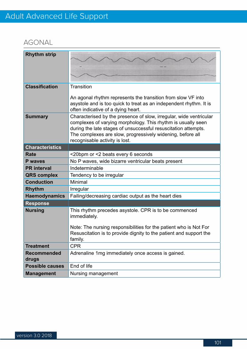
101version 3.0 2018
Adult Advanced Life Support
Rhythm strip
Classification Transition
An agonal rhythm represents the transition from slow VF into asystole and is too quick to treat as an independent rhythm. It is often indicative of a dying heart.
Summary Characterised by the presence of slow, irregular, wide ventricular complexes of varying morphology. This rhythm is usually seen during the late stages of unsuccessful resuscitation attempts. The complexes are slow, progressively widening, before all recognisable activity is lost.
CharacteristicsRate <20bpm or <2 beats every 6 secondsP waves No P waves, wide bizarre ventricular beats presentPR interval IndeterminableQRS complex Tendency to be irregularConduction MinimalRhythm IrregularHaemodynamics Failing/decreasing cardiac output as the heart diesResponseNursing This rhythm precedes asystole. CPR is to be commenced
immediately.
Note: The nursing responsibilities for the patient who is Not For Resuscitation is to provide dignity to the patient and support the family.
Treatment CPRRecommended drugs
Adrenaline 1mg immediately once access is gained.
Possible causes End of lifeManagement Nursing management
AGONAL
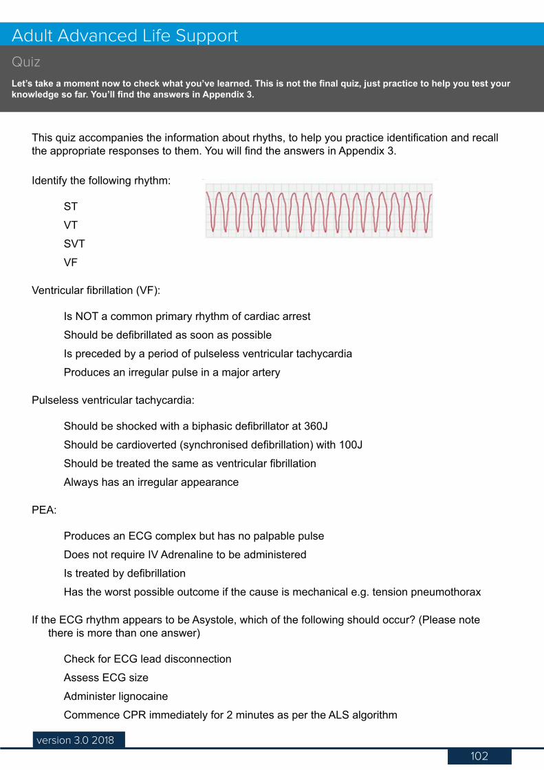
102version 2.0 2016
Advanced Life Support
102version 3.0 2018
Adult Advanced Life SupportQuizLet’s take a moment now to check what you’ve learned. This is not the final quiz, just practice to help you test your knowledge so far. You’ll find the answers in Appendix 3.
This quiz accompanies the information about rhyths, to help you practice identification and recall the appropriate responses to them. You will find the answers in Appendix 3.
Identify the following rhythm:
STVTSVTVF
Ventricular fibrillation (VF):
Is NOT a common primary rhythm of cardiac arrestShould be defibrillated as soon as possibleIs preceded by a period of pulseless ventricular tachycardiaProduces an irregular pulse in a major artery
Pulseless ventricular tachycardia:
Should be shocked with a biphasic defibrillator at 360JShould be cardioverted (synchronised defibrillation) with 100JShould be treated the same as ventricular fibrillationAlways has an irregular appearance
PEA:
Produces an ECG complex but has no palpable pulseDoes not require IV Adrenaline to be administeredIs treated by defibrillationHas the worst possible outcome if the cause is mechanical e.g. tension pneumothorax
If the ECG rhythm appears to be Asystole, which of the following should occur? (Please note there is more than one answer)
Check for ECG lead disconnectionAssess ECG sizeAdminister lignocaineCommence CPR immediately for 2 minutes as per the ALS algorithm
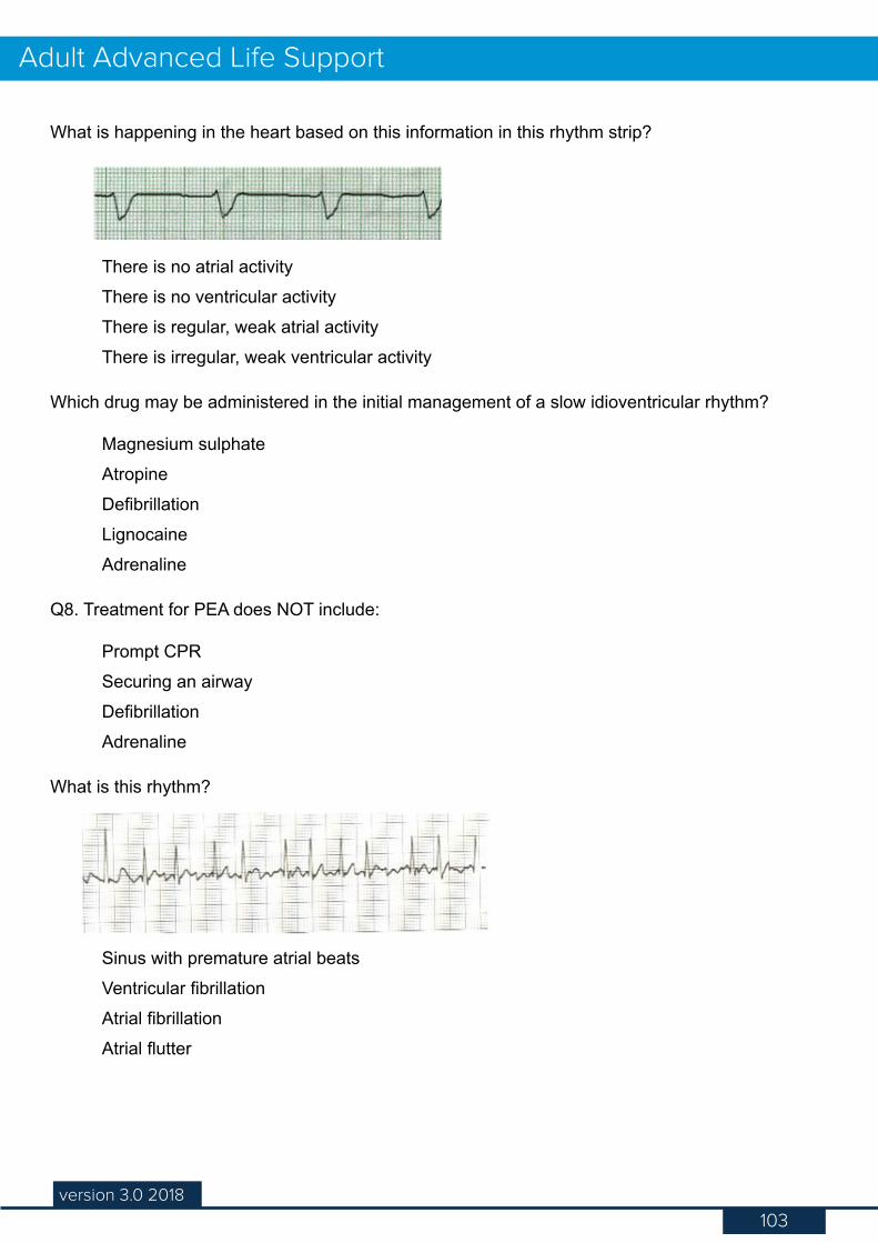
103version 3.0 2018
Adult Advanced Life Support
What is happening in the heart based on this information in this rhythm strip?
There is no atrial activityThere is no ventricular activityThere is regular, weak atrial activityThere is irregular, weak ventricular activity
Which drug may be administered in the initial management of a slow idioventricular rhythm?
Magnesium sulphateAtropineDefibrillationLignocaineAdrenaline
Q8. Treatment for PEA does NOT include:
Prompt CPRSecuring an airwayDefibrillationAdrenaline
What is this rhythm?
Sinus with premature atrial beatsVentricular fibrillationAtrial fibrillationAtrial flutter

104version 3.0 2018
Adult Advanced Life Support
During an emergency defibrillation, the energy level selected for the monophasic/biphasic defibrillators are:
360/360 joules200/200 joules200/360 joules360/200 joules
105version 3.0 2018
Adult Advanced Life Support
This resource lists the drugs used in resuscitation.
First line drugs ........................................................................................................................ 105
Adrenaline ......................................................................................................................... 105
Amiodarone ....................................................................................................................... 106
Second line and/or peri-arrest drugs.................................................................................... 107
Adenosine ......................................................................................................................... 107
Atropine ............................................................................................................................. 108
Calcium ............................................................................................................................. 109
Isoprenaline .......................................................................................................................110
Lignocaine ..........................................................................................................................111
Magnesium ........................................................................................................................112
Potassium ..........................................................................................................................113
Sodium Bicarbonate ...........................................................................................................114
Thrombolytics .....................................................................................................................115
Appendix 2 - Resuscitation Drugs
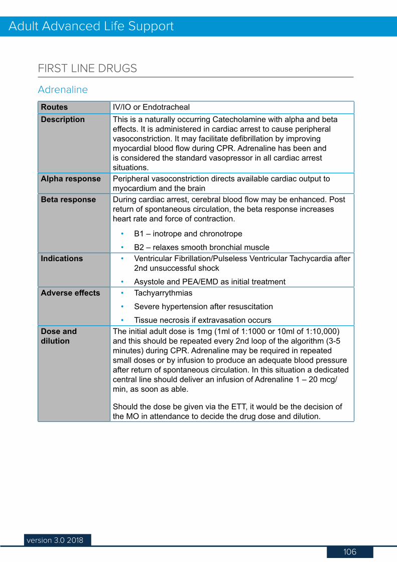
106version 3.0 2018
Adult Advanced Life Support
Routes IV/IO or EndotrachealDescription This is a naturally occurring Catecholamine with alpha and beta
effects. It is administered in cardiac arrest to cause peripheral vasoconstriction. It may facilitate defibrillation by improving myocardial blood flow during CPR. Adrenaline has been and is considered the standard vasopressor in all cardiac arrest situations.
Alpha response Peripheral vasoconstriction directs available cardiac output to myocardium and the brain
Beta response During cardiac arrest, cerebral blood flow may be enhanced. Post return of spontaneous circulation, the beta response increases heart rate and force of contraction.
• B1 – inotrope and chronotrope• B2 – relaxes smooth bronchial muscle
Indications • Ventricular Fibrillation/Pulseless Ventricular Tachycardia after 2nd unsuccessful shock
• Asystole and PEA/EMD as initial treatmentAdverse effects • Tachyarrythmias
• Severe hypertension after resuscitation• Tissue necrosis if extravasation occurs
Dose and dilution
The initial adult dose is 1mg (1ml of 1:1000 or 10ml of 1:10,000) and this should be repeated every 2nd loop of the algorithm (3-5 minutes) during CPR. Adrenaline may be required in repeated small doses or by infusion to produce an adequate blood pressure after return of spontaneous circulation. In this situation a dedicated central line should deliver an infusion of Adrenaline 1 – 20 mcg/min, as soon as able.
Should the dose be given via the ETT, it would be the decision of the MO in attendance to decide the drug dose and dilution.
FIRST LINE DRUGS
Adrenaline
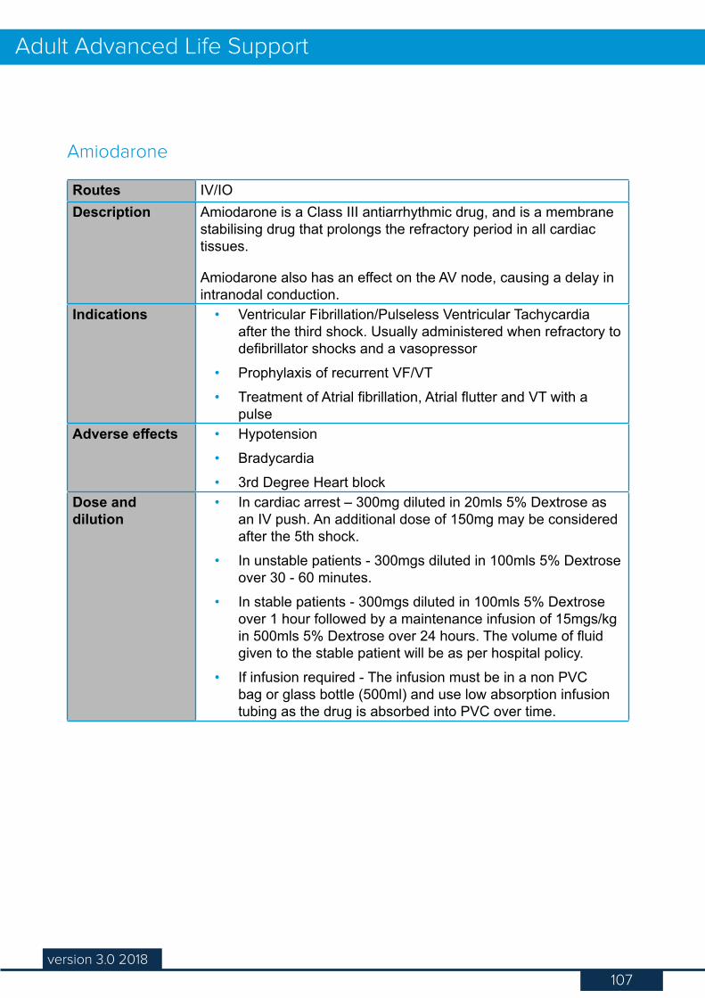
107version 3.0 2018
Adult Advanced Life Support
Routes IV/IODescription Amiodarone is a Class III antiarrhythmic drug, and is a membrane
stabilising drug that prolongs the refractory period in all cardiac tissues.
Amiodarone also has an effect on the AV node, causing a delay in intranodal conduction.
Indications • Ventricular Fibrillation/Pulseless Ventricular Tachycardia after the third shock. Usually administered when refractory to defibrillator shocks and a vasopressor
• Prophylaxis of recurrent VF/VT• Treatment of Atrial fibrillation, Atrial flutter and VT with a
pulseAdverse effects • Hypotension
• Bradycardia• 3rd Degree Heart block
Dose and dilution
• In cardiac arrest – 300mg diluted in 20mls 5% Dextrose as an IV push. An additional dose of 150mg may be considered after the 5th shock.
• In unstable patients - 300mgs diluted in 100mls 5% Dextrose over 30 - 60 minutes.
• In stable patients - 300mgs diluted in 100mls 5% Dextrose over 1 hour followed by a maintenance infusion of 15mgs/kg in 500mls 5% Dextrose over 24 hours. The volume of fluid given to the stable patient will be as per hospital policy.
• If infusion required - The infusion must be in a non PVC bag or glass bottle (500ml) and use low absorption infusion tubing as the drug is absorbed into PVC over time.
Amiodarone
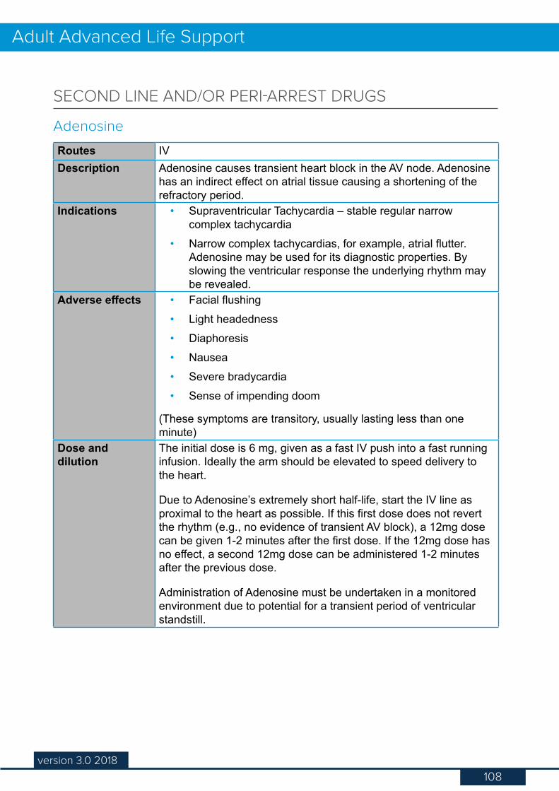
108version 3.0 2018
Adult Advanced Life Support
Routes IVDescription Adenosine causes transient heart block in the AV node. Adenosine
has an indirect effect on atrial tissue causing a shortening of the refractory period.
Indications • Supraventricular Tachycardia – stable regular narrow complex tachycardia
• Narrow complex tachycardias, for example, atrial flutter. Adenosine may be used for its diagnostic properties. By slowing the ventricular response the underlying rhythm may be revealed.
Adverse effects • Facial flushing• Light headedness• Diaphoresis• Nausea• Severe bradycardia• Sense of impending doom
(These symptoms are transitory, usually lasting less than one minute)
Dose and dilution
The initial dose is 6 mg, given as a fast IV push into a fast running infusion. Ideally the arm should be elevated to speed delivery to the heart.
Due to Adenosine’s extremely short half-life, start the IV line as proximal to the heart as possible. If this first dose does not revert the rhythm (e.g., no evidence of transient AV block), a 12mg dose can be given 1-2 minutes after the first dose. If the 12mg dose has no effect, a second 12mg dose can be administered 1-2 minutes after the previous dose.
Administration of Adenosine must be undertaken in a monitored environment due to potential for a transient period of ventricular standstill.
SECOND LINE AND/OR PERI-ARREST DRUGS
Adenosine
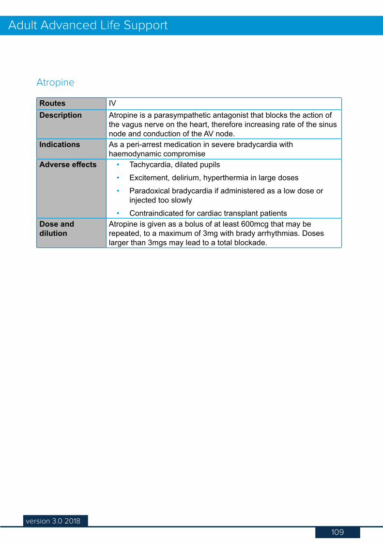
109version 3.0 2018
Adult Advanced Life Support
Routes IVDescription Atropine is a parasympathetic antagonist that blocks the action of
the vagus nerve on the heart, therefore increasing rate of the sinus node and conduction of the AV node.
Indications As a peri-arrest medication in severe bradycardia with haemodynamic compromise
Adverse effects • Tachycardia, dilated pupils• Excitement, delirium, hyperthermia in large doses• Paradoxical bradycardia if administered as a low dose or
injected too slowly• Contraindicated for cardiac transplant patients
Dose and dilution
Atropine is given as a bolus of at least 600mcg that may be repeated, to a maximum of 3mg with brady arrhythmias. Doses larger than 3mgs may lead to a total blockade.
Atropine
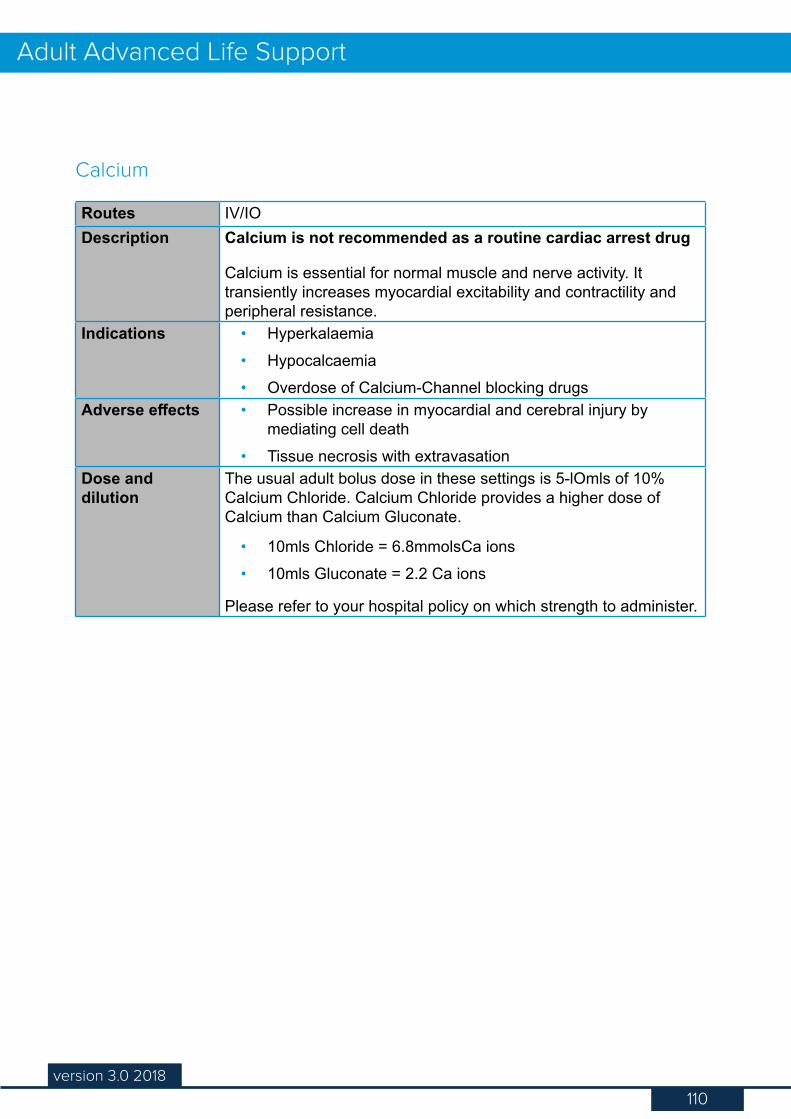
110version 3.0 2018
Adult Advanced Life Support
Routes IV/IODescription Calcium is not recommended as a routine cardiac arrest drug
Calcium is essential for normal muscle and nerve activity. It transiently increases myocardial excitability and contractility and peripheral resistance.
Indications • Hyperkalaemia• Hypocalcaemia• Overdose of Calcium-Channel blocking drugs
Adverse effects • Possible increase in myocardial and cerebral injury by mediating cell death
• Tissue necrosis with extravasationDose and dilution
The usual adult bolus dose in these settings is 5-lOmls of 10% Calcium Chloride. Calcium Chloride provides a higher dose of Calcium than Calcium Gluconate.
• 10mls Chloride = 6.8mmolsCa ions• 10mls Gluconate = 2.2 Ca ions
Please refer to your hospital policy on which strength to administer.
Calcium
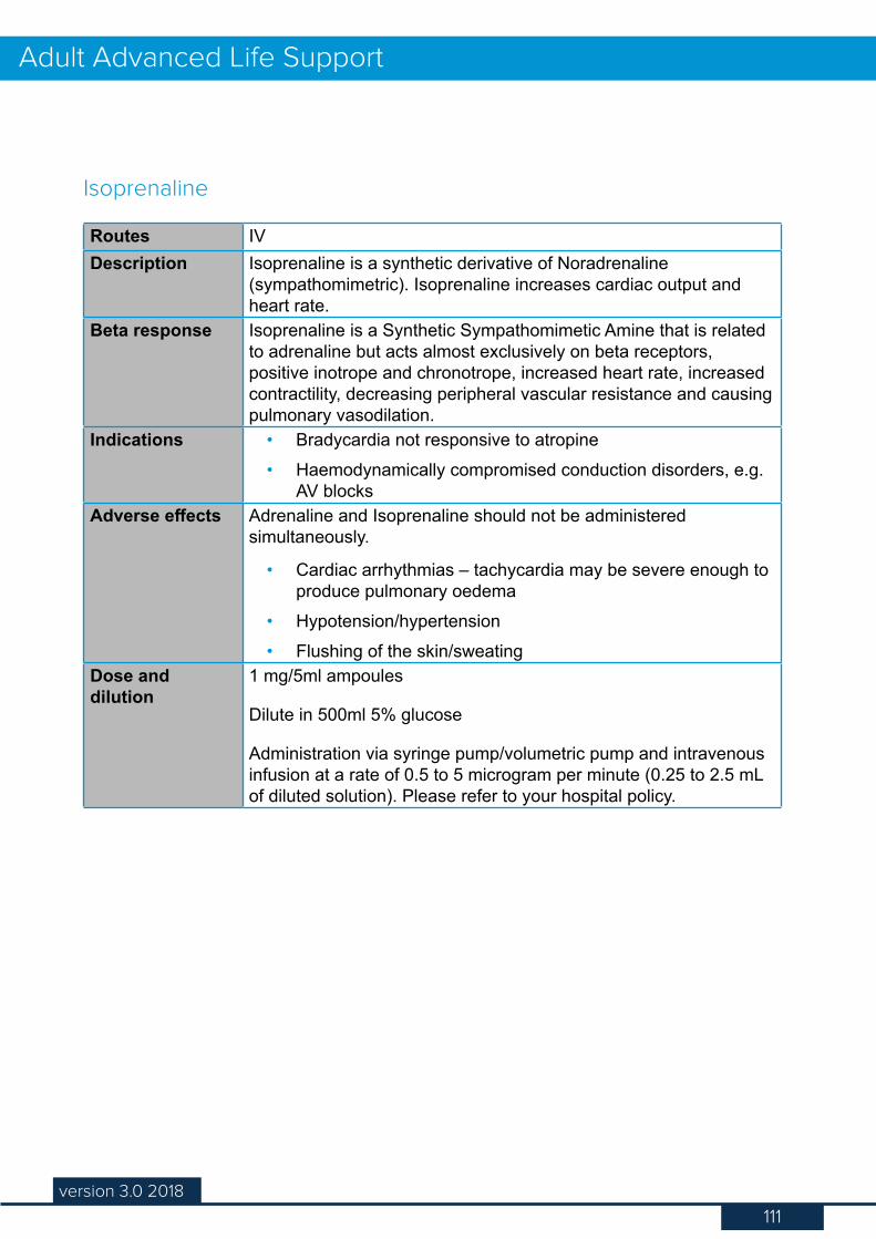
111version 3.0 2018
Adult Advanced Life Support
Routes IVDescription Isoprenaline is a synthetic derivative of Noradrenaline
(sympathomimetric). Isoprenaline increases cardiac output and heart rate.
Beta response Isoprenaline is a Synthetic Sympathomimetic Amine that is related to adrenaline but acts almost exclusively on beta receptors, positive inotrope and chronotrope, increased heart rate, increased contractility, decreasing peripheral vascular resistance and causing pulmonary vasodilation.
Indications • Bradycardia not responsive to atropine• Haemodynamically compromised conduction disorders, e.g.
AV blocksAdverse effects Adrenaline and Isoprenaline should not be administered
simultaneously.
• Cardiac arrhythmias – tachycardia may be severe enough to produce pulmonary oedema
• Hypotension/hypertension• Flushing of the skin/sweating
Dose and dilution
1 mg/5ml ampoules
Dilute in 500ml 5% glucose
Administration via syringe pump/volumetric pump and intravenous infusion at a rate of 0.5 to 5 microgram per minute (0.25 to 2.5 mL of diluted solution). Please refer to your hospital policy.
Isoprenaline
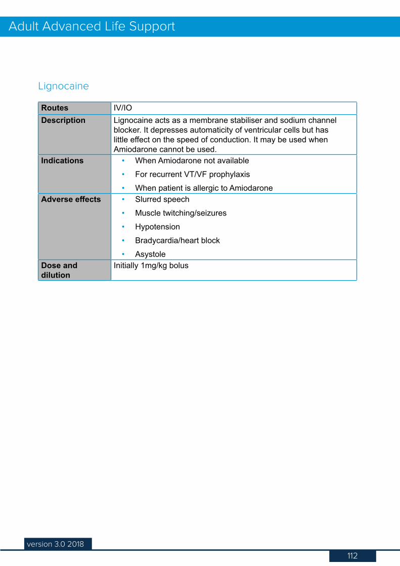
112version 3.0 2018
Adult Advanced Life Support
Routes IV/IODescription Lignocaine acts as a membrane stabiliser and sodium channel
blocker. It depresses automaticity of ventricular cells but has little effect on the speed of conduction. It may be used when Amiodarone cannot be used.
Indications • When Amiodarone not available• For recurrent VT/VF prophylaxis• When patient is allergic to Amiodarone
Adverse effects • Slurred speech• Muscle twitching/seizures• Hypotension• Bradycardia/heart block• Asystole
Dose and dilution
Initially 1mg/kg bolus
Lignocaine
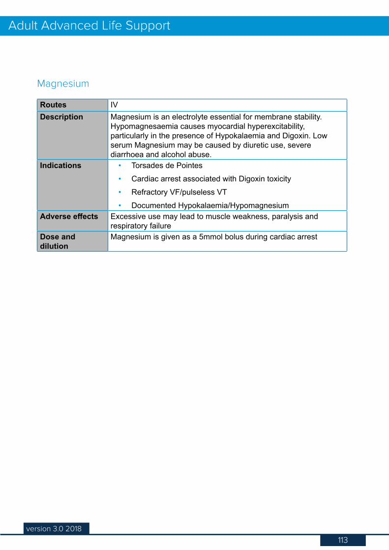
113version 3.0 2018
Adult Advanced Life Support
Routes IVDescription Magnesium is an electrolyte essential for membrane stability.
Hypomagnesaemia causes myocardial hyperexcitability, particularly in the presence of Hypokalaemia and Digoxin. Low serum Magnesium may be caused by diuretic use, severe diarrhoea and alcohol abuse.
Indications • Torsades de Pointes• Cardiac arrest associated with Digoxin toxicity• Refractory VF/pulseless VT• Documented Hypokalaemia/Hypomagnesium
Adverse effects Excessive use may lead to muscle weakness, paralysis and respiratory failure
Dose and dilution
Magnesium is given as a 5mmol bolus during cardiac arrest
Magnesium
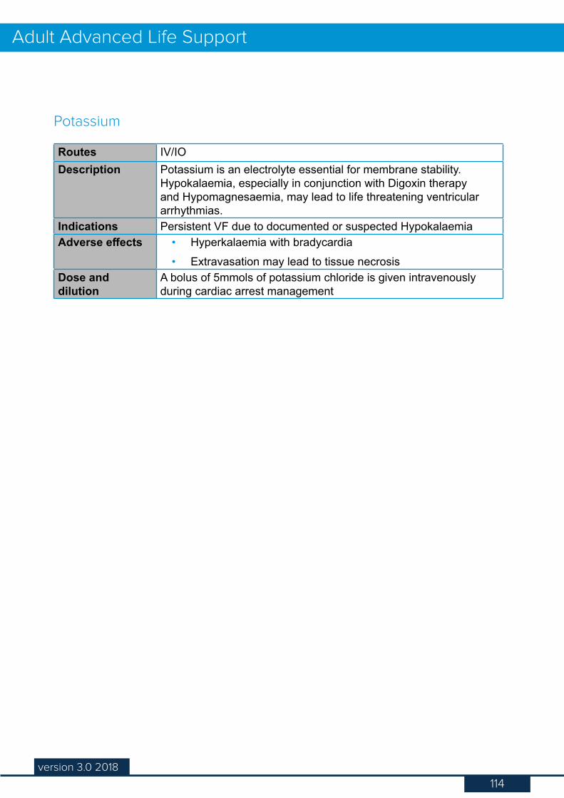
114version 3.0 2018
Adult Advanced Life Support
Routes IV/IODescription Potassium is an electrolyte essential for membrane stability.
Hypokalaemia, especially in conjunction with Digoxin therapy and Hypomagnesaemia, may lead to life threatening ventricular arrhythmias.
Indications Persistent VF due to documented or suspected HypokalaemiaAdverse effects • Hyperkalaemia with bradycardia
• Extravasation may lead to tissue necrosisDose and dilution
A bolus of 5mmols of potassium chloride is given intravenously during cardiac arrest management
Potassium

115version 3.0 2018
Adult Advanced Life Support
Routes IV/IODescription Routine Sodium Bicarbonate (NaHCO3) administration during an
arrest is not recommended. In most cardiac arrests early efficient CPR negates the need for NaHCO3.
Indications • Overdose tricyclics antidepressants• Hyperkalaemia• Documented metabolic acidosis• Protracted arrest – after arterial blood gas (ABG)
measurementAdverse effects • Metabolic Alkalosis, Hypokalaemia, Hypernatraemia
• Paradoxical intracellular acidosis due to carbon dioxide being liberated into the cells.
• When mixed with Adrenaline or Calcium the drugs are inactivated and or precipitate in the line. A dedicated line must be used during administration.
Dose and dilution
1mmol/kg over 2 – 3 mins. Then guided by ABG results.
Sodium Bicarbonate
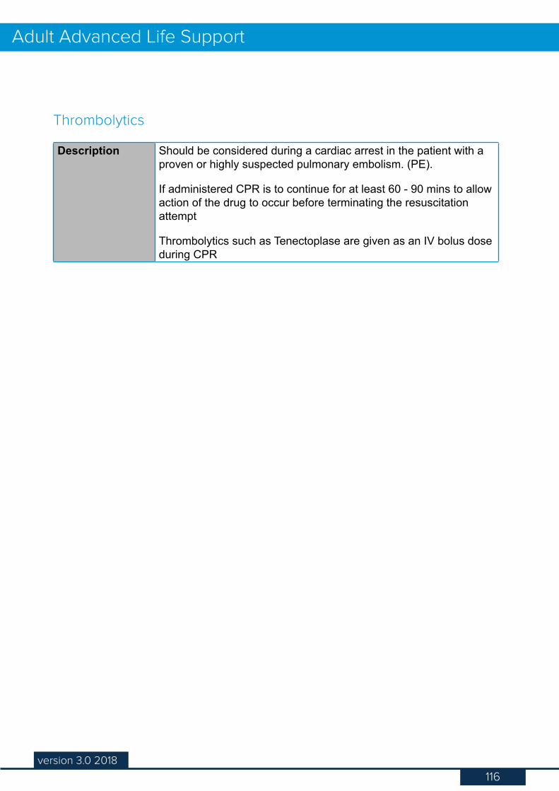
116version 3.0 2018
Adult Advanced Life Support
Description Should be considered during a cardiac arrest in the patient with a proven or highly suspected pulmonary embolism. (PE).
If administered CPR is to continue for at least 60 - 90 mins to allow action of the drug to occur before terminating the resuscitation attempt
Thrombolytics such as Tenectoplase are given as an IV bolus dose during CPR
Thrombolytics
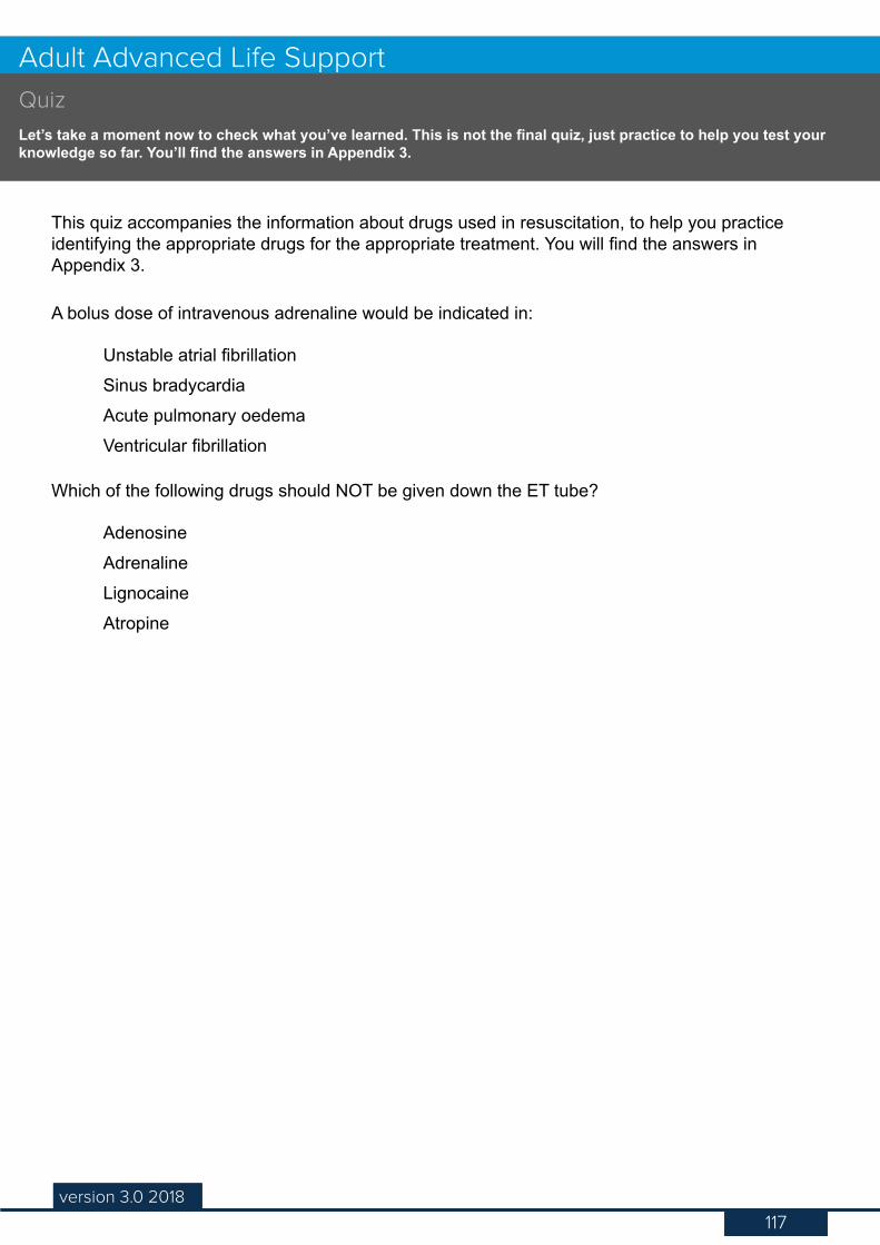
117version 2.0 2016
Advanced Life Support
117version 3.0 2018
Adult Advanced Life SupportQuizLet’s take a moment now to check what you’ve learned. This is not the final quiz, just practice to help you test your knowledge so far. You’ll find the answers in Appendix 3.
This quiz accompanies the information about drugs used in resuscitation, to help you practice identifying the appropriate drugs for the appropriate treatment. You will find the answers in Appendix 3.
A bolus dose of intravenous adrenaline would be indicated in:
Unstable atrial fibrillationSinus bradycardiaAcute pulmonary oedemaVentricular fibrillation
Which of the following drugs should NOT be given down the ET tube?
AdenosineAdrenalineLignocaineAtropine
118version 3.0 2018
Adult Advanced Life Support
Appendix 3 - Quiz Answers
Q1 What is the primary aim of the Chain of Survival?
b) To highlight the relationship between early detection and defibrillation and the rates of survival of cardiac arrests
Q2 Every minute the chances of survival after an arrest decrease by:
c) 7-10%Q3 The ethical considerations that need to be adhered to as an ALS trained nurse
in an emergency are:
d) Consent, NFR, Medical condition, Advanced Health DirectiveQ4 The legal requirements which have to be considered in a cardiac arrest situation
are:
c) Failure to start, documentation, cessation of CPR, Standard of care
Q1 The correct response to recognition of a non-shockable rhythm should be to:
c) Continue good quality CPR and reassess the rhythm after 2 minutesQ2 You are the ALS trained nurse responding to a call to resuscitate a patient on
the surgical ward. Ward staff found the patient collapsed, commenced CPR and attached the defibrillator. Your first priority is to:
d) Assess the effectiveness of the CPR being performed while planning for rhythm check/defibrillation
Q3 It is TRUE that failure to perform a visual sweep of the room and call “stand clear” (or similar) before pressing the Shock button constitutes a potential hazard to other staff members.
Q4 It is false that you should never charge the defibrillator pads on the patient’s chest wall as this may cause accidental discharge.
Quiz 1
Quiz 2
119version 3.0 2018
Adult Advanced Life Support
Q1 One of the conditions where it is recommended to deliver stacked shocks to a patient is:
a) Only when a monitored, witnessed patient develops a pulseless, shockable rhythm
Q2 It is true that time is a critical factor that affects defibrillation success.Q3 The patient’s chest wall size (b) is NOT a factor that influences transthoracic
impedance.
Q1 The size of the ET tube that you would expect to use for an average female during an arrest is:
b) 7.0 – 8.0Q2 The signs that an endotracheal intubation has been unsuccessful are:
c) Absence of audible breath sounds through auscultationQ3 The following statement regarding intubation during a cardiac arrest is false:
a) Endotracheal intubation can be performed by ALS trained nurses
Q1 Synchronised counter shock is delivered:
c) On the R wave of the ECGQ2 The following arrhythmia is an indication for the delivery of synchronised
cardioversion:
a) Atrial fibrillationQ3 It is True that to initiate pacing mode the patient must be connected to the
defibrillator ECG leads and pads.
Q1 The common signs of an anaphylaxis are:
c) Difficult/noisy breathing, hives/rash, wheezing, loss of consciousness
Quiz 3
Quiz 4
Quiz 5
Quiz 6
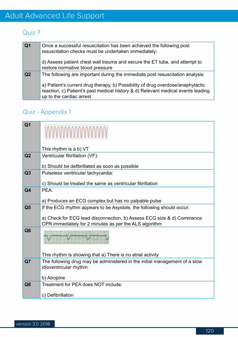
120version 3.0 2018
Adult Advanced Life Support
Q1 Once a successful resuscitation has been achieved the following post resuscitation checks must be undertaken immediately:
d) Assess patient chest wall trauma and secure the ET tube, and attempt to restore normative blood pressure
Q2 The following are important during the immediate post resuscitation analysis:
a) Patient’s current drug therapy, b) Possibility of drug overdose/anaphylactic reaction, c) Patient’s past medical history & d) Relevant medical events leading up to the cardiac arrest
Q1
This rhythm is a b) VTQ2 Ventricular fibrillation (VF):
b) Should be defibrillated as soon as possibleQ3 Pulseless ventricular tachycardia:
c) Should be treated the same as ventricular fibrillationQ4 PEA:
a) Produces an ECG complex but has no palpable pulseQ5 If the ECG rhythm appears to be Asystole, the following should occur:
a) Check for ECG lead disconnection, b) Assess ECG size & d) Commence CPR immediately for 2 minutes as per the ALS algorithm
Q6
This rhythm is showing that a) There is no atrial activityQ7 The following drug may be administered in the initial management of a slow
idioventricular rhythm:
b) AtropineQ8 Treatment for PEA does NOT include:
c) Defibrillation
Quiz 7
Quiz - Appendix 1
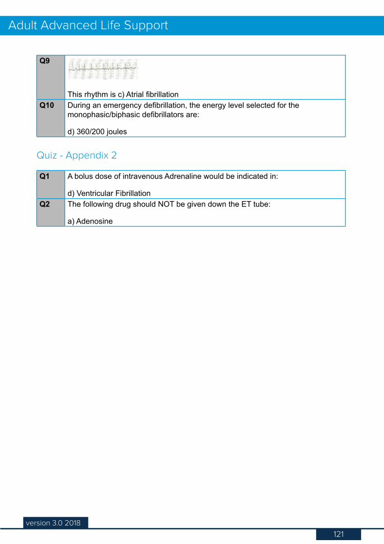
121version 3.0 2018
Adult Advanced Life Support
Q9
This rhythm is c) Atrial fibrillationQ10 During an emergency defibrillation, the energy level selected for the
monophasic/biphasic defibrillators are:
d) 360/200 joules
Q1 A bolus dose of intravenous Adrenaline would be indicated in:
d) Ventricular FibrillationQ2 The following drug should NOT be given down the ET tube:
a) Adenosine
Quiz - Appendix 2
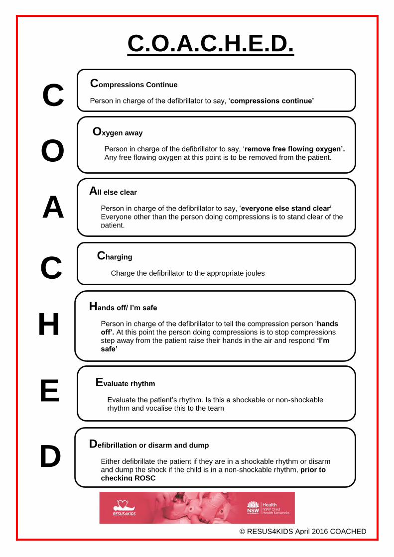
Compressions Continue
Person in charge of the defibrillator to say, ‘compressions continue’
Oxygen away
Person in charge of the defibrillator to say, ‘remove free flowing oxygen’. Any free flowing oxygen at this point is to be removed from the patient.
All else clear
Person in charge of the defibrillator to say, ‘everyone else stand clear’ Everyone other than the person doing compressions is to stand clear of the patient.
Charging
Charge the defibrillator to the appropriate joules
Defibrillation or disarm and dump
Either defibrillate the patient if they are in a shockable rhythm or disarm and dump the shock if the child is in a non-shockable rhythm, prior to checking ROSC
Hands off/ I’m safe
Person in charge of the defibrillator to tell the compression person ‘hands off’. At this point the person doing compressions is to stop compressions step away from the patient raise their hands in the air and respond ‘I’m safe’
Evaluate rhythm
Evaluate the patient’s rhythm. Is this a shockable or non-shockable rhythm and vocalise this to the team
C
E
A
C
H
O
D
C.O.A.C.H.E.D.
© RESUS4KIDS April 2016 COACHED
122version 3.0 2018
Adult Advanced Life Support
National Confidential Enquiry into Patient Outcome and Death (NCEPOD) 2012, CAP Summary. [ONLINE] Available at: http://www.ncepod.org.uk/2012report1/downloads/CAP_summary.pdf, [Accessed 19 October 2016].
3303.0 - Causes of Death, Australia, 2017. ABS cat. no. 3303.0 - September, 2018. [ONLINE] Available at: http://www.abs.gov.au/AUSSTATS/[email protected]/mf/3303.0/[Accessed 19 October 2018].
National Heart Foundation of Australia, Australian heard disease statistics 2015, [ONLINE] Available at: https://heartfoundation.org.au/images/uploads/publications/RES-115-Aust_heart_disease_statstics_2015_WEB.PDF [Accessed 20 October 2016].
The Australian Resuscitation Council Guidelines 2016, [ONLINE] Available at: https://resus.org.au/guidelines/ [Accessed 19 October 2016].
Yang, C,. Yen, Z., McGowan, J., Huiju, C., Chiang, W., Mancini, M., Soar, J., Lai, M. & Ma, M., A systematic review of retention of adult advanced life support knowledge and skills in healthcare providers, Resuscitation, vol. 83 (9), 2012, pp1055-1060 [ONLINE] Available via Athens @ Ramsay at: http://www.sciencedirect.com.proxy1.athensams.net/science/journal/03009572 [Accessed 19 February 2014]
Philips Healthcare Non-Invasive Transcutaneous Pacing – Application Note. 2013. Koninkliijke Philips Electronics N.V. [ONLINE] Available at: http://incenter.medical.philips.com/doclib/enc/fetch/2000/4504/577242/577243/577245/577817/577869/Non-Invasive_Transcutaneous_Pacing.pdf%3fnodeid%3d8615795%26vernum%3d-2 [Accessed 03 November 2016]
References