Embed Size (px)
Citation preview
Hindawi Publishing CorporationCase Reports in MedicineVolume 2010, Article ID 682081, 4 pagesdoi:10.1155/2010/682081
Case Report
Adrenocortical Secreting Mass in a Patient withGardner’s Syndrome: A Case Report
Nabila Mejdoub Rekik, Sourour Ben Salah, Nozha Kallel, Mahdi Kamoun, Nadia Charfi,and Mohamed Abid
Department of Diabetes and Endocrinology, Hedi Chaker Hospital, Magida Boulila Avenue. Sfax 3029, Tunisia
Correspondence should be addressed to Nabila Mejdoub Rekik, [email protected]
Received 16 August 2010; Accepted 15 November 2010
Academic Editor: Christian C. Apfel
Copyright © 2010 Nabila Mejdoub Rekik et al. This is an open access article distributed under the Creative Commons AttributionLicense, which permits unrestricted use, distribution, and reproduction in any medium, provided the original work is properlycited.
Gardner’s syndrome (GS) is a dysplasia characterized by neoformations of the intestine, soft tissue, and osseous tissue. Endocrineneoplasms have occasionally been reported in association with GS. Adrenal masses in GS are rare, and few have displayed clinicalmanifestations. In the current paper, The authors report a 37-year-old male patient with GS including familial adenomatouspolyposis (FAP) and mandible osteoma who presented with an incidental adrenal mass. Computerized tomography adrenal scanidentified bilateral masses. Functional analyses showed a hormonal secretion pattern consistent with pre-Cushing’s syndrome.Other extraintestinal manifestations were hypertrophy of the pigmented layer of the retina and histiocytofibroma in the right leg.This paper describes a rare association of adrenocortical secreting mass in an old male patient with Gardner syndrome.
1. Introduction
Gardner’s syndrome (GS) is a variant of familial adenomato-sis polyposis (FAP), which affects one in 8300 individualsand one in 7500 births in the United States [1]. Thedisease is characterized by colonic polyps and extracolonicmanifestations. The polyps typically develop in adolescenceand undergo malignant change between the third and fifthdecades of life. The extracolonic manifestations includeosteomas, desmoids tumors, epidermoid cysts, and malig-nancies [2].
It is believed that GS and familial adenomatous polyposisare variants of the same disorder, since they share the samegenetic alterations [3]. The fact that GS is associated withextracolonic manifestations may be explained by a variablepenetrance of a common mutation. The disorder is linked toband 5q21-q22, the adenomatous polyposis coli locus (APCgene) [4]. More than 1400 different mutations of this genehave been reported. These mutations have a nearly completepenetrance of the colonic phenotype, but a variable pene-trance of the extracolonic manifestations of the disease [4].
Among rare extracolonic manifestations of AFP,endocrine neoplasms of the adrenal cortex have occasionallybeen reported.
We describe a pre-Cushing’s syndrome in a 37-year-oldmale patient with GS who presented with an incidentaladrenal mass.
2. Case Report
A 37-year-old male with a 6-year history of AFP syndromewas referred to our endocrinologist outpatients for evalua-tion of an adrenal mass.
In 2003, the patient was found to have multiple ade-nomatous polyps in the colon as well as the rectum. Atotal proctocolectomy with ileal-anal pouch anastomosis wasperformed. The pathology of the specimen showed morethan 100 colorectal adenomatous polyps, several of themshowing carcinoma in situ and confirmed the diagnosis ofFAP.
The patient’s mother previously died of colon cancer atthe age of 39 years as well as his father and paternal uncle of
2 Case Reports in Medicine
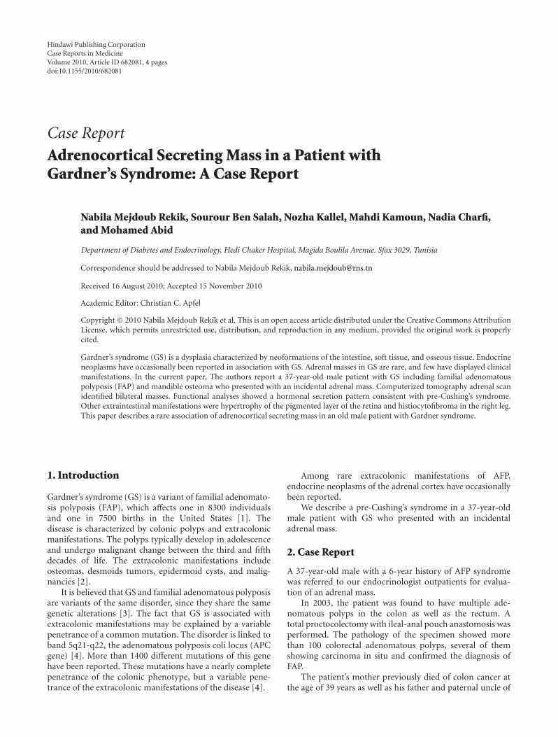
Figure 1: Partial pedigree of the family and mutation segregation.Square and circle symbols represent male and female, respectively.The genotypes of the exon ii polymorphism are shown under thesymbols. Arrow: proband. Grey symbols: asymptotic carriers of themutation.
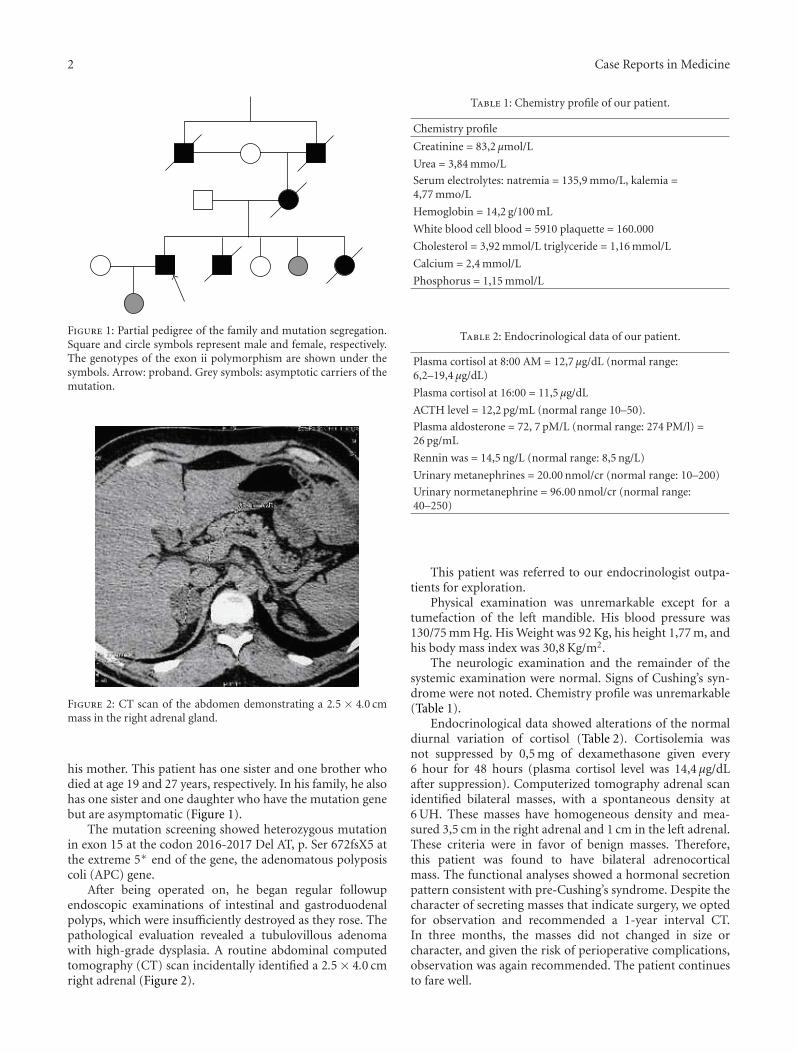
Figure 2: CT scan of the abdomen demonstrating a 2.5 × 4.0 cmmass in the right adrenal gland.
his mother. This patient has one sister and one brother whodied at age 19 and 27 years, respectively. In his family, he alsohas one sister and one daughter who have the mutation genebut are asymptomatic (Figure 1).
The mutation screening showed heterozygous mutationin exon 15 at the codon 2016-2017 Del AT, p. Ser 672fsX5 atthe extreme 5∗ end of the gene, the adenomatous polyposiscoli (APC) gene.
After being operated on, he began regular followupendoscopic examinations of intestinal and gastroduodenalpolyps, which were insufficiently destroyed as they rose. Thepathological evaluation revealed a tubulovillous adenomawith high-grade dysplasia. A routine abdominal computedtomography (CT) scan incidentally identified a 2.5 × 4.0 cmright adrenal (Figure 2).
Table 1: Chemistry profile of our patient.
Chemistry profile
Creatinine = 83,2 µmol/L
Urea = 3,84 mmo/L
Serum electrolytes: natremia = 135,9 mmo/L, kalemia =4,77 mmo/L
Hemoglobin = 14,2 g/100 mL
White blood cell blood = 5910 plaquette = 160.000
Cholesterol = 3,92 mmol/L triglyceride = 1,16 mmol/L
Calcium = 2,4 mmol/L
Phosphorus = 1,15 mmol/L
Table 2: Endocrinological data of our patient.
Plasma cortisol at 8:00 AM = 12,7 µg/dL (normal range:6,2–19,4 µg/dL)
Plasma cortisol at 16:00 = 11,5 µg/dL
ACTH level = 12,2 pg/mL (normal range 10–50).
Plasma aldosterone = 72, 7 pM/L (normal range: 274 PM/l) =26 pg/mL
Rennin was = 14,5 ng/L (normal range: 8,5 ng/L)
Urinary metanephrines = 20.00 nmol/cr (normal range: 10–200)
Urinary normetanephrine = 96.00 nmol/cr (normal range:40–250)
This patient was referred to our endocrinologist outpa-tients for exploration.
Physical examination was unremarkable except for atumefaction of the left mandible. His blood pressure was130/75 mm Hg. His Weight was 92 Kg, his height 1,77 m, andhis body mass index was 30,8 Kg/m2.
The neurologic examination and the remainder of thesystemic examination were normal. Signs of Cushing’s syn-drome were not noted. Chemistry profile was unremarkable(Table 1).
Endocrinological data showed alterations of the normaldiurnal variation of cortisol (Table 2). Cortisolemia wasnot suppressed by 0,5 mg of dexamethasone given every6 hour for 48 hours (plasma cortisol level was 14,4 µg/dLafter suppression). Computerized tomography adrenal scanidentified bilateral masses, with a spontaneous density at6 UH. These masses have homogeneous density and mea-sured 3,5 cm in the right adrenal and 1 cm in the left adrenal.These criteria were in favor of benign masses. Therefore,this patient was found to have bilateral adrenocorticalmass. The functional analyses showed a hormonal secretionpattern consistent with pre-Cushing’s syndrome. Despite thecharacter of secreting masses that indicate surgery, we optedfor observation and recommended a 1-year interval CT.In three months, the masses did not changed in size orcharacter, and given the risk of perioperative complications,observation was again recommended. The patient continuesto fare well.
Case Reports in Medicine 3
R L
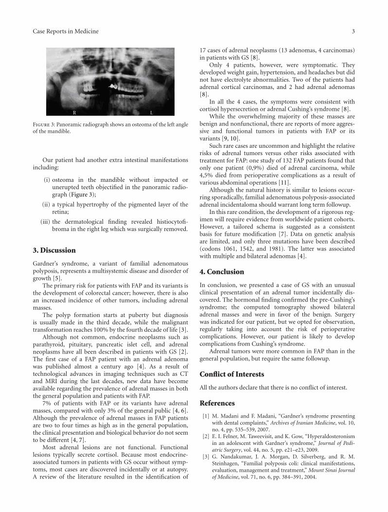
Figure 3: Panoramic radiograph shows an osteoma of the left angleof the mandible.
Our patient had another extra intestinal manifestationsincluding:
(i) osteoma in the mandible without impacted orunerupted teeth objectified in the panoramic radio-graph (Figure 3);
(ii) a typical hypertrophy of the pigmented layer of theretina;
(iii) the dermatological finding revealed histiocytofi-broma in the right leg which was surgically removed.
3. Discussion
Gardner’s syndrome, a variant of familial adenomatouspolyposis, represents a multisystemic disease and disorder ofgrowth [5].
The primary risk for patients with FAP and its variants isthe development of colorectal cancer; however, there is alsoan increased incidence of other tumors, including adrenalmasses.
The polyp formation starts at puberty but diagnosisis usually made in the third decade, while the malignanttransformation reaches 100% by the fourth decade of life [3].
Although not common, endocrine neoplasms such asparathyroid, pituitary, pancreatic islet cell, and adrenalneoplasms have all been described in patients with GS [2].The first case of a FAP patient with an adrenal adenomawas published almost a century ago [4]. As a result oftechnological advances in imaging techniques such as CTand MRI during the last decades, new data have becomeavailable regarding the prevalence of adrenal masses in boththe general population and patients with FAP.
7% of patients with FAP or its variants have adrenalmasses, compared with only 3% of the general public [4, 6].Although the prevalence of adrenal masses in FAP patientsare two to four times as high as in the general population,the clinical presentation and biological behavior do not seemto be different [4, 7].
Most adrenal lesions are not functional. Functionallesions typically secrete cortisol. Because most endocrine-associated tumors in patients with GS occur without symp-toms, most cases are discovered incidentally or at autopsy.A review of the literature resulted in the identification of
17 cases of adrenal neoplasms (13 adenomas, 4 carcinomas)in patients with GS [8].
Only 4 patients, however, were symptomatic. Theydeveloped weight gain, hypertension, and headaches but didnot have electrolyte abnormalities. Two of the patients hadadrenal cortical carcinomas, and 2 had adrenal adenomas[8].
In all the 4 cases, the symptoms were consistent withcortisol hypersecretion or adrenal Cushing’s syndrome [8].
While the overwhelming majority of these masses arebenign and nonfunctional, there are reports of more aggres-sive and functional tumors in patients with FAP or itsvariants [9, 10].
Such rare cases are uncommon and highlight the relativerisks of adrenal tumors versus other risks associated withtreatment for FAP: one study of 132 FAP patients found thatonly one patient (0,9%) died of adrenal carcinoma, while4,5% died from perioperative complications as a result ofvarious abdominal operations [11].
Although the natural history is similar to lesions occur-ring sporadically, familial adenomatous polyposis-associatedadrenal incidentaloma should warrant long term followup.
In this rare condition, the development of a rigorous reg-imen will require evidence from worldwide patient cohorts.However, a tailored schema is suggested as a consistentbasis for future modification [7]. Data on genetic analysisare limited, and only three mutations have been described(codons 1061, 1542, and 1981). The latter was associatedwith multiple and bilateral adenomas [4].
4. Conclusion
In conclusion, we presented a case of GS with an unusualclinical presentation of an adrenal tumor incidentally dis-covered. The hormonal finding confirmed the pre-Cushing’ssyndrome; the computed tomography showed bilateraladrenal masses and were in favor of the benign. Surgerywas indicated for our patient, but we opted for observation,regularly taking into account the risk of perioperativecomplications. However, our patient is likely to developcomplications from Cushing’s syndrome.
Adrenal tumors were more common in FAP than in thegeneral population, but require the same followup.
Conflict of Interests
All the authors declare that there is no conflict of interest.
References
[1] M. Madani and F. Madani, “Gardner’s syndrome presentingwith dental complaints,” Archives of Iranian Medicine, vol. 10,no. 4, pp. 535–539, 2007.
[2] E. I. Felner, M. Taweevisit, and K. Gow, “Hyperaldosteronismin an adolescent with Gardner’s syndrome,” Journal of Pedi-atric Surgery, vol. 44, no. 5, pp. e21–e23, 2009.
[3] G. Nandakumar, J. A. Morgan, D. Silverberg, and R. M.Steinhagen, “Familial polyposis coli: clinical manifestations,evaluation, management and treatment,” Mount Sinai Journalof Medicine, vol. 71, no. 6, pp. 384–391, 2004.
4 Case Reports in Medicine
[4] E. J. Groen, A. Roos, F. L. Muntinghe et al., “Extra-intestinalmanifestations of familial adenomatous polyposis,” Annals ofSurgical Oncology, vol. 15, no. 9, pp. 2439–2450, 2008.
[5] C. Fotiadis, D. K. Tsekouras, P. Antonakis, J. Sfiniadakis,M. Genetzakis, and G. C. Zografos, “Gardner’s syndrome: acase report and review of the literature,” World Journal ofGastroenterology, vol. 11, no. 34, pp. 5408–5411, 2005.
[6] T. G. P. Johnson Smith, S. K. Clark, D. E. Katz, R. H. Reznek,and R. K. S. Phillips, “Adrenal masses are associated withfamilial adenomatous polyposis,” Diseases of the Colon andRectum, vol. 43, no. 12, pp. 1739–1742, 2000.
[7] O. C. C. Will, A. Hansmann, R. K. S. Phillips et al., “Adrenalincidentaloma in familial adenomatous polyposis: a long-termfollow-up study and schema for management,” Diseases of theColon and Rectum, vol. 52, no. 9, pp. 1637–1644, 2009.
[8] P. Marchesa, V. W. Fazio, J. M. Church, and E. McGannon,“Adrenal masses in patients with familial adenomatous poly-posis,” Diseases of the Colon and Rectum, vol. 40, no. 9, pp.1023–1028, 1997.
[9] T. A. Painter and D. G. Jagelman, “Adrenal adenomas andadrenal carcinomas in association with hereditary adenomato-sis of the colon and rectum,” Cancer, vol. 55, no. 9, pp. 2001–2004, 1985.
[10] F. Beuschlein, M. Reincke, M. Koniger, D. D’Orazio, Z. Dob-bie, and L. C. Rump, “Cortisol producing adrenal adenoma—a new manifestation of Gardner’s syndrome,” EndocrineResearch, vol. 26, no. 4, pp. 783–790, 2000.
[11] M. L. Arvanitis, D. G. Jagelman, V. W. Fazio, I. C. Lavery,and E. McGannon, “Mortality in patients with familialadenomatous polyposis,” Diseases of the Colon and Rectum,vol. 33, no. 8, pp. 639–642, 1990.

Submit your manuscripts athttp://www.hindawi.com
Stem CellsInternational
Hindawi Publishing Corporationhttp://www.hindawi.com Volume 2014
Hindawi Publishing Corporationhttp://www.hindawi.com Volume 2014
MEDIATORSINFLAMMATION
of
Hindawi Publishing Corporationhttp://www.hindawi.com Volume 2014
Behavioural Neurology
EndocrinologyInternational Journal of
Hindawi Publishing Corporationhttp://www.hindawi.com Volume 2014
Hindawi Publishing Corporationhttp://www.hindawi.com Volume 2014
Disease Markers
Hindawi Publishing Corporationhttp://www.hindawi.com Volume 2014
BioMed Research International
OncologyJournal of
Hindawi Publishing Corporationhttp://www.hindawi.com Volume 2014
Hindawi Publishing Corporationhttp://www.hindawi.com Volume 2014
Oxidative Medicine and Cellular Longevity
Hindawi Publishing Corporationhttp://www.hindawi.com Volume 2014
PPAR Research
The Scientific World JournalHindawi Publishing Corporation http://www.hindawi.com Volume 2014
Immunology ResearchHindawi Publishing Corporationhttp://www.hindawi.com Volume 2014
Journal of
ObesityJournal of
Hindawi Publishing Corporationhttp://www.hindawi.com Volume 2014
Hindawi Publishing Corporationhttp://www.hindawi.com Volume 2014
Computational and Mathematical Methods in Medicine
OphthalmologyJournal of
Hindawi Publishing Corporationhttp://www.hindawi.com Volume 2014
Diabetes ResearchJournal of
Hindawi Publishing Corporationhttp://www.hindawi.com Volume 2014
Hindawi Publishing Corporationhttp://www.hindawi.com Volume 2014
Research and TreatmentAIDS
Hindawi Publishing Corporationhttp://www.hindawi.com Volume 2014
Gastroenterology Research and Practice
Hindawi Publishing Corporationhttp://www.hindawi.com Volume 2014
Parkinson’s Disease
Evidence-Based Complementary and Alternative Medicine
Volume 2014Hindawi Publishing Corporationhttp://www.hindawi.com