Embed Size (px)
Citation preview
7/30/2019 ADI 900 159 Insert Enzo
http://slidepdf.com/reader/full/adi-900-159-insert-enzo 1/12
1
FOR RESEARCH USE ONLY. NOT FOR USE IN DIAGNOSTIC PROCEDURES.
www.enzolifesciences.com
> Glutathione reductase acvity kitCatalog # ADI-900-159
Sucient Reagents for 480 tests with 5 x 96-well plates
For use with cell and ssue extracts
All reagents,
except Standard
and NADPH, should
be stored at 4˚C.
Store Standard and
NADPH at -20˚C.
For proper per-
formance, use the
insert provided with
each individual kit
received.
Check our website
for addional proto-
cols, technical notes
and FAQs.
Table of Contents
2 Introducon
2 Principle
3 Materials Supplied
3 Storage
3 Materials Needed but Not Supplied
4 Reagent Preparaon5 Sample Handling
7 Assay Procedure
8 Calculaon of Results
11 Trouble Shoong
11 References
12 Limited Warranty
7/30/2019 ADI 900 159 Insert Enzo
http://slidepdf.com/reader/full/adi-900-159-insert-enzo 2/12
2
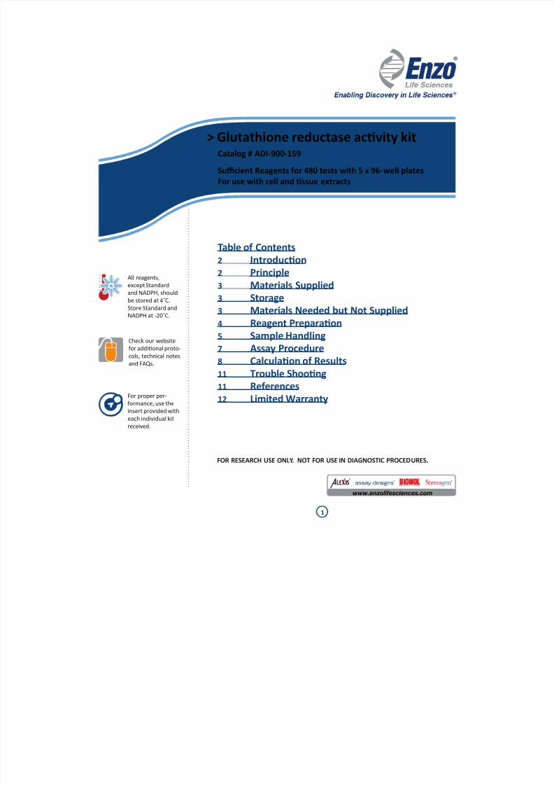
IntroduconThe Glutathione reductase acvity kit is a complete kit for the measurement of glutathione
reductase acvity in cell and ssue extracts.
Glutathione reductase (GR), a homodimeric avoprotein disulde oxidoreductase, plays
an indirect but essenal role in the prevenon of oxidave damage within the cell by
helping to maintain appropriate levels of intracellular reduced glutathione (GSH). Reduced
glutathione is essenal for maintaining the normal structure of red blood cells and for
keeping hemoglobin in the ferrous state1. Glutathione reductase together with its co-factor,
NADPH, catalyzes the reducon of oxidized glutathione (glutathione disulde, GSSG) to
glutathione (Figure 1). GSH is also a reactant for glutathione peroxidase, which converts
hydrogen peroxide (H2O2) into water.
In this assay the oxidaon of NADPH to NADP+ (Figure 1) is monitored by the decrease in
absorbance at 340 nm. This rate of decrease in absorbance at 340 nm is directly proporonal
to the glutathione reductase acvity in the sample because the enzyme is present at rate
liming concentraons2-4. The unit denion for glutathione reductase acvity may be
expressed in terms of the oxidaon of NADPH or the reducon of GSSG since their molar
rao is 1:1. One unit of glutathione reductase oxidizes 1 µmol of NADPH per minute at
25°C, pH 7.5.
Principle
1. Samples and standards are added to wells of a 96-well plate. GR Master mix and
buers are added.
2. NADPH soluon is added to the wells to iniate the reacon.
3. The plate is transferred to a plate reader and absorbance readings are taken at 340
nm every minute for 10 minutes.
GSSG + NADPH + H+ 2GSH + NADP+
GLUTATHIONEREDUCTASE
(Absorbs at
340 nm)
Figure 1. Reducon of glutathione disulde (GSSG) by glutathione reductase and NADPH.
7/30/2019 ADI 900 159 Insert Enzo
http://slidepdf.com/reader/full/adi-900-159-insert-enzo 3/12
3
Materials Supplied
1. Clear Microter Plate
Five Plates of 96 Wells, Catalog No. 80-1639Clear uncoated solid plates.
2. GR Standard
1 mL, Catalogue No. 80-1640
Glutathione reductase standard with an acvity of 1 unit/mL. One unit of GR oxidizes
1 µmole of NADPH per minute at 25°C, pH 7.5.
3. 10X GR Buer
20 mL, Catalog No. 80-1637
4. NADPH
5 lyophilized vials, Catalog No. 80-1638
One vial is sucient for one 96-well plate.
5. GSSG
3 mL, Catalog No. 80-1636
6. 20% Triton X-100
1 mL, Catalog No. 80-1641
7. Glutathione Reductase Assay Layout Sheet
1 each, Catalog No. 30-0239
Storage
The GR Standard and NADPH should be stored at -20°C. All other components of this kit are
stable at 4°C. All kit components are stable at their recommended storage temperatures
unl the kit expiraon date.
Materials Needed but Not Supplied
1. PBS
2. Dislled water
3. Protease inhibitors (oponal) such as phenylmethylsulfonyl uoride (PMSF), Sigma
P7626 or equivalent
4. Reagents to determine protein concentraon
5. Ficoll-Hypaque™ (erythrocyte, lymphocyte and monocyte preparaons)6. Microtubes, 0.5 and 1.5 mL
7. 15 mL conical tubes (adherent and suspension cell preparaon)
8. 50 mL conical tubes (ssue preparaon)
9. Precision pipees for volumes between 5-1000 µL
10. Mulchannel pipeor for volumes between 1 - 50 µL and 50 µL – 200 µL
11. Microplate reader capable of reading at 340 nm and taking readings every minute
for ten minutes and exporng data to an Excel spreadsheet
12. Centrifuge and microfuge for processing samples
Do not mix compo-
nents from dierent
kit lots or use
reagents beyond the
expiraon date of
the kit.
The physical, chemi-
cal, and toxicologi-
cal properes of the
chemicals and
reagents contained
in this kit may not
yet have been
fully invesgated.
Therefore, we
recommend the use
of gloves, lab coats,
and eye protecon
while using any
of these chemical
reagents.
Reagents require
separate storage
condions.
7/30/2019 ADI 900 159 Insert Enzo
http://slidepdf.com/reader/full/adi-900-159-insert-enzo 4/12
4
Reagent Preparaon
1. 1X GR Buer
Dilute the 10X GR Buer to 1X (1:10) with dislled water. The 1X GR Buer is usedto prepare diluons of GR Standard Curve and to prepare the NADPH reagent. The
10X GR Buer is used directly to prepare 1X Cell Extracon Buer and Master Mix.
2. GR Standard Curve
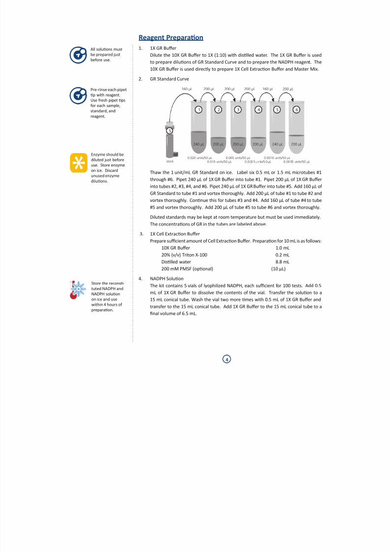
Thaw the 1 unit/mL GR Standard on ice. Label six 0.5 mL or 1.5 mL microtubes #1
through #6. Pipet 240 µL of 1X GR Buer into tube #1. Pipet 200 µL of 1X GR Buer
into tubes #2, #3, #4, and #6. Pipet 240 µL of 1X GR Buer into tube #5. Add 160 µL of
GR Standard to tube #1 and vortex thoroughly. Add 200 µL of tube #1 to tube #2 and
vortex thoroughly. Connue this for tubes #3 and #4. Add 160 µL of tube #4 to tube
#5 and vortex thoroughly. Add 200 µL of tube #5 to tube #6 and vortex thoroughly.
Diluted standards may be kept at room temperature but must be used immediately.
The concentraons of GR in the tubes are labeled above.
3. 1X Cell Extracon Buer
Prepare sucient amount of Cell Extracon Buer. Preparaon for 10 mL is as follows:
10X GR Buer 1.0 mL
20% (v/v) Triton X-100 0.2 mL
Dislled water 8.8 mL
200 mM PMSF (oponal) (10 µL)
4. NADPH SoluonThe kit contains 5 vials of lyophilized NADPH, each sucient for 100 tests. Add 0.5
mL of 1X GR Buer to dissolve the contents of the vial. Transfer the soluon to a
15 mL conical tube. Wash the vial two more mes with 0.5 mL of 1X GR Buer and
transfer to the 15 mL conical tube. Add 1X GR Buer to the 15 mL conical tube to a
nal volume of 6.5 mL.
160 µL 200 µL 200 µL 200 µL 160 µL
450 µL 200 µL 200 µL 200 µL240 µL 450 µL240 µL 200 µL
0.020 units/50 µL0.010 units/50 µLstock
0.005 units/50 µL0 .0025 un its/50 µL 0.0005 units/50 µL
0.0010 units/50 µL
200 µL
1 2 3 4 5 6
S
Enzyme should be
diluted just before
use. Store enzyme
on ice. Discard
unused enzyme
diluons.
Pre-rinse each pipet
p with reagent.
Use fresh pipet ps
for each sample,
standard, andreagent.
Store the recons-
tuted NADPH and
NADPH soluon
on ice and use
within 4 hours of
preparaon.
All soluons must
be prepared justbefore use.
7/30/2019 ADI 900 159 Insert Enzo
http://slidepdf.com/reader/full/adi-900-159-insert-enzo 5/12
5
5. Master Mix
Count the total number of wells needed for the samples and add 21 (for the complete
standard curve and Blank wells in triplicate). Use the following formula to calculate
the volume of Master Mix required.
A. Total volume required
[Total number of wells needed + 21] x 100 µL = __________ µL
B. Volume of 10X GR Buer required
[Total Volume Required (from A. above)] x 0.1 = __________ µL
C. Volume of GSSG Reagent required
[Total Volume required (from A. above)] x 0.04 = __________ µL
D. Volume of dislled water required
[Total Volume required (from A. above)] x 0.86 = __________ µL
Prepare Master Mix by combining the appropriate reagent volumes calculated in B,
C, and D above. For example, to prepare 100 µL of Master Mix the following volumes
are combined: 10 µL 10X GR Buer, 4 µL GSSG reagent, and 86 µL dislled water.
6. Biological Extracts
Aer preparing the samples as outlined in the Sample Handling secon that follows,
make serial diluons of cell or ssue extracts with 1X GR Buer. Inial concentraons
between 0.5 µg/50 µL to 50 µg/50 µL are recommended.
Sample Handling
Choose the appropriate protocol in Secon A to process sample before proceeding to
Secon B. Keep samples on ice to maintain enzyme acvity.
Secon A. Processing SamplesSuspension cells:
1. Centrifuge 2 to 6 x 10⁶ suspension cells at 250 x g for 10 minutes at 4ºC. Discard the
supernatant.
2. Suspend the cell pellet in 1 mL of ice-cold 1X PBS and transfer to a 1.5 mL microtube
on ice. Centrifuge, discard supernatant, and place on ice.
3. Proceed to Secon B. Preparaon of Cytosolic Extracts
Adherent cells:
1. Wash 2 to 6 x 10⁶ adherent cells with 1X PBS. Adherent cells may be harvested by
gentle trypsinizaon.
2. Transfer to a 15 mL tube on ice. Centrifuge at 250 x g for 10 minutes at 4ºC and discard
the supernatant.
3. Suspend the cell pellet in 1 mL of ice-cold 1X PBS and transfer to 1.5 mL microtube
on ice. Centrifuge, discard supernatant, and place on ice..
4. Proceed to Secon B. Preparaon of Cytosolic Extracts.
Samples must
be kept on ice to
maintain enzyme
acvity.
Thaw the GSSG
stock on ice. Keep
the stock on icewhile using.
7/30/2019 ADI 900 159 Insert Enzo
http://slidepdf.com/reader/full/adi-900-159-insert-enzo 6/12
6
Erythrocytes:
1. Dilute ancoagulated blood with an equal volume of PBS. Layer over Ficoll-Hypaque™
or similar reagent and centrifuge at 800 x g for 25 min at 12°C with the BRAKE OFF in
a swinging bucket rotor.2. Collect the mononuclear cells (lymphocytes and monocytes) at the interphase and
transfer to another tube.
3. Remove the remaining liquid from above the red blood cell pellet. Wash the pellet
with 10 cell volumes of PBS.
4. Determine the packed cell volume and add 10 cell volumes of cold dislled water. Mix
well and incubate on ice for 10-15 minutes to lyse the red blood cells. Lysis occurs
when the opaque soluon changes to a brilliant clear red soluon, indicang the
release of hemoglobin.5. Centrifuge at 10,000 x g for 10 minutes at 4˚C to remove cell debris. Transfer the
supernatant to a fresh tube and store on ice.
Lymphocytes and Monocytes:
1. Dilute ancoagulated blood with an equal volume of PBS. Layer over Ficoll-Hypaque™
or similar reagent and centrifuge at 800 x g for 25 min at 12°C with the BRAKE OFF in
a swinging bucket rotor.
2. Collect the mononuclear cells (lymphocytes and monocytes) at the interphase and
transfer to another tube.
3. Dilute the blood mononuclear cells with 5 volumes of PBS and centrifuge at 400 x g
for 10 minutes at 4ºC. Discard the supernatant
4. Suspend the cell pellet in 1 mL of ice-cold 1X PBS and transfer to a pre-chilled 1.5
mL microtube. Centrifuge, discard supernatant, and place on ice.
5. Proceed to Secon B. Preparaon of Cytosolic Extracts.
Tissue
1. Remove ssue and place in cold PBS in a 50 mL conical tube. Repeatedly wash thessue with PBS to remove blood clots and other debris.
2. Transfer the ssue to a Petri dish on ice and mince the ssue to small pieces with
surgical scissors.
3. Transfer the ssue pieces to a clean stainless steel sieve. Place the sieve with the
ssue pieces in a Petri dish which contains about 20 mL of cold 1X PBS.
4. Create a single cell suspension of the ssue as follows: Using a pestle or a round
boom tube, grind the ssue pieces thoroughly unl the bulk of the ssue passes
through the sieve.5. Transfer the PBS containing the single cell suspension to a 50 mL conical tube. Fill
with cold PBS and mix by inverng the tube several mes. Let the tube stand on ice
for 1 minute to allow large aggregates of ssue to sele out of soluon.
6. Carefully transfer the supernatant containing the single cell suspension to a clean 50
mL conical centrifuge tube. Centrifuge at 400 x g for 10 minutes at 4ºC. Discard the
supernatant. Suspend the cell pellet in 1 mL of ice-cold 1X PBS and transfer to a pre-
chilled 1.5 mL microtube on ice. Centrifuge, discard the supernatant, and place on ice.
7. Proceed to Secon B. Preparaon of Cytosolic Extracts.
Samples must
be kept on ice to
maintain enzyme
acvity.
7/30/2019 ADI 900 159 Insert Enzo
http://slidepdf.com/reader/full/adi-900-159-insert-enzo 7/12
7
Secon B. Preparaon of Cytosolic Extracts from Cells and Tissue
1. Measure the approximate volume of the cell pellets prepared above (except for
erythrocytes) and suspend the cells in 5-10 volumes of cold 1X Cell Extracon Buer.
Incubate the cell suspensions on ice, with periodic vortexing, for 30 minutes.
2. Microcentrifuge the disrupted cell suspension at 10,000 x g for 10 minutes at 4ºC to
remove insoluble material. Recover the supernatant to a fresh tube pre-chilled on
ice. Occasionally, the pellet may oat and can easily be removed with a pipet p.
3. Determine the protein concentraon of the cleared cell lysate. Note that the 1X GR
Buer contains BSA at a concentraon of 0.1 mg/mL and this should be subtracted
from your observed protein concentraon.
4. If not assaying for GR immediately, snap-freeze the cleared cell extract in 100 µL
aliquots by immersing the aliquots in liquid nitrogen and store at -80ºC. Avoid repeated
freezing and thawing of the extract.
Assay Procedure
Refer to the Assay layout Sheet to determine the number of wells to be used.
1. Pipet 50 µL of the 1X GR Buer to the boom of the Blank wells.2. Pipet 50 µL of the prepared GR Standards #1 through #6 to the boom of the
appropriate wells.
3. Pipet 50 µL of the diluted sample to the boom of the appropriate wells.
4. Pipet 100 µL of Master Mix into each well.
5. Iniate the reacon by adding 50µL of NADPH Soluon to all the wells using a
mulchannel pipet.
6. Immediately transfer the plate to a microter plate reader and take absorbance
readings at 340 nm every minute for 10 minutes at room temperature. If possible,include a 10 second orbital shake prior to the rst read.
All standards, con-
trols, and samples
should be run in
triplicate.
Pre-rinse each pipet
p with reagent.
Use fresh pipet ps
for each sample,
standard, and
reagent.
Pipet the reagents
to the sides of the
wells to avoid pos-
sible contaminaon.
7/30/2019 ADI 900 159 Insert Enzo
http://slidepdf.com/reader/full/adi-900-159-insert-enzo 8/12
8
Calculaon of Results
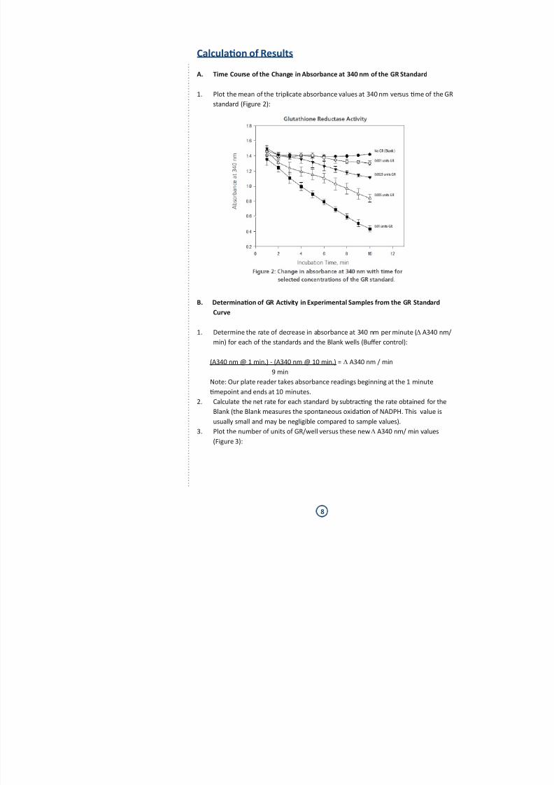
A. Time Course of the Change in Absorbance at 340 nm of the GR Standard
1. Plot the mean of the triplicate absorbance values at 340 nm versus me of the GR
standard (Figure 2):
B. Determinaon of GR Acvity in Experimental Samples from the GR Standard
Curve
1. Determine the rate of decrease in absorbance at 340 nm per minute (∆ A340 nm/
min) for each of the standards and the Blank wells (Buer control):
(A340 nm @ 1 min.) - (A340 nm @ 10 min.) = ∆ A340 nm / min
9 min
Note: Our plate reader takes absorbance readings beginning at the 1 minute
mepoint and ends at 10 minutes.
2. Calculate the net rate for each standard by subtracng the rate obtained for the
Blank (the Blank measures the spontaneous oxidaon of NADPH. This value is
usually small and may be negligible compared to sample values).
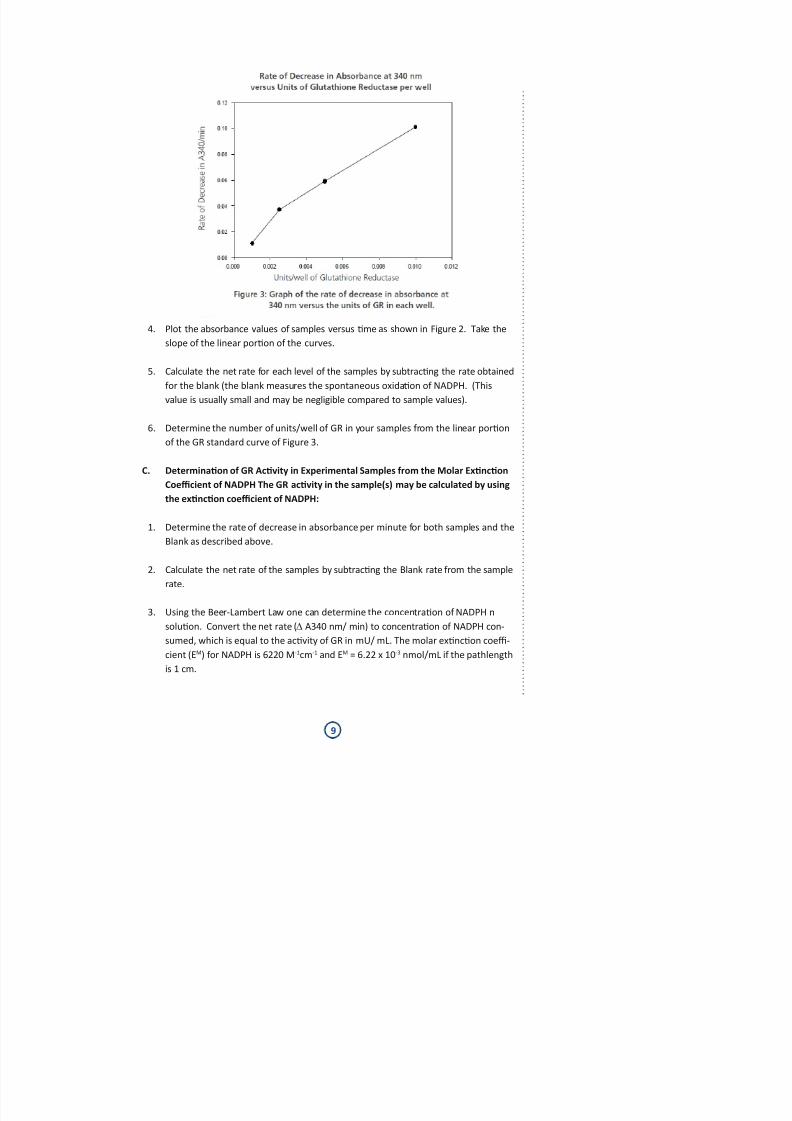
3. Plot the number of units of GR/well versus these new ∆ A340 nm/ min values
(Figure 3):
7/30/2019 ADI 900 159 Insert Enzo
http://slidepdf.com/reader/full/adi-900-159-insert-enzo 9/12
9
4. Plot the absorbance values of samples versus me as shown in Figure 2. Take the
slope of the linear poron of the curves.
5. Calculate the net rate for each level of the samples by subtracng the rate obtained
for the blank (the blank measures the spontaneous oxidaon of NADPH. (This
value is usually small and may be negligible compared to sample values).
6. Determine the number of units/well of GR in your samples from the linear poron
of the GR standard curve of Figure 3.
C. Determinaon of GR Acvity in Experimental Samples from the Molar Exncon
Coecient of NADPH The GR acvity in the sample(s) may be calculated by using
the exncon coecient of NADPH:
1. Determine the rate of decrease in absorbance per minute for both samples and the
Blank as described above.
2. Calculate the net rate of the samples by subtracng the Blank rate from the samplerate.
3. Using the Beer-Lambert Law one can determine the concentraon of NADPH n
soluon. Convert the net rate (∆ A340 nm/ min) to concentraon of NADPH con-
sumed, which is equal to the acvity of GR in mU/ mL. The molar exncon coe-
cient (EM) for NADPH is 6220 M-1cm-1 and EM = 6.22 x 10⁻³ nmol/mL if the pathlength
is 1 cm.
7/30/2019 ADI 900 159 Insert Enzo
http://slidepdf.com/reader/full/adi-900-159-insert-enzo 10/12
10
One unit of glutathione reductase is dened as the amount of enzyme required to cata-
lyze the reducon of one μmole of GSSG per minute at pH 7.5 and 25°C. One molecule
of NADPH is oxidized per molecule of GSSG reduced. Therefore, the oxidaon of NADPH
(measured by loss of A340 nm) directly correlates with GSSG reducon.
1 U of GR = 1 μmol GSSG reduced/min
= 1 μmol NADPH oxidized/min
1 mU of GR = 1 x 10⁻³ μmol NADPH oxidized/min
1 mU of GR = 1 nmol NADPH oxidized/min
If EM is the molar exncon coecient for NADPH at 340 nm,
EM = 6220 M-1cm-1
= 6220 x 10-6 μM-1cm-1
= 6.22 x 10⁻³ μM-1cm-1
= 6.22 x 10⁻³ L/μmol/cm
= 6.22 x 10⁻³ mL/nmol/cm
Note: The path length for 200 µL of reacon volume in the 96 well plate is
0.6 cm.
Therefore,
EM
= 6.22 x 10⁻³
mL/nmol /cm x 0.6 cm= 3.732 x 10⁻³ mL/nmole
∆ A340 nm/ min = ∆ A340 nm/ min
= 3.732 x 10⁻³ mL/nmole NADPH
= Y nmole NADPH/min/mL
= Y mU/mL GR
4. Correct for the sample diluon in the assay and for the sample diluon per formed
prior to the assay. For example: If the sample volume was 50 µL and was diluted
1/50 prior to the assay:
Mean ∆ A340 nm/min (Sample) = 0.0325/min
Mean ∆ A340 nm/min (Blank) = 0.0005/ min
Net Rate, ∆ A340 nm/min = 0.0320/ min
Glutathione Reductase Acvity = 0.0320/min/3.732x10-3mL/nmolNADPH/min
= 8.57 mU/mL
Assay Diluon Correcon = 200 µL/50 µL x 8.57 mU/mL
= 34.30 mU/mL
Sample Diluon Correcon = 50 x 34.30 mU/mL
= 1715 mU/mL
5. Divide the GR acvity (mU/mL) by the protein concentraon to determine the spe-
cic acvity of GR in your sample (mU GR/mg protein).
7/30/2019 ADI 900 159 Insert Enzo
http://slidepdf.com/reader/full/adi-900-159-insert-enzo 11/12
11
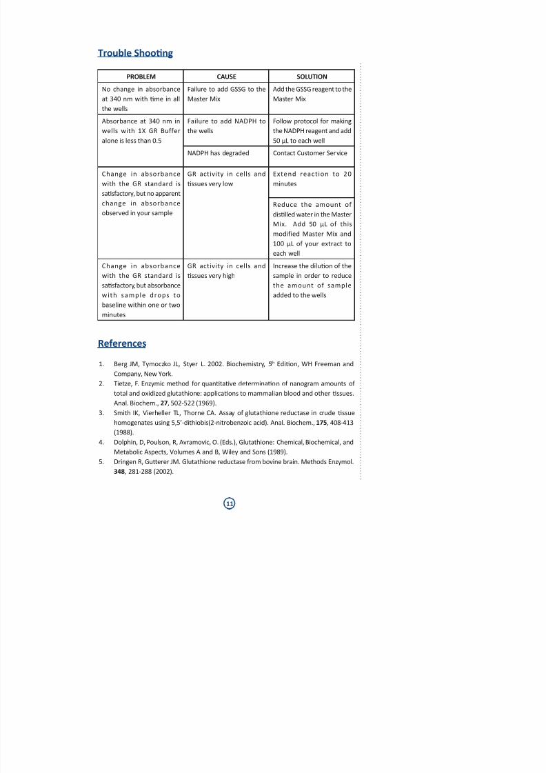
Trouble Shoong
PROBLEM CAUSE SOLUTION
No change in absorbance
at 340 nm with me in all
the wells
Failure to add GSSG to the
Master Mix
Add the GSSG reagent to the
Master Mix
Absorbance at 340 nm in
wells with 1X GR Buffer
alone is less than 0.5
Failure to add NADPH to
the wells
Follow protocol for making
the NADPH reagent and add
50 µL to each well
NADPH has degraded Contact Customer Service
Change in absorbance
with the GR standard is
sasfactory, but no apparent
change in absorbance
observed in your sample
GR activity in cells and
ssues very low
Extend reaction to 20
minutes
Reduce the amount of
dislled water in the Master
Mix. Add 50 µL of this
modified Master Mix and
100 µL of your extract to
each well
Change in absorbance
with the GR standard is
sasfactory, but absorbance
with sample drops to
baseline within one or two
minutes
GR activity in cells and
ssues very high
Increase the diluon of the
sample in order to reduce
the amount of sample
added to the wells
References
1. Berg JM, Tymoczko JL, Styer L. 2002. Biochemistry, 5th Edion, WH Freeman and
Company, New York.
2. Tietze, F. Enzymic method for quantave determinaon of nanogram amounts of
total and oxidized glutathione: applicaons to mammalian blood and other ssues.Anal. Biochem., 27, 502-522 (1969).
3. Smith IK, Vierheller TL, Thorne CA. Assay of glutathione reductase in crude ssue
homogenates using 5,5’-dithiobis(2-nitrobenzoic acid). Anal. Biochem., 175, 408-413
(1988).
4. Dolphin, D, Poulson, R, Avramovic, O. (Eds.), Glutathione: Chemical, Biochemical, and
Metabolic Aspects, Volumes A and B, Wiley and Sons (1989).
5. Dringen R, Guerer JM. Glutathione reductase from bovine brain. Methods Enzymol.
348, 281-288 (2002).
7/30/2019 ADI 900 159 Insert Enzo
http://slidepdf.com/reader/full/adi-900-159-insert-enzo 12/12
Use of ProductThis product contains research chemicals. As such, they should be used and handled only by or under the
supervision of technically qualied individuals. This product is not intended for diagnosc or human use.
WarrantyEnzo Life Sciences Internaonal, Inc. makes no warranty of any kind, expressed or implied, which extends
beyond the descripon of the product in this brochure, except that the material will meet our specica-
ons at the me of delivery. Enzo Life Sciences Internaonal, Inc. makes no guarantee of results and
assumes no liability for injuries, damages or penales resulng from product use, since the condions
of handling and use are beyond our control.
North/South America Germany UK & Ireland
ENZO LIFE SCIENCES INT’L, INC. ENZO LIFE SCIENCES GmbH ENZO LIFE SCIENCES (UK) LTD.
5120 Butler Pike Marie-Curie-Strasse 8 Palane House
Plymouth Meeng, PA 19462-1202/USA DE-79539 Lorrach / Germany Maord Court
Tel. 1-800-942-0430/(610)941-0430 Tel. +49/0 7621 5500 526 Exeter EX2 8NL / UK
Fax (610) 941-9252 Toll Free 0800 664 9518 Tel. 0845 601 1488 (UK customers)
[email protected] Fax +49/0 7621 5500 527 Tel. +44/0 1392 825900 (overseas)
[email protected] Fax +44/0 1392 825910
Switzerland & Rest of Europe Benelux France
ENZO LIFE SCIENCES AG ENZO LIFE SCIENCES BVBA ENZO LIFE SCIENCES
Industriestrasse 17, Posach Melkerijweg 3 c/o Covalab s.a.s.
CH-4415 Lausen / Switzerland BE-2240 Zandhoven / Belgium 13, Avenue Albert Einstein
Tel. +41/0 61 926 89 89 Tel. +32/0 3 466 04 20 FR-69100 Villeurbanne / France
Fax +41/0 61 926 89 79 Fax +32/0 3 466 04 29 Tel. +33 472 440 655
[email protected] [email protected] Fax +33 437 484 239
12
www.enzolifesciences.com
Enabling Discovery in Life Science®
www.enzolifesciences.com
Catalog No. 25-0599 August 3, 2010 © 2007
MSDS (Material
Safety Data Sheet)
available online


![FLUID CHILLERS 28 TO 150 TONS - Delta Inddeltaind.net/wp-content/uploads/2019/08/012617_Chase... · 2019. 8. 21. · Tank Capacity [gal] 124 124 124 124 159 159 159 159 159 159 159](https://img.pdfslide.us/doc/110x75/613777b90ad5d2067648a37d/fluid-chillers-28-to-150-tons-delta-2019-8-21-tank-capacity-gal-124-124.jpg)


![Aritmética Elemental [Enzo R Gentile]](https://img.pdfslide.us/doc/110x75/55cf9904550346d0339b0fc8/aritmetica-elemental-enzo-r-gentile.jpg)