Embed Size (px)
Citation preview
J o u r n a l o f R h e u m a t i c D i s e a s e sV o l . 2 1 , N o . 1 , F e b r u a r y , 2 0 1 4h ttp ://d x .d o i.o rg /1 0 .4 07 8 /jrd .20 1 4 .2 1 .1 .2 5
□ Case Report □
25
<Received:March 12, 2013, Revised (1st: April 15, 2013, 2nd: May 10, 2013), Accepted:May 12, 2013>Corresponding to:Hyoun-Ah Kim, Department of Rheumatology, Ajou University School of Medicine, Woncheon-dong San 5,
Youngtong-gu, Suwon 443-721, Korea. E-mail: [email protected]
pISSN: 2093-940X, eISSN: 2233-4718Copyright ⓒ 2014 by The Korean College of RheumatologyThis is a Free Access article, which permits unrestricted non-commerical use, distribution, and reproduction in any medium, provided the original work is properly cited.
Acute Polymyositis/systemic Lupus Erythematosus Overlap Syndrome with Severe Subcutaneous Edema and Interstitial Lung Disease
U-ram Jin1, Kyu-Sung Kwack2, Kyung-Joo Park2, Ji-Eun Kwon3, Si-Yeon Kim1, Ki-Chan Kim1
, Ga-Yong Ban1, Ju-Yang Jung
1, Chang-Hee Suh
1, Hyoun-Ah Kim
1
Departments of Rheumatology1, Radiology2, Pathology3, Ajou University School of Medicine, Suwon, Korea
Inflammatory myopathy is characterized by symmetrical
proximal muscle weakness, elevated muscle enzyme levels
and favorable response to glucocorticoids therapy.
Although periorbital edema is a common manifestation of
inflammatory myopathy, generalized subcutaneous edema
is very rare. We report here a case of a 47-year-old female
patient with acute polymyositis/systemic lupus eryth-
ematosus overlap syndrome with generalized subcutaneous
edema and interstitial lung disease. We aggressively treat-
ed the disease with high-dose glucocorticoids, intravenous
immunoglobulin, and immunosuppressive agents.
Key Words. Inflammatory myopathy, Overlap syndrome,
Subcutaneous edema, Interstitial lung disease
Introduction
Inflammatory myopathy is usually characterized by in-
flammation of proximal muscle, which results in muscle
weakness and elevated muscle enzyme levels. As the disease
progresses, esophageal and respiratory muscles may be
affected. Other manifestations, such as skin lesions, arthritis,
calcinosis, vasculitis and interstitial lung disease are relatively
common (1,2).
Periorbital edema is not an uncommon manifestation of the
disease, but generalized edema is rarely reported (3-13). There
are no guidelines for the treatment of this condition and the
clinical courses of reported cases have been variable.
We describe a case of polymyositis/systemic lupus eryth-
ematosus (SLE) overlap syndrome with severe generalized
subcutaneous edema and interstitial lung disease. Magnetic
resonance imaging (MRI) presented myositis and generalized
edema, and chest computed tomography (CT) scan presented
interstitial lung disease. The patient was treated successfully
with corticosteroid, human intravenous immunoglobulin
(IVIg), and immunosuppressive agents.
Case Report
A 47-year-old woman was admitted with progressive prox-
imal muscle weakness, generalized swelling, fever, dysphagia,
and dyspnea for 7 days. She had no history of diabetes melli-
tus, hypertension, or pulmonary tuberculosis, but had a
2-month history of rheumatoid arthritis. She was treated with
hydroxychloroquine and nonsteroidal anti-inflammatory drug.
She had cancer screening including esophagogastroduodeno-
scopy and colonoscopy about 1 month ago, and no evidence
was found of an underlying malignancy. Examination revealed
a blood pressure of 130/64 mmHg, pulse rate of 105/min, res-
piratory rate of 20/min, and body temperature of 37.6oC. A
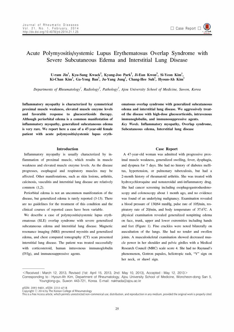
physical examination revealed generalized nonpitting edema
on face, trunk, upper and lower extremities including hands
and feet (Figure 1). Fine crackles were noted bilaterally on
auscultation of the lungs. She had no tender and swollen
joints. A musculoskeletal examination showed decreased mus-
cle power in her shoulder and pelvic girdles with a Medical
Research Council (MRC) scale score 4. She had no Raynaud’s
phenomenon, Gottron papules, heliotropic rash, “V” sign on
her neck, or shawl sign.
26 U-ram Jin et al.
Figure 1. Gross appearance of generalized edema of upper (A) and lower extremities (B) in a 47-year-old woman with acute
polymyositis.
Her initial laboratory findings showed; white blood cell
11,000/mm3, hemoglobin 11.2 g/dL, platelet 221,000/mm3, er-
ythrocyte sedimentation rate (ESR) 103 mm/hr, and C-reactive
protein (CRP) 6.91 mg/dL. The muscle enzymes were ele-
vated: creatine kinase (CK) 12,176 U/L (normal 29∼145
U/L), myoglobin >3,000 ng/mL (normal 0∼51 ng/mL), aldo-
lase 47.4 U/L (normal 0∼7.6 U/L), aspartate aminotransferase
565 U/L, alanine aminotransferase 374 U/L, and lactate de-
hydrogenase 1,597 U/L (normal 100∼200 U/L). Antinuclear
antibody (ANA) was positive (1:2,560, speckled and cyto-
plasmic mixed pattern), and complement 3 was reduced (C3
52 mg/dL, C4 13 mg/dL). Anti-Jo-1 antibody, anti-Ro anti-
body, anti-La antibody, anti-double stranded DNA antibody,
and anti-cardiolipin antibody were all negative. However, an-
ti-Sm antibody and anti-RNP antibody were positive, and
rheumatoid factor and anti-cyclic citrullinated peptide anti-
body were elevated with 83.6 IU/mL and 43 U/mL. She had
subclinical hypothyroidism with TSH level 12.34 uIU/mL, T3
60.4 ng/dL and free T4 0.72 ng/dL. Chest radiography showed
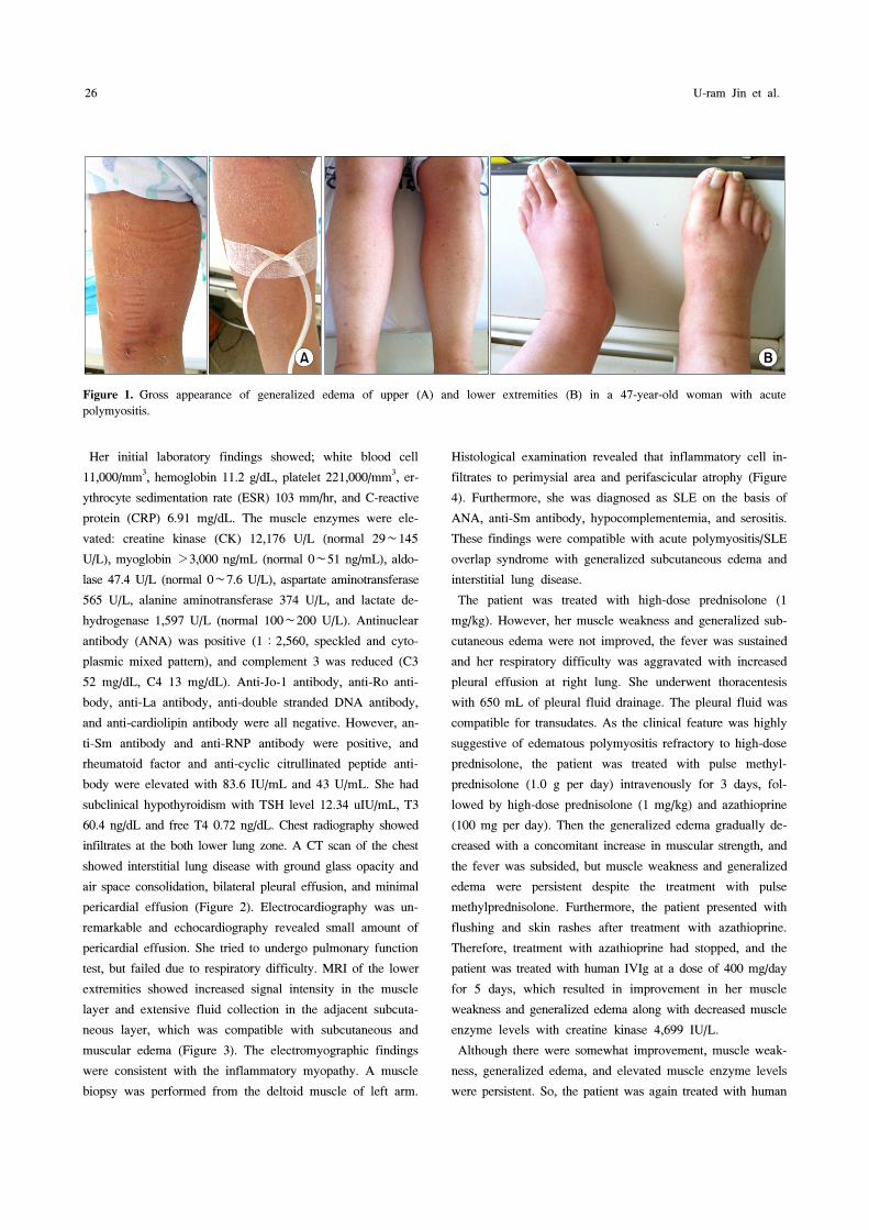
infiltrates at the both lower lung zone. A CT scan of the chest
showed interstitial lung disease with ground glass opacity and
air space consolidation, bilateral pleural effusion, and minimal
pericardial effusion (Figure 2). Electrocardiography was un-
remarkable and echocardiography revealed small amount of
pericardial effusion. She tried to undergo pulmonary function
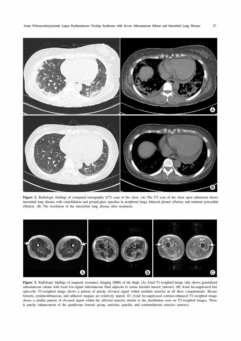
test, but failed due to respiratory difficulty. MRI of the lower
extremities showed increased signal intensity in the muscle
layer and extensive fluid collection in the adjacent subcuta-
neous layer, which was compatible with subcutaneous and
muscular edema (Figure 3). The electromyographic findings
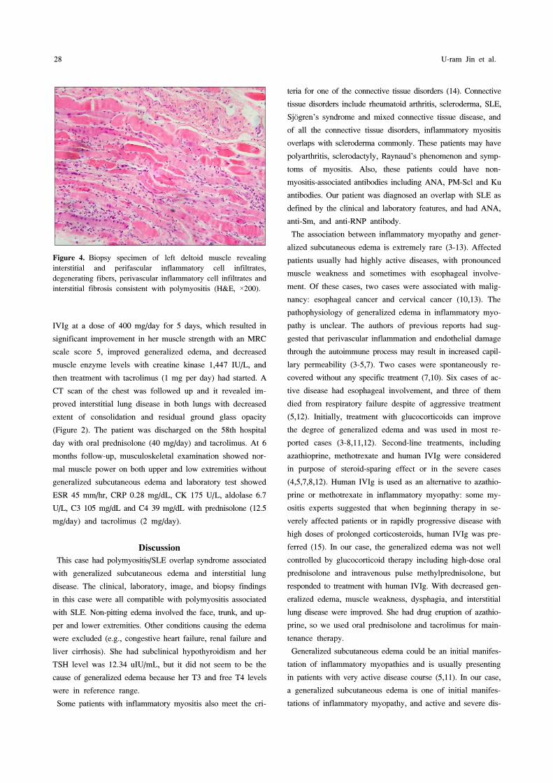
were consistent with the inflammatory myopathy. A muscle
biopsy was performed from the deltoid muscle of left arm.
Histological examination revealed that inflammatory cell in-
filtrates to perimysial area and perifascicular atrophy (Figure
4). Furthermore, she was diagnosed as SLE on the basis of
ANA, anti-Sm antibody, hypocomplementemia, and serositis.
These findings were compatible with acute polymyositis/SLE
overlap syndrome with generalized subcutaneous edema and
interstitial lung disease.
The patient was treated with high-dose prednisolone (1
mg/kg). However, her muscle weakness and generalized sub-
cutaneous edema were not improved, the fever was sustained
and her respiratory difficulty was aggravated with increased
pleural effusion at right lung. She underwent thoracentesis
with 650 mL of pleural fluid drainage. The pleural fluid was
compatible for transudates. As the clinical feature was highly
suggestive of edematous polymyositis refractory to high-dose
prednisolone, the patient was treated with pulse methyl-
prednisolone (1.0 g per day) intravenously for 3 days, fol-
lowed by high-dose prednisolone (1 mg/kg) and azathioprine
(100 mg per day). Then the generalized edema gradually de-
creased with a concomitant increase in muscular strength, and
the fever was subsided, but muscle weakness and generalized
edema were persistent despite the treatment with pulse
methylprednisolone. Furthermore, the patient presented with
flushing and skin rashes after treatment with azathioprine.
Therefore, treatment with azathioprine had stopped, and the
patient was treated with human IVIg at a dose of 400 mg/day
for 5 days, which resulted in improvement in her muscle
weakness and generalized edema along with decreased muscle
enzyme levels with creatine kinase 4,699 IU/L.
Although there were somewhat improvement, muscle weak-
ness, generalized edema, and elevated muscle enzyme levels
were persistent. So, the patient was again treated with human
Acute Polymyositis/systemic Lupus Erythematosus Overlap Syndrome with Severe Subcutaneous Edema and Interstitial Lung Disease 27
Figure 2. Radiologic findings of computed tomography (CT) scan of the chest. (A) The CT scan of the chest upon admission shows
interstitial lung disease with consolidation and ground-glass opacities in peripheral lungs, bilateral pleural effusion, and minimal pericardial
effusion. (B) The resolution of the interstitial lung disease after treatment.
Figure 3. Radiologic findings of magnetic resonance imaging (MRI) of the thigh. (A) Axial T1-weighted image only shows generalized
subcutaneous edema with focal low-signal subcutaneous fluid adjacent to vastus lateralis muscle (arrows). (B) Axial fat-suppressed fast
spin-echo T2-weighted image shows a pattern of patchy elevated signal within multiple muscles in all three compartments. Biceps
femoris, semimembranosus, and adductor magnus are relatively spared. (C) Axial fat-suppressed contrast-enhanced T1-weighted image
shows a similar pattern of elevated signal within the affected muscles similar to the distribution seen on T2-weighted images. There
is patchy enhancement of the quadriceps femoris group, sartorius, gracilis, and semitendinosus muscles (arrows).
28 U-ram Jin et al.
Figure 4. Biopsy specimen of left deltoid muscle revealing
interstitial and perifascular inflammatory cell infiltrates,
degenerating fibers, perivascular inflammatory cell infiltrates and
interstitial fibrosis consistent with polymyositis (H&E, ×200).
IVIg at a dose of 400 mg/day for 5 days, which resulted in
significant improvement in her muscle strength with an MRC
scale score 5, improved generalized edema, and decreased
muscle enzyme levels with creatine kinase 1,447 IU/L, and
then treatment with tacrolimus (1 mg per day) had started. A
CT scan of the chest was followed up and it revealed im-
proved interstitial lung disease in both lungs with decreased
extent of consolidation and residual ground glass opacity
(Figure 2). The patient was discharged on the 58th hospital
day with oral prednisolone (40 mg/day) and tacrolimus. At 6
months follow-up, musculoskeletal examination showed nor-
mal muscle power on both upper and low extremities without
generalized subcutaneous edema and laboratory test showed
ESR 45 mm/hr, CRP 0.28 mg/dL, CK 175 U/L, aldolase 6.7
U/L, C3 105 mg/dL and C4 39 mg/dL with prednisolone (12.5
mg/day) and tacrolimus (2 mg/day).
Discussion
This case had polymyositis/SLE overlap syndrome associated
with generalized subcutaneous edema and interstitial lung
disease. The clinical, laboratory, image, and biopsy findings
in this case were all compatible with polymyositis associated
with SLE. Non-pitting edema involved the face, trunk, and up-
per and lower extremities. Other conditions causing the edema
were excluded (e.g., congestive heart failure, renal failure and
liver cirrhosis). She had subclinical hypothyroidism and her
TSH level was 12.34 uIU/mL, but it did not seem to be the
cause of generalized edema because her T3 and free T4 levels
were in reference range.
Some patients with inflammatory myositis also meet the cri-
teria for one of the connective tissue disorders (14). Connective
tissue disorders include rheumatoid arthritis, scleroderma, SLE,
Sjögren’s syndrome and mixed connective tissue disease, and
of all the connective tissue disorders, inflammatory myositis
overlaps with scleroderma commonly. These patients may have
polyarthritis, sclerodactyly, Raynaud’s phenomenon and symp-
toms of myositis. Also, these patients could have non-
myositis-associated antibodies including ANA, PM-Scl and Ku
antibodies. Our patient was diagnosed an overlap with SLE as
defined by the clinical and laboratory features, and had ANA,
anti-Sm, and anti-RNP antibody.
The association between inflammatory myopathy and gener-
alized subcutaneous edema is extremely rare (3-13). Affected
patients usually had highly active diseases, with pronounced
muscle weakness and sometimes with esophageal involve-
ment. Of these cases, two cases were associated with malig-
nancy: esophageal cancer and cervical cancer (10,13). The
pathophysiology of generalized edema in inflammatory myo-
pathy is unclear. The authors of previous reports had sug-
gested that perivascular inflammation and endothelial damage
through the autoimmune process may result in increased capil-
lary permeability (3-5,7). Two cases were spontaneously re-
covered without any specific treatment (7,10). Six cases of ac-
tive disease had esophageal involvement, and three of them
died from respiratory failure despite of aggressive treatment
(5,12). Initially, treatment with glucocorticoids can improve
the degree of generalized edema and was used in most re-
ported cases (3-8,11,12). Second-line treatments, including
azathioprine, methotrexate and human IVIg were considered
in purpose of steroid-sparing effect or in the severe cases
(4,5,7,8,12). Human IVIg is used as an alternative to azathio-
prine or methotrexate in inflammatory myopathy: some my-
ositis experts suggested that when beginning therapy in se-
verely affected patients or in rapidly progressive disease with
high doses of prolonged corticosteroids, human IVIg was pre-
ferred (15). In our case, the generalized edema was not well
controlled by glucocorticoid therapy including high-dose oral
prednisolone and intravenous pulse methylprednisolone, but
responded to treatment with human IVIg. With decreased gen-
eralized edema, muscle weakness, dysphagia, and interstitial
lung disease were improved. She had drug eruption of azathio-
prine, so we used oral prednisolone and tacrolimus for main-
tenance therapy.
Generalized subcutaneous edema could be an initial manifes-
tation of inflammatory myopathies and is usually presenting
in patients with very active disease course (5,11). In our case,
a generalized subcutaneous edema is one of initial manifes-
tations of inflammatory myopathy, and active and severe dis-
Acute Polymyositis/systemic Lupus Erythematosus Overlap Syndrome with Severe Subcutaneous Edema and Interstitial Lung Disease 29
ease course including esophageal and respiratory involvement
were shown. Although there are few case reports of this con-
dition, the prognosis of this disease seems to be usually
favorable. But in some cases, it may have a fatal course, espe-
cially when the patient has esophageal and respiratory in-
volvement (5,12). Recent studies reported favorable clinical
and biochemical response after treatment with rituximab in re-
fractory inflammatory myopathy (15). So treatment with ritux-
imab may be helpful in refractory inflammatory myopathy
with generalized edema, especially involving esophageal and
respiratory muscle (4).
Summary
We report a rare patient with polymyositis/SLE overlap syn-
drome associated with generalized subcutaneous edema. Her
muscle power and edema were much improved after the third
course of IVIg and maintenance therapy of tacrolimus. IVIg
was an effective treatment in this case.
References
1. Miller FW. New approaches to the assessment and treat-
ment of the idiopathic inflammatory myopathies. Ann
Rheum Dis 2012;71 Suppl 2:i82-5.
2. Dimachkie MM. Idiopathic inflammatory myopathies. J
Neuroimmunol 2011;231:32-42.
3. Jung KD, Kim PS, Park HY, Kim CR, Byun JY, Lee DY,
et al. Dermatomyositis associated with generalized sub-
cutaneous edema and Evans syndrome. J Am Acad
Dermatol 2012;66:144-7.
4. Lee KH, Lim SR, Kim YJ, Lee KJ, Myung DS, Jeong HC,
et al. Acute dermatomyositis associated with generalized
subcutaneous edema. Rheumatol Int 2008;28:797-800.
5. Werner de Castro GR, Appenzeller S, Bértolo MB,
Costallat LT. Acute dermatomyositis with subcutaneous
generalized edema. Clin Rheumatol 2006;25:898-900.
6. Mroué KH, Sharara NH, Rbeiz JG, Arayssi TK. A case
of edematous dermatomyositis. J Rheumatol 2003;30:
2722-3.
7. Gorelik O, Almoznino-Sarafian D, Alon I, Rapoport MJ,
Goltsman G, Herbert M, et al. Acute inflammatory myo-
pathy with severe subcutaneous edema, a new variant?
Report of two cases and review of the literature.
Rheumatol Int 2001;20:163-6.
8. Smyth AE, Bell AL, Crone M. Acute oedematous
dermatomyositis. Ann Rheum Dis 2000;59:575.
9. Nitsche A, San Agustín PG, Amado V, Prina AP, Corsaro
G. Trunk and abdominal wall edema in dermatomyositis.
Medicina (B Aires) 1988;48:331-2.
10. Lyon-Caen O, Bouche P, Chaunu MP, Duyckaerts C,
Vitoux JF. Acute polymyositis with spontaneously re-
gressive subcutaneous edema. Apropos of a case. Rev
Neurol (Paris) 1985;141:749-52.
11. Andonopoulos AP, Gogos CA, Tzanakakis G.
Subcutaneous edema: an "unrecognized" feature of acute
polymyositis. Rheumatol Int 1993;13:159-61.
12. Venables GS, Bates D, Cartlidge NE, Hudgson P. Acute
polymyositis with subcutaneous oedema. J Neurol Sci
1982;55:161-4.
13. Haroon M, Eltahir A, Harney S. Generalized subcuta-
neous edema as a rare manifestation of dermatomyositis:
clinical lesson from a rare feature. J Clin Rheumatol
2011;17:135-7.
14. Koler RA, Montemarano A. Dermatomyositis. Am Fam
Physician 2001;64:1565-72.
15. Castro C, Gourley M. Diagnosis and treatment of in-
flammatory myopathy: issues and management. Ther Adv
Musculoskelet Dis 2012;4:111-20.