Embed Size (px)
Citation preview

Acute Coronary Syndrome Pathway
NT Perspective
DR MARCUS ILTON

CVD
▪ CVD
▪ Ischaemic Heart Disease
▪Acute Coronary Syndromes
▪ Cerebrovascular Disease
▪Strokes
▪ Peripheral Vascular Disease
▪Aortic aneurysm

• Is a complication of atherosclerosis.
• Remains a major cause of death in our society.
• Considerable morbidity.
• Improved survival over the last 20 years – 60% reduction in mortality.
Ischaemic Heart Disease
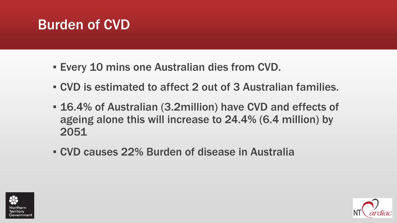
Burden of CVD
▪ Every 10 mins one Australian dies from CVD.
▪ CVD is estimated to affect 2 out of 3 Australian families.
▪ 16.4% of Australian (3.2million) have CVD and effects of ageing alone this will increase to 24.4% (6.4 million) by 2051
▪ CVD causes 22% Burden of disease in Australia
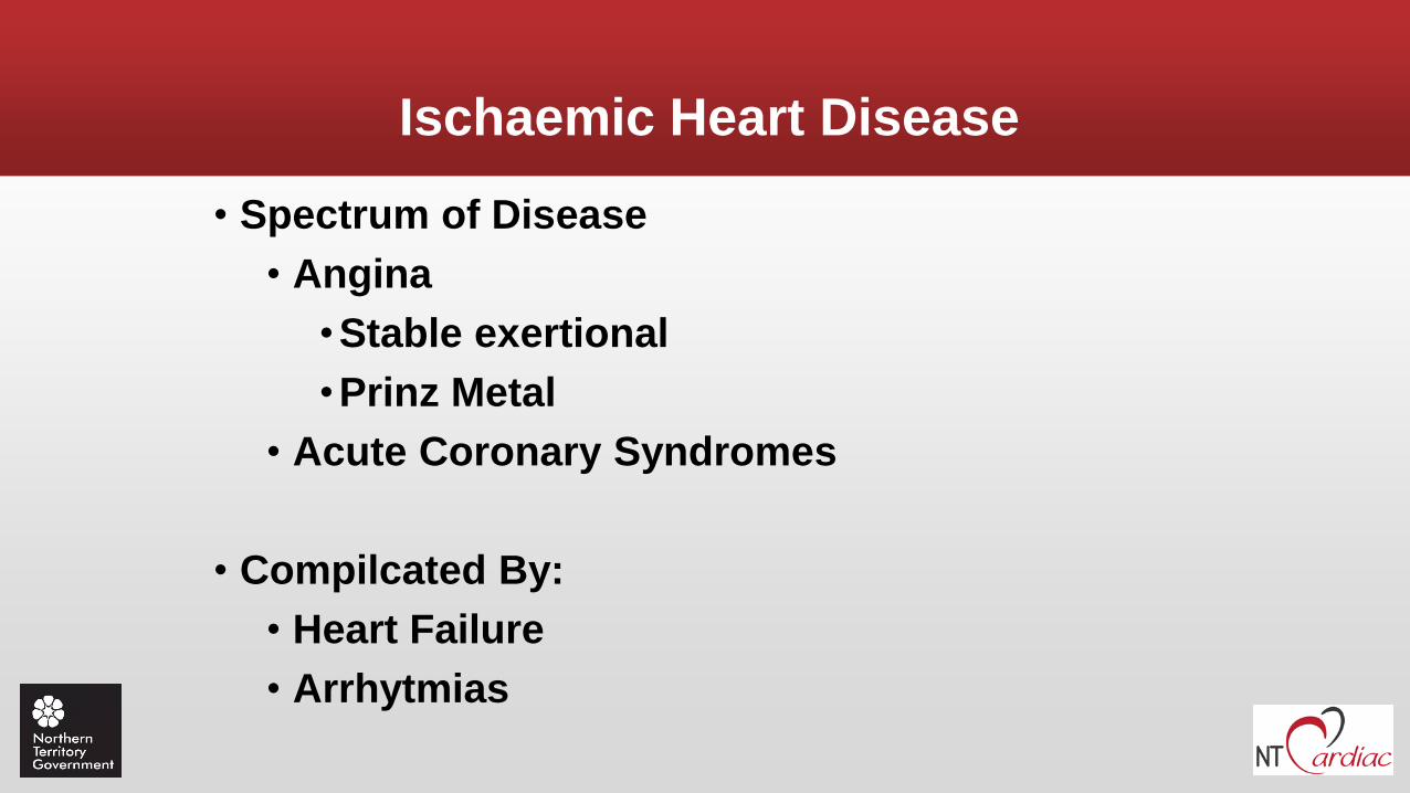
• Spectrum of Disease
• Angina
•Stable exertional
•Prinz Metal
• Acute Coronary Syndromes
• Compilcated By:
• Heart Failure
• Arrhytmias
Ischaemic Heart Disease

NSTEACS

Types of Myocardial infarction
▪ Type 1: Ischemic myocardial necrosis due to plaque rupture (ACS)
▪ Type 2: Ischemic myocardial necrosis due to supply-demand mismatch, e.g. coronary spasm, embolism, low or high blood pressures, anemia, or arrhythmias.
▪ Type 3: sudden cardiac death (no cTr values)
▪ Type 4: procedure related, post PCI or stent thrombosis ( cTr > 5X Decision Level).
▪ Type 5 post CABG (cTr > 10X Decision Level).

Acute coronary syndromes
▪ Unstable angina 2/1000 admissions per year
▪ Infarct risk 15-20%
▪ Death 15%
▪ Pathogenesis Endothelial disruption
Plaque erosion
Rupture
Platelet adhesion
Thrombus

Acute Coronary Syndromes
▪ 33% w/ confirmed MI have no CP on presentation (esp older, female, DM, CHF)
▪ 5% of NSTEMI will develop Cardiogenic Shock (60% mortality)
▪ Association between quantity of troponin and risk of death
▪ NSTEMI includes Type 2 -Type 5 biomarker elevations

Pathophysiology of ACS
Fuster V et al NEJM 1992; 326: 310–318.
Davies MJ et al Circulation 1990; 82(Suppl 3): II38-46.
Lipid pool
Macrophages
Stress, tensile, internal
Shear forces, external Fissure
Large fissure
Small fissure
Mural thrombus (NSTEACS- unstable angina vs NSTEMI)
Occlusive thrombus (STEMI)
Atherosclerotic plaque
Plaque disruption
Thrombus
One example of atherothrombotic
disease progression

18 hrs after admission

Coronary Angiography

Subsequent PTCA - 3.5x13 mm stent - IV abciximab

What is the Problem we are trying to address
▪ Despite our relatively younger population, the Northern Territory has the highest death rates from coronary artery disease (CAD) in both males and females.
▪ The higher death rates are seen in both the indigenous and non-indigenous populations.
▪ Complex pathway for patient care for ACS management in NT.
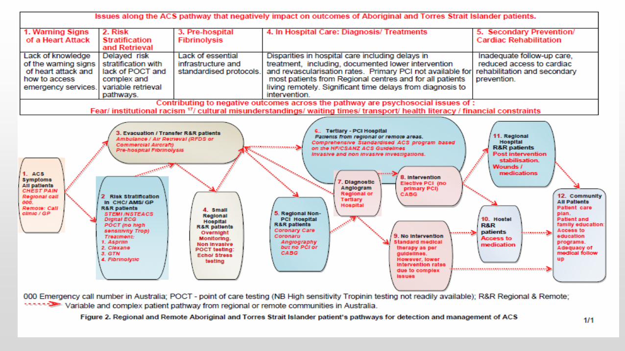
Diagram From HF ACSIAWG ATSI paper 2011

Case 1.
▪ 30 year old male from Millingimbi
▪ History:
▪ 2 hours of typical sounding chest pain presented to clinic
▪ CRF- Smoker, mild dyslipidaemia HDL 0.7, LDL 2.8
▪ Exam:
▪ Pulse 85 regular Normotensive
▪ No murmurs no failure
▪ ECG:
▪ Faxmate to oncall Cardiologist and RMP
▪ Dx – Inferior STEMI


Case 1. - cont
▪ Initial Treatment
▪ Aspirin
▪ Clopidogrel
▪ IV clexane
▪ Tenecteplase in community – given by RN
▪ Outcome:
▪ Resolution of pain and ECG changes
▪ Further Treatment
▪ Transferred to Darwin via Careflight
▪ Monitored CCU
▪ Angiogram – moderate ectasis all vessels with 70% RCA -4-5mm vessel diameter
▪ Transferred to FMC (nurse and family escort) – 5mm stent 6 days after STEMI
▪ Home in community – for teleconference post procedure after 1 week

Case 2.
▪ 37 year old lady from Yeundemu
▪ History:
▪ 7-10 hours of typical sounding chest pain presented to clinic
▪ CRF- Type II diabetes (HbA1c -14%), Smoker, dyslipidaemia LDL 3.8/HDL 0.9
▪ Exam:
▪ Pulse 60 regular Normotensive
▪ No murmurs no failure
▪ ECG:
▪ Faxmate to on call Cardiologist and RMP
▪ T wave inversion in anterior leads
▪ Dx. NSTEAC – Unstable angina – no tropinin rise

Case 2. - cont
▪ Initial Treatment ▪ Aspirin
▪ Clopidogrel
▪ SC clexane
▪ Outcome: ▪ Resolution of pain and ECG changes
▪ Further Treatment ▪ Transferred to ASH (01 May 2015)
▪ Monitored CCU – no Tropinin rise
▪ Angiogram – Severe 3 vessel disease – totally occluded RCA- collaterals from left, 70-90% proximal and mid LAD and 60-70% proximal LCX. Preserved LV function.
▪ Accepted for CABG surgery
▪ Patient wanted to return home to community – for discussion with family
▪ Further delay – risk of event 10-20%

Case 3.
▪ 59 year old male from Katherine
▪ History:
▪ 4-5 hours of typical sounding chest pain presented to Katherine Hospital 8/08/15
▪ CRF- Previous LAD stent 1998 RPH,Type II diabetes (HbA1c -14%), Smoker, dyslipidaemia LDL 2.4/HDL 0.9 (on treatment)
▪ Exam:
▪ Pulse 75 regular BP 138/85
▪ No murmurs no failure
▪ ECG:
▪ Faxmate to on call Cardiologist – RBBB with T wave inversion in inferior leads
▪ Dx. NSTEMI – troponin rise 3126

Case 3. - cont
▪ Initial Treatment
▪ Aspirin
▪ Clopidogrel
▪ SC clexane
▪ Standard medications
▪ Outcome:
▪ Resolution of pain and ECG changes
▪ Further Treatment
▪ Transferred to RDH 08/08/15
▪ Angiogram – Hazy 60-80% proximal LCX –modest OM , 40-50% instent stenosis Preserved LV function. 11/08/15
▪ For FFR - ? PCI in Darwin
▪ FFR +ve 0.76 (eg <0.85) – Therefore stented proceeded to DES 13/08/15

Issues
▪ Dual anti platelet therapy
▪ Tigagrelor vs. clopidogrel
▪ Case 1 – no data yet on safety with use with thrombolytic
▪ Case 2- good evidence to suggest better efficacy than clopidogrel
▪ Case 3- Assessed as Intermediate risk – significance of
▪ Early Revascularisation
▪ PCI Darwin vs. Adelaide –early PCI indicated in all 3 as per guidelines.
▪ Case 1. - ? Not a low risk procedure – large stent risk of slow flow
▪ Case 2- Bifurcating lesion triple vessel– therefore CABG is best option
▪ Case 3- Met low risk guidelines for PCI in Darwin – FFR guide therapy more regular use
▪ All delays form initial presentation

2011 addendum to 2006 Guidelines
The 2011 addendum to the 2006 Guidelines provides updates
to:
1. Systems of care to support delivery of ACS services
2. Early response
3. Management of patients with STEMI
4. Management of patients with NSTEACS
5. Long-term management (after control of myocardial ischaemia).1
Reference
1. Chew DP, Aroney CN, Aylward PE, et al. 2011 addendum to the National Heart Foundation of Australia/Cardiac Society of Australia and New Zealand guidelines for the management of acute
coronary syndromes (ACS) 2006. Heart Lung Circ 2011; 20(8):487–502.

Formal systems of care:
• defined continuum of care – from presentation to long-term management
• system-based approaches to deliver timely reperfusion at a local level (Grade B)
• routine audit integrated into all clinical ACS services (Grade B)
• training GPs/health workers to initiate fibrinolysis (if primary percutaneous coronary intervention [PCI]
services are not readily accessible)
• practitioners are supported by ready access to expert cardiology consultation (Consensus)
• cardiac clinical networks established with appropriate protocols (Grade B).
1. Systems of care to support delivery of ACS services
For example: iCCnet CHSA network links > 70 hospitals, health centres and general
practitioner [GP] surgeries across SA, aligned to the Health Reform Agenda principles.

2. Early response: treatment is time critical
Time from symptom onset and likely outcome
< 1 hour
Aborted heart attack or only little heart muscle damage
1–2 hours
Minor heart muscle damage only
2–4 hours
Some heart muscle damage with moderate heart muscle salvage
4–6 hours
Significant heart muscle damage with only minor heart muscle salvage
6–12 hours
No heart muscle salvage (permanent loss) with potential infarct
healing benefit
> 12 hours
Reperfusion is not routinely recommended if the patient is
asymptomatic and haemodynamically stable
In cases of major delay to hospitalisation (> 30 minutes) ambulance
crews should consider pre-hospital fibrinolysis.

STEMI - Definition
An ST-segment elevation myocardial infarction (STEMI) is confirmed by an ECG.
STEMI is defined as presentation with clinical symptoms consistent with an ACS with ECG
features including any of:
• persistent ST-segment elevation ≥ 1 mm in two contiguous limb leads
• ST-segment elevation ≥ 2 mm in two contiguous chest leads
• new left bundle branch block (LBBB) pattern.

3. Management of patients with STEMI

Early response
• Implement reperfusion strategy for patients presenting within 12 hours of onset of
ischaemic symptoms consistent with ACS (determined by physical examination):
immediate 12-lead ECG
insert cannulae
pain relief
blood tests.
• Give aspirin 150–300 mg (unless already given, or contraindicated).
• Doctor sees patient within 10 minutes of arrival (Australasian Triage Scale
Category 2).
• Oxygen therapy indicated only for patients with hypoxia (oxygen saturation < 93%) and those
with evidence of shock (Consensus).

NSTEACS – Definition
• Non-ST-elevation ACS (NSTEACS) applies to patients with suspected ACS in the absence
of other plausible causes of troponin elevation (e.g. sepsis, pulmonary embolus).
• Patients with NSTEACS may have a ‘normal’ ECG reading, or show minor changes
(occurs in up to 50% of patients).
• All patients with NSTEACS should have their risk stratified to direct management
decisions.
• The management of patients with NSTEACS requires evolving risk stratification: clinical
assessment, assessment of cardiac biomarkers and time.

4. Management of patients with NSTEACS
.
• Clinical assessment: careful clinical history,
ECG, chest X-ray and investigations to
diagnose other causes of chest pain and
evaluate the likelihood of evolving ACS.
• Troponin assessment: to assess the
likelihood of MI.
• Stratify risk.

Evolving risk stratification
Admit to coronary care unit or high
dependency unit:
• estimate ischaemic risk, estimate
bleeding risk, choose augmented
antithrombotic therapy
→refer for angiography to determine
surgery/PCI, or medical therapy.

Evolving risk stratification

Evolving risk stratification
Intermediate-risk NSTEACS
Recurrent ischaemia or elevated troponin?
YES
• admit to CCU or high dependency unit:
estimate ischaemic risk, estimate bleeding
risk, choose augmented antithrombotic
therapy
→refer for angiography to determine
surgery/PCI, or medical therapy.
NO
• undertake stress test (e.g. exercise ECG):
→positive – refer for angiography to
determine surgery/PCI, or medical therapy
→negative – proceed to discharge patient
with urgent cardiac follow-up (on upgraded
medical therapy) according to long-term
management after control of myocardial
ischaemia.

Evolving risk stratification
Appropriate period of
observation. Consider if stress
test (e.g. exercise ECG)
needed?
Stress test (e.g. exercise ECG)
using treadmill.
YES
Proceed to discharge patient with urgent
cardiac follow-up (on upgraded medical
therapy) according to long-term management
after control of myocardial ischaemia.
NO

Framework for Enabling Streamlined Patieny Care
Creating individual, family and community awareness of warning signs of heart attack and what action to take
The ACS pathway
Acute Care Management
Secondary Prevention & 3 Phase Cardiac Rehab.


High
Risk
Inter
media
te
Risk
Low
Risk
•Typical chest pain/discomfort (>10mins)
•ST elevation or depression (≥0.5 mm) or deep T wave inversion in 3
or more leads (Note: Thrombolysis for STEMI)
•Elevated serum markers (Troponin)
•Syncope
•Associated: heart failure, mitral regurgitation or gallop rhythm
•Haemodynamic compromise:
systolic blood pressure <90 mmHg
cool peripheries
diaphoresis
•Prior PCI within 6 months or prior CABG
•Presence of known diabetes (with typical Symptoms of ACS)
•CRD – eGFR<60ml/min (with typical Symptoms of ACS)
•Admit Cardiology, monitored bed, plan for angiography
•Aspirin - 300mg, then 100mg daily
•GTN 300 to 600mg SL (if BP ≥ 90mmHg)
•Morphine IV
•LMW heparin -1 mg/kg SC
•Consider Beta blocker –metoprolol 25 mg PO
•GTN IV if ongoing pain
•Repeat Troponin level and ECG at 8-24hrs
•Clopidogrel 300mg mg stat, then 75 mg daily
OR
•Tirofiban with unfractionated heparin in patients with:
•Pain/ischemia refractory to medical therapy;
•ST-segment depression or T- wave inversion >3mm in multiple leads
Assess bleeding risk in all high risk NSTEACS
Yes to ANY
Manage as Intermediate
Risk
If no to ALL
Proceed to Low risk
•Resolved but prolonged (>10mins) or repetitive chest pain
•Nocturnal pain
•New onset exertional chest pain/discomfort
•Age ≥65 years Caucasian (or ≥45 Indigenous)
•Known IHD: prior MI, LVEF ≤40% or known coronary lesion >50%
stenosed
•Patient history of 2 or more of:
•hypertension,
•family history
•active smoking,
•hyperlipidaemia
•Diabetes and/or Chronic Renal Disease – eGFR <60ml/min (with
atypical symptoms of ACS)
•Onset of angina within the last month.
•Worsening of the severity or frequency of angina
•Lowering in the anginal threshold.
•Short duration (<1min)
•Atypical pain
and
•No risk factors
•Admit Cardiology, monitored bed,
•Morphine & Aspirin as per High Risk
•Repeat Troponin and ECG 9 hr from pain onset
•Repeat ECG if ongoing pain.
•Review results of repeat Troponin and reclassify as high risk on
basis of troponin elevation, ECG changes or clinical status.
•In-patient exercise stress test if 9hr troponin negative, no further
chest pain, and no high risk ECG changes.
RDH ECG Department
Phone 8922 8468 Fax 8944 8070
•Discharge for early Cardiology review if negative.
•Discharge for outpatient assessment.
+/-Outpatient exercise test
NT Cardiac 8920 6250 Fax 8945 1365
•Re-consider other diagnoses/ 8hr TnI
•No further investigation required. D/c
Yes to ANY
Manage as High Risk
If no to ALL
Proceed to intermediate
risk
Yes to ANY
and no higher risk
features
Manage as Low Risk
Standardised Guideline RDH - Chest Pain Risk Stratification


Chest pain /Suspected Heart Attack – In Community
1) All CP patients perform ECG +
Troponin. (As per CARPA page 59)
2) ECG sent to Epiphany or Fax mate
8918000 and Call RMP if no answer
call on call Cardiologist 1300000324
As per ACS protocol
1. If no ECG Changes, no Troponin rise after 4
hours and no further chest pain use heart
score and discuss with cardiologist
2. If score >3 and pain in last 24 hours – for
acute transfer to nearest hospital
3. If score >3 and pain >24 hours previously for
EST and review at nearest regional hospital
4. If score less <3 and pain atypical – For
review RMP – consider alternative diagnosis

ACS Network
• NT Remote communities and GDH, KDH, TCH ▪ digital ECG’s
▪ Phone / Fax System
▪ Dedicated Fax system ▪ 8918 8000 – Fax ECG (Faxmate)
▪ ECG is emailed to on call Cardiologist ( 4 local Cardiologists)
▪ Phone: ▪ Dedicated Phone system: 1300 000ECG – 1300 000324
▪ RDH switch to transfer calls to on call Cardiologist - Escalation to 2nd on call -24/7
▪ Flow Chart
▪ Cardiologist informs treatment plan
▪ Community clinic treats and arranges transfer as per current protocols.
• Commenced October 26th 2013
• 1860 ECG’s reviewed in first 18 monthe
• Increased Thrombolysis in communities – no complications

ACS Network Cont
▪ Final Phase
• Digital ECG Service:
• Activate Epiphiny Server
• Data storage, transfer and retrieval system (CVIS)
• Ongoing Training program for all other communities.
• Flow Chart Modification – as now direct ECG transmission
• Dedicated Phone system – 1300 000 ECG (324)
• Faxmate will remain as backup
• Further Developments
• Incorporate Heart Foundations Warning Signs AMI into CARPA
• Change from Clopidogrel to Ticagrelor for all High Risk ACS patients (not STEMI)
• Develop protocols for ? PCI within 24 hours of thrombolysis.

Epiphany - Main Screen

1. Site Codes and Locations
Site Code Location Site
Code Location
Site
Code Location
Site
Code Location
Amoonguna Amoonguna RAL Alyangula RJH Jabiru RTJ Titjikala
Ampilatwatja Ampilatwatja RAN Angurugu RLN Lake Nash RTR Tara
Areyonga Areyonga RAR Adelaide River RLR Laramba RTT Ti Tree
Bulman Bulman RBI Bickerton Island RMA Yuelamu RUH Umbakumba
Galiwinku Galiwinku RBL Belyuen RMG Milingimbi RWA Kings Canyon
Kalkarindji Kalkarindji RBO Bonya (Baikal) RML Mt Liebig RWB Wagait Beach
Kintore Kintore RBR Borroloola RMN Maningrida RWD Woodycupaldiya
Lajamanu Lajamanu RCC Canteen Creek RNB Numbulwar RWH Warruwi
Mataranka Mataranka RCI Minjilang RNC Nguiu RWI Willowra
Minyerri Minyerri RDO Docker River RNY Nyirripi RWL Wilora (Stirling)
Ngukurr Ngukurr RDR Nauiyu
Nambiyu ROP Gunbalanya RWR
Wallace Rock
Hole
Pigeon Hole Pigeon Hole REL Elliott RPC Pine Creek RYD Yuendumu
Santa Teresa Santa
Teresa REN
Alcoota
(Engawala) RPG Pirlangimpi RYU Yulara
Timber Creek Timber Creek
REP Epenarra (Wutunurrgurru)
RPK Wadeye (Port Keats)
Utopia Utopia RFN Aputula RPL Palumpa
Wugularr (Beswick)
Wugularr (Beswick)
RGP Gapuwiyak RPP Papunya
Yarralin Yarralin RHB Hermannsburg RRM Ramingining
Yirrkala Yirrkala RHM Haast Bluff RRR Robinson
River
RAB Batchelor RHR Harts Range RSB Milikapiti
RAC Ali Curung RIM Imanpa RTH Ti Tree6Mile
Version number Purpose Changes Author Date
0.1 Initial draft SO 30/01/2015
0.2 Draft reviewed RN 01/02/2015
0.3 Draft finalised RN 02/02/2015
1 Final draft approved MI 03/02/2015
1.1 Amendment for Trial Period SO 03/03/2015
1.2 Amendment for Trial Period RN 10/04/2015

Rapid Chest Pain Assessment Unit
▪ Principle:
▪ Chest Pain/ ? Angina symptoms -Diagnosis and further risk stratification within 2 weeks of onset • Darwin
• Located ground floor DPH
• Stress test, Stress Echo, Nuclear Perfusion Scans, CT CA, Coronary angiography and PCI (angioplasty)
• Cardiology Consult – Cardiologist or Registrar.
• Inpatient exercise testing post negative troponins.
• Out Patients referrals GP’s
• Katherine and Gove District Hospitals • Stress Test inpatient troponin negative and GP referral
• Stress Echo / Cardiologist or Registrar review 4 weekly clinics
• Groote Eylandt • Planned Stress test for trop negative patients and for GP referral – Oct 2017

Rapid Chest Pain Assessment Unit
▪ Principle:
▪ Chest Pain/ ? Angina symptoms -Diagnosis and further risk stratification within 2 weeks of onset
• Alice Springs Hospital • Located ground floor ASH -
• Stress test, Stress Echo, CT CA
• Cardiology Consult – Cardiologist (3-4 days 3 weeks out of 4) or Registrar.
• Inpatient exercise testing post negative troponins.
• Out Patients referrals GP’s
• Tennant Creek Hospital • Stress Test inpatient troponin negative and GP referral
• Stress Echo / Cardiologist or Registrar review 8 weekly clinics

ACS – Pathway On going developments
▪ Expanded PCE
▪ Secondary Prevention
▪ Patient Centered Care
▪ Hospital based at time of acute care Phase 1
▪ Phase 2-3
▪ Out reach services
▪ Multi Disciplinary

Framework for ongoing development and improvement of patient pathway with ACS
Creating individual, family and community awareness of warning signs of heart attack and what action to take
The ACS pathway
Acute Care Management
Secondary Prevention & 3 Phase Cardiac Rehab.