Embed Size (px)
DESCRIPTION
Acute Coronary Syndrome. APS Fleming College. What is an ACS?. A sudden event during which the myocardium suffers from a relative or a complete lack of perfusion Covers the continuum between angina and MI This is reflected in: Signs and symptoms Electrocardiographic changes - PowerPoint PPT Presentation
Citation preview
Acute Coronary Syndrome
APS
Fleming College

What is an ACS?
A sudden event during which the myocardium suffers from a relative or a complete lack of perfusion
Covers the continuum between angina and MI
This is reflected in:Signs and symptomsElectrocardiographic changesBiochemical changes
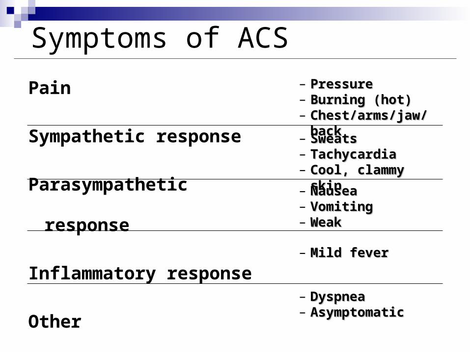
Symptoms of ACS
Pain
Sympathetic response
Parasympathetic response
Inflammatory response
Other
– PressurePressure– Burning (hot)Burning (hot)– Chest/arms/jaw/backChest/arms/jaw/back
– SweatsSweats– TachycardiaTachycardia– Cool, clammy skinCool, clammy skin
– NauseaNausea– VomitingVomiting– WeakWeak
– Mild feverMild fever
– DyspneaDyspnea– AsymptomaticAsymptomatic

Physical FindingsPhysical Findings
Inspection
BP - often increase anterior MI
- often decrease inferior MI
HR - often increase anterior MI
- often decrease inferior MI
RA po - increase in RV MI
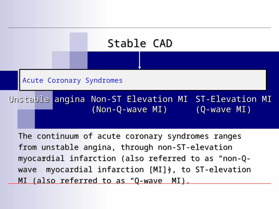
Acute Coronary Syndromes
Unstable anginaUnstable angina ST-Elevation MIST-Elevation MI(Q-wave MI)(Q-wave MI)
Non-ST Elevation MINon-ST Elevation MI(Non-Q-wave MI)(Non-Q-wave MI)
Stable CADStable CAD
The continuum of acute coronary syndromes ranges from unstable The continuum of acute coronary syndromes ranges from unstable
angina, through non-ST-elevation myocardial infarction (also referred angina, through non-ST-elevation myocardial infarction (also referred
to as to as ““non-Q-wavenon-Q-wave”” myocardial infarction [MI]), to ST-elevation MI myocardial infarction [MI]), to ST-elevation MI
(also referred to as (also referred to as ““Q-waveQ-wave”” MI). MI).

Platelets & ACS
Platelets become activated by various stimuli
Binds to fibrinogen and serves to cross-link and aggregate platelets
Platelet plug becomes the centre of a larger thrombus

Triggers to Plaque Rupture
Inflammatorycytokines
Plaque RupturePlaque Rupture
Physical Stress
VulnerablePlaque
EmotionalStress

Extent of Myocardial Injury
muscle mass perfused by vessel
Magnitude/Duration of flow
Oxygen demand of affected tissue
Adequacy of collaterals
Tissue response to ischemia
Determined by:Determined by:

Preventing ACS / Reducing Infarct Size
Primary prevention -lifestyle Stabilizing plaque Preventing platelet aggregation Decreasing preload and afterload Decreasing cardiac workload Reperfusion Treating arrhythmia
Which ones do you as medics do??
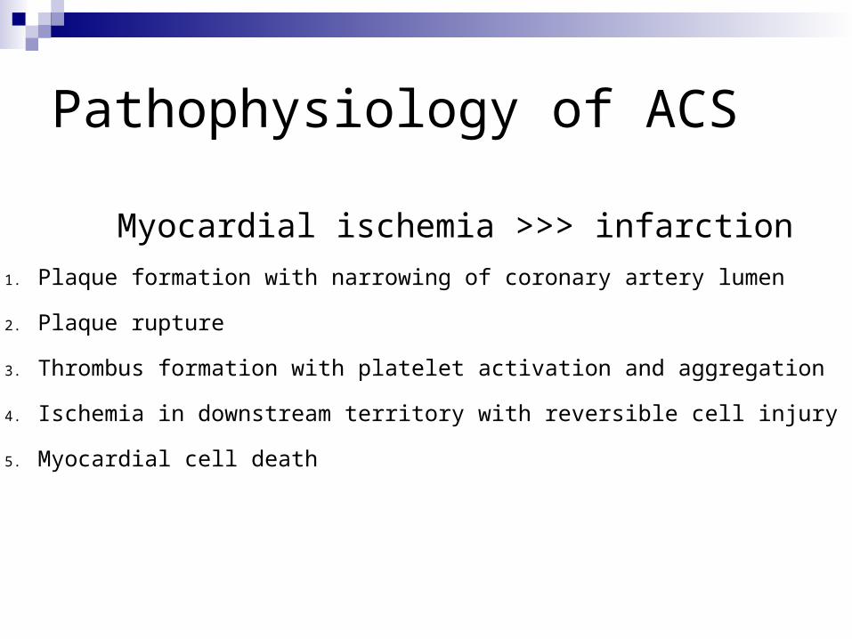
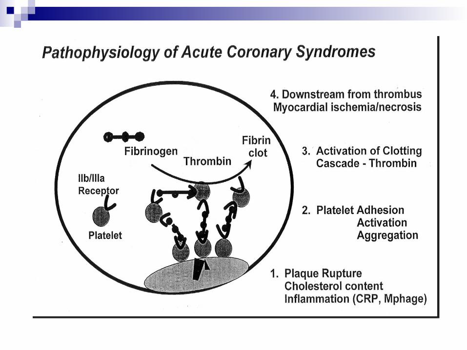
Pathophysiology of ACS
Myocardial ischemia >>> infarction
1. Plaque formation with narrowing of coronary artery lumen
2. Plaque rupture
3. Thrombus formation with platelet activation and aggregation
4. Ischemia in downstream territory with reversible cell injury
5. Myocardial cell death

Cardiovascular Pathology Angina Unstable angina Myocardial Infarction Congestive heart failure Valvular dysfunction Cardiogenic shock Aneurysms Deep vein thrombosis/arterial occlusion

Ischemic Chest Pain Good history and physical exam
3 & 12/15lead ECG
OPQRST to guide history investigation
Differential diagnosis
Chapter 27 27.24-27.36

OPQRST
Onset - when did it start? Provoked - at rest, exertion, better or
worse? Quality - sharp, dull, ache, heaviness? Radiating - to shoulder, back, jaw? Severity - on a scale of 1-10? Time - does it come and go?

Myocardial Ischemia Blood supply and
demand Causes of ischemia
HR – too slow or too fast vasospasm (Prinzmetals) coronary artery occlusion narrowing of coronary arteries low blood pressure hypoxemia

Coronary Artery Disease
Poor dietary habits
Imbalance between good cholesterol (HDL)
and bad cholesterol (LDL)
Atherosclerosis
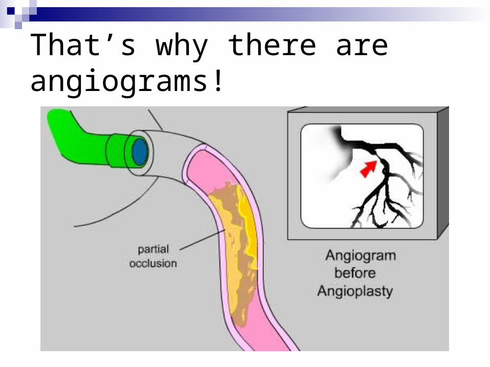
That’s why there are angiograms!
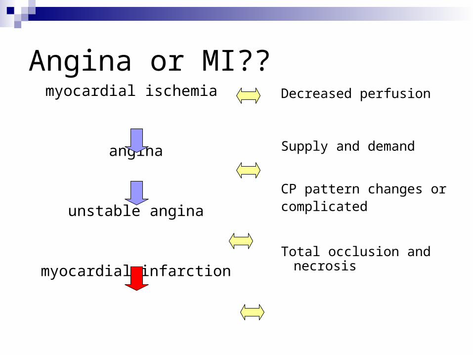
Angina or MI??myocardial ischemia
angina
unstable angina
myocardial infarction
Decreased perfusion
Supply and demand
CP pattern changes orcomplicated
Total occlusion and necrosis

Consequences ofCoronary Thrombosis
Lilly. Pathophysiology of Heart Disease, 4th Ed. Lippincott Williams, 2007. Page 173

The Importance of History Someone with angina knows their typical
pattern In addition to OPQRST, take an AMPLE history “are you SOB?” Similar pain? MI in past? Is this pain similar? Other cardiac Hx? e.g. CABG,
angioplasty ,stress testing, hospitalizations etc.
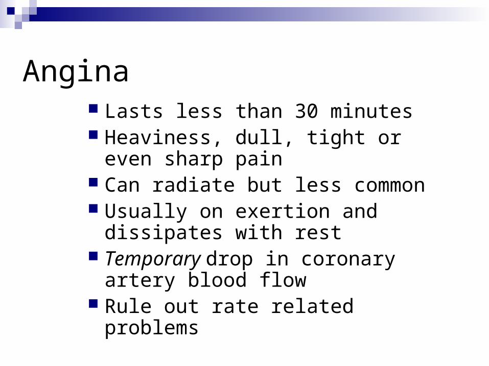
Angina Lasts less than 30 minutes Heaviness, dull, tight or even sharp
pain Can radiate but less common Usually on exertion and dissipates
with rest Temporary drop in coronary artery
blood flow Rule out rate related problems

Angina Management Rest/relaxation (Be calm!) O2 therapy IV access ? ASA 160mg chewed & swallowed Vitals Nitroglycerin 0.4mg SL q 5 min. if SBP >100 and
HR > 60 but less than 160 bpmHR and blood pressure parameters…why?

Hemodynamic Parameters
SBP < 100preload reduction - vasodilationdecreased coronary perfusion
HR < 60 bpm or > 160 bpmrate related ischemia?
Ischemic Chest Pain
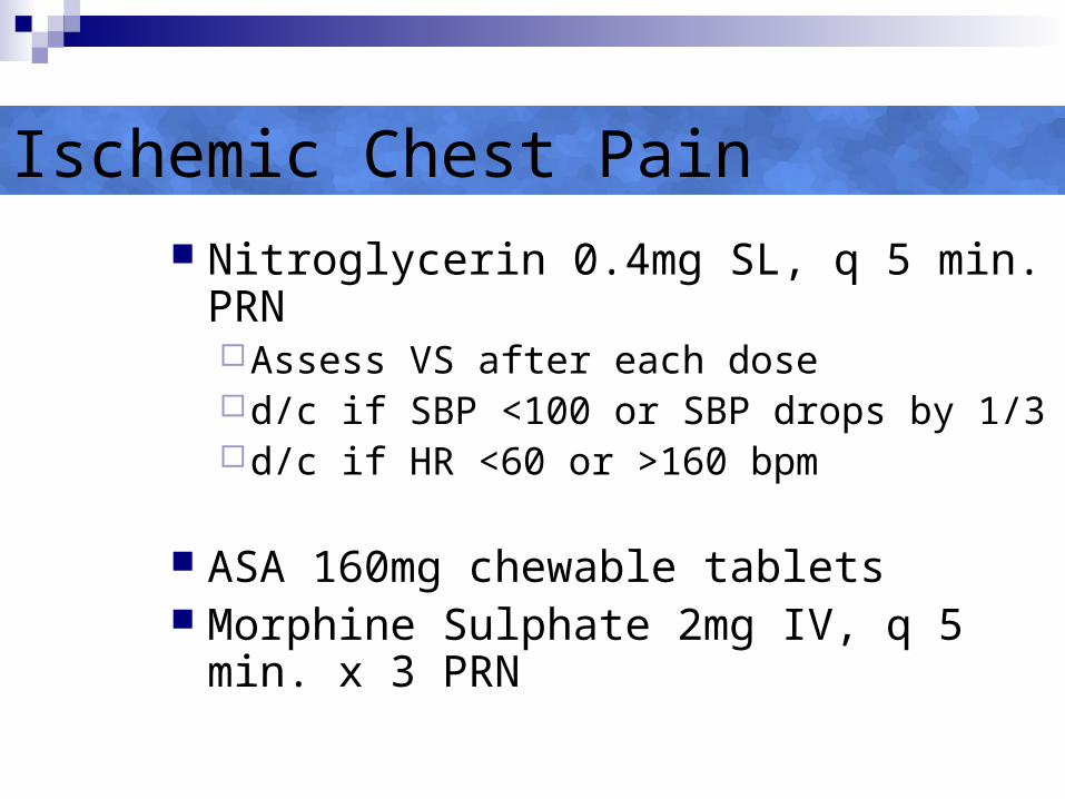
Nitroglycerin 0.4mg SL, q 5 min. PRNAssess VS after each dosed/c if SBP <100 or SBP drops by 1/3d/c if HR <60 or >160 bpm
ASA 160mg chewable tablets Morphine Sulphate 2mg IV, q 5 min. x 3
PRN
Unstable Angina
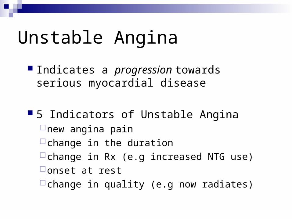
Indicates a progression towards serious myocardial disease
5 Indicators of Unstable Anginanew angina pain change in the durationchange in Rx (e.g increased NTG use)onset at rest change in quality (e.g now radiates)
Acute Myocardial Infarction
Definition:Necrosis of heart muscle due to absolute
or relative lack of blood supply to the myocardium.
The site of infarction is determined by the location of the arterial occlusion.

Myocardial Infarction
Killing of myocardial tissue Conventional treatment will only save
ischemic zone Thrombolytics
Ischemic zone
Necrotic zone

Presentation of Myocardial Infarction
Varies widely from patient to patient Typical vs. Atypical (e.g.. weakness or
SOB) The elderly, alcoholics, and women CP unresolved with rest or NTG 12 Lead shows acute ST elevation,
Flipped T waves or Q waves (old)

Electrocardiographic Changes
Change in rate and rhythm
Most often sinus with:No discernible changeHyper-acute T-wavesT wave flattening or inversionST segments up or downQ waves

Biochemical Markers of ACS
Enzymes which are unique to cardiac myocytes
Released into the circulation by dead cells
Thus a rise in these indicates that myocardium has suffered damage
Biochemical Markers
Troponin (remember its role in actin/myosin binding??)
CPK (Creatine Phosphokinase) Specifically one isoenzyme -MB band
LDH (Lactose dehydrogenase) AST
Hospital staff will draw blood for these tests early but do NOT generally help in the decision making
Cardiac Markers
Myoglobin is found in cardiac and skeletal muscle
Very sensitive if measured early
Not specific
Not often used

Use of Nitrates in ACS
Nitrates, typically nitroglycerin
Nitrous Oxide acts as a smooth muscle relaxant leading to vasodilatation
Transdermally, sublingually and/or parenterally
Benefits of Nitrates
Reduce preload and afterloadDilate coronary arteriesAssists coronary perfusion

Disadvantages of Nitrates
Not useful in patients who are reperfused
May cause hypotensionSevere hypotension in patients with RV
dysfunctionMay cause hypotension in inferior MI
30% have RV involvement-check!!!
-blockers
Multi-purpose in the setting of ACS
Anti-arrhythmicAnti-ischemicAnti-hypertensive

-blockers
Decrease myocardial oxygen demand
Decreased heart rate – increases diastoleDecreased myocardial contractilityDecreased MAP

Advantages of -blockers
Reduction in pain Decreased morbidity and mortality Decreased risk of arrhythmia Decreased infarct size Decreased risk of re-infarction
Treatment with a -blocker is a standard of care

ASA
Anti-inflammatory prevents the formation of arachidonic acid
A pathway that can be blocked to prevent platelet aggregation
Does not block all platelet activators
Other Treatment Modalities in ACS
Heparins (LMWH) Reperfusion
PCIMechanical (the digger!)Thrombolytic agents
Antiarrhythmic agents
Focus of ACS
Common reason for transport Much can be done during transfer Reduce risk of morbidity and mortality
The first step = recognizing the ACS Signs and symptoms ECG changes Biochemical changes

Summary
Strategies for reducing morbidity and mortality
Reduce cardiac workload Improve perfusion to cardiac tissueReduce risk of fatal arrhythmiasReduce extension of clot formationReperfuse the ischemic myocardium

Myocardial Infarction ASA (2 x 80 mg) P.O. O2 therapy IV access NTG via SL, transdermal, and/or IV Morphine (ACP) Heparin and/or Beta blockers (Hosp) 12/15 Lead ECG as soon as possible Angioplasty (hosp) Thrombolytics if PCI not available or contraindicated

PCI

Pre-hospital Thrombolysis
Oshawa Land ALS Northern Ornge Bases
Frequent use Southern Ornge Bases
Carry it Positive empirical trends

Pre-hospital Thrombolysis
prolonged transport time
no thrombolysis at the sending facility
Long delays

Indications - Thrombolysis
Ischemic C.P.
Less than 6 hours duration

Ischemic Chest Pain?
O - at rest or with exertion
P – better or worse
Q - heaviness, tightening, sharp, weakness etc
R - neck, jaw and/or left arm
S - varies
T - consistent, does NOT come & go

12 Lead ECG Criteria
ST segment elevation New onset Left Bundle Branch Block
with S&S? Some acute coronary syndromes
(A.C.S.) do not benefit from thrombolysis

Next
We will be looking at 12 leads specifically Look at the hand powerpoint (12lead) Read the sample handouts Read your text 12 lead ECG (big book)