Embed Size (px)
Citation preview
RESEARCH ARTICLE
Activation of c-Jun by human cytomegalovirus
UL42 through JNK activation
Tetsuo KoshizukaID*, Naoki Inoue
Microbiology and Immunology, Gifu Pharmaceutical University, Gifu, Japan
Abstract
c-Jun is a major component of the AP-1 transactivator complex. In this report, we demon-
strated that AP-1 was activated by the expression of UL42, a human cytomegalovirus-
encoded membrane protein that has two PPXY (PY) motifs and a C-terminal transmem-
brane domain (TMD). Although UL42 interacts with Itch, an ubiquitin E3 ligase, through the
PY motifs, UL42 phosphorylated c-Jun and c-Jun N-terminal kinase (JNK) in the absence of
any interaction with Itch. Experiments using mutated versions of UL42 suggest the impor-
tance of the carboxyl half (a.a. 52–124) of UL42 for the activation of the JNK signaling, while
C-terminal TMD alone is not sufficient. Thus, we hypothesize that UL42 plays a role in the
activation of JNK signaling in HCMV-infected cells. (118 words).
Introduction
The proto-oncogene c-Jun, one of the most studied transactivator proteins, is a major compo-
nent of the heterodimeric AP-1 transcription factor family [1]. Activated c-Jun is transported
into the nucleus, where it forms the AP-1 heterodimer complex and binds to promoter regions
of target genes. Phosphorylation of c-Jun at Serine 63 (Ser63) and Ser73 by c-Jun N-terminal
kinase (JNK) regulates c-Jun transcription activities [2, 3]. The c-Jun/JNK pathway is activated
by various extracellular stimuli, including infection, inflammation, oxidative stress, DNA dam-
age, osmotic stress, and cytoskeletal changes [4]. As JNK is a key component of the innate
immunity pathways, pathogens have developed strategies to modulate the JNK signaling events
[4]. While suppression of JNK signaling has some advantages to many pathogens, other patho-
gens activate the JNK pathway. For example, Epstein-Barr virus (EBV) LMP1 activates JNK
through TRAF signaling [5]. Human cytomegalovirus (HCMV) IE1 activates the phosphoryla-
tion of c-Jun [6]. Further, the activation of JNK is essential for effective viral protein expression
and replication in varicella-zoster virus-infected neuronal cells [7]. Therefore, the regulation of
c-Jun/JNK signaling by viral proteins is important for the replication of some viruses.
The UL42 gene product of HCMV is a membrane protein that contains two PPXY (PY)
motifs to interact with Itch, a member of the ubiquitin E3 ligase Nedd4 family [8]. UL42 and its
alpha- and beta-herpesvirus homologs share a number of conserved structures including the
PY motifs in their N-terminal domain and the C-terminal transmembrane domain (TMD), but
the function of other domains remains to be elucidated [9–11]. All these homologs interact
with Itch through their PY motifs. As Itch ubiquitinates various substrates, it plays multiple
roles in signal transduction, intracellular trafficking, cell survival and immune responses [12].
PLOS ONE
PLOS ONE | https://doi.org/10.1371/journal.pone.0232635 May 5, 2020 1 / 12
a1111111111
a1111111111
a1111111111
a1111111111
a1111111111
OPEN ACCESS
Citation: Koshizuka T, Inoue N (2020) Activation of
c-Jun by human cytomegalovirus UL42 through
JNK activation. PLoS ONE 15(5): e0232635.
https://doi.org/10.1371/journal.pone.0232635
Editor: Michael Nevels, University of St Andrews,
UNITED KINGDOM
Received: January 24, 2020
Accepted: April 17, 2020
Published: May 5, 2020
Peer Review History: PLOS recognizes the
benefits of transparency in the peer review
process; therefore, we enable the publication of
all of the content of peer review and author
responses alongside final, published articles. The
editorial history of this article is available here:
https://doi.org/10.1371/journal.pone.0232635
Copyright: © 2020 Koshizuka, Inoue. This is an
open access article distributed under the terms of
the Creative Commons Attribution License, which
permits unrestricted use, distribution, and
reproduction in any medium, provided the original
author and source are credited.
Data Availability Statement: All relevant data are
within the paper and its Supporting Information
files.
Funding: This work was partly supported by the
Grants-in-Aid for Scientific Research from the
Japan Society for the Promotion of Science (JSPS

Indeed, Itch is involved in the negative regulation of c-Jun/JNK signaling through ubiqutina-
tion of c-Jun [13]. Fu and colleagues have recently reported that UL42 inhibits DNA binding,
oligomerization and enzymatic activity of cyclic GMP-AMP synthase to antagonize innate anti-
viral responses in a Nedd4 family- independent manner [14].
In the present study, we investigated whether UL42 regulated c-Jun activation through
its interaction with Itch. For this purpose, we performed mapping of the UL42 functional
domains for AP-1 transcriptional activation, nuclear localization of c-Jun, and phosphoryla-
tion of c-Jun and JNK. Unexpectedly, we found that UL42 activated c-Jun in an Itch-inde-
pendent manner. Thus, UL42 has the ability to regulate JNK signaling among HCMV-
encoded proteins.
Materials and methods
Cells
The HEK293T cells (RIKEN Cell Bank, Tsukuba, Japan) were cultured in Dulbecco’s mini-
mum essential medium (DMEM) supplemented with 10% fetal bovine serum (FBS), 100U/ml
penicillin and 100U/ml streptomycin. Immortalized human fibroblasts (hTERT-BJ1) were cul-
tured in DMEM-medium 199 (4:1) supplemented with 10% FBS, 2 mM L-glutamine, 1 mM
sodium pyruvate, 100 U/ml penicillin, and 100 μg/ml streptomycin.
Plasmids and transfection
pCAGGS-HAUL42WT, pCAGGS expressing wild-type UL42 with an HA-tag at the N-ter-
minus, was described previously [8, 15]. Primers P1-13 used in this report were shown in
Supporting Information (S1 Table). pCAGGS-HAUL42ΔN and -HAUL42ΔI, pCAGGS
expressing HA-tagged UL42 lacking the amino acid (a.a.) 1–50 and 51–86 regions, respec-
tively (Fig 1A), were constructed by the inverse PCR-based method using pCAGG-
S-HAUL42WT as a template along with primer pairs P1 and P2 for ΔN and P3 and P4 for
ΔI, respectively.
A PCR-amplified fragment encoding the UL42 open reading frame using with primers P5
and P6 was inserted between the BamHI and XhoI sites of pEGFP-C1 (Takara Bio, Shiga,
Japan) to construct pEGFP-UL42WT. pEGFP-UL42AY, a PY motif-disrupted (PPXY to
AAXY alteration) mutant, was constructed by the QuickChange site-directed mutagenesis
(Agilent technologies, St Clara, CA) of pEGFP-UL42WT using primers P8-P11.
The C-terminal TMD of UL42 was amplified by PCR using primers P4 and P7 and
inserted between the BglII and SalI sites of pEGFP-C1, generating pEGFP-UL42Ct. Integri-
ties of all inserts were confirmed by DNA sequencing. All plasmids used were purified with
a Qiagen plasmid plus midi kit (Qiagen, Venlo, Netherlands). HEK293T cells were trans-
fected with the indicated plasmids using ScreenFect A (Fuji-Film Wako Pure Chemical,
Osaka, Japan).
Luciferase assay
The control and reporter plasmids, 0.01 μg/well of pRL-TK (Promega, Madison, WI) and
0.1μg/well of pAP1(PMA)-TA-Luc, which contains the firefly luciferase gene under the control
of a minimal promoter with multiple copies of the AP-1 enhancer elements (Takara Bio), and
0.1 μg/well of UL42 expression plasmids were transfected to HEK293T cells in 96-well plates.
At 48h post-transfection, the luciferase activity of the cells was analyzed with a Dual-Glo Lucif-
erase assay kit (Promega). The ratios of firefly luciferase activities to Renilla luciferase activities
were obtained in triplicated wells in each experiment.
PLOS ONE Activation of c-Jun by HCMV UL42
PLOS ONE | https://doi.org/10.1371/journal.pone.0232635 May 5, 2020 2 / 12
KAKENHI 17K10185) to T.K. and a grant from the
Takeda Science Foundation to T.K.
Competing interests: The authors have declared
that no competing interests exist.

Fig 1. UL42 mutant constructs and their ability to activate AP-1-dependent transcription. A. HCMV UL42 has two PY motifs
(closed boxes) and a C-terminal transmembrane domain (TMD; a closed box in gray). In the PA and AY mutants, the PPXY sequences
were substituted to PPXA (open boxes) and AAXY (hatched boxes), respectively. UL42 and its mutated forms were fused with HA or
EGFP tags at the N-terminus of UL42. Residue numbers are based on the position in wild-type (WT) UL42. B and C. Indicated plasmid
expressing HA-tagged (B) or EGFP-tagged (C) UL42 mutants and the luciferase reporter and control plasmids, pAP1(PMA)-TA-Luc
and pRL-TK, were transfected into HEK293T cells. Means ± SEMs of the ratios of firefly luciferase activities to Renilla luciferase
activities obtained in three independent experiments (S1 Fig) are shown. The p-values were determined by the one-way ANOVA
followed by the Tukey’s multiple comparison test. ��p<0.01, ���p<0.005, ����p<0.001. (B) pCAGGS (Vec), pCAGGS-HAUL42WT
(WT), -HAUL42PA (PA), -HAUL42ΔN (ΔN), and -HAUL42ΔI (ΔI). (C) pEGFP-C1 (Vec), -UL42WT (WT), -UL42AY (AY), and
-UL42Ct (Ct).
https://doi.org/10.1371/journal.pone.0232635.g001
PLOS ONE Activation of c-Jun by HCMV UL42
PLOS ONE | https://doi.org/10.1371/journal.pone.0232635 May 5, 2020 3 / 12

Antibodies
The anti-HA rabbit polyclonal antibody, and anti-GFP monoclonal antibody (dilution ratio
1:1000) (MBL, Nagoya, Japan), anti-c-Jun (dilution ratio 1:1000), anti-phospho-c-Jun (S63)
(dilution ratio 1:1000), and anti-Itch monoclonal antibodies (dilution ratio 1:1000) (BD Bio-
science, Franklin Lakes, NJ), anti-JNK and phospho-JNK rabbit polyclonal antibodies (dilu-
tion ratio 1:250)(Cell Signaling Technology, Danvers, MA), and anti-actin monoclonal
antibody (dilution ratio 1:5000) (Merck, Darmstadt, Germany) were purchased as indicated.
Anti-UL42 rabbit polyclonal antibody was raised against GST-UL42 (dilution ratio 1:2000).
Peroxidase-conjugated anti-mouse or anti-rabbit IgG antibodies (dilution ratio 1:2000 and
1:5000, respectively) (GE healthcare, Chicago, IL), and Alexa Fluor 488-, 594- or 647-conju-
gated antibodies (Thermo Fisher Scientific, Waltham, MA) were used as the secondary
antibodies.
Immunoblots and immunofluorescence analysis
Immunoblotting analyses were performed essentially as described elsewhere [9]. In brief,
HEK293T cells were transfected with 0.6 μg/well of plasmids, cultured for 48h and lysed in the
Laemmli’s sample buffer. After boiling at 98 ˚C for 5 min and brief sonication, cell lysates were
separated on sodium dodecyl sulfate-poly-acrylamide gels and transferred to PVDF mem-
branes (Millipore). After blocking with PBS-T (0.05% Tween 20 in PBS) containing 5% skim
milk (Fuji-Film Wako Pure Chemical), the membranes were washed three times with PBS-T
and incubated with primary antibodies diluted in PBS-T containing 1% bovine serum albumin
(BSA) at 4 ˚C for overnight. After washing three times with PBS-T, membranes were incubated
with peroxidase-conjugated secondary antibodies diluted into PBS-T containing 5% skim-
milk at room temperature for 3 h, and then washed three times with PBS-T. After reaction
with Immunostar LD reagent (Fuji-Film Wako Pure Chemical), signals were detected in Che-
miDoc system (BioRad Laboratories, Hercules, CA).
Cells were grown on coverslips and fixed with 4% paraformaldehyde in PBS at 48 h post-
transfection. The cells were permeabilized with PBS containing 0.05% Triton X-100 and
stained with DAPI (Thermo Fisher Scientific) and the indicated antibodies at room tempera-
ture for 30 min. Samples were mounted on slide glass with antifade (Thermo Fisher Scientific)
and analyzed with confocal microscopy (LSM700, Carl Zeiss, Oberkochen, Germany). Data
capture was done under conditions identical among series of the samples.
Recombinant HCMV strains
The recombinant viruses were constructed using the two-step Red-mediated mutagenesis [16].
To recover the epithelial and endothelial cell tropisms of HCMV strain Towne, the UL130
gene was repaired as described previously [17] to generate TowBACdTT-WT genome. The
UL42 open reading frame was deleted from TowBACdTT-WT genome to construct Tow-
BACdTT-ΔUL42 as described previously [8]. Briefly, a DNA fragment containing kanamycin
resistant gene (Kmr) and I-SceI site was amplified with primers P12 and P13 and inserted into
pCAGGS-UL42WT and pCAGGS-UL42PA. I-SceI-Kmr-UL42 fragments were amplified with
primers P13 and P14, inserted into TowBACdTT-ΔUL42 genome, and Kmr sequence was
removed to generate TowBACdTT-UL42R and TowBACdTT-UL42PA. Successful recombi-
nation was confirmed by PCR, Southern blotting and DNA sequencing. Those BAC genomes
were purified using a Nucleobond BAC 100 kit (TaKaRa Bio) and transfected to human fibro-
blasts to reconstitute infectious recombinant viruses.
PLOS ONE Activation of c-Jun by HCMV UL42
PLOS ONE | https://doi.org/10.1371/journal.pone.0232635 May 5, 2020 4 / 12

Statistical analysis
Data of luciferase assays and cell counting were based on three independent experiments, and
each set of experimental results was analyzed statistically with GraphPad PRISM software
ver.8.3 (GraphPad Software, San Diego, CA). The luciferase data obtained in triplicated wells
were analyzed with the one-way ANOVA test followed by the Tukey’s multiple comparison
test. Immunofluorescence assay data were digitalized with Photoshop software (Adobe sys-
tems, San Jose, CA), and the percentages of nuclear c-Jun-positive cells among the cells
expressing UL42 derivatives or EGFP were obtained in three random fields in each experi-
ment. Then, the percentages obtained in three independent experiments were analyzed with
the one-way ANOVA test followed by the Tukey’s multiple comparison test.
Results
AP-1 signaling activation by UL42 expression
To measure the AP-1-dependent transcriptional activities, HEK293T cells were transfected
with a UL42-expressing plasmid and a reporter plasmid, pAP1(PMA)-TA-Luc, for a luciferase
assay. Expression of UL42 wild type (WT) enhanced luciferase activities (Fig 1B). Expression
of UL42 mutant protein UL42PA, which contains alterations of the two PPXY (PY) motifs to
the PPXA (PA) motifs to abolish binding activity to the Nedd4 family, yielded the same lucifer-
ase activity as UL42WT. We constructed several deletion mutants of UL42 (Fig 1A) to identify
the domain responsible for AP-1 transcriptional activation. The deletion of the N-terminal
region (ΔN) induced luciferase activity as that of UL42WT, but the deletion of the internal
region (ΔI) showed reduced activity, suggesting the requirement of the C-terminal TMD for
the AP-1 signaling. To confirm two observations described above, i.e. no involvement of the
Nedd4 family E3 ligases and the requirement of the C-terminal TMD for induction of the AP-
1 signaling, we constructed EGFP-UL42AY and EGFP-UL42Ct for the respective purposes.
EGFP-tagged UL42 derivatives EGFP-UL42WT and -UL42AY but not EGFP-UL42Ct which
contains only C-terminal TMD activated AP-1 signaling (Fig 1C), indicating that the C-termi-
nal half of UL42 rather than the PY motifs is essential for the activation of AP-1-dependent
transcription.
The nuclear accumulation of c-Jun by UL42 expression
As it is well known that the activated form of c-Jun is translocated to the nucleus for AP-1 for-
mation, we examined the intracellular localization of c-Jun in UL42-expressing cells. As shown
in Fig 2, transfection with a plasmid expressing UL42WT, PA orΔN increased the numbers of
c-Jun-positive nuclei in comparison with transfection with a control vector plasmid. C-Jun
was only weakly detectable in cells expressing UL42ΔI. As reported previously, UL42WT and
UL42PA were localized in cytoplasmic membranous structures [8]. The mutant proteins con-
taining the C-terminal TMD were localized in the cytoplasmic structures and such localizing
patterns resembled that of UL42WT. In addition, the percentages of nuclear c-Jun-positive
cells in cells expressing HA-tagged UL42 derivatives were evaluated as described in Materials
and Methods. C-Jun was highly accumulated in the nuclei of cells expressing HA-tagged
UL42WT, UL42PA and UL42ΔN but not those expressing HA-tagged UL42ΔI (Fig 2A), and
the defect of UL42ΔI was statistically significant (Fig 2B).
To confirm the results based on HA-tagged UL42 derivatives, we evaluated the nuclear
accumulation of c-Jun using EGFP-tagged UL42 derivatives (Fig 3A and 3B). EGFP-UL42WT
was localized in the cytoplasmic membranous structures as HA-tagged UL42WT (S2A and
S2B Fig). Although the subcellular localization of EGFP-UL42AY was similar to that of
PLOS ONE Activation of c-Jun by HCMV UL42
PLOS ONE | https://doi.org/10.1371/journal.pone.0232635 May 5, 2020 5 / 12

Fig 2. Intracellular localization of HA-tagged UL42 mutants and c-Jun. A. Detection of c-Jun and HA-tagged UL42 derivatives. The HEK293T cells
were transfected with pCAGGS, pCAGGS-HAUL42WT (WT), -HAUL42PA (PA), -HAUL42ΔN (ΔN), or -HAUL42ΔI (ΔI), and reacted with anti-c-Jun
and anti-HA antibodies, and then with Alexa Fluor 488- and 594-conjugated secondary antibodies for c-Jun (shown in green) and UL42 (in red). The
nuclei of the cells were stained with DAPI (blue). B. The percentages of nuclear c-Jun-positive cells among UL42 or its derivative expressing cells.
Mean ± SEM of the percentages obtained in three independent experiments are shown. Statistical differences were determined by the one-way ANOVA
followed by the Tukey’s multiple comparison test. ��p<0.01, ���p<0.005.
https://doi.org/10.1371/journal.pone.0232635.g002
PLOS ONE Activation of c-Jun by HCMV UL42
PLOS ONE | https://doi.org/10.1371/journal.pone.0232635 May 5, 2020 6 / 12
EGFP-UL42WT, EGFP-UL42Ct was detected in a mesh-like localization pattern in the cyto-
plasm (S2B Fig), which is the characteristic of ER localization. As shown in Fig 3A and 3B, the
expression of EGFP-UL42WT and -UL42AY but not of EGFP-UL42Ct induced the nuclear
localization of c-Jun in HEK293T cells. The percentages of nuclear c-Jun-positive cells were
increased by expression of EGFP-UL42WT and -UL42AY but not of EGFP-UL42Ct (Fig 3B).
These results indicate that the C-terminal TMD alone was not sufficient for the nuclear accu-
mulation of c-Jun.
JNK and c-Jun phosphorylation by UL42 expression
The phosphorylation status of c-Jun, JNK and Itch were analyzed by immunoblotting using
lysates of cells expressing UL42 or its mutated forms (Fig 4). As shown in Fig 4A, UL42ΔN
could be more sensitive to proteolytic cleavage, as 17 kDa and 8 kDa bands were detected in
addition to the 24 kDa full-length product. As reported previously [8], the amount of Itch was
decreased by the expression of UL42WT but not of UL42PA. The Ser63 residue of c-Jun was
phosphorylated by the expression of UL42WT and UL42PA (Fig 4A). The phosphorylation
was further increased by UL42ΔN expression. In addition, JNK was highly phosphorylated by
UL42WT, PA and ΔN. In contrast, ΔI only weakly induced the phosphorylation of c-Jun and
JNK, while ΔI, which possesses the PPXY motifs, significantly decreased Itch amount. C-Jun
and JNK were phosphorylated by the expression of EGFP-UL42WT and -UL42AY but not by
EGFP-UL42Ct.
In spite of the evident effect of UL42 on c-Jun in transfection assays, the phosphorylation
status of c-Jun was not affected by UL42 expression in HCMV-infected cells (S3 Fig).
Discussion
Our results revealed that HCMV UL42 induced AP-1 signaling by the activation of the c-Jun/
JNK pathway. UL42 expression increased the phosphorylation of JNK as well as that of c-Jun
Ser63, one of two JNK-mediated phosphorylation sites required for the promotion of c-Jun
transactivation activity [1]. UL42 reduced Itch amount, a member of the ubiquitin E3 ligase
Nedd4 family [8]. Itch belongs to the negative feedback mechanism of JNK, as activated Itch
ubiquitinates c-Jun to induce their degradation [12, 13]. Importantly, however, the lack of the
PY motif for binding to Itch in UL42PA and ΔN mutants did not decrease JNK signaling, indi-
cating that Itch is not involved in the UL42-mediated JNK activation. Although the phosphor-
ylation levels of c-Jun seems weak in EGFP-UL42WT and -AY expressing cells as compared to
those in cells expressing HA-tagged UL42WT and PA, the phosphorylation of JNK was signifi-
cantly increased in EGFP-UP42WT and -AY expressing cells (Fig 3B). We assume that steric
hindrance due to EGFP fusion reduced the levels partially. In fact, the results of luciferase
assay (Fig 1C) and nuclear translocation of c-Jun (Fig 3) supported the notion that the phos-
phorylation of c-Jun and activation of AP-1 signaling occurred by expression of EGF-
P-UL42WT and -AY but not -Ct. As JNK is a member of MAPK, which is activated by various
stimuli, including infection, inflammation, oxidative stress, DNA damage, stimulation, if at all,
of these signaling pathways.
The results of our domain mapping experiments suggest that the a.a. 52–86 region of UL42
is responsible for the c-Jun nuclear localization, as the a.a. 52–124 region, but not the C-termi-
nal TMD itself, activated AP-1 signaling (Fig 1), re-localized c-Jun to the nucleus (Figs 2 and
3), and c-Jun phosphorylation (Fig 4). Although EGFP-UL42Ct expressed a lower level of its
UL42 product than the other constructs in both immunofluorescence and immunoblotting
assays, c-Jun was not re-localized to the nuclei in the EGFP-UL42Ct expressing cells, suggest-
ing that the C-terminus of UL42 was not responsible for the activation of c-Jun. It is unlikely
PLOS ONE Activation of c-Jun by HCMV UL42
PLOS ONE | https://doi.org/10.1371/journal.pone.0232635 May 5, 2020 7 / 12
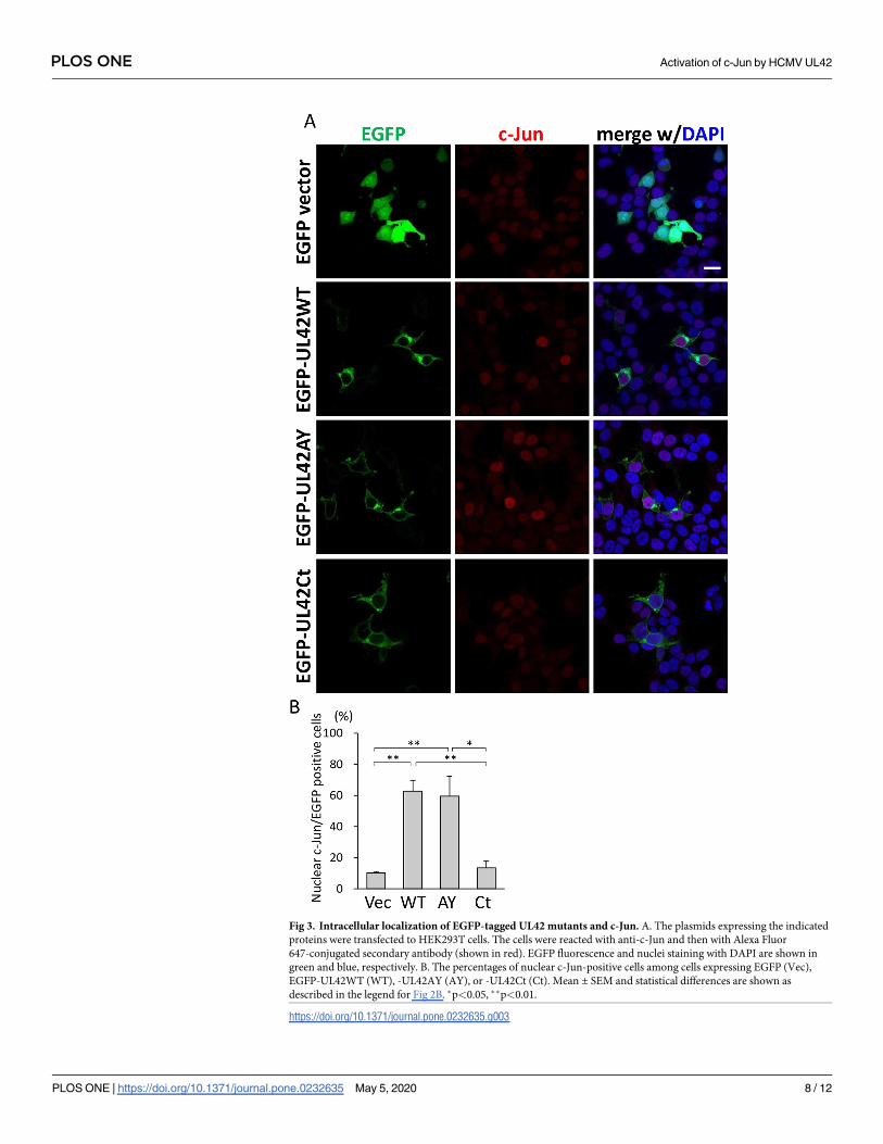
Fig 3. Intracellular localization of EGFP-tagged UL42 mutants and c-Jun. A. The plasmids expressing the indicated
proteins were transfected to HEK293T cells. The cells were reacted with anti-c-Jun and then with Alexa Fluor
647-conjugated secondary antibody (shown in red). EGFP fluorescence and nuclei staining with DAPI are shown in
green and blue, respectively. B. The percentages of nuclear c-Jun-positive cells among cells expressing EGFP (Vec),
EGFP-UL42WT (WT), -UL42AY (AY), or -UL42Ct (Ct). Mean ± SEM and statistical differences are shown as
described in the legend for Fig 2B. �p<0.05, ��p<0.01.
https://doi.org/10.1371/journal.pone.0232635.g003
PLOS ONE Activation of c-Jun by HCMV UL42
PLOS ONE | https://doi.org/10.1371/journal.pone.0232635 May 5, 2020 8 / 12

that the expression levels of EGFP-UL42 variants affected the nuclear localization and activa-
tion of c-Jun, because nuclear accumulation of c-Jun was observed in all cells expressing
HA-UL42WT, -UL42PA or -UL42ΔN but not those expressing HA-UL42ΔI, while the expres-
sion levels of HA-tagged proteins in individual cells varied significantly. The N-terminal region
is thought to contain a regulatory domain for the control of JNK signaling, as the deletion of
the N-terminal region enhanced AP-1 activity more than that of UL42WT. Interestingly, the
expression of the UL42ΔI mutant did not increase c-Jun nuclear import but induced phosphor-
ylation of c-Jun to the same degree as UL42WT, which is consistent with a previous report that
the nuclear import of c-Jun is independent of its phosphorylation [18]. Further investigation
will be needed to elucidate the precise functional domains of UL42.
In contrast to outcomes in transient transfection assays which performed in HEK293T cells,
the lack of UL42 did not affect the phosphorylation status of c-Jun in HCMV-infected fibroblasts
(S3 Fig). As we did not examine the effects of UL42 constructs in fibroblasts due to a low transfec-
tion efficiency, one potential explanation of the discrepancy would be a cell-type specific function
of UL42. However, it is more plausible that the presence of multiple mechanisms of the c-Jun
phosphorylation in HCMV-infected cells results in the discrepancy. Indeed, the activation of c-
Fig 4. Detection of phosphorylated JNK, c-Jun and Itch in cells expressing UL42 derivatives. A. Lysates of HEK293T cells
transfected with pCAGGS (Vec), pCAGGS-HAUL42WT (WT), -HAUL42PA (PA), -HAUL42ΔN (ΔN), or -HAUL42ΔI (ΔI) were
analyzed by immunoblotting using antibodies for the detection of the indicated forms of proteins. Ratios of band intensities between
the forms of the indicated protein without and with phosphorylation were analyzed with NIH imageJ software, and indicated
beneath the panels. B. Lysates of HEK293T cells transfected with pEGFP-C1 (Vec), pEGFP-UL42WT (WT), -UL42AY (AY), or
-UL42Ct (Ct) were analyzed by immunoblotting using antibodies for the detection of the indicated forms of proteins.
https://doi.org/10.1371/journal.pone.0232635.g004
PLOS ONE Activation of c-Jun by HCMV UL42
PLOS ONE | https://doi.org/10.1371/journal.pone.0232635 May 5, 2020 9 / 12

Jun/JNK signaling occurs immediately after HCMV infection and a JNK inhibitor, SP600125,
inhibits HCMV replication by the suppression of immediate-early gene expression [19]. HCMV
encodes many c-Jun/JNK signal-modulating proteins, including IE1 and UL38 [6, 20]. IE1, a
transactivator encoded by HCMV, promotes phosphorylation of c-Jun through a cellular protein
kinase [6, 21]. On the other hand, another HCMV-encoded protein, UL38, which is classified as
an early gene product [22], reduces JNK phosphorylation to suppress ER stress-induced cell
death [20]. Previous studies demonstrated that UL42 was classified as a dispensable gene [8, 23],
which is consistent with the notion that HCMV has several c-Jun modifying genes. As UL42 is
known to be expressed at least 1 day post-infection in HCMV-infected fibroblasts [8], we hypoth-
esize that UL42 would contribute to regulate c-Jun/JNK signaling in some manner during the
early phase of infection with some HCMV encoded protein(s).
In conclusion, UL42 activated c-Jun/JNK signaling in an Itch-independent manner. The
results presented herein indicate that the a.a. 52–86 regions of UL42 is responsible for c-Jun
nuclear translocation. This region is important for induction of AP-1 signaling and JNK acti-
vation. In the future, it would be interesting to see the Nedd4 family-independent inhibitory
effect of UL42 on cyclic GMP-AMP synthase [14]. Further research is still required, however,
to elucidate the precise mechanisms of the UL42-mediated activation of c-Jun/JNK signaling.
Supporting information
S1 Data.
(PDF)
S1 Table. Primer list.
(DOCX)
S1 Fig. Luciferase assay results of UL42 derivatives. The luciferase assay results of three inde-
pendent experiments are shown. A plasmid expressing the indicated UL42 mutant tagged with
HA (A) or with EGFP (B), the luciferase reporter plasmid pAP1(PMA)-TA-Luc, and the con-
trol plasmid pRL-TK were transfected into HEK293T cells. Ratios of firefly luciferase activities
to Renilla luciferase activities obtained in triplicated wells are shown as the means ± SEMs. (A)
pCAGGS (Vec), pCAGGS-HAUL42WT (WT), -HAUL42PA (PA), -HAUL42ΔN (ΔN), and
-HAUL42ΔI (ΔI). (B) pEGFP-C1 (Vec), -UL42WT (WT), -UL42AY (AY), and -UL42Ct (Ct).
(PPTX)
S2 Fig. Intracellular localization of UL42 derivatives. HA-tagged UL42 proteins (A) and
EGFP-tagged UL42 proteins (B) were expressed in HEK293T cells and analyzed with immuno-
fluorescence assay. A. The cells expressing HA-tagged UL42 derivatives were stained with
anti-HA (red) and anti-c-Jun (green) antibodies. The nuclei were stained with DAPI (blue). B.
The cells expressing EGFP-tagged UL42 derivatives were reacted with anti c-Jun antibody and
then with Alexa Flora 647-conjugated secondary antibody (red). EGFP fluorescence and nuclei
staining with DAPI are shown in green and blue, respectively. Bar = 10μm.
(PPTX)
S3 Fig. The phosphorylation status of c-Jun in fibroblasts infected with HCMV wild-type
or UL42 mutants. Fibroblasts, hTERT-BJ1 cell- were mock infected (m) or infected with the
following HCMV strains at a multiplicity of infection (MOI) of 5, harvested at 3 day post-
infection, and their lysates were analyzed by immunoblotting with the indicated antibodies.
WT: HCMV encoding wild-type UL42, R: HCMV encoding rescued UL42, PA: HCMV
encoding UL42PA, Δ: HCMV lacking UL42.
(PPTX)
PLOS ONE Activation of c-Jun by HCMV UL42
PLOS ONE | https://doi.org/10.1371/journal.pone.0232635 May 5, 2020 10 / 12

Author Contributions
Conceptualization: Tetsuo Koshizuka.
Data curation: Tetsuo Koshizuka.
Funding acquisition: Tetsuo Koshizuka.
Investigation: Tetsuo Koshizuka.
Project administration: Tetsuo Koshizuka.
Supervision: Tetsuo Koshizuka, Naoki Inoue.
Writing – original draft: Tetsuo Koshizuka.
Writing – review & editing: Tetsuo Koshizuka, Naoki Inoue.
References1. Meng Q, Xia Y. c-Jun, at the crossroad of the signaling network. Protein Cell. 2011; 2(11):889–98.
https://doi.org/10.1007/s13238-011-1113-3 PMID: 22180088.
2. Smeal T, Binetruy B, Mercola DA, Birrer M, Karin M. Oncogenic and transcriptional cooperation with
Ha-Ras requires phosphorylation of c-Jun on serines 63 and 73. Nature. 1991; 354(6353):494–6.
https://doi.org/10.1038/354494a0 PMID: 1749429.
3. Pulverer BJ, Kyriakis JM, Avruch J, Nikolakaki E, Woodgett JR. Phosphorylation of c-jun mediated by
MAP kinases. Nature. 1991; 353(6345):670–4. https://doi.org/10.1038/353670a0 PMID: 1922387.
4. Zeke A, Misheva M, Remenyi A, Bogoyevitch MA. JNK Signaling: Regulation and Functions Based on
Complex Protein-Protein Partnerships. Microbiol Mol Biol Rev. 2016; 80(3):793–835. https://doi.org/10.
1128/MMBR.00043-14 PMID: 27466283.
5. Busch LK, Bishop GA. Multiple carboxyl-terminal regions of the EBV oncoprotein, latent membrane pro-
tein 1, cooperatively regulate signaling to B lymphocytes via TNF receptor-associated factor (TRAF)-
dependent and TRAF-independent mechanisms. J Immunol. 2001; 167(10):5805–13. https://doi.org/
10.4049/jimmunol.167.10.5805 PMID: 11698454.
6. Wang X, Sonenshein GE. Induction of the RelB NF-kappaB subunit by the cytomegalovirus IE1 protein
is mediated via Jun kinase and c-Jun/Fra-2 AP-1 complexes. J Virol. 2005; 79(1):95–105. https://doi.
org/10.1128/JVI.79.1.95-105.2005 PMID: 15596805.
7. Kurapati S, Sadaoka T, Rajbhandari L, Jagdish B, Shukla P, Ali MA, et al. Role of the JNK Pathway in
Varicella-Zoster Virus Lytic Infection and Reactivation. J Virol. 2017; 91(17). https://doi.org/10.1128/
JVI.00640-17 PMID: 28637759.
8. Koshizuka T, Tanaka K, Suzutani T. Degradation of host ubiquitin E3 ligase Itch by human cytomegalo-
virus UL42. The Journal of general virology. 2016; 97(1):196–208. https://doi.org/10.1099/jgv.0.000336
PMID: 26555021.
9. Koshizuka T, Goshima F, Takakuwa H, Nozawa N, Daikoku T, Koiwai O, et al. Identification and charac-
terization of the UL56 gene product of herpes simplex virus type 2. Journal of virology. 2002; 76
(13):6718–28. https://doi.org/10.1128/JVI.76.13.6718-6728.2002 PMID: 12050385.
10. Koshizuka T, Kobayashi T, Ishioka K, Suzutani T. Herpesviruses possess conserved proteins for inter-
action with Nedd4 family ubiquitin E3 ligases. Sci Rep. 2018; 8(1):4447. https://doi.org/10.1038/
s41598-018-22682-2 PMID: 29535361.
11. Koshizuka T, Ota M, Yamanishi K, Mori Y. Characterization of varicella-zoster virus-encoded ORF0
gene—comparison of parental and vaccine strains. Virology. 2010; 405(2):280–8. https://doi.org/10.
1016/j.virol.2010.06.016 PMID: 20598727.
12. An H, Krist DT, Statsyuk AV. Crosstalk between kinases and Nedd4 family ubiquitin ligases. Molecular
bioSystems. 2014; 10(7):1643–57. https://doi.org/10.1039/c3mb70572b PMID: 24457516.
13. Ahn YH, Kurie JM. MKK4/SEK1 is negatively regulated through a feedback loop involving the E3 ubiqui-
tin ligase itch. J Biol Chem. 2009; 284(43):29399–404. https://doi.org/10.1074/jbc.M109.044958 PMID:
19737936.
14. Fu YZ, Guo Y, Zou HM, Su S, Wang SY, Yang Q, et al. Human cytomegalovirus protein UL42 antago-
nizes cGAS/MITA-mediated innate antiviral response. PLoS Pathog. 2019; 15(5):e1007691. https://doi.
org/10.1371/journal.ppat.1007691 PMID: 31107917.
PLOS ONE Activation of c-Jun by HCMV UL42
PLOS ONE | https://doi.org/10.1371/journal.pone.0232635 May 5, 2020 11 / 12
15. Niwa H, Yamamura K, Miyazaki J. Efficient selection for high-expression transfectants with a novel
eukaryotic vector. Gene. 1991; 108(2):193–9. https://doi.org/10.1016/0378-1119(91)90434-d PMID:
1660837.
16. Tischer BK, von Einem J, Kaufer B, Osterrieder N. Two-step red-mediated recombination for versatile
high-efficiency markerless DNA manipulation in Escherichia coli. BioTechniques. 2006; 40(2):191–7.
https://doi.org/10.2144/000112096 PMID: 16526409.
17. Koshizuka T, Sato Y, Kobiyama S, Oshima M, Suzutani T. A two-step culture method utilizing secreted
luciferase recombinant virus for detection of anti-cytomegalovirus compounds. Microbiol Immunol.
2018; 62(10):651–8. https://doi.org/10.1111/1348-0421.12645 PMID: 30187945.
18. Schreck I, Al-Rawi M, Mingot JM, Scholl C, Diefenbacher ME, O’Donnell P, et al. c-Jun localizes to the
nucleus independent of its phosphorylation by and interaction with JNK and vice versa promotes
nuclear accumulation of JNK. Biochem Biophys Res Commun. 2011; 407(4):735–40. https://doi.org/10.
1016/j.bbrc.2011.03.092 PMID: 21439937.
19. Zhang H, Niu X, Qian Z, Qian J, Xuan B. The c-Jun N-terminal kinase inhibitor SP600125 inhibits
human cytomegalovirus replication. J Med Virol. 2015; 87(12):2135–44. https://doi.org/10.1002/jmv.
24286 PMID: 26058558.
20. Xuan B, Qian Z, Torigoi E, Yu D. Human cytomegalovirus protein pUL38 induces ATF4 expression,
inhibits persistent JNK phosphorylation, and suppresses endoplasmic reticulum stress-induced cell
death. Journal of virology. 2009; 83(8):3463–74. https://doi.org/10.1128/JVI.02307-08 PMID:
19193809.
21. Kim S, Yu SS, Lee IS, Ohno S, Yim J, Kim S, et al. Human cytomegalovirus IE1 protein activates AP-1
through a cellular protein kinase(s). J Gen Virol. 1999; 80 (Pt 4):961–9. https://doi.org/10.1099/0022-
1317-80-4-961 PMID: 10211966.
22. Tenney DJ, Colberg-Poley AM. Human cytomegalovirus UL36-38 and US3 immediate-early genes:
temporally regulated expression of nuclear, cytoplasmic, and polysome-associated transcripts during
infection. J Virol. 1991; 65(12):6724–34. PMID: 1658371.
23. Dunn W, Chou C, Li H, Hai R, Patterson D, Stolc V, et al. Functional profiling of a human cytomegalovi-
rus genome. Proceedings of the National Academy of Sciences of the United States of America. 2003;
100(24):14223–8. https://doi.org/10.1073/pnas.2334032100 PMID: 14623981.
PLOS ONE Activation of c-Jun by HCMV UL42
PLOS ONE | https://doi.org/10.1371/journal.pone.0232635 May 5, 2020 12 / 12