Embed Size (px)
Citation preview
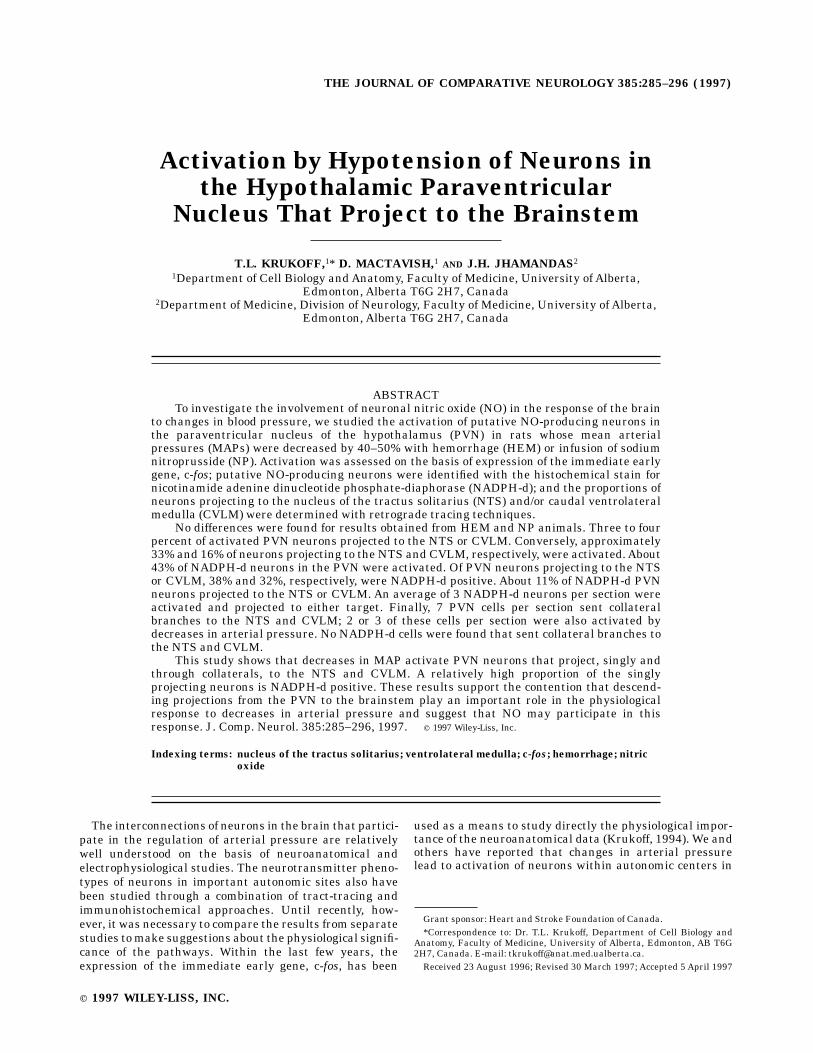
Activation by Hypotension of Neurons inthe Hypothalamic Paraventricular
Nucleus That Project to the Brainstem
T.L. KRUKOFF,1* D. MACTAVISH,1 AND J.H. JHAMANDAS2
1Department of Cell Biology andAnatomy, Faculty of Medicine, University of Alberta,Edmonton, Alberta T6G 2H7, Canada
2Department of Medicine, Division of Neurology, Faculty of Medicine, University of Alberta,Edmonton, Alberta T6G 2H7, Canada
ABSTRACTTo investigate the involvement of neuronal nitric oxide (NO) in the response of the brain
to changes in blood pressure, we studied the activation of putative NO-producing neurons inthe paraventricular nucleus of the hypothalamus (PVN) in rats whose mean arterialpressures (MAPs) were decreased by 40–50% with hemorrhage (HEM) or infusion of sodiumnitroprusside (NP). Activation was assessed on the basis of expression of the immediate earlygene, c-fos; putative NO-producing neurons were identified with the histochemical stain fornicotinamide adenine dinucleotide phosphate-diaphorase (NADPH-d); and the proportions ofneurons projecting to the nucleus of the tractus solitarius (NTS) and/or caudal ventrolateralmedulla (CVLM) were determined with retrograde tracing techniques.
No differences were found for results obtained from HEM and NP animals. Three to fourpercent of activated PVN neurons projected to the NTS or CVLM. Conversely, approximately33% and 16% of neurons projecting to the NTS and CVLM, respectively, were activated.About43% of NADPH-d neurons in the PVN were activated. Of PVN neurons projecting to the NTSor CVLM, 38% and 32%, respectively, were NADPH-d positive. About 11% of NADPH-d PVNneurons projected to the NTS or CVLM. An average of 3 NADPH-d neurons per section wereactivated and projected to either target. Finally, 7 PVN cells per section sent collateralbranches to the NTS and CVLM; 2 or 3 of these cells per section were also activated bydecreases in arterial pressure. No NADPH-d cells were found that sent collateral branches tothe NTS and CVLM.
This study shows that decreases in MAP activate PVN neurons that project, singly andthrough collaterals, to the NTS and CVLM. A relatively high proportion of the singlyprojecting neurons is NADPH-d positive. These results support the contention that descend-ing projections from the PVN to the brainstem play an important role in the physiologicalresponse to decreases in arterial pressure and suggest that NO may participate in thisresponse. J. Comp. Neurol. 385:285–296, 1997. r 1997 Wiley-Liss, Inc.
Indexing terms: nucleus of the tractus solitarius; ventrolateral medulla; c-fos; hemorrhage; nitric
oxide
The interconnections of neurons in the brain that partici-pate in the regulation of arterial pressure are relativelywell understood on the basis of neuroanatomical andelectrophysiological studies. The neurotransmitter pheno-types of neurons in important autonomic sites also havebeen studied through a combination of tract-tracing andimmunohistochemical approaches. Until recently, how-ever, it was necessary to compare the results from separatestudies tomake suggestions about the physiological signifi-cance of the pathways. Within the last few years, theexpression of the immediate early gene, c-fos, has been
used as a means to study directly the physiological impor-tance of the neuroanatomical data (Krukoff, 1994). We andothers have reported that changes in arterial pressurelead to activation of neurons within autonomic centers in
Grant sponsor: Heart and Stroke Foundation of Canada.*Correspondence to: Dr. T.L. Krukoff, Department of Cell Biology and
Anatomy, Faculty of Medicine, University of Alberta, Edmonton, AB T6G2H7, Canada. E-mail: [email protected] 23 August 1996; Revised 30 March 1997; Accepted 5 April 1997
THE JOURNAL OF COMPARATIVE NEUROLOGY 385:285–296 (1997)
r 1997 WILEY-LISS, INC.

the brain (Badoer et al., 1992, 1993; McAllen et al., 1992;Shen et al., 1992a,b; Chan et al., 1993; Dun et al., 1993;Chan and Sawchenko, 1994; Li and Dampney, 1994;Murphy et al., 1994; Krukoff et al., 1995b; Petrov et al.,1995; Krukoff and Khalili, 1997). Combination of c-fosimmunohistochemistry with other neuroanatomical tech-niques offers a powerful tool for understanding the func-tional significance of identified autonomic pathways in thebrain.The paraventricular nucleus of the hypothalamus (PVN)
is well known for its role in the homeostatic control of bloodpressure. Baroreceptor information reaches the PVN viathe nucleus of the tractus solitarius (NTS) and otherbrainstem regions including the ventrolateral medulla(VLM; Swanson and Sawchenko, 1983). We have shownthat approximately 24% and 34% of PVN-projecting neu-rons in the NTS and VLM, respectively, are activated bydecreases in arterial pressure (Krukoff et al., 1995b).Neurons within the PVN in turn project to many siteswithin the brain, including to the NTS and VLM them-selves (Swanson and Sawchenko, 1983). It is not known,however, whether PVN neurons that project to these twoareas are specifically activated by changes in arterialpressure. Thus, the goal of the present series of experi-ments was to identify neurons in the PVN that areactivated by decreases in arterial pressure, to determinethe proportions of these neurons that project to the NTSand/or the caudal subdivision of the VLM (CVLM), and toelucidate the contribution of PVN neurons that putativelyproduce the gaseous neurotransmitter, nitric oxide (NO),to these populations. Furthermore, because hemorrhage(HEM) causes a decrease in blood volume in addition to adecrease in arterial pressure, infusions of the vasodilatoryagent, sodium nitroprusside, were used in a second groupof animals to decrease pressure without affecting bloodvolume. The results from the two groups of animals werecompared to differentiate between populations of neurons
in the PVN, which may be activated by changes in bloodvolume.
MATERIALS AND METHODS
Male Sprague-Dawley rats (250–280 g) were purchasedfrom the Biological Animal Center, University of Alberta.They were housed in a 12-hour:12-hour light:dark cycle(lights on at 0800 hours) at 20°C and given free access tofood and water. All protocols used in these experimentswere approved by the local Animal Welfare Committee.
Instrumentation and injectionof retrograde tracer
Animals were anesthetized with sodium pentobarbital(50 mg/kg, i.p.; Somnotol, M.T.C. Pharmaceuticals, Hamil-ton, Canada). Rhodamine-labeled and fluorescein-labeledmicrospheres (Lumafluor, New York) were injected unilat-erally (10–20 nl) into the NTS and VLM, respectively, asdescribed previously (Krukoff et al., 1992b; Petrov et al.,1993). Injections into the NTS were centered at a point0.50 mm rostral to the calamus scriptorius, 0.50 mmlateral to the midline, and 0.80 mm below the dorsalsurface. In the VLM, this point was in the CVLM 0.50 mmcaudal to the calamus scriptorius, 2.0 mm lateral to themidline, and 2.1 mm from the dorsal surface. The descend-ing aorta and inferior vena cava were cannulated andexteriorized as described previously (Krukoff et al., 1995b).Wounds were sutured closed, and rats were allowed torecover from anesthesia.Animals were handled individually and placed in a
metabolic cage on a daily basis so that they becameaccustomed to these procedures. Five to seven days afterinitial surgery, arterial and venous lines were flushed withsaline. The arterial line of each rat was connected forcontinuous recording of arterial pressure, and the venousline was attached to an empty 10-ml syringe. The rat wasplaced into ametabolic cage for 1 hour before experimenta-tion.
Hemorrhage and nitroprusside infusions
Between 5 and 7 ml of venous blood were removedthrough the venous catheter so that mean arterial pres-sure (MAP) was lowered by 40–50%. Additional 1–2 ml ofblood were withdrawn in small volumes over the next 90minutes to maintain the reducedMAP in this range.At theend of the experiment, rats received an overdose of sodiumpentobarbital and were prepared for perfusion fixation.Control rats were treated similarly, but no blood wasremoved.In another set of rats handled the same way, sodium
nitroprusside (NP; 2.5 mg/ml saline; Sigma Chemicals, St.Louis, MO) was infused into the venous line with aHarvard pump at a rate of 30 µl/minute or until MAPdropped by 40–50%. The NP was infused at the same ratefor short periods of time during the next 90 minutes tomaintain MAP at the same reduced level. At the end of theexperiments, rats were anesthetized and prepared forperfusion fixation. Control rats were treated similarlyexcept that only vehicle was infused.
Tissue processing
Animals were immediately perfused transcardially with100ml saline followed by 500ml ice-cold 4% paraformalde-hyde in 0.1 M phosphate buffer (pH 7.2). Brains were
Abbreviations
A1 noradrenergic cell groupAP area postremaap anterior parvocellular division (PVN)C1 adrenergic cell groupcc central canalCVLM caudal ventrolateral medulladp dorsal parvocellular division (PVN)FLI Fos-like immunoreactivityHEM hemorrhageIO inferior olivary nucleuslp lateral parvocellular division (PVN)LRt lateral reticular nucleusMAP mean arterial pressuremp medial parvocellular division (PVN)NA nucleus ambiguusNADPH-d nicotinamide adenine dinucleotide phosphate-diaphoraseNO nitric oxideNP sodium nitroprussideNTS nucleus of the tractus solitariusPBS phosphate buffered salinepm posterior magnocellular division (PVN)pv periventricular division (PVN)PVN paraventricular nucleus of the hypothalamuspy pyramidal tracttr tract of NTSVLM ventrolateral medullaVsp spinal trigeminal nucleusX dorsal motor nucleus of the vagusXII hypoglossal nucleus3v third ventricle
286 T.L. KRUKOFF ET AL.

postfixed in half-strength fixative: 10% sucrose for 1 hourand cryoprotected in 30% sucrose (aqueous) overnight at4°C.Frozen coronal sections (50 µm) were cut in two sets. The
first set was used to reveal Fos-like immunoreactivity andnicotinamide adenine dinucleotide phosphate-diaphorase(NADPH-d) staining, and the second set of sections wasused as a control for the immunohistochemical reaction.Sections were incubated for 30 minutes in a solutioncontaining 1 mg b-NADPH, 0.1 mg/ml nitroblue tetra-zolium (Sigma), and 0.6% Triton X-100 (Sigma) in phos-phate buffered saline (PBS; pH 7.2). After a rinse in PBS,sections were incubated overnight at 4°C in sheep antiseraagainst Fos (catalogue no. 0A-11-823; Cambridge ResearchBiochemicals, Valley Stream, NY) diluted 1:2,000 in 0.3%Triton X-100/PBS. According to the supplier’s specifica-tions, this affinity-purified antibody recognizes Fos andFos-related proteins. Thus, the staining we obtained isdescribed as Fos-like immunoreactivity (FLI).Tissues were processed the following day by using the
biotin-avidin immunoperoxidasemethod (ABCVecta StainKit, Vector Laboratories, Burlingame, CA). The FLI wasvisualized as a brown reaction product with the chroma-gen diaminobenzidine (Sigma). Sections were mountedonto glass microscope slides, coverslipped with Elvanol(Moviol, Calbiochem Corporation, La Jolla, CA; dissolvedin 40 ml PBS and 20 ml glycerol), and examined with aZeiss light/fluorescence microscope. The brain from anonexperimental rat was sectioned, stained with thionin,and used to prepare projection drawings of the brainstemand PVN.
Analysis
In animals in which injections of latex microsphereswere centered within the NTS or CVLM, neuronal profileswith FLI, those positive for NADPH-d, and those contain-ing either or both of the retrograde tracers were counted inevery second section of the PVN ipsilateral to the sites ofinjection. Only neuronal profiles with visible nuclei wereincluded in the counts, thereby ensuring that no neuronwas counted twice. For each of the two groups of animals(injections in NTS or CVLM), data for each animal werepooled to obtain (1) total numbers of neurons per section inthe PVNwith each of the labels, (2) percentages of neuronswith FLI as a proportion of neurons with either retrogradetracer, (3) percentages of neurons with either retrogradetracer as a proportion of neurons with FLI, (4) percentagesof neurons with FLI as a proportion of NADPH-d neurons,(5) percentages of NADPH-d neurons as a proportion ofneurons with FLI, (6) percentages of NADPH-d neurons asa proportion of neurons with either retrograde tracer, (7)percentages of neurons with either retrograde tracer as aproportion of NADPH-d neurons, and (8) numbers ofNADPH-d neurons that contained FLI and the retrogradetracer (triple labeled). Finally, when successful injectionsin both the NTS and VLM were obtained in the sameanimal, the number of PVN neurons containing bothretrograde tracers was counted; the presence of FLI,NADPH-d (triple labeling), or both (quadruple labeling)was recorded.Counts for neuronswith FLI and both FLI andNADPH-d
were combined for all animals subjected to either HEM orNP to compare data between the two treatments. Eachpair of data (HEM vs. NP) was subjected to Student’st-test; P , 0.05 was considered to be significant.
RESULTS
Arterial blood pressure
Figure 1 shows the effects on MAP of hemorrhage ornitroprusside infusions. MAP was decreased by approxi-mately 50% within the first 5 minutes of the experimentand was maintained at this level for the duration of theexperiment.
Injection sites
Figures 2 and 3 illustrate the extent of the six retrogradetracer injection sites in the NTS or CVLM in HEM and NPanimals, respectively. For convenience, three injectionssites are shown on each side of the brainstem. Figure 4shows photographic examples of injection sites in the NTSand CVLM.
PVN labeling
In all control rats, very small numbers of neurons in thePVN (,10 cells/section) expressed FLI. Therefore, controlswere not included in the subsequent analyses.HEM versus NP. The following are the data per sec-
tion for combined counts from the PVN of HEM and NPanimals: FLI: 202 6 16 (HEM) vs. 197 6 26 (NP); FLI andNADPH-d: 42 6 5 (HEM) vs. 39 6 5 (NP). No significantdifferences (P . 0.05) were found between any of thesepairs of data from HEM and NP animals.Animals with injections of retrograde tracer in NTS.
Figure 5 shows the distributions of singly and multiplylabeled neurons at three levels of the PVN from a HEMand an NP animal, respectively. The location of retro-gradely labeled neurons in the PVN is similar to thatdescribed by others (Ricardo and Koh, 1978; Ross et al.,1981) and is not repeated here.Tables 1 and 2 show the data for HEM and NP animals
in which the retrograde tracer was injected into the NTS.The data can be summarized as follows: (1) approximately4% (HEM and NP) of neurons with FLI contained thetracer; (2) approximately 18% (HEM and NP) of neuronswith FLI were NADPH-d positive; (3) 38% (HEM) and 28%(NP) of retrogradely labeled neurons contained FLI; (4)43% (HEM) and 32% (NP) of retrogradely labeled neuronswere NADPH-d positive; (5) approximately 12% (HEMandNP) of NADPH-d positive neurons contained retrogradelabel; (6) approximately 42% (HEM and NP) of NADPH-dpositive neurons contained FLI; and (7) 3 cells per section(HEM and NP) contained FLI and retrograde tracer andwere NADPH-d positive.Animalswith injections of retrograde tracer inCVLM.
Figure 6 shows the distributions of singly and multiplylabeled neurons at three levels of the PVN from a HEMand an NP animal, respectively. The location of retro-gradely labeled neurons in the PVN is similar to thatdescribed by others (Tribollet and Dreifuss, 1981; Blessinget al., 1982) and is not repeated here.Tables 3 and 4 show the data for HEM and NP animals
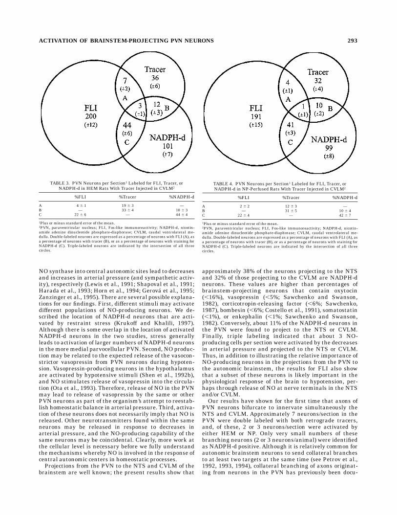
in which the retrograde tracer was injected into theCVLM. The data can be summarized as follows: (1) 4%(HEM and NP) of neurons with FLI contained the tracer;(2) 22% (HEMandNP) of neuronswith FLIwereNADPH-dpositive; (3) 19% (HEM) and 12% (NP) of retrogradelylabeled neurons contained FLI; (4) approximately 32%(HEM and NP) of retrogradely labeled neurons wereNADPH-d positive; (5) 10% (HEM and NP) of NADPH-dpositive neurons contained retrograde label; (6) approxi-
ACTIVATION OF BRAINSTEM-PROJECTING PVN NEURONS 287

mately 43% (HEM and NP) of NADPH-d positive neuronscontained FLI; (7) approximately 2 cells per sections (HEMand NP) contained FLI and retrograde tracer and wereNADPH-d positive. Figure 7 shows examples of double-(FLI and tracer) and triple-(FLI, NADPH-d, and tracer)labeled neurons in the PVN after an injection of tracer intothe VLM.
Collaterals from PVN neurons
In one HEM and one NP animal, all aspects of the tissueprocessing were successful, and transport of both retro-grade tracers enabled visualization of PVN neurons thatproject to both the NTS and CVLM. In the HEM animal, 6neurons/section contained both retrograde tracers. Twoneurons per section contained both retrograde tracers andFLI; an average of 0.5 neuron/section contained bothretrograde tracers andwere NADPH-d positive. Finally, noneurons were found which contained all four labels.In the NP animal, 7 neurons/section contained both
retrograde tracers. Three neurons per section containedboth retrograde tracers and FLI; 0.5 neuron/section con-tained both retrograde tracers and were NADPH-d posi-tive. No neurons were found which contained all fourlabels.
DISCUSSION
We have used a multilabeling approach to investigatethe responses of neuronal elements within the PVN todecreases in arterial pressure. We describe the activationof PVN neurons by HEM or infusions of the vasodilator,NP, and the proportions of these neurons that project tothe NTS or CVLM in the brainstem. In addition, we havedetermined the proportions of NADPH-d neurons in thePVN that are activated by HEM or NP and the proportionsof activated and nonactivated NADPH-d neurons thatproject to the NTS or CVLM. Finally, we describe popula-tions of activated or NADPH-d PVN neurons that sendcollateral branches to the NTS and CVLM.
Several technical issues relating to the present studymust be considered. First, because NADPH-d staining isconsidered to be indicative of NO synthase activity in thecentral nervous system when fixation has been employed(Matsumoto et al., 1993; Norris et al., 1995), our discussionwill be in terms of NO-producing neurons. Our results donot, however, conclusively prove that the positively stainedneurons produce NO. Second, our injections of retrogradetracer necessarily did not fill entirely either the NTS or
Fig. 2. Schematic drawings through three levels of the brainstem(A–C) illustrating unilateral injections sites in the NTS and CVLM ofhemorrhaged rats. Six injection sites each are shown for the NTS andCVLM. For convenience, three are shown on each side of the brainstem,and injections are depicted with different types of shading. Numbersindicate rostrocaudal coordinates relative to the bregma (Paxinos andWatson, 1986). See Abbreviations list for definitions of legend in allfigures.
Fig. 1. Changes in mean arterial pressure produced by hemor-rhage (n 5 12) or sodium nitroprusside infusion (n 5 12) vs. that seenin control (n 5 2) rats. Pressures included for hypotensive rats arefrom the same animals that were analyzed for retrograde transport oftracers.
288 T.L. KRUKOFF ET AL.

CVLM. Thus, our data for PVN neurons that project toeither of these targets are likely underestimated. Third,although HEM and NP treatments are commonly dis-cussed in terms of activating baroreceptors, we cannotexclude the possibility that chemoreceptors also may beactivated due to changes in blood gases that may accom-pany decreases in MAP.A comparison of the results obtained from HEM and NP
animals in the present study reveals that a decrease in
blood volume did not activate larger numbers of neurons inthe PVN compared with animals in which only the MAPwas decreased. Furthermore, there were no differences inactivation of NO-producing neurons in the PVN betweenHEM and NP animals. Others have suggested that HEMactivates slightly increased numbers of oxytocin-produc-ing PVN neurons compared with NP (Shen et al., 1992b),but it was not stated whether these differences werestatistically significant. In general, our results suggest
Fig. 3. Schematic drawings through three levels of the brainstem(A–C) illustrating unilateral injections sites in the NTS and CVLM ofnitroprusside-infused rats. Six injection sites each are shown for theNTS and CVLM. For convenience, three are shown on each side of thebrainstem, and injections are depicted with different types of shading.Numbers indicate rostrocaudal coordinates relative to the bregma(Paxinos and Watson, 1986). Fig. 4. Photographic examples of injection sites in the NTS (A) and
CVLM (B). Scale bars 5 300 µm inA, 275 µm in B.
ACTIVATION OF BRAINSTEM-PROJECTING PVN NEURONS 289

Fig.5.
Com
posite
draw
ings
show
inglabelingat
fourlevels
ofthePVN
inrats
withinjectionsof
retrograde
tracer
intheNTS.SeriesAisfrom
HEM
ratsandseries
Bisfrom
NP-infusedrats.N
umbers
indicaterostrocaudalcoordinatesrelative
tothebregma(PaxinosandWatson,1986).

that a decrease in blood volume is not reflected in recruit-ment of PVN neurons beyond those already activated bythe decrease in MAP. Oxytocin-producing neurons may berecruited by HEM, but, because the total numbers ofactivated neurons are not different, this recruitment wouldhave to be at the expense of some other population ofneurons in the PVN.In agreement with previous studies (Badoer et al., 1992,
1993; Shen et al., 1992a,b; Li and Dampney, 1994; Grahamet al., 1995; Petrov et al., 1995), we show that a decrease inarterial pressure leads to the activation of relatively largenumbers of neurons in both the parvo- and magnocellulardivisions of the PVN. Our results also show that 3–4% ofactivated neurons also project to theNTS and that approxi-mately an equal number of neurons projects to the CVLM.Stated conversely, approximately 33% of neurons project-ing to the NTS and approximately 16% of neurons project-ing to the CVLM were activated by decreases in arterialpressure. Our values are likely underestimates of theabsolute numbers of PVN neurons that project to each ofthese targets because our injections did not encompass theentire extent of either nucleus. Nevertheless, these valuesindicate that larger numbers of brainstem-projecting neu-rons are activated by decreases in arterial pressure thanthe reverse. These values also emphasize that PVN neu-rons activated by changes in arterial pressure likely have avariety of targets throughout the central nervous systemin addition to those in the brainstem. The targets ofactivated PVN neurons in the NTS or CVLM coincide withthe location of brainstem neurons that were activated bydecreases in arterial pressure and that project to the PVN(Krukoff et al., 1995b), suggesting that neurons in the
brainstem and PVNmay reciprocally conduct cardiovascu-lar-related information.Because approximately 22% of activated PVN neurons
produced were NADPH-d positive, the stimuli in our studymust activate PVN neurons that produce substances inaddition to NO. Indeed, it has been shown that HEM ordecreased arterial pressure also activates neurons in thePVN, which produces vasopressin or oxytocin (Badoer etal., 1993; Roberts et al., 1993; Shen et al., 1993b; Petrov etal., 1995). We have shown that many of the activatedneurons are situated not only in the magnocellular divi-sions of the PVN, where the majority of vasopressin- andoxytocin-producing neurons are found, but also in theparvocellular divisions. Therefore, other candidates forneurotransmitters in activated PVN neurons include themany substances that have been described in parvocellu-lar neurons. Another point that should be considered isthat activated NADPH-d neuronsmay themselves produceother neuroactive substances because the presence ofvasopressin, oxytocin, angiotensin-(1-7), enkephalin, dynor-phin, and pituitary adenylate cyclase-activating polypep-tide has been demonstrated in NO-producing neurons ofthe PVN (Calka and Block, 1993; Miyagawa et al., 1994;Murakami, 1994; Okamura et al., 1994).Approximately 43% of NADPH-d neurons in the PVN
were activated by the decreases in MAP. The majority ofactivated NADPH-d neurons was found in the parvocellu-lar divisions, with smaller numbers found in themagnocel-lular divisions. These results are especially interesting inlight of recent evidence that suggests that NO acts atseveral sites in the brain to decrease sympathetic drive.For example, microinjections of NO donors or inhibitors of
TABLE 1. PVN Neurons per Section1 Labeled for FLI, Tracer, orNADPH-d in HEM Rats With Tracer Injected in NTS2
%FLI %Tracer %NADPH-d
A 4 6 1 38 6 2 —B — 43 6 5 11 6 3C 19 6 4 — 45 6 4
1Plus or minus standard error of the mean.2PVN, paraventricular nucleus; FLI, Fos-like immunoreactivity; NADPH-d, nicotin-amide adenine dinucleotide phosphate-diaphorase; NTS, nucleus of the tractus soli-tarius. Double-labeled neurons are expressed as a percentage of neurons with FLI (A),as a percentage of neurons with tracer (B), or as a percentage of neurons with stainingfor NADPH-d (C). Triple-labeled neurons are indicated by the intersection of all threecircles.
TABLE 2. PVN Neurons per Section1 Labeled for FLI, Tracer, orNADPH-d in NP-Perfused Rats With Tracer Injected in NTS2
%FLI %Tracer %NADPH-d
A 5 6 1 28 6 4 —B — 32 6 6 13 6 4C 18 6 5 — 39 6 6
1Plus or minus standard error of the mean.2PVN, paraventricular nucleus; FLI, Fos-like immunoreactivity; NADPH-d, nicotin-amide adenine dinucleotide phosphate-diaphorase; NTS, nucleus of the tractus soli-tarius. Double-labeled neurons are expressed as a percentage of neurons with FLI (A),as a percentage of neurons with tracer (B), or as a percentage of neurons with stainingfor NADPH-d (C). Triple-labeled neurons are indicated by the intersection of all threecircles.
ACTIVATION OF BRAINSTEM-PROJECTING PVN NEURONS 291

Fig.6.
Com
posite
draw
ings
show
inglabelingat
fourlevels
ofthePVN
inrats
withinjectionsof
retrograde
tracerintheCVLM.SeriesAisfrom
HEMratsandseriesBisfrom
NP-infusedrats.N
umbers
indicaterostrocaudalcoordinatesrelative
tothebregma(PaxinosandWatson,1986).
NO synthase into central autonomic sites lead to decreasesand increases in arterial pressure (and sympathetic activ-ity), respectively (Lewis et al., 1991; Shapoval et al., 1991;Harada et al., 1993; Horn et al., 1994; Gerova et al., 1995;Zanzinger et al., 1995). There are several possible explana-tions for our findings. First, different stimuli may activatedifferent populations of NO-producing neurons. We de-scribed the location of NADPH-d neurons that are acti-vated by restraint stress (Krukoff and Khalili, 1997).Although there is some overlap in the location of activatedNADPH-d neurons in the two studies, stress generallyleads to activation of larger numbers of NADPH-d neuronsin themoremedial parvocellular PVN. Second, NO produc-tion may be related to the expected release of the vasocon-strictor vasopressin from PVN neurons during hypoten-sion. Vasopressin-producing neurons in the hypothalamusare activated by hypotensive stimuli (Shen et al., 1992b),and NO stimulates release of vasopressin into the circula-tion (Ota et al., 1993). Therefore, release of NO in the PVNmay lead to release of vasopressin by the same or otherPVN neurons as part of the organism’s attempt to reestab-lish homeostatic balance in arterial pressure. Third, activa-tion of these neurons does not necessarily imply that NO isreleased. Other neurotransmitters found within the sameneurons may be released in response to decreases inarterial pressure, and the NO-producing capability of thesame neurons may be coincidental. Clearly, more work atthe cellular level is necessary before we fully understandthe mechanisms whereby NO is involved in the response ofcentral autonomic centers in homeostatic processes.Projections from the PVN to the NTS and CVLM of the
brainstem are well known; the present results show that
approximately 38% of the neurons projecting to the NTSand 32% of those projecting to the CVLM are NADPH-dneurons. These values are higher than percentages ofbrainstem-projecting neurons that contain oxytocin(,16%), vasopressin (,5%; Sawchenko and Swanson,1982), corticotropin-releasing factor (,6%; Sawchenko,1987), bombesin (,6%; Costello et al., 1991), somatostatin(,1%), or enkephalin (,1%; Sawchenko and Swanson,1982). Conversely, about 11% of the NADPH-d neurons inthe PVN were found to project to the NTS or CVLM.Finally, triple labeling indicated that about 3 NO-producing cells per section were activated by the decreasesin arterial pressure and projected to the NTS or CVLM.Thus, in addition to illustrating the relative importance ofNO-producing neurons in the projections from the PVN tothe autonomic brainstem, the results for FLI also showthat a subset of these neurons is likely important in thephysiological response of the brain to hypotension, per-haps through release of NO at nerve terminals in the NTSand/or CVLM.Our results have shown for the first time that axons of
PVN neurons bifurcate to innervate simultaneously theNTS and CVLM. Approximately 7 neurons/section in thePVN were double labeled with both retrograde tracers,and, of these, 2 or 3 neurons/section were activated byeither HEM or NP. Only very small numbers of thesebranching neurons (2 or 3 neurons/animal) were identifiedas NADPH-d positive. Although it is relatively common forautonomic brainstem neurons to send collateral branchesto at least two targets at the same time (see Petrov et al.,1992, 1993, 1994), collateral branching of axons originat-ing from neurons in the PVN has previously been docu-
TABLE 3. PVN Neurons per Section1 Labeled for FLI, Tracer, orNADPH-d in HEM Rats With Tracer Injected in CVLM2
%FLI %Tracer %NADPH-d
A 4 6 1 19 6 3 —B — 33 6 4 10 6 3C 22 6 6 — 44 6 4
1Plus or minus standard error of the mean.2PVN, paraventricular nucleus; FLI, Fos-like immunoreactivity; NADPH-d, nicotin-amide adenine dinucleotide phosphate-diaphorase; CVLM, caudal ventrolateral me-dulla. Double-labeled neurons are expressed as a percentage of neurons with FLI (A), asa percentage of neurons with tracer (B), or as a percentage of neurons with staining forNADPH-d (C). Triple-labeled neurons are indicated by the intersection of all threecircles.
TABLE 4. PVN Neurons per Section1 Labeled for FLI, Tracer, orNADPH-d in NP-Perfused Rats With Tracer Injected in CVLM2
%FLI %Tracer %NADPH-d
A 2 6 2 12 6 3 —B — 31 6 5 10 6 4C 22 6 4 — 42 6 7
1Plus or minus standard error of the mean.2PVN, paraventricular nucleus; FLI, Fos-like immunoreactivity; NADPH-d, nicotin-amide adenine dinucleotide phosphate-diaphorase; CVLM, caudal ventrolateral me-dulla. Double-labeled neurons are expressed as a percentage of neurons with FLI (A), asa percentage of neurons with tracer (B), or as a percentage of neurons with staining forNADPH-d (C). Triple-labeled neurons are indicated by the intersection of all threecircles.
ACTIVATION OF BRAINSTEM-PROJECTING PVN NEURONS 293

Fig. 7. The FLI, NADPH-d staining, and retrograde labeling of theneurons in the PVN of an NP-infused rat. The cell indicated by thearrowhead in A contains retrograde tracer (B, arrowhead) and stainspositively for FLI and NADPH-d (C, arrowhead). The arrow in A
indicates a singly labeled NADPH-d neuron. The cell shown in D andE (curved arrows) is taken from a field not shown inA; the cell containsFLI (D) and FLI (E) but is not NADPH-d positive. Scale bars 5 60 µminA, 12 µm for B–E.

mented only for magnocellular vasopressinergic neurons,which send collateral branches to the posterior pituitaryand back onto other vasopressin-producing neurons (Rayand Choudhury, 1990). Because nearly one-half of theneurons with collaterals destined for the NTS and VLM inthe present study were activated by decreases in arterialpressure, these pathways are likely important for convey-ing cardiovascular information to more than one brain-stem target in a synchronous manner.In conclusion, we have shown that PVN neurons are
activated by decreases in arterial pressure and that manyof the activated neurons project to the NTS, CVLM, or boththrough collateral branches. Finally, we provide data thatshow that significant proportions of activated PVN neu-rons are NADPH-d positive and that some of them projectto the NTS or CVLM. These results show that descendingprojections from the PVN to the brainstem play an impor-tant role in the response to hypotension and suggest thatNOmay participate in this function.
ACKNOWLEDGMENTS
We are grateful to Mr. Kim Harris for graphics assis-tance.
LITERATURE CITED
Badoer, E., M.J. McKinley, F.B. Oldfield, and R.M. McAllen (1992) Distribu-tion of hypothalamic, medullary and lamina terminalis neurons express-ing Fos after hemorrhage in conscious rats. Brain Res. 582:323–328.
Badoer, E., M.J. McKinley, B.J. Oldfield, and R.M. McAllen (1993) Acomparison of hypotensive and non-hypotensive hemorrhage on Fosexpression in spinally projecting neurons of the paraventricular nucleusand rostral ventrolateral medulla. Brain Res. 610:216–223.
Blessing, W.W., C.B. Jaeger, R.A. Ruggiero, and D.J. Reis (1982) Hypotha-lamic projections of medullary catecholamine neurons in the rabbit: Acombined catecholamine fluorescence and HRP transport study. BrainRes. Bull. 9:279–286.
Calka, J., and C.H. Block (1993)Angiotensin-(1-7) and nitric oxide synthasein the hypothalamo-neurohypophysial system. Brain Res. Bull. 30:677–685.
Chan, R.K.W., and P.E. Sawchenko (1994) Spatially and temporally differ-entiated patterns of c-fos expression in brainstem catecholaminergiccell groups induced by cardiovascular challenges in the rat. J. Comp.Neurol. 348:433–460.
Chan, R.K.W., E.R. Brown, A. Ericsson, K.J. Kovacs, and P.E. Sawchenko(1993) A comparison of two immediate-early genes, c-fos and NGFI-B,as markers for functional activation in stress-related neuroendocrinecircuitry. J. Neurosci. 13:5126–5138.
Costello, J.F., M.R. Brown, and T.S. Gray (1991) Bombesin immunoreactiveneurons in the hypothalamic paraventricular nucleus innervate thedorsal vagal complex in the rat. Brain Res. 542:77–82.
Dun, N.J., S.L. Dun, and N.L. Chiaia (1993) Hemorrhage induces Fosimmunoreactivity in rat medullary catecholaminergic neurons. BrainRes. 608:223–232.
Gerova, M., C. Masanova, and J. Pavlasek (1995) Inhibition of NO synthasein the posterior hypothalamus increases blood pressure in the rat.Physiol. Res. 44:131–134.
Graham, J.C., G.E. Hoffman, andA.F. Sved (1995) c-Fos expression in brainin response to hypotension and hypertension in conscious rats. J.Auton.Nerv. Syst. 55:92–104.
Harada, S., S. Tokunga, M. Momohara, H. Masaki, T. Tagawa, T. Imaizumi,and A. Takshita (1993) Inhibition of nitric oxide formation in thenucleus tractus solitarius increases renal sympathetic nerve activity inrabbits. Circ. Res. 72:511–516.
Horn, T., P.M. Smith, B.E. McLaughlin, L. Bauce, G.S. Marks, Q.J.Pittman, andA.V. Ferguson (1994) Nitric oxide actions in paraventricu-lar nucleus: Cardiovascular and neurochemical implications. Am. J.Physiol. 266:R306–R313.
Krukoff, T.L. (1994) Expression of c-fos in studies of central autonomic andsensory systems. Mol. Neurobiol. 7:247–263.
Krukoff, T.L., and P. Khalili (1997) Stress-induced activation of nitricoxide-producing neurons in the rat brain. J. Comp. Neurol. 377:509–519.
Krukoff, T.L., T.L. Morton, K.H. Harris, and J.H. Jhamandas (1992a)Expression of c-fos protein in rat brain elicited by electrical stimulationof the pontine parabrachial nucleus. J. Neurosci. 12:3582–3590.
Krukoff, T.L., T. Vu, K.H. Harris, and J.H. Jhamandas (1992b) Neurons inthe rat medulla oblongata containing neuropeptide Y-, angiotensin II-,or galanin-like immunoreactivity project to the parabrachial nucleus.Neuroscience 47:175–184.
Krukoff, T.L., F. Gehlen, D. Ganten, and J. Wagner (1995a) Gene expressionof brain nitric oxide synthase and soluble guanylyl cyclase in hypothala-mus and medulla of 2 kidney–1 clip hypertensive rats. Hypertension27:171–176.
Krukoff, T.L., D. MacTavish, K.H. Harris, and J.H. Jhamandas (1995b)Changes in blood volume and pressure induce c-fos expression inbrainstem neurons that project to the paraventricular nucleus of thehypothalamus. Mol. Brain Res. 34:99–108.
Lewis, S.J., H. Ohta, B. Machado, J.N. Bates, and W.T. Talman (1991)Microinjection of S-nitrosocysteine into the nucleus tractus solitariidecreases arterial pressure and heart rate via activation of solubleguanylyl cyclase. Eur. J. Pharmacol. 202:135–136.
Li, Y.-W., and R.A.L. Dampney (1994) Expression of Fos-like protein inbrain following sustained hypertension and hypotension in consciousrabbit. Neuroscience 61:613–634.
McAllen, R.M., E. Badoer, A.D. Shafton, B.J. Oldfield, and M.J. McKinley(1992) Hemorrhage induces c-fos immunoreactivity in spinally project-ing neurons of cat subretrofacial nucleus. Brain Res. 575:329–332.
Matsumoto, T., M. Nakane, J.S. Pollock, J.E. Kuk, and U. Forstermann(1993) A correlation between soluble brain nitric oxide snythase andNADPH-diaphorase activity is only seen after exposure of the tissue tofixative. Neurosci. Lett. 155:61–64.
Miyagawa,A., H. Okamura, andY. Ibata (1994) Coexistence of oxytocin andNADPH-diaphorase in magnocellular neurons of the paraventricularand the supraoptic nuclei of the rat hypothalamus. Neurosci. Lett.171:13–16.
Murakami, T. (1994) Expression of nitric oxide synthase in enkephalin anddynorphin systems of the rat hypothalamus [Japanese]. Folia Endocri-nol. Jap. 70:967–978.
Murphy,A.Z.,M. Ennis,M.T. Shipley, andM.M. Behbehani (1994) Direction-ally specific changes in arterial pressure induce differential patterns ofFos expression in discrete areas of the rat brainstem: A double-labelingstudy for Fos and catecholamines. J. Comp. Neurol. 349:36–50.
Norris, P.J., I.G. Charles, C.A. Scorer, and P.C. Emson (1995) Studies on thelocalization and expression of nitric oxide synthase using histochemicaltechniques. Histochem. J. 27:745–756.
Okamura, H., A. Miyagawa, H. Takagi, H. Esumi, N. Yanaihara, and Y.Ibata (1994) Co-existence of PACAP and nitric oxide synthase in the rathypothalamus. Neuroreport 5:1177–1180.
Ota, M., J.T. Crofton, G.T. Festavan, and L. Share (1993) Evidence thatnitric oxide can act centrally to stimulate vasopressin release. Neuroen-docrinology 57:955–959.
Paxinos, G., and C. Watson (1986) The Rat Brain in Stereotaxic Coordi-nates. San Diego, CA: Academic Press.
Petrov, T., T.L. Krukoff, and J.H. Jhamandas (1992) The hypothalamicparaventricular and lateral parabrachial nuclei receive collaterals fromraphe nucleus neurons:Acombined double retrograde and immunocyto-chemical study. J. Comp. Neurol. 318:18–26.
Petrov, T., T.L. Krukoff, and J.H. Jhamandas (1993) Branching projectionsof catecholaminergic brainstemneurons to the paraventricular hypotha-lamic nucleus and the central nucleus of the amygdala in the rat. BrainRes. 609:81–92.
Petrov, T., T.L. Krukoff, and J.H. Jhamandas (1994) Chemically definedcollateral projections from the pons to the central nucleus of theamygdala and the hypothalamic paraventricular nucleus in the rat.Cell Tissue Res. 277:289–295.
Petrov, T., K. Harris, D. McTavish, T.L. Krukoff, and J.H. Jhamandas(1995) Hypotension induces fos-like immunoreactivity in NADPH-diaphorase positive neurons in the paraventricular and supraoptichypothalamic nuclei of the rat. Neuropharmacology 34:509–514.
Ray, P.K., and S.R. Choudhury (1990) Vasopressinergic axon collaterals andaxon terminals in the magnocellular neurosecretory nuclei of the rathypothalamus. ActaAnat. 137:37– 44.
Ricardo, J.A., and E.T. Koh (1978)Anatamical evidence of direct projectionsfrom the nucleus of the solitary tract to the hypothalamus, amygdala,and other forebrain structures in the rat. Brain Res. 153:1–26.
ACTIVATION OF BRAINSTEM-PROJECTING PVN NEURONS 295

Roberts, M.M., A.G. Robinson, M.D. Fitzsimmons, F. Grant, W.-S. Lee, andG.E. Hoffman (1993) C-fos expression in vasopressin and oxytocinneurons reveals functional heterogeneity within magnocellular neu-rons. Neuroendocrinology 57:338–400.
Ross, C.A., D.A. Ruggiero, and D.J. Reis (1981) Afferent projections tocardiovascular portions of the tractus solitarius in the rat. Brain Res.223:402–408.
Sawchenko, P.E. (1987) Evidence for differential regulation of corticotropin-releasing factor and vasopressin immunoreactivities in parvocellularneurosecretory and autonomic-related projections of the paraventricu-lar nucleus. Brain Res. 437:253–263.
Sawchenko, P.E., and L.W. Swanson (1982) Immunohistochemical identifi-cation of neurons in the paraventricular nucleus of the hypothalamusthat project to the medulla or to the spinal cord in the rat. J. Comp.Neurol. 205:260–272.
Shapoval, L.N., V.F. Sagach, and L.S. Pobegailo (1991) Nitric oxide influ-
ences ventrolateral medullary mechanisms of vasomotor control in thecat. Neurosci. Lett. 86:9030–9033.
Shen, E., S.L. Dun, C. Ren, and N.J. Dun (1992a) Hypovolemia inducesFos-like immunoreactivity in neurons of the rat supraoptic and paraven-tricular nuclei. J. Auton. Nerv. Syst. 37:227–230.
Shen, E., S.L. Dun, C. Ren, C. Bennett-Clarke, and N.J. Dun (1992b)Hypotension preferentially induces c-fos immunoreactivity in supraop-tic vasopressin neurons. Brain Res. 593:136–139.
Swanson, L.W., and P.E. Sawchenko (1983) Hypothalamic integration:Organization of the paraventricular and supraoptic nuclei. Annu. Rev.Neurosci. 6: 269–324.
Tribollet, E., and J.J. Dreifuss (1981) Localization of neurones projecting tothe hypothalamic paraventricular nucleus area of the rat: A horserad-ish peroxidase study. Neuroscience 6:1315–1328.
Zanzinger, J., J. Czachurski, and H. Seller (1995) Inhibition of basal andreflex-mediated sympathetic activity in the RVLM by nitric oxide. Am.J. Physiol. 268:R958–R962.
296 T.L. KRUKOFF ET AL.