Embed Size (px)
Citation preview
Life Science Journal 2013;10(3) http://www.lifesciencesite.com
Acrylamide Disrupts the Ontogeny of Neurobehaviour in Albino Rats
Ahmed A Allam 1,2
*, Manal Abdul-Hamid 2, Ahlam Bakry
2, Abdelwahb El-Ghareeb
3, Jamaan S Ajarem
1,
Mohamed Sabri 4
1 King Saud University, College of Science, Zoology Department, Riyadh11451, Saudia Arabia.
2 Department of Zoology, Faculty of Science, Beni-suef University, Egypt.
3 Department of Zoology, Faculty of Science, Cairo University, Egypt. 4Oregon Health and Science University, Portland OR, USA.
[email protected] Abstract: The present study aimed to elucidate abnormalities in the ontogeny of sensorimotor reflexes in developing rats after prenatal and perinatal acrylamide or saline intoxicat ion of pregnant rats. Acrylamide was used as an experimental probe to investigate neurobehavioural and morphological changes in developing rats after administration of the toxin to pregnant mothers. Acrylamide was administered to non-anaesthetised pregnant rats by gastric intubation at a dose of 10 mg/kg/day. Rat pups were assigned to one of three groups: Group A, which comprised pups whose mothers were treated with saline (control group); Group B, which comprised pups whose mothers were treated with acrylamide from day D7 of gestation to birth (prenatal intoxication); and Group C, which comprised pups whose mothers were treated with acrylamide from D7 of gestation to D28 after birth (perinatal intoxication). This study has been conducted recently in Beni-Suef University (Egypt) and Kig Saud University (Saudi Arabia) by May 2012. Acrylamide-induced morphological changes (CNS aberration) and neurobehavioural changes (sensorimotor reflex retardation) were studied. The reflexes tested included rooting, forelimb (FL) grasping, hind limb (HL) grasping, surface body righting, air body righting, FL hopping, HL hopping, chin tactile placing and visual placing. These reflexes were tested in newborns in all groups from postnatal day 2 (D2) until reflex maturation. The appearance of select external features was recorded. administration of acrylamide in pregnant albino rats disrupts the ontogeny of sensorimotor reflexes and morphological changes in the CNS of developing albino rats. [Allam A, Abdul-Hamid M, Bakery A, El-gareeb A, Ajarem J, Sabri M. Acrylamide Disrupts the Ontogeny of Neurobehaviour in Albino Rats. Life Sci J 2013;10(3):1760-1771] (ISSN:1097-8135). http://www.lifesciencesite.com. 265
Key words: Acrylamide; Neurotoxicity; Sensorimotor reflexes; Ontogeny 1. Introduction
While a large number of studies have investigated the neurotoxic and carcinogenic effects of acrylamide, data on acrylamide-induced
neurobehavioural and teratogenic effects during embryonic and postnatal development of the albino rat are relatively limited. Acrylamide is an industrial
agent used in the manufacture of polymers and synthetic organic chemicals. Polymeric acrylamide is used as a filtration and flocculation aid in water
treatment and waste processing, mining and paper mills (Seale etal., 2012). Acrylamide is metabolised to glycidamide (Sumner et al., 2001).
Acrylamide exposure at doses as low as 1.0
mg/kg/day leads to developmental retardation and
decreased body weight gain (Garey et al., 2005; Allam
et al., 2012) while other studies have documented body
weight reductions induced by acrylamide administered
intrauterinely. The lowest dose reported to cause
significant reductions in body weight was 5.0
mg/kg/day (Allam et al., 2011). Body weight loss is the
most sensitive indicator of developmental toxicity
(Sumner et al., 2001). Acrylamide has also been
detected in breast milk and can cross the human
placenta to induce toxicity ( Allam et al., 2010).
The signs of acrylamide toxicity include decreased body weight and decreased food and
water consumption. These toxic effects are also
associated with a decrease in the lactation index at high doses of acrylamide (Allam et al., 2011).
In previous developmental studies of
acrylamide pinnae detachment was not reported;
however, we recently demonstrated that acrylamide
delays pinnae detachment by approximately 2 days
(Allam et al., 2012). The delay in pinnae detachment is
intriguing given that the inner ear develops in part from
the same embryonic germ cell layer (i.e., the
ectoderm) as the outer ear (Sorgel et al., 2002). The term reflex refers to a stereotyped distinct
neural response that occurs largely at the spinal level
and evoked by a fixed and usually peripheral stimulus.
These peripheral sensory inputs modify various
characteristics of animal behaviours. Impaired reflex
input can contribute to severe dysfunction during
human walking. The appearance and maturation of a
number of sensorimotor reflexes are components of an
animal’s mature motor repertoire, and the expression
of these reflexes can be correlated with the
1760
Life Science Journal 2013;10(3) http://www.lifesciencesite.com development and maturation of the nervous
system (Cassidy et al., 1994). The ontogeny of sensorimotor reflexes (e.g.,
visual and tactile orientation, forelimb (FL) and hind limb (HL) hopping and righting) in albino rats follows a rostral-caudal developmental pattern from birth through adulthood. The neurobehavioural evolution
of a normally developing rat may be investigated by means of a series of reflex, motor and sensory tests from birth to weaning. The second week following
birth is important in the maturation of the abilities assessed via neurobehavioural tests (ten et al,. 2003; Seale et al., 2012). Such tests may serve as useful baseline references to evaluate changes that
may be induced by pharmacological and toxicological agents in developing rats and mice (Gold et al., 2000). Smart and Dobbing (1971)
studied the effect of early nutritional deprivation on reflex ontogeny and recorded that the development of physical features and reflexes were significantly
retarded in malnourished rats. Acrylamide adversely affects the CNS, FL hang
reflex time, open field activity and other behavioural
measures in rats; it also produces detectable effects on
rotarod performance prior to the occurrence of other
observable effects (Garey et al., 2005). These authors
suggested that motor deficits may have just been
beginning to appear in the rats at the time they were
tested on the rotarod. The performance of multi-model
negative geotaxis and rotarod tasks requires the
involvement of numerous central and peripheral
nervous system components. Thus, the effect of
acrylamide on these tasks may involve a variety of
systems and/or regions, such as muscle strength,
response to fatigue and cerebellar function. However,
studies that have correlated behavioural abnormalities
with early pathological findings and their evolution have
been limited (Allam et al., 2011). High doses of
acrylamide exposure have been shown to affect the
dopaminergic system. However, appropriate
functioning of the vestibular system is essential for
negative geotaxis and rotarod performance.
Acrylamide exposure alters the trajectory of normal ear
development, and the righting reflexes also depend
upon vestibular function (Smart and Dobbing, 1971). A
dose of 15 mg/kg/day acrylamide produced significant
decreases in open field activity at postnatal day 21 in
female rats only. The same dose also produced
significant decreases in the auditory startle response at
postnatal day 22 in male and female Sprague-Dawley
rats, although the lower dose (10 mg/kg/day) produced
no significant effect (Allam et al., 2010).
The present study was designed to determine
the teratogenic effects of oral (gavage) exposure of
acrylamide monomer to albino rats during pregnancy
and lactation on the development of body weight,
external features, the ontogeny sensorimotor reflexes
and structural changes in different CNS regions. 2. Materials and Methods
Chemicals: Acrylamide (99% pure) and other chemicals were purchased from Sigma
Chemical Company (St Louis, MO, USA). All other chemicals used were of analytical grade.
Animals and dosing schedule: The current
study has been conducted recently in Beni-Suef
University (Egypt) and Kig Saud University (Saudi
Arabia) by May 2012. A total of 60 albino rats (Rattus
norvegicus) were used in this study. A total of 45
mature virgin females and 15 mature males weighing
140-150 g were purchased from the Organization for
Vaccine and Biological Preparations (Helwan
laboratory farms, Egypt). The animals were marked,
housed with 4 individuals per cage and were fed a
standard rodent pellet diet manufactured by the
Egyptian Company for Oil and Soap. Food and tap
water were provided ad lib itum. Vaginal smears of
each virgin female were examined daily to determine
the oestrous cycle. Oestrous females exhibited
cornified cells in vaginal smears. Mating was
conducted via overnight housing of 2 pro-oestrous
females with 1 male in separate cages. The presence
of sperm in the vaginal smear was used to determine
D1 of gestation. Acrylamide was dissolved in distilled
water and orally administered to non-anaesthetised
pregnant rats by gastric intubation at a dose of 10
mg/kg/day. Pregnant females were observed daily to
record the day of birth. Three groups of animals were labelled as
follows: Group A: Pregnant rats that were given
saline (Control group). Group B: Pregnant rats that were given
acrylamide from D7 of gestation to birth (prenatal intoxicated group).
Group C: Pregnant rats that were given acrylamide from D7 of gestation to D28 after birth (perinatal intoxicated group).
Postnatal investigations: The following
observations were recorded daily for pups. a. Body weight b. The time of fur appearance.
c. The time of ear opening. d. The time of eye opening. Sensorimotor reflexes (Cassidy et al., 1994):
at birth, each mother was housed with its pups in a
large cage in a ventilated room at a constant
temperature (25oC) with a 12:12 h light/ dark cycle. A
total of 50 to 80 pups from the first generation of each
animal were used from postnatal D2 to D28. Testing
was conducted in an open field on a 90×60 cm plastic
cart with a 6-cm lip around the edge. The cart was
1761
Life Science Journal 2013;10(3) http://www.lifesciencesite.com covered with a sheet of plastic that was wiped with
ethanol swabs between testing. Each test was
conducted on 6 animals beginning on postnatal D2
until the reflex was at the adult level up to D28. A reflex
was considered stable when it was expressed at the
adult level for 3 consecutive days. Animals were tested
5 times for a given reflex at a given age, and the
positive response was recorded (scores of 0/5 to 5/5).
The average score was calculated and converted to
the percentage of the adult level, and the data are
presented as the per cent reflex. The period between
the appearance of a reflex and its stabilisation was
considered the period of maturation. The animals were
brought to a room that was reserved for testing at a
temperature of 25oC. All tests were conducted by the
same individual to avoid experimental variation. The
Statistical Package for the Social Sciences (SPSS for
windows version 11.0; SPSS Inc., Chicago) was used
for the statistical analysis. Comparative analysis was
conducted by paired samples t-test according to the
general linear model (SPSS, Inc.). Values of P > 0.05
were considered statistically insignificant. Values of P <
0.05, P < 0.01 and P < 0.001 were considered
statistically significant, highly significant and very highly
significant, respectively. Rooting: The animal was placed on the
test surface, and the experimenter formed a cone with the fingers around the snout of the animal.
The reflex was considered present when the animal followed the movement of the experimenter as he slowly withdrew his hands.
Grasp: The animal was held in the air, and the sole of the hand or foot was gently touched with the tip of a fine brush. The left and right
sides were tested equally. The reflex was considered present when the animal closed the stimulated hand or foot around the brush.
Hopping: The animal was held so that only one hand or foot touched the test surface, and the animal was moved forward as the tested limb
dragged on the surface. The left and right sides were tested equally. The reflex was considered present when the animal lifted its limb and hopped
in the direction of the movement. The reflex was tested only in the forward direction.
Body righting on the surface: The animal
was held in a supine position so that the dorsum of the head and of the trunk was in contact with the
test surface. The reflex was considered present when the animal turned on its ventrum or limbs within 15 seconds after release by the experimenter.
Body righting in the air: The animal was held supine in the air at a distance of 30 cm from a soft surface. The reflex was considered present when, upon release, the animal turned in the air
and landed on its ventrum or limbs.
Chin tactile placing: The animal was held by
the posterior half of its body, and the skin of the chin
was gently rubbed on the edge of the test surface. The
reflex was considered present when the animal lifted
one (or both) of the FLs and placed it on the surface. Visual placing: The animal was held by the
tail at 10 cm from the test surface and was slowly brought closer to the test surface. The reflex was considered present when the animal raised its head and extended its arm towards the surface before the
arms came in contact with the surface. Light microscopy: After sacrifice, the
cerebrum, cerebellum, brachial spinal cord and lumber
spinal cord were immediately cut into 5 mm3 pieces
and fixed in 20% neutral buffered formalin for 24 h.
The tissues were washed to remove excess fixative
and then dehydrated in ascending concentrations of
ethyl alcohol of 70, 80, 90 and 95% for 45 min each,
followed by two changes of absolute ethyl alcohol for
30 min each. This was followed by clearing in two
changes of xylene for 30 min each. The tissues were
then impregnated with paraplast plus (three changes)
at 60ºC for 3 h and then embedded in paraplast plus.
Sections (4 to 5 µm) were prepared using a microtome,
de-waxed, hydrated and stained in Mayer’s haemalum
solution for 3 min. The sections were stained in eosin
for one min, washed in tap water and dehydrated in
ethanol as described above. Haematoxylin- and eosin-
stained sections were prepared (Abdul-Hamid et al.,
2007). 3. Results
The pups in group B were exposed to prenatal
acrylamide, while pups in group C were exposed to
perinatal acrylamide. Signs of acrylamide toxicity in
postnatally treated mothers included ataxia, splayed
HLs, weakness of HL muscles and paralysis causing
alterations in maternal behaviour. Consequently, the
pups of these mothers suffered from poor lactation and
malnutrition, particularly those in group C. The time of
fur appearance and ear and eye opening was delayed
in groups B and C (Table 1). The mean weights of
these pups varied between D1 and D28 (Table 2). The
pups in group B exhibited marked decreases in body
weight gain, whereas the newborns of group C
exhibited even greater decreases in body weight gain
(Table 2). Rooting was absent at all postnatal stages for
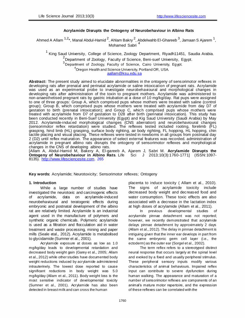
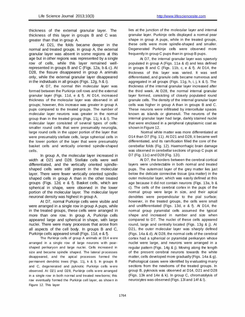
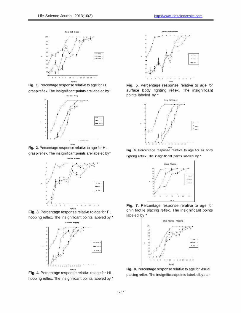
all individuals across all groups between D2 and D28. FL grasp reflex was absent in all groups at D2.
In normal pups, FL grasp was 31.67% at D4 and
increased rapidly to 100% by D10. The pattern of the
reflex curve indicated high activation of the reflex
during development. In group B, FL grasp was 8.33%
at D4 and increased to 100% by D15. In group C, it
was ~15% at D5 and increased to 100% by D17. Large
1762
Life Science Journal 2013;10(3) http://www.lifesciencesite.com fluctuations were observed in the activation of FL
grasp in group B (Fig. 1). In normal pups, HL grasp appeared after FL
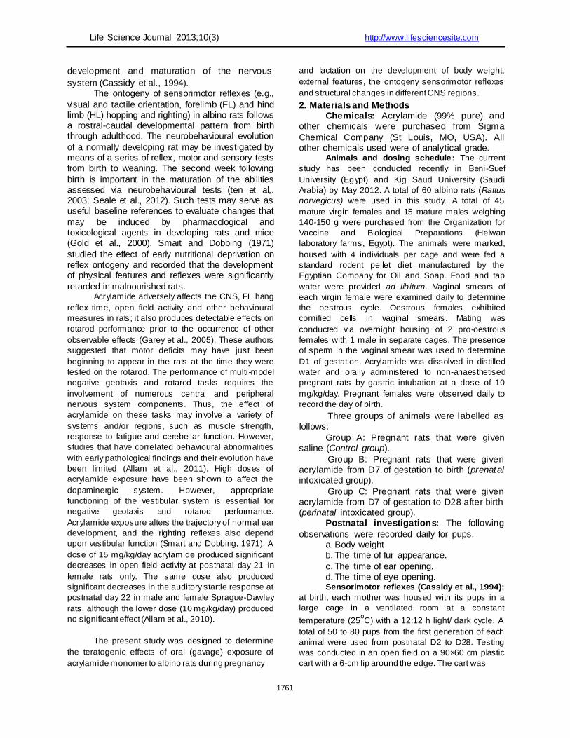
grasp. HL grasp was 18.33% at D10 and increased to 100% by D16 (Fig. 2). In groups B and C, HL grasp was 11.67% and 6.67%, respectively, at D13 and reached 100% by D19 and D21, respectively. In
group C, reflex development in the first two days was very slow (Fig. 2).
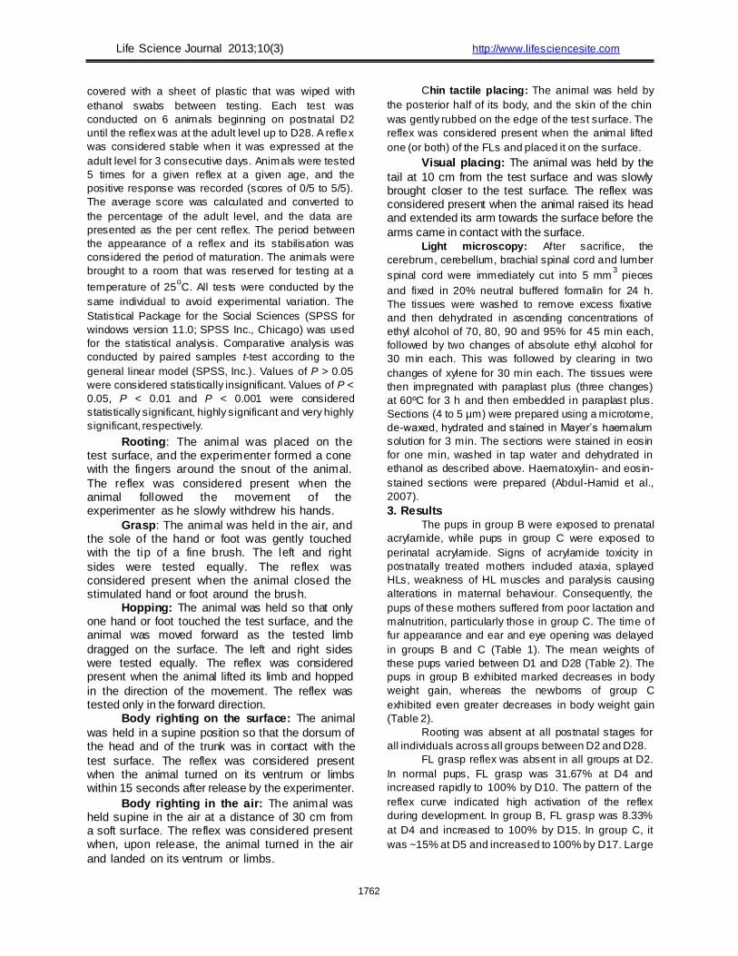
FL hopping was 48.33% at D3 in normal pups
and increased rapidly to 100% by D8. In groups B and C, the expression of this reflex was 13.33% and 43.33% at D4 and D5, respectively. The reflex
increased slowly to 100% by D14 in group B and by D15 in group C. Figure 3 shows the level of
significance between the normal and treated groups. HL hopping reflex was 38.33% at D3 in the
normal group, and its expression increased
quickly to 100% by D10. In groups B and C, HL hopping was 21.67% and 11.67%, respectively, at D5 and increased slowly to 100% by D15 and D18 in groups B and C, respectively. The pattern
of reflex curves of the acrylamide-treated groups revealed abnormalities in groups B and C when compared to the normal group (Fig. 4).
Body righting on the surface is precocious reflex was already 50% at D2 in normal pups and
increased quickly to 100% by D6. However, in groups B and C, this reflex was 10% at D2. Its expression increased to 100% in group B and group C by D12 and D13, respectively. The irregularities in
the reflex curve indicate weakness in the pups of groups B and C. The difference between normal and treated groups was significant (Fig. 5).
Body righting in the air, in normal pups, this reflex was ~30% at D11 and increased to 100% by D17. The development of the air body righting
reflex in treated groups was 16.67% in group B at D15 and 18.33% at D17 in group C. Expression increased slowly to 100% by D20 in group B and
by D22 in group C (Fig. 6). Chin tactile placing expression was 8.33% at
D4 and 100% by D11 in normal pups. Expression of
this reflex in groups B and C was 21.67% and 8.33%,
respectively, by D8 and increased rapidly to 100% in
group B at D16 and in group C at D17 (Fig. 7). Visual placing reflex was 46% at D15 and
matured rapidly to 100% by D17 in the normal
group. It was 28.33% and 27% in groups B and C at D16, respectively. Its expression increased to 100% by D19 in group B and by D21 in group C (Figure 8).
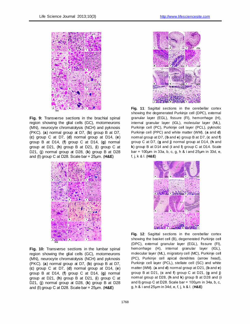
The brachial spinal cord consists of centrally
located grey matter and peripheral white matter.
Neurons and glial cells were distributed in the grey
matter. The white matter consisted of neuronal axons
and some glial (neuroglia) cells (Fig. 9). At D7,
normal motor neurons were large in size, with many
processes and oval nuclei (Fig. 9a). In group B, most motor neurons appeared small in size.
Pyknosis was also noted (Fig. 9b). In group C, motor neuron chromatolysis was observed (Fig. 9c).
Between D14 and D28, the motor neurons in individuals in group A increased in size, with well-
differentiated chromatin and a distinct thick coat of cytoplasm (Figs. 9d, g & j). The acrylamide-treated
groups exhibited chromatolysis and pyknosis in neurocytes (Figs. 9e, f, i & l). However, group B
pups had almost normally structured motor neurons at D21 and D28 (Figs. 9h & k).
The histological structure of the lumbar spinal cord is similar to that of the brachial spinal cord; the difference between these regions depended on the
degree of neuronal development. The lumbar motor neurons were relatively smaller than the brachial motor neurons at the same age (Fig. 10). At D7,
most lumbar motor neurons appeared small and less differentiated in groups B and C when compared to those of group A. Pyknosis and neurocyte chromatolysis were noted (Figs. 10a, b &
c). Between D14 and D28, normal motor neurons increased in size in group A pups, while pyknosis and neurocyte chromatolysis were still present in
the acrylamide-treated groups (Figs. 10e, f, i & l). Group B displayed improvement in lumbar motor neuron structures at D21 and D28, as was noted in
the brachial region (Figs. 10h & k). In general, during the first four weeks after birth,
the cerebellum of the normal and treated pups
consisted of grey and white matter. Grey matter was
arranged in four layers. These layers included the
superficial external granular layer, the molecular layer
with few neurons (stratum molecular), the deep internal
granular layer (stratum granulosum) and the ganglionic
(Purkinje) cell layer that was present between the
molecular and the deep internal granular layers. The
white matter was composed of corticonuclear
projections; it comprised afferent and efferent fibres. In
normal pups, the transverse fissures subdivided the
cerebellum into groups of folds (Figs. 11 & 12). These
fissures migrated vertically along the antero-posterior
axis of the brain. At D7, the folds and fissures were defined
in normal and acrylamide-treated pups. The folds were deeper in the normal group than in the treated groups (Figs. 11a, b & d). The normal group fissures were narrow, while in groups B
and C, they were wide and crude. The folds had a prominent external granular layer. This layer was thicker in group A than in groups B and C.
At D14, the folds became deep, and all layers
were represented in the normal and treated pups (Figs.
11g, h & i). In group A, there was a decline in the
1763
Life Science Journal 2013;10(3) http://www.lifesciencesite.com thickness of the external granular layer. The
thickness of this layer in groups B and C was
greater than that in group A. At D21, the folds became deeper in the
normal and treated groups. In group A, the external granular layer was absent in some regions at this age but in other regions was represented by a single
row of cells, while this layer remained well-represented in groups B and C (Figs. 12a, b & c). At D28, the fissure disappeared in group A animals
only, while the external granular layer disappeared in the individuals in all groups (Figs. 12g, h & i).
At D7, the normal thin molecular layer was
formed between the Purkinje cell rows and the external
granular layer (Figs. 11d, e & f). At D14, increased
thickness of the molecular layer was observed in all
groups; however, this increase was greater in group A
pups compared to the treated groups. The number of
molecular layer neurons was greater in the normal
group than in the treated groups (Figs. 11j, k & l). The
molecular layer consisted of several types of cells:
smaller round cells that were presumably neuroglia,
large round cells in the upper portion of the layer that
were presumably stellate cells, large spherical cells in
the lower portion of the layer that were presumably
basket cells and vertically oriented spindle-shaped
cells. In group A, the molecular layer increased in
width at D21 and D28. Stellate cells were well differentiated, and the vertically oriented spindle-shaped cells were still present in the molecular
layer. There were fewer vertically oriented spindle-shaped cells in group A than in the other treated groups (Figs. 12d, e & f). Basket cells, which are
spherical in shape, were observed in the lower portion of the molecular layer. The molecular layer neuronal density was highest in group A.
At D7, normal Purkinje cells were visible and were arranged in a single row in group A pups, while in the treated groups, these cells were arranged in
more than one row. In group A, Purkinje cells appeared large and spherical in shape, with large nuclei. There were many processes that arose from
all aspects of the cell body. In groups B and C, Purkinje cells appeared small (Figs. 11d, e & f).
The Purkinje cells of group A animals at D14 w ere
arranged in a single row of large neurons w ith pear-
shaped perikaryon and large nuc lei. Cells increased in
size and became spindle shaped. The lateral processes
disappeared, and the apical processes formed the
permanent dendrit ic trees (Figs. 11j, k & l). In groups B
and C, degenerated and pyknotic Purkinje cells w ere
observed. At D21 and D28, Purkinje cells w ere arranged
in a single row in both normal and treated new borns; this
row eventually formed the Purkinje cell layer, as show n in
Figure 12. This layer
lies at the junction of the molecular layer and internal
granular layer. Purkinje cells displayed a normal pear
shape in group A pups, while in the treated groups,
these cells were more spindle-shaped and smaller.
Degenerated Purkinje cells were observed more
frequently in group C pups than in group B pups. At D7, the internal granular layer was sparsely
populated in group A (Figs. 11a & d) and less defined
in groups B and C (Figs. 11b, c, e & f). At D14, the
thickness of this layer was varied. It was well
differentiated, and granule cells became numerous and
aggregated in all groups (Figs. 11g, h, i, j, k & l). The
thickness of the internal granular layer increased after
the third week. At D28, the normal internal granular
layer formed, consisting of closely populated round
granule cells. The density of the internal granular layer
cells was higher in group A than in groups B and C.
These neurons were infiltrated by intercellular spaces
known as islands or glomeruli. The neurons of the
internal granular layer had large, darkly stained nuclei
that were enclosed in a peripheral cytoplasmic coat as
shown in Figure 12. Normal white matter was more differentiated at
D14 than D7 (Fig. 11). At D21 and D28, it became well
differentiated, and its ramification filled the core of the
cerebellar folds (Fig. 12). Haemorrhagic brain damage
was observed in cerebellar sections of group C pups at
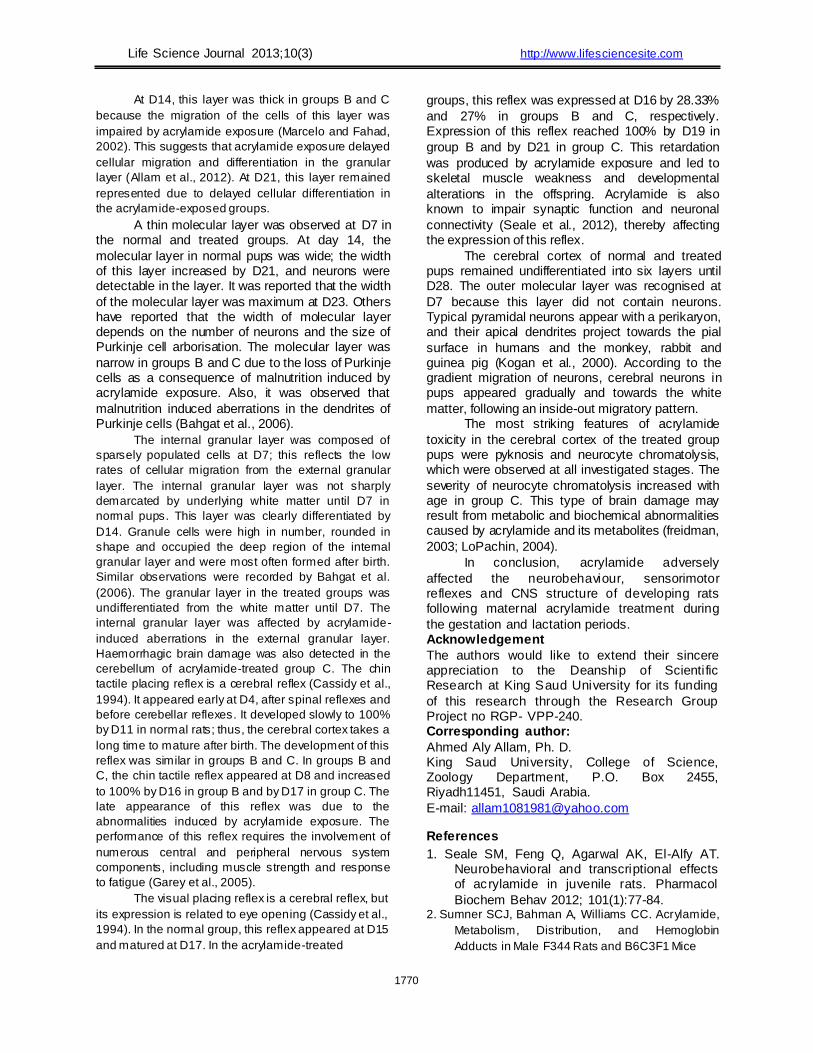
D7 (Fig. 11c) and D28 (Fig. 12i). At D7, the borders between the cerebral cortical
layers were undetectable in both normal and treated
pups. The outermost layer of the grey matter lies just
below the delicate connective tissue (pia matter) in the
outer molecular layer, which was easily defined at this
age because it did not contain neurons (Figs. 13a, b &
c). The cells of the cerebral cortex in the pups of the
normal group were large in size, and their apical
dendrites were perpendicular to the pial surface;
however, in the treated groups, the cells were small
and undifferentiated (Figs. 13d, e & f). At D14, the
normal group pyramidal cells assumed the typical
shape and increased in number and size when
compared to D7. The nuclei of these cells appeared
round, large and centrally located (Figs. 13g & j). At
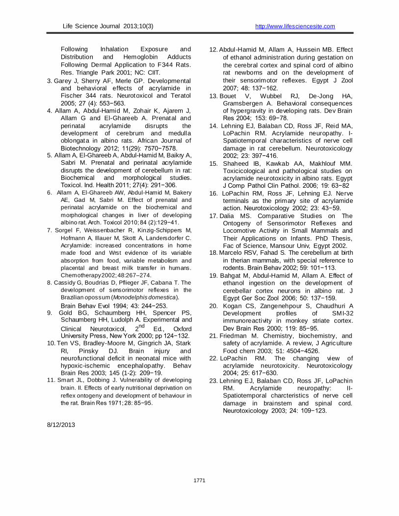
D21, the outer molecular layer was sharply defined
(Figs. 14a & d). At D28, the normal cells of the cerebral
cortex had a spherical or pyramidal perikaryon whose
nuclei were large, and neurons were arranged in a
regular pattern (Figs. 14g & j). Moving along the length
of the present cerebral neurons towards the white
matter, cells developed more gradually (Figs. 14a & g).
Pathological cases were identified by evaluating many
sections from the newborns of the treated groups. In
group B, pyknosis was observed at D14, D21 and D28
(Figs. 13k and 14e & k). In group C, chromatolysis of
neurocytes was observed (Figs. 13l and 14f & l).
1764
Life Science Journal 2013;10(3) http://www.lifesciencesite.com
4. Discussion The present study was designed to study the
ontogenesis of sensorimotor reflexes and structural
changes in the cerebral cortex, cerebellar cortex and
spinal cord of developing rats after maternal
acrylamide exposure. The effect of prenatal and
perinatal acrylamide exposure was investigated in the
developing rat embryo. Water-soluble acrylamide and
its metabolite, glycidamide, pass readily through the
placenta (Allam et al., 2010) and are distributed in
various foetal tissues during gestation (Sumner et al.,
2001). Acrylamide is also passed through the mother's
milk to pups during lactation (Allam et al., 2011). In
addition, acrylamide treatment leads to poor lactation;
this presumably results from poor maternal behaviours,
which consequently cause postnatal malnutrition in
developing pups(Bouet et al., 2004). Thus, group B
pups that were exposed to prenatal acrylamide and
group C pups that were exposed to acrylamide during
the gestation and lactation periods were exposed to
malnutrition and other toxic effects of acrylamide.
In the normal group A pups, fur appeared at D9. Fur
appearance was delayed in both acrylamide-treated
groups, consistent with Gold [9] who demonstrated that
acrylamide causes growth retardation due to protein
deficiencies resulting from malnutrition during development
(Garey et al., 2005). Compared to the normal group A
pups, ear opening was delayed in acrylamide-treated pups
(Allam et al., 2012). This suggests that acrylamide
exposure impaired organogenesis, as reported by Allam et
al. (2010). These results are also in agreement with those
reported by Garey et al. (2005). Eye opening may also be
delayed due to acrylamide exposure, as observed in
groups B and C. This effect of acrylamide supports results
reported by Sumner et al. (2001), who observed slowed
developmental changes in pups following maternal
acrylamide exposure. The newborn pups of acrylamide-treated dams
exhibited body weight loss. The mean birth weight of
pups was 5.08±0.03 g in group B and 3.88±0.11 g in
group C; the mean normal group A pup birth weight
was 6.31±0.12 g, a very highly significant difference.
Garey et al. (2005) recorded body weight reductions in
pups when female rats were exposed to acrylamide
during pregnancy. Prenatal weight reduction may be
caused by intrauterine acrylamide exposure, which
retards the growth of the developing foetus. Body
weight has been reported to be the most sensitive
indicator of developmental toxicity (Allam et al., 2011).
The toxic effects of acrylamide on embryos must be
intrauterine in nature because foetuses lack the
enzymes required to degrade the toxin once it has
entered the blood supply (Allam et al., 2010). In group
A pups, body weight increased with age, and the mean
body weight was 43.17±0.99 g at D28. In the treated
groups, the increase in body w eight w as quite slow :
26.62±0.62 g and 24.21±1.11 g at D28 in groups B and C,
respectively. In the treated groups, acrylamide affects the
function of the mammary glands by reducing prolactin
levels in animals, thus impairing lactation. It w as
demonstrated that the main reason for postnatal w eight
reduction in treated pups w as related to changes in
maternal behaviours as w ell as decreased food and w ater
consumption and lactation index. The expression of each reflex may be
dependent on the state of developmental maturation of
a specific portion of the CNS and the rate of its
development (Cassidy et al., 1994). The retarded
development of sensorimotor reflexes in the treated
groups represents a feature of acrylamide neurotoxicity
that was observed and previously described by Gary et
al., (2005). Acrylamide exposure leads to prenatal and
postnatal malnutrition through its effects on maternal
behaviours (Bouet et al., 2004). Smart and Dobbing
(1971) demonstrated that malnutrition retards the
development of sensorimotor reflexes. The results of
this study demonstrated that acrylamide induces
neuropathy and neuronal loss, leading to the
behavioural abnormalities reported by Lehning et al.
(2002). Cassidy et al. (1994) suggested that
sensorimotor reflex expression is mediated by CNS
neuronal activity, and therefore the loss of these
neurons would have an adverse effect on the
expression of these reflexes. Acrylamide impairs
synaptic function and neuronal connectivity and the
present results indicate that acrylamide caused
weakness, ataxia and malformations in the pups in the
treated groups (Shaheed et al., 2006). Similar
observations were reported by Lopachin et al. (2002). The rooting reflex was not observed in normal
and treated groups, as previously reported by Abdul-
Hamid et al. (2007). Rooting is innate in mammals
because it enables young individuals to find warmth
and a nipple for feeding (Dalia, 2002). As this need
decreases, the reflex also decreases, as reported by
Cassidy et al. (1994). The disappearance of rooting in
rat pups may be due to the inhibition of the subcortical
regions responsible for the reflex by the cerebral
cortex. The present study revealed that the laminar
organisation of the cerebral cortex was not detectable
in the rats at D7 & D14 of the experimental timeline. FL grasping, HL grasping, FL hopping, HL
hopping and surface body righting are spinal reflexes
(Cassidy et al., 1994). These behaviours appear early
in normal pups as the neuronally mediated reflexes
(motor neurons) are differentiated by this age, as
supported by the present study. Reflex expression
occurs after differentiation of the mediating neurons is
complete ( Dalia, 2002). In normal pups, expression of
the spinal reflexes increased regularly to reach mature
and well-formed motor neurons. The FL reflexes
1765
Life Science Journal 2013;10(3) http://www.lifesciencesite.com appeared and matured earlier than the HL reflexes.
The spinal histological sections revealed that
development of the spinal brachial regions occurred
earlier than the lumbar region (rostral-caudal
development). The ontogeny of sensorimotor reflexes
from birth through adulthood has been elaborated, and
mature characteristics are observed at various ages
during rostral-caudal development. In group B, the
spinal reflexes were detected late due to weakness
resulting from malnutrition and poor behaviour of
acrylamide-treated dams (Bouet et al., 2004). In group
C, these reflexes were detected later than in groups A
and B as a result of the retarded development and
differentiation of reflex-mediated neurons. Reflex
development in group C was slow and irregular due to
decreased activation of neurons, as previously
reported by Garey et al. (2005). With increasing age,
these reflexes attained 100% maturation due to
persisting small neurons. The late appearance and
maturation of grasping and hopping in the treated
groups resulted from malformations of the limbs and
spinal cord. Acrylamide exposure leads to malnutrition,
which delays the appearance and maturation of
sensorimotor reflexes (Ten et al., 2003). In addition,
acrylamide delayed the development of the reflexes
and motor neurons. Dalia (2002) reported that the
motor neurons of the ventral horn of the rat spinal cord
are already well-differentiated at birth; however, the
neurons of the dorsal horn are smaller, less
differentiated and more densely packed. The brachial and lumbar motor neurons
that were present were well differentiated at D7. Their number and size increased with age; therefore, the spinal reflexes appeared and
matured early after neuronal maturation. Brachial motor neurons appeared to be more differentiated than lumbar neurons. Dalia (2002) reported that
brachial motor neurons are large in size and number compared to lumbar motor neurons.
The motor neurons of the acrylamide-treated groups were small and less differentiated than those of the normal pups. Between D7 and D28, the normal pups’ motor neurons increased in size,
although pyknosis and neurocyte chromatolysis were observed. Rats in group B showed some improvement in the structure of motor neurons at
D21 and D28 during lactation. Acrylamide exposure impaired motor neuron functions, including motor coordination, motor control and the ability to
generate action potentials (Shaheed et al. 2006). Air body righting is a cerebellar reflex that
appears late in the developmental course of normal
pups because cerebellum maturation occurs similarly
late in development (Cassidy et al., 1994). This reflex
is normally detected at D11 and increases regularly to
reach 100% at D17. This pattern of reflex
development occurs due to the continuous
differentiation and maturation of cerebellar neurons.
These results parallel those of Allam et al. (2012).
Maturation was reported to reach 100% at D22 in the
Mongolian gerbil (Cassidy et al., 1994) and at D21 in
both the rabbit and guinea pig (Dalia, 2002). In group
B, this reflex was detected at D15 and developed to
maturation by D20. In group C, it was detected later, at
D17, and reached 100% by D22. This reflex developed
earlier in group B than group C because the pups of
group C were exposed to acrylamide for a longer
period of time during lactation. In both treated groups,
retardation of reflex development resulted from either
late maturation and differentiation of the cerebellar
neurons or from high levels of neuronal loss caused by
acrylamide. Similar results were reported by Garey et
al. (2005). At D7, the external granular layer of normal
pups was thick due to high cellular proliferation rates. Marcelo and Fahad (2002) observed that the anlage fold appears at birth in normal and acrylamide-treated pups and was represented by
the external granular layer. This layer forms by cellular migration in an inside-out pattern (Marcelo and Fahad, 2002). These cells proliferate rapidly in
the external granular layer and then migrate inward and differentiate into granular neurons in the internal granular layer (Allam et al., 2011).
The cerebellar cortex of the normal rat
newborns at D14 consisted of four layers: the
superficial external granular layer, the molecular layer,
the Purkinje cell layer and the internal granular layer.
The external granular layer was present in normal rats
at D14 but was thin because the cells had migrated to
the internal granular layer. This layer remained
represented at D21 by one row of cells or was
completely absent in some regions; it disappeared
completely at D28 in the rat, D21 in the guinea pig
(Dalia, 2002) and D18 in the mouse (Abdul-Hamid et
al., 2007). The disappearance of this layer was
attributed to the migration of its cells into the
neighbouring molecular layer, which in turn, increased
in width via resorption of the external granular layer. In the acrylamide-treated groups, the external
granular layer was thin at D7 due to chronic acrylamide
exposure in pre- and postnatal pups; this in turn
delayed the proliferation of the cells in the granular
layer. Marcelo and Fahad (2002) reported that the
external granular layer is the external germinal layer
that generates the granule cells; the cells then begin to
migrate through the Purkinje cell layer to form the
internal granular layer. Therefore, the internal granular
layer will be affected by aberrations in the external
granular layer (Allam et al., 2012).
1766
Life Science Journal 2013;10(3) http://www.lifesciencesite.com
Fore-limb Grasp
100
90 * * *
80
70 *
60 Gro u p A
%
50 Gro u p B
40 Gro u p C
30
20
10
0
4 5 6 7 8 9 10 11 12 13 14 15 16 17
Age (D) Fig. 1. Percentage response relative to age for FL
grasp reflex. The insignificant points are labeled by *
Hind-limb Grasp
Surface Body Righting
100
90
* * *
80
70
60
%
50 G roup A
G roup B
40
G roup C
30
20
10
0
2 3 4 5 6 7 8 9 10 11 12 13
Age (D) Fig. 5. Percentage response relative to age for
surface body righting reflex. The insignificant
points labeled by *
100 *
90 80
70
60
50
%
40
30
20
10
0 10 11 12 13 14 15 16 17 18 19 20 21 22
Age (D)
Group A G roup B Group C
Body Righting Air
100
90
*
80
70
60
%
50 Group A
40
Group B
Group C 30
20
10
0
11 12 13 14 15 16 17 18 19 20 21 22 23
Fig. 2. Percentage response relative to age for HL
grasp reflex. The insignificant points are labeled by *
Fore-limb Hopping
100
90 * * * *
*
80
70
60
% 50 G roup A
40 G roup B
30
G roup C
20
10
0
3 4 5 6 7 8 9 10 11 12 13 14 15
Age (D) Fig. 3. Percentage response relative to age for FL
hooping reflex. The insignificant points labeled by *
Hind-limb Hopping
Age (D)
Fig. 6. Percentage response relative to age for air body
righting reflex. The insignif icant points labeled by *
Visual Placing
100 *
90 *
80
70 G roup A
60 G roup B
%
50 G roup C
40
30
20
10
0
15 16 17 18 19 20 21
Age (D)
Fig. 7. Percentage response relative to age for
chin tactile placing reflex. The insignificant points
labeled by *
Chin Tactile Placing
100 100
90
80
70
60
% 50 40 30
20 10
0
* *
Group A
Group B
Group C
90 * *
80 *
70
60 Group A
%
50 Group B
40 Group C
30
20
10
0
4 5 6 7 8 9 10 11 12 13 14 15 16 17
Age (D)
3 4 5 6 7 8 9 10 11 12 13 14 15 16 17 18 19 20 Age (D)
Fig. 4. Percentage response relative to age for HL
hooping reflex. The insignificant points labeled by *
Fig. 8. Percentage response relative to age for visual
placing reflex. The insignificant points labeled by star
1767
Life Science Journal 2013;10(3) http://www.lifesciencesite.com
Fig. 9: Transverse sections in the brachial spinal region showing the glial cells (GC), motorneurons
(MN), neurocyte chromatolysis (NCH) and pyknosis
(PKC). (a) normal group at D7, (b) group B at D7, (c) group C at D7, (d) normal group at D14, (e)
group B at D14, (f) group C at D14, (g) normal
group at D21, (h) group B at D21, (i) group C at
D21, (j) normal group at D28, (k) group B at D28 and (l) group C at D28. Scale bar = 25μm. (H&E) Fig. 10: Transverse sections in the lumbar spinal
region showing the glial cells (GC), motorneurons
(MN), neurocyte chromatolysis (NCH) and pyknosis
(PKC). (a) normal group at D7, (b) group B at D7, (c) group C at D7, (d) normal group at D14, (e)
group B at D14, (f) group C at D14, (g) normal
group at D21, (h) group B at D21, (i) group C at D21, (j) normal group at D28, (k) group B at D28
and (l) group C at D28. Scale bar = 25μm. (H&E)
Fig. 11: Sagittal sections in the cerebellar cortex
showing the degenerated Purkinje cell (DPC), external
granular layer (EGL), fissure (FI), hemorrhage (H),
internal granular layer (IGL), molecular layer (ML),
Purkinje cell (PC), Purkinje cell layer (PCL), pyknotic
Purkinje cell (PPC) and white matter (WM). (a and d)
normal group at D7, (b and e) group B at D7, (c and f)
group C at D7, (g and j) normal group at D14, (h and
k) group B at D14 and (i and l) group C at D14. Scale
bar = 100μm in 33a, b, c, g, h & i and 25μm in 33d, e,
f, j, k & l. (H&E)
Fig. 12: Sagittal sections in the cerebellar cortex
showing the basket cell (B), degenerated Purkinje cell
(DPC), external granular layer (EGL), fissure (FI),
hemorrhage (H), internal granular layer (IGL),
molecular layer (ML), migratory cell (MC), Purkinje cell
(PC), Purkinje cell apical dendrites (arrow head),
Purkinje cell layer (PCL), stellate cell (SC) and white
matter (WM). (a and d) normal group at D21, (b and e)
group B at D21, (c and f) group C at D21, (g and j)
normal group at D28, (h and k) group B at D28 and (i
and l) group C at D28. Scale bar = 100μm in 34a, b, c,
g, h & i and 25μm in 34d, e, f, j, k & l. (H&E)
1768
Life Science Journal 2013;10(3) http://www.lifesciencesite.com
Fig. 13: Sagittal sections in the cerebral cortex
showing the outer molecular layer (OML), pyramidal cells distribution (PYC), neurocyte
chromatolysis (NCH) and pyknosis (PKC). (a and
d) normal group at D7, (b and e) group B at D7, (c and f) group C at D7, (g and j) normal group at
D14, (h and k) group B at D14 and (i and l) group
C at D14. Scale bar = 100μm in 29a, b, c, g, h & i and 25μm in 29d, e, f, j, k & l. (H&E) Fig. 14: Sagittal sections in the cerebral cortex
showing the neurocyte chromatolysis (NCH), outer molecular layer (OML), pyramidal cells
distribution (PYC) and pyknosis (PKC). (a and d)
normal group at D21, (b and e) group B at D21, (c and f) group C at D21, (g and j) normal group
at D28, (h and k) group B at D28 and (i and l) group C at D28. Scale bar = 100μm in 30a, b, c,
g, h & i and 25μm in 30d, e, f, j, k & l. (H& E)
Table 1. External features appearance in rat newborns.
Features/Groups A B C
Fur appearing D9 D11-12 D12-13
Ear opening D12-13 D15 D15
Eye opening D14-15 D16-17 D16-17
Table 2. Changes of body weights in rat newborns.
/ Groups
.31±0.12 .08±0.13*** .88±0.11***
.4±0.26 .15±0.08*** .47±0.5 0* *
.33±0.19 .53±0.16*** .27±0.3 8* *
.15±0.15 .57±0.11*** .83±0.53***
.58±0.28 .8±0.13*** .48±0.29***
0.65±0.23 ±0.21*** .23±0.40***
2.13±0.16 .83±0.27*** .18±0.27***
3.57±0.1 .85±0.41*** .75±0.31***
4.27±0.13 .23±0.68*** .13±0.38***
0 6.1±0.15 0.9±0.3 36 * ** .9±0.30***
1 7.23±0.13 0.47± 0.63 * ** .33±0.77***
2 7.33±0.07 0.77± 0.41 * ** .85±0.80***
3 8.25±0.09 2.27± 0.81 * ** .27±0.65***
4 9.35±0.15 1.58± 0.72 * ** .33±0.67***
5 0.35±0.08 3.28± 0.68 * ** 0.72±0.6 0* **
6 1.60±0.15 5.38± 0.42 * ** 0.39±0.3 4* **
7 3.67±0.36 6.93±0.7 6* * 2.93±0.6 1* **
8 3.97±0.36 7.36± 0.78 * ** 1.3±0.56***
9 5.22±0.51 9.73± 0.40 * ** 1.63±0.6 0* **
0 5.8±0.64 0.68± 0.69 * ** 2.47±0.7 0* **
1 7.75±0.60 0.9±0.38*** 4.18±0.5 0* **
2 8.47±0.52 1.2±0.51*** 6.48±0.4 7* **
3 1.42±0.45 2.85± 0.71 * ** 0.12±0.9 5* **
4 4.52±0.35 3.68± 0.99 * ** 0.97±0.8 7* **
5 6.07±0.37 3.55± 0.77 * ** 1.55±1.1 3* **
6 8.45±0.57 4.65± 0.99 * ** 2.40±1.1 2* **
7 9.65±0.62 5.87±77*** 3.25±0.6 4* **
8 3.17±0.99 6.62± 0.62 * ** 4.21±1.1 1* **
Data are expressed as a mean ± S.E. (N =6) Values significantly compared to the control
newborns; p*≤0.05, p
**≤0.01and p
***≤0.001.
1769
Life Science Journal 2013;10(3) http://www.lifesciencesite.com
At D14, this layer was thick in groups B and C
because the migration of the cells of this layer was
impaired by acrylamide exposure (Marcelo and Fahad,
2002). This suggests that acrylamide exposure delayed
cellular migration and differentiation in the granular
layer (Allam et al., 2012). At D21, this layer remained
represented due to delayed cellular differentiation in
the acrylamide-exposed groups. A thin molecular layer was observed at D7 in
the normal and treated groups. At day 14, the
molecular layer in normal pups was wide; the width of this layer increased by D21, and neurons were detectable in the layer. It was reported that the width
of the molecular layer was maximum at D23. Others have reported that the width of molecular layer depends on the number of neurons and the size of Purkinje cell arborisation. The molecular layer was
narrow in groups B and C due to the loss of Purkinje cells as a consequence of malnutrition induced by acrylamide exposure. Also, it was observed that
malnutrition induced aberrations in the dendrites of Purkinje cells (Bahgat et al., 2006).
The internal granular layer was composed of
sparsely populated cells at D7; this reflects the low
rates of cellular migration from the external granular
layer. The internal granular layer was not sharply
demarcated by underlying white matter until D7 in
normal pups. This layer was clearly differentiated by
D14. Granule cells were high in number, rounded in
shape and occupied the deep region of the internal
granular layer and were most often formed after birth.
Similar observations were recorded by Bahgat et al.
(2006). The granular layer in the treated groups was
undifferentiated from the white matter until D7. The
internal granular layer was affected by acrylamide-
induced aberrations in the external granular layer.
Haemorrhagic brain damage was also detected in the
cerebellum of acrylamide-treated group C. The chin
tactile placing reflex is a cerebral reflex (Cassidy et al.,
1994). It appeared early at D4, after spinal reflexes and
before cerebellar reflexes. It developed slowly to 100%
by D11 in normal rats; thus, the cerebral cortex takes a
long time to mature after birth. The development of this
reflex was similar in groups B and C. In groups B and
C, the chin tactile reflex appeared at D8 and increased
to 100% by D16 in group B and by D17 in group C. The
late appearance of this reflex was due to the
abnormalities induced by acrylamide exposure. The
performance of this reflex requires the involvement of
numerous central and peripheral nervous system
components, including muscle strength and response
to fatigue (Garey et al., 2005). The visual placing reflex is a cerebral reflex, but
its expression is related to eye opening (Cassidy et al.,
1994). In the normal group, this reflex appeared at D15
and matured at D17. In the acrylamide-treated
groups, this reflex was expressed at D16 by 28.33%
and 27% in groups B and C, respectively. Expression of this reflex reached 100% by D19 in
group B and by D21 in group C. This retardation
was produced by acrylamide exposure and led to skeletal muscle weakness and developmental
alterations in the offspring. Acrylamide is also known to impair synaptic function and neuronal
connectivity (Seale et al., 2012), thereby affecting the expression of this reflex.
The cerebral cortex of normal and treated pups remained undifferentiated into six layers until D28. The outer molecular layer was recognised at
D7 because this layer did not contain neurons. Typical pyramidal neurons appear with a perikaryon, and their apical dendrites project towards the pial
surface in humans and the monkey, rabbit and guinea pig (Kogan et al., 2000). According to the gradient migration of neurons, cerebral neurons in pups appeared gradually and towards the white
matter, following an inside-out migratory pattern. The most striking features of acrylamide
toxicity in the cerebral cortex of the treated group pups were pyknosis and neurocyte chromatolysis, which were observed at all investigated stages. The
severity of neurocyte chromatolysis increased with age in group C. This type of brain damage may result from metabolic and biochemical abnormalities caused by acrylamide and its metabolites (freidman,
2003; LoPachin, 2004). In conclusion, acrylamide adversely
affected the neurobehaviour, sensorimotor reflexes and CNS structure of developing rats following maternal acrylamide treatment during
the gestation and lactation periods. Acknowledgement The authors would like to extend their sincere appreciation to the Deanship of Scientific Research at King Saud University for its funding
of this research through the Research Group Project no RGP- VPP-240. Corresponding author: Ahmed Aly Allam, Ph. D. King Saud University, College of Science, Zoology Department, P.O. Box 2455, Riyadh11451, Saudi Arabia. E-mail: [email protected] References 1. Seale SM, Feng Q, Agarwal AK, El-Alfy AT.
Neurobehavioral and transcriptional effects of acrylamide in juvenile rats. Pharmacol
Biochem Behav 2012; 101(1):77-84. 2. Sumner SCJ, Bahman A, Williams CC. Acrylamide,
Metabolism, Distribution, and Hemoglobin
Adducts in Male F344 Rats and B6C3F1 Mice
1770
Life Science Journal 2013;10(3) http://www.lifesciencesite.com
Following Inhalation Exposure and
Distribution and Hemoglobin Adducts
Following Dermal Application to F344 Rats.
Res. Triangle Park 2001; NC: CIIT. 3. Garey J, Sherry AF, Merle GP. Developmental
and behavioral effects of acrylamide in Fischer 344 rats. Neurotoxicol and Teratol
2005; 27 (4): 553−563. 4. Allam A, Abdul-Hamid M, Zohair K, Ajarem J,
Allam G and El-Ghareeb A. Prenatal and
perinatal acrylamide disrupts the development of cerebrum and medulla oblongata in albino rats. African Journal of
Biotechnology 2012; 11(29): 7570−7578. 5. Allam A, El-Ghareeb A, Abdul-Hamid M, Baikry A,
Sabri M. Prenatal and perinatal acrylamide
disrupts the development of cerebellum in rat: Biochemical and morphological studies. Toxicol. Ind. Health 2011; 27(4): 291−306.
6. Allam A, El-Ghareeb AW, Abdul-Hamid M, Bakery
AE, Gad M, Sabri M. Effect of prenatal and
perinatal acrylamide on the biochemical and
morphological changes in liver of developing
albino rat. Arch. Toxicol 2010; 84 (2):129−41.
7. Sorgel F, Weissenbacher R, Kinzig-Schippers M,
Hofmann A, Illauer M, Skott A, Landersdorfer C.
Acrylamide: increased concentrations in home
made food and Wrst evidence of its variable
absorption from food, variable metabolism and
placental and breast milk transfer in humans.
Chemotherapy 2002; 48:267–274. 8. Cassidy G, Boudrias D, Pflieger JF, Cabana T. The
development of sensorimotor reflexes in the
Brazilian opossum (Monodelphis domestica).
Brain Behav Evol 1994; 43: 244−253. 9. Gold BG, Schaumberg HH, Spencer PS,
Schaumberg HH, Ludolph A. Experimental and
Clinical Neurotoxicol, 2nd
Ed., Oxford University Press, New York 2000; pp 124−132.
10. Ten VS, Bradley-Moore M, Gingrich JA, Stark
RI, Pinsky DJ. Brain injury and neurofunctional deficit in neonatal mice with hypoxic-ischemic encephalopathy. Behav Brain Res 2003; 145 (1-2): 209−19.
11. Smart JL, Dobbing J. Vulnerability of developing
brain. II. Effects of early nutritional deprivation on
reflex ontogeny and development of behaviour in
the rat. Brain Res 1971; 28: 85−95.
8/12/2013
12. Abdul-Hamid M, Allam A, Hussein MB. Effect
of ethanol administration during gestation on
the cerebral cortex and spinal cord of albino rat newborns and on the development of
their sensorimotor reflexes. Egypt J Zool
2007; 48: 137−162. 13. Bouet V, Wubbel RJ, De-Jong HA,
Gramsbergen A. Behavioral consequences of hypergravity in developing rats. Dev Brain Res 2004; 153: 69−78.
14. Lehning EJ, Balaban CD, Ross JF, Reid MA, LoPachin RM. Acrylamide neuropathy. I-Spatiotemporal characteristics of nerve cell
damage in rat cerebellum. Neurotoxicology 2002; 23: 397−416.
15. Shaheed IB, Kawkab AA, Makhlouf MM. Toxicicological and pathological studies on acrylamide neurotoxicity in albino rats. Egypt J Comp Pathol Clin Pathol. 2006; 19: 63−82
16. LoPachin RM, Ross JF, Lehning EJ. Nerve terminals as the primary site of acrylamide action. Neurotoxicology 2002; 23: 43−59.
17. Dalia MS. Comparative Studies on The Ontogeny of Sensorimotor Reflexes and Locomotive Activity in Small Mammals and
Their Applications on Infants. PhD Thesis, Fac of Science, Mansour Univ, Egypt 2002.
18. Marcelo RSV, Fahad S. The cerebellum at birth
in therian mammals, with special reference to rodents. Brain Behav 2002; 59: 101−113.
19. Bahgat M, Abdul-Hamid M, Allam A. Effect of ethanol ingestion on the development of
cerebellar cortex neurons in albino rat. J Egypt Ger Soc Zool 2006; 50: 137−159.
20. Kogan CS, Zangenehpour S, Chaudhuri A Development profiles of SMI-32 immunoreactivity in monkey striate cortex.
Dev Brain Res 2000; 119: 85−95. 21. Friedman M. Chemistry, biochemistry, and
safety of acrylamide. A review, J Agriculture
Food chem 2003; 51: 4504−4526. 22. LoPachin RM. The changing view of
acrylamide neurotoxicity. Neurotoxicology 2004; 25: 617−630.
23. Lehning EJ, Balaban CD, Ross JF, LoPachin RM. Acrylamide neuropathy: II- Spatiotemporal charcteristics of nerve cell
damage in brainstem and spinal cord. Neurotoxicology 2003; 24: 109−123.
1771