Embed Size (px)
Citation preview
A LARGE AMELOBLASTOMA OF THE MANDIBLE: A CASE REPORT
Keywords:Ameloblastoma, Reconstruction, 3D CBCT
Key Message:Ameloblastoma is a anatomically benign, clinical-
ly persistent and locally aggressive tumor with high recurrence
rates. Radiographically it may present as a multilocular radiolus-
cency with characteristic "soap bubble or honey comb appearance
" or as a unilocular lesion. Conservative approaches of treatment
include curettage and enucleation with reported high recurrence
rates. Radical treatment involves wide margin excisions of the
tumor mass. Reconstruction of the resected defect is a challenge
for the surgeon to provide a favorable functional and aesthetic
outcome to the patient.
How to cite this Article:
Bhargava A,Soni S,Tyagi.A Large Ameloblastoma of the
Mandible: A Case Report.Arch CranOroFac Sc 2014;1(5):66-72
Source of Support: Nil
Conflict of Interest:No
INTRODUCTION
Ameloblastoma is a rare benign true neoplasm of odontogenic
origin. It is derived from the word of English literature "Amel"
which means enamel.In Greek "Blastos" refers to
germ[1].Ameloblastoma is known for its locally invasive and
aggressive character. It has a strong tendency to recur which is
well evidenced by its high recurrence rates[2].
This odontogenic tumor has been considered to be of varied
origin by many authorities, Thus the tumor might be conceivably
thought to be derived from cell rest of the enamel organ,either
remnants of dental lamina or remnants of Hertwig's sheaths,
epithelial rest of mallassez, epithelium of odontogenic cysts, par-
ticularly the dentigerous or the odontomas,disturbance of the
developing enamel organ, basal cells of the surface epithelium of
the jaws or the heterotopic epithelium in others parts of the body,
especially the pituitary gland[3].
In 1827 Cusack was the first person to describe this tumor[4].
The name "Admantinoma" was introduced by Mallassez in
1885,which nowadays is used to describe a rare type of bone can-
cer[5].Churchill coined the term "Ameloblastoma" in
1930[6].According to WHO the tumor is a benign, but locally
invasive neoplasm that often has diverse histologic patterns[7].
This group of tumors comprise of various histopathological
types and clinical behavior[8]. Ameloblastoma accounts for about
1% of all the cyst and tumors of the jaws and 18% of the differ-
ent odontogenic tumors[9].
The mandible has five times higher occurrence of the tumor as
compared to the maxilla[10]. The average age of occurrence as
reported in literature is 38.9 years[11]. It occurs with equal fre-
quency in both sexes[12].If we review the literature, In 52% of
the cases it occurred in men,48% in women[11].Small and
Waldron[11] have reported that it is the molar region where 47%
of Maxillary Ameloblastomas occur, a lesser percentage of 33%
occur in the antrum and floor of the nose, only 9% occur in both
the premolar and canine regions.2% occurrence was noted in the
palatal region of the maxilla according their study. In the
mandible most common site is molar and ascending ramus region
accounting for 39%, and 16% occurred in molar premolar region
and 9% in the anterior region[13].
Clinically the tumor can be classified in four distinct types:
unicystic, solid or multicystic, peripheral and malignant.The uni-
cystic type usually presents as a "cystic" lesion with either an
intraluminal or an intramural proliferation of the cystic lin-
ing[14]. Radiographically, it is seen as a well-circumscribed
slow- growing radioluscency.Multicystic Ameloblastomas can
show infiltration into the neighbouring tissues with ability to
recur.They can sometimes show metastasis. Radiographically, it
www.acofs.com
Use the QR Code scanner to access
this article online in our databse
Article Code: ACOFS0021
66
ACOFS VOL I ISSUE VCASE REPORT
This Article Published by BPH,India is licensed under a Creative Commons Attribution-Non Commercial-Share Alike 3.0 Unported License.
Anuj Bhargava1,Smita Soni2,Amit Tyagi3
ABSTRACT
Ameloblastoma is a slow growing, and locally aggressive
tumor with high recurrence rates. The tumor can assume large
sizes. It arises from epithelium of the dental lamina.
Clinically the tumor can presents itself as swelling which are
generally asymptomatic. Histologically there are many vari-
ants. Radiographically the tumor can occur either as mutiloc-
ular radioluscent lesion giving a peculiar honey comb appear-
ance or as a unicystic variety. There are many treatment
options available which range from conservative treatment of
curettage, enucleation to radical surgical approaches of wide
margin excision. Radical treatment approaches have the
advantage of lowering the recurrence rates but at the same
time pose extremely difficult challenges of reconstruction of
the surgical defects. We are reporting a case of a 20 year old
young female diagnosed with a large multicystic ameloblas-
toma of the mandible in which wide margin surgical excision
of the tumor by segmental resection of the left hemimandible
was performed with spanning of the boney defect with titani-
um reconstruction plates to achieve a favorable aesthetic and
functional outcome for the patient.
www.acofs.com
may appear as a unilocular or multilocular lesion[15]. Peripheral
ameloblastoma is a soft tissue variant of ameloblastoma which
mostly presents in the alveolar mucoas. Although this lesion can
also involve the underlying bone[16].
Malignant Ameloblastoma although a rare type is defined as an
ameloblastoma which has already undergone malignant metas-
tases but still has its classical histological microscopic fea-
tures[17].Other histological variants have also been described for
ameloblastoma like follicular, plexiform, basal, granular and
acanthomathous[18].Desmoplastic Ameloblastoma is a rare vari-
ant which has been described in The World Health
Organization(WHO) classification of odontogenic tumors late-
ly[19].
Clinically, ameloblastomas appears as an aggressive odonto-
genic tumor, often asymptomatic and slow growing, with no sign
of swelling. It can sometimes cause symptoms such as swelling,
dental malocclusion, pain, paresthesia of the affected area[20].It
spreads by forming pseudopods in marrow spaces without con-
comitant resorption of the trabacular bone. Beacause of this, the
tumor margins are not clearly seen on radiographs or during sur-
gery and the tumor frequently recurs after inadequate surgical
removal[21,22].The appearance of septae on the radiograph usu-
ally represents differential resorption of the cortical plate by the
tumor and not actual separation of tumor portions[23].Because of
its slow growth, recurrence of ameloblatoma generally present
many years and decades after primary surgery[22].When treated
inadequately , malignant development is a possibility[20].
In most cases ameloblastoma has a characteristic but not diag-
nostic radiographic appearance[21].The neoplasm usually
appears as a unilocular radioluscent area or a multilocular radio-
luscent area with honey comb appearance[20,21].Adjacent tooth
roots might show evidence of resorption[21].The tumor in many
cases might be accompanied with an unerupted tooth, most com-
monly a mandibular third molar[24].
Treatment of mandibular ameloblatomas continues to be con-
troversial. It can change with clinicoradiographic variant,
anatomic location and clinical behavior of the
tumor[25].Treatment consists of wide resection, enucleation and
curettage [22,26].Rates of recurrence may be as high as 15 % to
25% after radical treatment and 75% to 90% after conservative
treatment[26].According to Reichart PA, Philipsen HP[13]and
associates the rate of recurrence demonstrated were 17.7% for
enbloc resection to 34.7% for conservative therapy.
The aim of this article is to present a case of a large mandibu-
lar ameloblastoma which was treated with radical treatment
approach of segmental resection of the left hemimandible fol-
lowed by reconstruction with Titanium plates to achieve a par-
tially favorable aesthetic and functional outcome. The article
briefly elaborates the clinical and histological types of the tumor
along with the recurrence rates associated with the tumor because
of its unique biologic behavior. The article also discusses the
importance of reconstruction especially in cases of large tumor
resections.
CASE REPORT
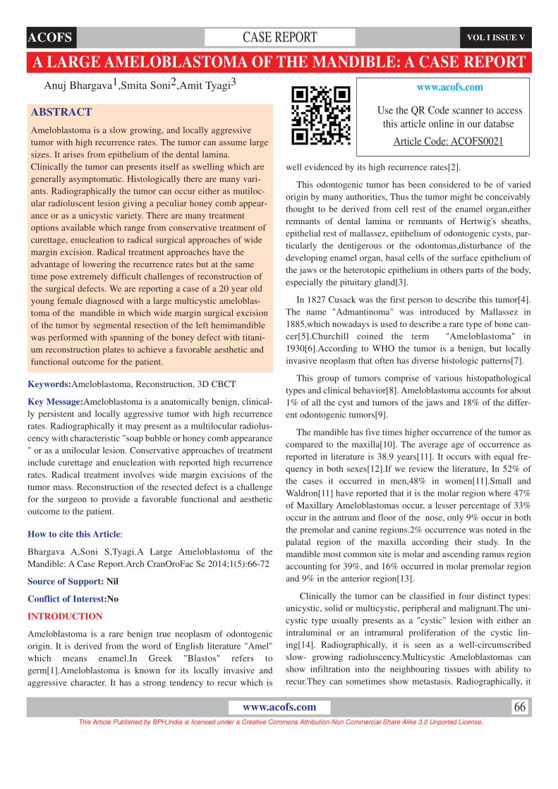
A 20 year old young female
patient [Figure.1] reported to the
outpatient department of our
institute around one year back
with a chief complaint of slowly
increasing large painless
swelling over left side of the
lower jaw since last one year of
her first visit to the institute.
There were no signs of difficul-
ty in deglutition or breathing.
No underlying significant med-
ical history was elicited.
Physical examination
revealed a large non tender,
bony hard 6cmX 6cm asympto-
matic well demarcated swelling anteriorly extending from left
angle of the mandible to the left corner of the mouth. The
swelling extended superiorly from the left malar region inferior-
ly into the left hyoid bone region of the neck causing gross facial
disfigurement. On palpation regional cervical lymph nodes were
non tender and not enlarged. There was no evidence of any neu-
rosensory deficit especially along the distribution of the trigemi-
nal nerve.
The swelling was non pulsatile with normal overlying skin
color and texture. Few dilated superficial veins were prominent
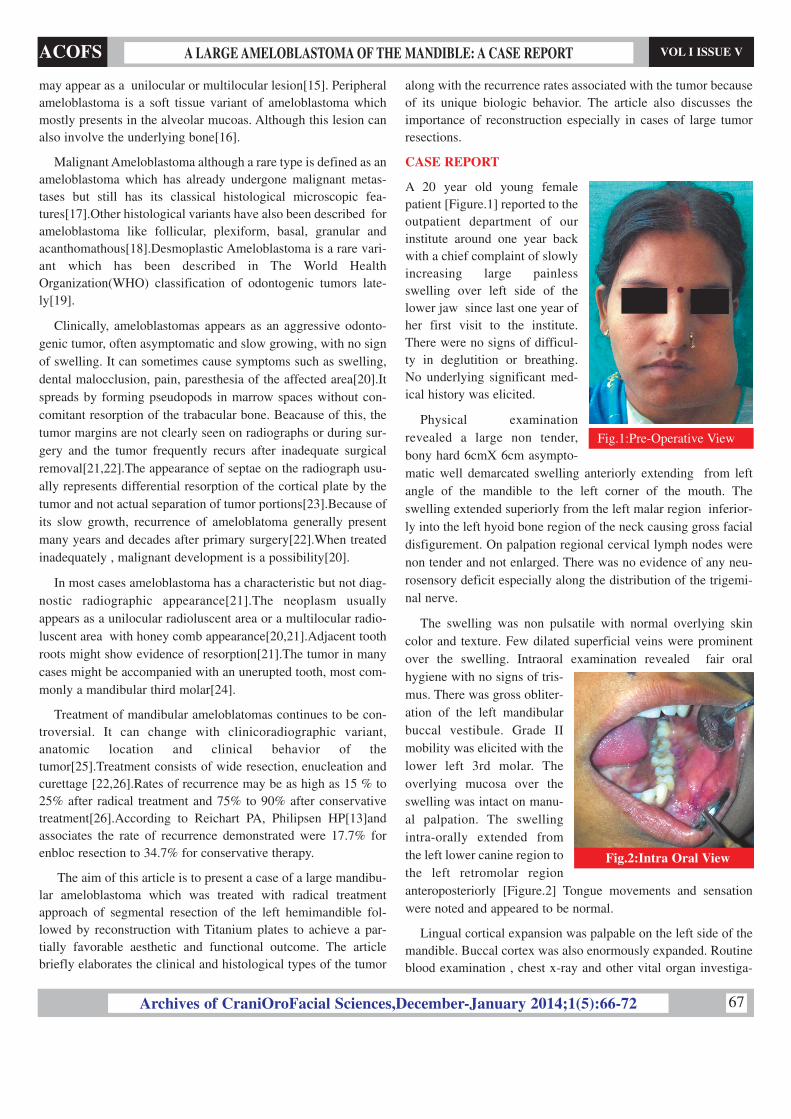
over the swelling. Intraoral examination revealed fair oral
hygiene with no signs of tris-
mus. There was gross obliter-
ation of the left mandibular
buccal vestibule. Grade II
mobility was elicited with the
lower left 3rd molar. The
overlying mucosa over the
swelling was intact on manu-
al palpation. The swelling
intra-orally extended from
the left lower canine region to
the left retromolar region
anteroposteriorly [Figure.2] Tongue movements and sensation
were noted and appeared to be normal.
Lingual cortical expansion was palpable on the left side of the
mandible. Buccal cortex was also enormously expanded. Routine
blood examination , chest x-ray and other vital organ investiga-
ACOFS VOL I ISSUE V
67
A LARGE AMELOBLASTOMA OF THE MANDIBLE: A CASE REPORT
Fig.1:Pre-Operative View
Fig.2:Intra Oral View
Archives of CraniOroFacial Sciences,December-January 2014;1(5):66-72
68
ACOFS VOL I ISSUE V
tions were also per-
formed which were
also normal.
Left and Right
Lateral oblique 30
Degree views
[Figure.3] along with
PosteroAnterior and
AnteroPosteror view
of the mandible were
also advised. Ortho-
pantograph facility is
not available at our centre. A plain and contrast CT scan of the
mandible with 4mm collimation was advised which revealed
[Figure.4] multilocu-
lated expansile lytic
lesion (7.3 cm X 6.2
cm X 7.3cm) show-
ing multiple internal
osseous trabuculae
giving honey comb
appearance involving
the left body of the
mandible and the
ramus.There was cor-
tical thinning, with
multiple areas of cor-
tical breach and adja-
cent small soft tissue
component. Left side
of the condyle and
Tempor-omandibular
Joint were free of the
mass. Differential
diagnosis as per the
radiographic report included odontogenic fibroma, odontogenic
myxoma, fibroma , ameloblastoma and central giant cell granulo-
ma. An intra-oral aspiration biopsy was also performed which
revealed yellowish straw colored fluid.
Although biopsy results did not show any specific features of
ameloblastoma, the clinical presentation, history along with radi-
ographic diagnosis was quite suggestive of ameloblastoma. The
patient was planned for wide margin surgical excision of the
tumor mass under general anesthesia. Nasotracheal intubation
was performed and Erisch arch bars were placed on the contralat-
eral side. The tumor site was reached by the traditional lip split-
ting and the submandibular incisions. Soft tissue dissection was
performed carefully and the skin flaps were raised, marginal
mandibular nerve was protected to expose the tumor mass.
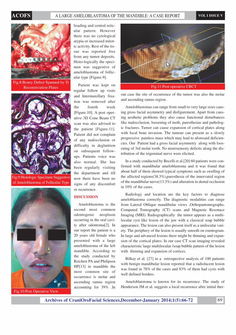
Intraoperatively the tumor was found to be around 7cm X 6cm
in size [Figure.5] with anteroposterior extensions from the left
A LARGE AMELOBLASTOMA OF THE MANDIBLE: A CASE REPORT
incisal region up to the
left ascending rami of
the mandible sparring
the coronoid and the
condyle. Segmental
resection of the left
hemimandible was done
from the left incisal
region to the left ascend-
ing ramus of the
mandible leaving the
coronoid process and the
bony chunk of sub-
condylar region of the
mandible. The tumor
was freed from all its
soft tissue attachments
and removed in totality
[Figure.6].
The specimen was
sent for histopathologic
diagnosis[Figure.7].
The bony defect created
by the resected mass
was spanned with titani-
um reconstruction
plates [Figure.8] taking
due consideration of the
occlusion of the con-
tralateral side which
was previously secured
with arch bars. Hemo-
stasis was achieved.
Surgical drains were
placed. Wound closure was done in layers. Post operative
recovery was uneventful. The patient was kept on intermaxil-
lary fixation from the second post operative day. The extraoral
sutures were removed after 1 week. The patient was also kept
on ryles tube feeding
for 2 weeks and later
instructed to start liquid
diet orally. Patient was
discharged after 2
weeks.
Post operative histo-
pathologic report sug-
gested of bone tissue
with tumor composed
of growth and islands of
odontogenic epithelium
with peripheral pal-
Fig.3:Preoperative Left & Right
Lat.Oblique 30 Degree View
Fig.4:CT Scan Mandible
Fig.5:Intraoperative Tumor
measuring 7cm X 6 cm in Size
Fig.6:Tumor mass Resected
Fig.7:Tumor Specimen
www.acofs.com
lisading and central retic-
ular pattern. However
there was no cytological
atypia or increased mitot-
ic activity. Rest of the tis-
sue was reported free
from any tumor deposits.
Histo-logically the speci-
men was suggestive of
ameloblastoma of follic-
ular type [Figure.9].
Patient was kept on
regular follow up visits
and Intermaxillary fixa-
tion was removed after
the fourth week
[Figure.10]. A post oper-
ative 3D Cone Beam CT
scan was also advised to
the patient [Figure.11].
Patient did not complain
of any malocclusion or
difficulty in deglutition
on subsequent follow-
ups. Patients voice was
also normal. She has
been regularly visiting
the department and till
now there have been no
signs of any discomfort
or recurrence.
DISCUSSION
Ameloblastoma is the
second most common
odontogenic neoplasm
occurring in the oral cavi-
ty after odontoma[2]. In
our report the patient is a
20 years old female who
presented with a large
ameloblastoma of the left
mandible. According to
the study conducted by
Reichert PA and Philipsen
HP[13] in mandible the
most common site of
occurrence is molar and
ascending ramus region
accounting for 39% .In
our case the site of occurrence of the tumor was also the molar
and ascending ramus region.
Ameloblastomas can range from small to very large sizes caus-
ing gross facial asymmetry and disfigurement. Apart from caus-
ing aesthetic problems they also cause functional disturbances
like malocclusion, loosening of teeth, paresthesias and patholog-
ic fractures. Tumor can cause expansion of cortical plates along
with local bone invasion. The tumour can present as a slowly
progressive painless mass which may lead to aforesaid deficien-
cies. Our Patient had a gross facial asymmetry along with loos-
ening of 3rd molar tooth. No neurosensory deficits along the dis-
tribution of the trigeminal nerve were elicited.
In a study conducted by Becelli et al.[20] 60 patients were con-
firmed with mandibular ameloblastoma and it was found that
about half of them showed typical symptoms such as swelling of
the affected regions(38.3%),paresthesia of the innervated region
of the mandibular nerve(13.3%) and alteration in dental occlusion
in 10% of the cases.
Radiology and location are the key factors to diagnose
ameloblastoma correctly. The diagnostic modalities can range
from Lateral Oblique mandibular views ,Orthopantomographs,
Computed Tomography (CT) scans and Magnetic Resonace
Imaging (MRI). Radiographically the tumor appears as a multi-
locular cyst like lesion of the jaw with a classical soap bubble
appearance. The lesion can also present itself as a unilocular vari-
ety. The periphery of the lesion is usually smooth on roentogram.
In large and advanced lesions there might be thinning and expan-
sion of the cortical plates. In our case CT scan imaging revealed
characteristic large multilocular /soap bubble pattern of the lesion
with thinning and expansion of cortices.
Bilkay et al. [27] in a retrospective analysis of 100 patients
with benign mandibular lesion reported that a radiolucent lesion
was found in 78% of the cases and 83% of them had cysts with
well defined borders.
Ameloblatstoma is known for its recurrence. The study of
Henderson JM et al. suggests a local recurrence after initial ther-
Fig.8:Boney Defect Spanned by Ti
Reconstrcution Plates
Fig.9:Histologic Specimen Suggestive
of Ameloblastoma of Follicular Type
Fig.10:Post Operative View
Fig.11:Post operative CBCT
Archives of CraniOroFacial Sciences,December-January 2014;1(5):66-72 69
ACOFS VOL I ISSUE VA LARGE AMELOBLASTOMA OF THE MANDIBLE: A CASE REPORT
apy. A recurrence rate of 50% to 72 % has been reported by
them[28].Recurrence may be attributed to method of treatment of
the primary lesion, extent, site of origin.
Surgery is the treatment of choice for ameloblatomas.
Treatment ranges from conservative approaches like curettage
and enucleation to radical approaches by removal of some
amount of normal bone beyond the tumor margins. Radiotherapy
can also be an option but rarely used as primary line of treatment,
it can be used for some inoperable cases.
In our case segmental resection of the left hemimandible was
performed using the traditional approach for hemi mandibulecto-
my through a lip splitting and submandibular incision. The surgi-
cal access to remove a large tumor like our case could not be sat-
isfied with other reported techniques in literature.Shirani et
al[29]in a series of 7 patients have elaborated a new technique
which is indicated for removal of large ameloblastomas of the
mandible with immediate reconstruction only by using an intra
oral incision. Its obvious advantage is maneuvering and reposi-
tioning of the mandible to remove the mass and to perform recon-
struction simultaneously. This technique avoids facial scars and
also bypasses the marginal mandibular nerve that innervates the
lip.
Eppley et al. [30] in a comparative analysis reviewed 60
mandibular ameloblastoma cases and demonstrated that there was
no recurrence in cases treated via en-bloc resection as compared
to enucleation and curettage. The latter showed recurrence rate as
high as 25% to 50%.Our case was a large ameloblastoma of the
mandible with significant bone destruction visible on CT which
required a more aggressive approach. A regular follow up of the
patient has not revealed any signs of recurrence till date.
Surgical removal of large ameloblastomas leave large defects
which are a challenge to repair. Mandible has to be reconstructed
not only for aesthetics reasons especially in females but to
improve overall functionality post surgery. A large untreated
defect leads to severe midline shifts towards the operated sides
leading to gross malocclusion. One of the biggest challenges in
cases left without reconstruction is mastication and speech defi-
ciency which have to addressed immediately in the postoperative
recovery phase to improve the quality of life of the patient.
There have been number of reconstruction procedures to treat
large defects in literature. An ideal Treatment for large
Ameloblastomas has been suggested by Chana et al. [31] in a
series of 10 cases utilizing vascularized fibula flap and simulta-
neous placement of dental implants. Becelli et al.[27] describe
two phases of reconstruction process in their study. The first
phase comprises of reconstruction of the surgical defect by free or
autogenous bone graft or revascularized autogenous bone graft.
The second subsequent phase is of prosthetic rehabilitation by
placement of dental implants. Another method of reconstruction
has been demonstrated by Mcarthy et al. [32] by demonstrating
internal distraction osteogenesis.
Cloke and Sandor[33] in a series of 10 patients with large
mandibular defect have shown a latest technique of reconstruc-
tion by spanning the defects with rigid reconstruction plates to
hold the remaining bone segments in position. The defects was
stuffed with a bioimplant containing bone morphogenic protein-
7(BMP-7) in a demineralized bone matrix(DBM)suspended in a
reverse -phase medium to enable sustained BMP delivery.
In our case the resected defect was spanned with Titanium
Reconstruction plates which provided a rigid support to both the
lesser and the remaining mandibular segment. This provided our
case with a descent acceptable aesthetic profile and functionality.
CONCLUSION
Large ameloblatomas are always challenging to treat especial-
ly with conservative procedures like enucleation and curettage.
Free wide marginal surgical excision of the tumor is the treat-
ment of choice. This has both advantages and drawbacks. Radical
approaches to treat large mandibular ameloblastomas on one hand
reduce the recurrence rates but at the same time they create chal-
lenging tasks of reconstruction. A thorough assessment and algo-
rithm of treatment plan has to be discussed along with Head &
Neck ,maxillofacial and plastic surgeons to attain the best clinical
outcome .
REFERENCES
1. Brazis PW, Miller NR, Lee AG, Holliday MJ: Neuro-oph-
thalmologic aspects of ameloblastoma. Skull Base Surg
1995, 5(4):233-244.
2. Jordan RCK, Speight PM: Current concepts of odontogenic
tumours.Diagn Histopathol 2009, 15(6):303-310.
3. Hennry M. Cherrick .Odontogenic tumors of the jaw.In:
Daniel M Laskin. Ed. Oral and Maxillofacial surgery, Delhi,
India: AITBS. 2009. P. 626-636.
4. Cusack JW: Report of the amputations of the lower jaw.
Dublin Hosp Rec 1827, 4:1-38.
5. Malassez L: Sur Le role des debris epitheliaux papdentaires.
Arch Physiol Norm Pathol 1885, 5:309-340, 6:379-449.
6. Ivery RH, Churchill L: The need of a standardized surgical
and pathological classification of tumors and anomalies of
dental origin. Am Assoc Dent Sch Trans 1930, 7:240-245.
7. Lagares DT,Cossio PI,Jose M Mandibular Amelob las-
toma.Med Oral Patol Oral Cir Bucal (Ed.impr.)Valencia
mayo 2005;10:231-8
8. Neville BW,Damn DD,Allen CM,editors,Oral and maxillo-
facial pathology.2nd ed Philedelphia:WB Saunders
Co;2002. P.610-5.
www.acofs.com 70
ACOFS VOL I ISSUE VA LARGE AMELOBLASTOMA OF THE MANDIBLE: A CASE REPORT
9. Shashikanth MC, Neetha MC, Ali IM, Shambulingappa P.
Desmoplastic ameloblastoma in the maxilla: a case report
and review of literature. Indian J Dent Res 2007;18:214-7.
10. Chaudhuri P: Ameloblastoma of the upper jaw.J Laryngol
Otol. 1975;89:457-465.
11. Small IA, Waldron CA. Ameloblastoma of the Jaws.Oral
Surg Oral Med Oral Pathol 1995;8:281-297.
12. Mehlisch DR, Dahlin DC, Masson JK.Ameloblastoma:A
clinicopathologic report. J Oral Surg 1972;30:9-22
13. Reichart PA, Philipsen HP, Sonner S. Ameloblast
oma:Biological profile of 3677 cases. Oral Oncol, Eur 'jCan
cer. 1995;31:86-99.
14. Robinson L,Martinez MG.Unicystic ameloblastoma:A prog-
nostically distinct entity.Cancer 1977;40:2278-82.
15. Williams TP.Management of ameloblastoma:A changing
perspective.J Oral Maxillofac Surg 1993;51:1064-70
16. Stanley HR Jr, Krogh HW.Peripheral Ameloblast
oma;Report a case Oral Surg Oral Med Oral Pathol
1959;12:760- 5
17. Corio RL,Goldblatt LI,Edwards PA,Hartman KS.Amelob
lastoma Carcinoma:A clinicopathologic study and ass ess-
ment of eight cases.Oral Surg Oral Med Oral Pathol 1987;
64:570-6
18. Desai H, Sood R, Shah R, Cawda J, Pandya H. Desmoplastic
ameloblastoma: report of a unique case and review of liter-
ature. Indian J Dent Res 2006;17:45-9.
19. Kishino M, Murakami S, Fukuda Y, Ishida T. Pathology of
the desmoplastic ameloblastoma. J Oral Pathol Med 2001
;30:35-40.
20. Becelli R, Carboni A, Cerulli G, Perugini M, Iannetti G.
Mandibular ameloblastoma: analysis of surgical treatment
carried out in 60 patients between 1977 and 1998. J
Craniofac Surg 2002; 13 (3):395-400.
21. Iordanidis S, Makos C, Dimitrakopoulos J, Kariki H.
Amelob lastoma of the maxilla :case report. Aust Dent J
1999; 44(1): 51-5.
22. Ferretti C, Polakow R, Coleman H. Recurrent ameloblas-
toma: report of 2 cases. J Oral Maxillofac Surg 2000;
58(7):800-4.
23. Asseal LA. Surgical management of odontogenic cysts and
tumors. In: Peterson LJ, editor. Principals of oral and max-
illofacial surgery. Vol 2. Philadelphia: Lippincott- Raven;
1997. p. 694-8.
24. Hollows P, Fasanmade A, Hayter JP. Ameloblastoma - a
diagnostic problem. Br Dent J 2000; 188(5):243-4.
25. Sampson DE, Pogrel MA. Management of mandibular
ameloblastoma: the clinical basis for a treatment algorithm.
J Oral Maxillofac Surg 1999; 57(9):1074-7.
26. Nakamura N, Higuchi Y, Mitsuyasu T, Sandra F, Ohishi M.
Comparison of long-term results between different
approaches to ameloblastoma. Oral Surg Oral Med Oral
Pathol Oral Radiol Endod 2002; 93(1):13-20.
27. Bilkay U, Tokat C, Helvaci E, Ozek C and Alper M :Free
fibula flap mandible reconstruction in benign mandibular
lesions. J Craniofac Surg, 2004;15(6):1002- 1009.
28. Henderson JM,Sonner JR,Ord RA:Pulmonary metastasis of
ameloblastoma:case report and review of literature.Oral
Surg Oral Med Oral Pathol Oral Radiol Endod 1999,88
(2):170-176
29. Shirani G, Arshad M and Mohammadi F :Immediate recon-
struction of a large mandibular defect of locally invasive
benign lesions (a new method). J Craniofac Surg, 2007; 18
(6): 1422-1428.
30. Eppley BL :Re: Mandibular ameloblastoma:analysis of sur-
gical treatment carried out in 60 patients between 1977 and
1998. J Craniofac Surg, 2002;13(3): 400.
31. Chana JS, Chang YM, Wei FC, et al. : Segmental
mandibulectomy and immediate free fibula osteoseptocuta-
neous flap reconstruction with endosteal implants: An ideal
treatment method for mandibular ameloblastoma. Plast
Reconstr Surg, 2004; 113(1): 80-87.
32. McCarthy JG, Schreiber J, Karp N, Thorne CH and Grayson
BH :Lengthening the human mandible by gradual distrac-
tion. Plast Reconstr Surg,1992; 89(1): 1-8.
33. Clokie CM and Sándor GK :Reconstruction of 10 major
mandibular defects using bioimplants containing BMP-7. J
Can Dent Assoc, 2008;74(1): 67-72
Authors
1. Dr.Anuj Bhargava
MDS(Oral & Maxillofacial Surgery)
Assistant Professor
Department of Dentistry
Index Medical College
Nemawar Road,Indore
2. Dr.Smita Soni
M.S(E.N.T)
Associate Professor
Department of Ear,Nose & Throat
Gandhi Medical College
Sultania Road,Bhopal-462001
M.P,India
Archives of CraniOroFacial Sciences,December-January 2014;1(5):66-72 71
ACOFS VOL I ISSUE VA LARGE AMELOBLASTOMA OF THE MANDIBLE: A CASE REPORT
3. Dr.Amit Tyagi
M.S(E.N.T)
Post Graduate Resident
Department of Ear,Nose & Throat
Gandhi Medical College
Sultania Road,Bhopal-462001
M.P,India
Correspondence Address
Dr.Anuj Bhargava
MDS(Oral & Maxillofacial Surgery)
Assistant Professor
Department of Dentistry
Index Medical College
Nemawar Road,Indore
E-Mail:[email protected]
Mobile:09303917171
www.acofs.com 72
ACOFS VOL I ISSUE VA LARGE AMELOBLASTOMA OF THE MANDIBLE: A CASE REPORT


![ACOFS Case Report VOLIII ISSUE I Rhinoscleroma :ACase ...acofs.weebly.com/uploads/2/3/6/9/23692028/acofs0032.pdf · Unusual sites are the middle ear[10] and the lower respiratory-tract[11]](https://img.pdfslide.us/doc/110x75/5f7d95cdaacca4656259e847/acofs-case-report-voliii-issue-i-rhinoscleroma-acase-acofs-unusual-sites-are.jpg)