Embed Size (px)
Citation preview
molecules
Article
Acetylharpagide Protects Mice from StaphylococcusAureus-Induced Acute Lung Injury by InhibitingNF-κB Signaling Pathway
Zhaoxin Zhang, Yun Wang, Yating Shan and Wu Yin *
The State Key Lab of Pharmaceutical Biotechnology, College of life Sciences, Nanjing University, Nanjing 210023,China; [email protected] (Z.Z.); [email protected] (Y.W.); [email protected] (Y.S.)* Correspondence: [email protected]; Tel.:+86-139-1476-1940
Received: 8 October 2020; Accepted: 24 November 2020; Published: 25 November 2020�����������������
Abstract: Staphylococcus aureus (S. aureus)-induced acute lung injury (ALI) is a serious disease thathas a high risk of death among infants and teenagers. Acetylharpagide, a natural compound ofAjuga decumbens Thunb. (family Labiatae), has been found to have anti-tumor, anti-inflammatoryand anti-viral effects. This study investigates the therapeutic effects of acetylharpagide onS. aureus-induced ALI in mice. Here, we found that acetylharpagide alleviated S. aureus-inducedlung pathological morphology damage, protected the pulmonary blood-gas barrier and improvedthe survival of S. aureus-infected mice. Furthermore, S. aureus-induced myeloperoxidase (MPO)activity of lung homogenate and pro-inflammatory factors in bronchoalveolar lavage (BAL) fluidwere suppressed by acetylharpagide. Mechanically, acetylharpagide inhibited the interaction betweenpolyubiquitinated receptor interacting protein 1 (RIP1) and NF-κB essential modulator (NEMO),thereby suppressing NF-κB activity. In summary, these results show that acetylharpagide protectsmice from S. aureus-induced ALI by suppressing the NF-κB signaling pathway. Acetylharpagide isexpected to become a potential treatment for S. aureus-induced ALI.
Keywords: acute lung injury; Staphylococcus aureus; acetylharpagide; NF-κB signaling
1. Introduction
Acute lung injury (ALI) is caused by severe infection, trauma and sepsis, and is mainly shown asnon-cardiogenic pulmonary edema, progressive dyspnea and refractory hypoxemia [1,2]. In somecircumstances, ALI deteriorates into acute respiratory distress syndrome (ARDS) [3]. After years of basicand clinical research, the diagnosis and treatment of ALI are improving day by day. However, due to itscomplicated pathogenesis and poor treatment effect, the mortality rate still remains high. It is generallyconsidered that microbial infection is the main cause of ALI [4]. Staphylococcus aureus (S. aureus) isregarded as a highly infectious Gram-positive bacterium for ALI [5,6]. S. aureus infection activates thehost’s immune system, causing infiltration of inflammatory cells (like neutrophils and macrophages),inducing expression of cytokines and chemokines, leading to uncontrollable inflammatory response [7].Many studies have found that excessive lung inflammation was the main cause of ALI and alleviatinginflammation was an effective way to treat ALI [8–10]. Recently, many studies have reported thattraditional Chinese medicines, such as Scutellaria baicalensis, Lonicera japonica, Ajuga decumbens andForsythia suspensa can effectively relieve inflammation and defense against infection [11–14].
Ajuga decumbens Thunb. (family Labiatae) is a common traditional Chinese medicine fordispelling internal heat and toxic material. It can be used for respiratory tract infections, such astonsillitis, pharyngitis and bronchitis [15]. Its active ingredients have many biological effects suchas anti-inflammatory, bacteriostatic, immune function regulation, anti-fibrosis, anti-tumor and so
Molecules 2020, 25, 5523; doi:10.3390/molecules25235523 www.mdpi.com/journal/molecules
Molecules 2020, 25, 5523 2 of 15
on [16,17]. Acetylharpagide is one of the wonderful active ingredients derived from Ajuga decumbensand was first discovered to have vasoconstrictive activity in guinea pig [18]. Recently, some studieshave reported that acetylharpagide had outstanding antibacterial, anti-inflammatory, and antiviralactivities [19–21]. However, the effects of acetylharpagide on S. aureus-induced pneumonia arestill unclear.
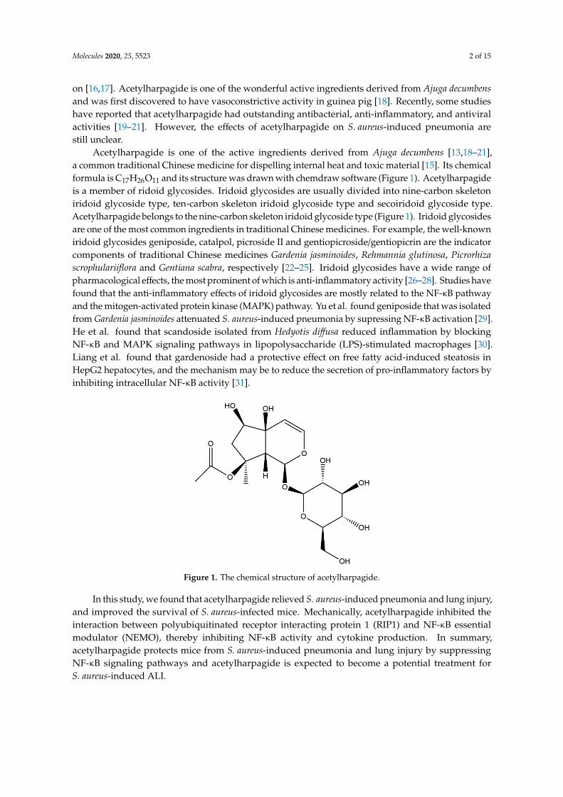
Acetylharpagide is one of the active ingredients derived from Ajuga decumbens [13,18–21],a common traditional Chinese medicine for dispelling internal heat and toxic material [15]. Its chemicalformula is C17H26O11 and its structure was drawn with chemdraw software (Figure 1). Acetylharpagideis a member of ridoid glycosides. Iridoid glycosides are usually divided into nine-carbon skeletoniridoid glycoside type, ten-carbon skeleton iridoid glycoside type and secoiridoid glycoside type.Acetylharpagide belongs to the nine-carbon skeleton iridoid glycoside type (Figure 1). Iridoid glycosidesare one of the most common ingredients in traditional Chinese medicines. For example, the well-knowniridoid glycosides geniposide, catalpol, picroside II and gentiopicroside/gentiopicrin are the indicatorcomponents of traditional Chinese medicines Gardenia jasminoides, Rehmannia glutinosa, Picrorhizascrophulariiflora and Gentiana scabra, respectively [22–25]. Iridoid glycosides have a wide range ofpharmacological effects, the most prominent of which is anti-inflammatory activity [26–28]. Studies havefound that the anti-inflammatory effects of iridoid glycosides are mostly related to the NF-κB pathwayand the mitogen-activated protein kinase (MAPK) pathway. Yu et al. found geniposide that was isolatedfrom Gardenia jasminoides attenuated S. aureus-induced pneumonia by supressing NF-κB activation [29].He et al. found that scandoside isolated from Hedyotis diffusa reduced inflammation by blockingNF-κB and MAPK signaling pathways in lipopolysaccharide (LPS)-stimulated macrophages [30].Liang et al. found that gardenoside had a protective effect on free fatty acid-induced steatosis inHepG2 hepatocytes, and the mechanism may be to reduce the secretion of pro-inflammatory factors byinhibiting intracellular NF-κB activity [31].
Molecules 2020, 25, x FOR PEER REVIEW 2 of 15
anti-inflammatory, bacteriostatic, immune function regulation, anti-fibrosis, anti-tumor and so on [16,17]. Acetylharpagide is one of the wonderful active ingredients derived from Ajuga decumbens and was first discovered to have vasoconstrictive activity in guinea pig [18]. Recently, some studies have reported that acetylharpagide had outstanding antibacterial, anti-inflammatory, and antiviral activities [19–21]. However, the effects of acetylharpagide on S. aureus-induced pneumonia are still unclear.
Acetylharpagide is one of the active ingredients derived from Ajuga decumbens [13,18–21], a common traditional Chinese medicine for dispelling internal heat and toxic material [15]. Its chemical formula is C17H26O11 and its structure was drawn with chemdraw software (Figure 1). Acetylharpagide is a member of ridoid glycosides. Iridoid glycosides are usually divided into nine-carbon skeleton iridoid glycoside type, ten-carbon skeleton iridoid glycoside type and secoiridoid glycoside type. Acetylharpagide belongs to the nine-carbon skeleton iridoid glycoside type (Figure 1). Iridoid glycosides are one of the most common ingredients in traditional Chinese medicines. For example, the well-known iridoid glycosides geniposide, catalpol, picroside II and gentiopicroside/gentiopicrin are the indicator components of traditional Chinese medicines Gardenia jasminoides, Rehmannia glutinosa, Picrorhiza scrophulariiflora and Gentiana scabra, respectively [22–25]. Iridoid glycosides have a wide range of pharmacological effects, the most prominent of which is anti-inflammatory activity [26–28]. Studies have found that the anti-inflammatory effects of iridoid glycosides are mostly related to the NF-κB pathway and the mitogen-activated protein kinase (MAPK) pathway. Yu et al. found geniposide that was isolated from Gardenia jasminoides attenuated S. aureus-induced pneumonia by supressing NF-κB activation [29]. He et al. found that scandoside isolated from Hedyotis diffusa reduced inflammation by blocking NF-κB and MAPK signaling pathways in lipopolysaccharide (LPS)-stimulated macrophages [30]. Liang et al. found that gardenoside had a protective effect on free fatty acid-induced steatosis in HepG2 hepatocytes, and the mechanism may be to reduce the secretion of pro-inflammatory factors by inhibiting intracellular NF-κB activity [31].
Figure 1. The chemical structure of acetylharpagide.
In this study, we found that acetylharpagide relieved S. aureus-induced pneumonia and lung injury, and improved the survival of S. aureus-infected mice. Mechanically, acetylharpagide inhibited the interaction between polyubiquitinated receptor interacting protein 1 (RIP1) and NF-κB essential modulator (NEMO), thereby inhibiting NF-κB activity and cytokine production. In summary, acetylharpagide protects mice from S. aureus-induced pneumonia and lung injury by suppressing NF-κB signaling pathways and acetylharpagide is expected to become a potential treatment for S. aureus-induced ALI.
Figure 1. The chemical structure of acetylharpagide.
In this study, we found that acetylharpagide relieved S. aureus-induced pneumonia and lung injury,and improved the survival of S. aureus-infected mice. Mechanically, acetylharpagide inhibited theinteraction between polyubiquitinated receptor interacting protein 1 (RIP1) and NF-κB essentialmodulator (NEMO), thereby inhibiting NF-κB activity and cytokine production. In summary,acetylharpagide protects mice from S. aureus-induced pneumonia and lung injury by suppressingNF-κB signaling pathways and acetylharpagide is expected to become a potential treatment forS. aureus-induced ALI.
Molecules 2020, 25, 5523 3 of 15
2. Results
2.1. Acetylharpagide Protects Mice from S. aureus-Induced ALI
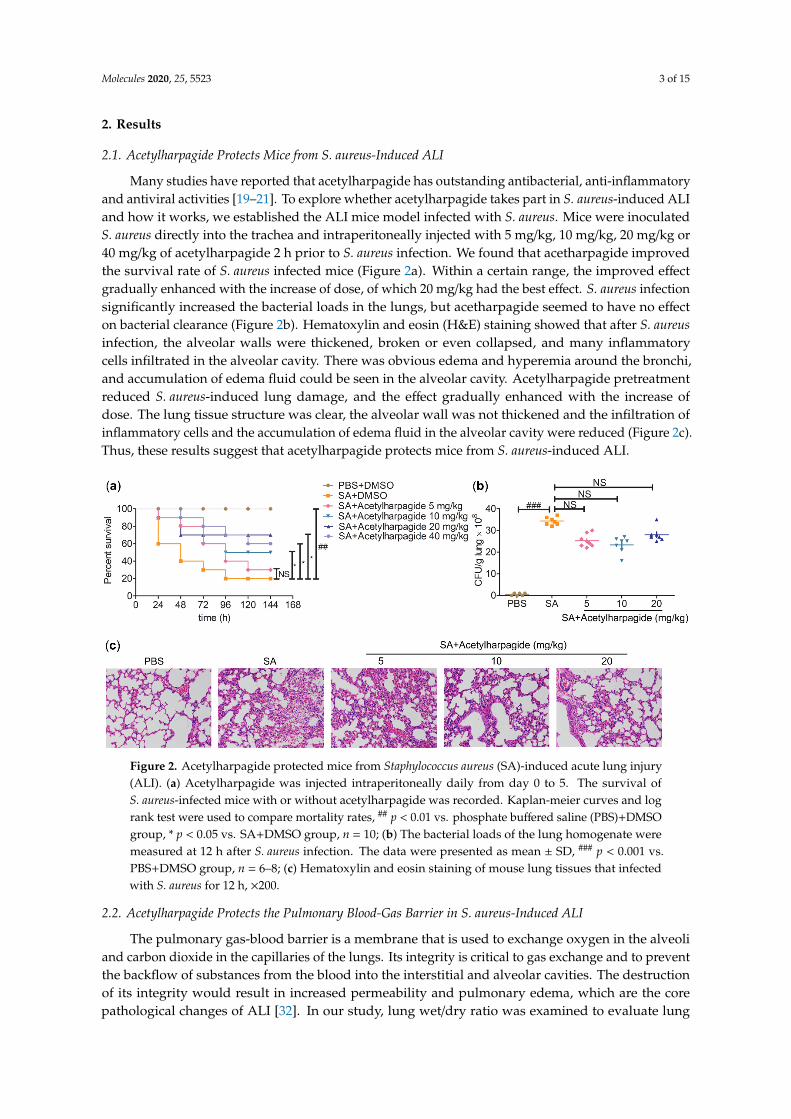
Many studies have reported that acetylharpagide has outstanding antibacterial, anti-inflammatoryand antiviral activities [19–21]. To explore whether acetylharpagide takes part in S. aureus-induced ALIand how it works, we established the ALI mice model infected with S. aureus. Mice were inoculatedS. aureus directly into the trachea and intraperitoneally injected with 5 mg/kg, 10 mg/kg, 20 mg/kg or40 mg/kg of acetylharpagide 2 h prior to S. aureus infection. We found that acetharpagide improvedthe survival rate of S. aureus infected mice (Figure 2a). Within a certain range, the improved effectgradually enhanced with the increase of dose, of which 20 mg/kg had the best effect. S. aureus infectionsignificantly increased the bacterial loads in the lungs, but acetharpagide seemed to have no effecton bacterial clearance (Figure 2b). Hematoxylin and eosin (H&E) staining showed that after S. aureusinfection, the alveolar walls were thickened, broken or even collapsed, and many inflammatorycells infiltrated in the alveolar cavity. There was obvious edema and hyperemia around the bronchi,and accumulation of edema fluid could be seen in the alveolar cavity. Acetylharpagide pretreatmentreduced S. aureus-induced lung damage, and the effect gradually enhanced with the increase ofdose. The lung tissue structure was clear, the alveolar wall was not thickened and the infiltration ofinflammatory cells and the accumulation of edema fluid in the alveolar cavity were reduced (Figure 2c).Thus, these results suggest that acetylharpagide protects mice from S. aureus-induced ALI.
Molecules 2020, 25, x FOR PEER REVIEW 3 of 15
2. Results
2.1. Acetylharpagide Protects Mice from S. aureus-Induced ALI
Many studies have reported that acetylharpagide has outstanding antibacterial, anti-inflammatory and antiviral activities [19–21]. To explore whether acetylharpagide takes part in S. aureus-induced ALI and how it works, we established the ALI mice model infected with S. aureus. Mice were inoculated S. aureus directly into the trachea and intraperitoneally injected with 5 mg/kg, 10 mg/kg, 20 mg/kg or 40 mg/kg of acetylharpagide 2 h prior to S. aureus infection. We found that acetharpagide improved the survival rate of S. aureus infected mice (Figure 2a). Within a certain range, the improved effect gradually enhanced with the increase of dose, of which 20 mg/kg had the best effect. S. aureus infection significantly increased the bacterial loads in the lungs, but acetharpagide seemed to have no effect on bacterial clearance (Figure 2b). Hematoxylin and eosin (H&E) staining showed that after S. aureus infection, the alveolar walls were thickened, broken or even collapsed, and many inflammatory cells infiltrated in the alveolar cavity. There was obvious edema and hyperemia around the bronchi, and accumulation of edema fluid could be seen in the alveolar cavity. Acetylharpagide pretreatment reduced S. aureus-induced lung damage, and the effect gradually enhanced with the increase of dose. The lung tissue structure was clear, the alveolar wall was not thickened and the infiltration of inflammatory cells and the accumulation of edema fluid in the alveolar cavity were reduced (Figure 2c). Thus, these results suggest that acetylharpagide protects mice from S. aureus-induced ALI.
Figure 2. Acetylharpagide protected mice from Staphylococcus aureus (SA)-induced acute lung injury (ALI). (a) Acetylharpagide was injected intraperitoneally daily from day 0 to 5. The survival of S. aureus-infected mice with or without acetylharpagide was recorded. Kaplan-meier curves and log rank test were used to compare mortality rates, ## p < 0.01 vs. phosphate buffered saline (PBS)+DMSO group, * p < 0.05 vs. SA+DMSO group, n = 10; (b) The bacterial loads of the lung homogenate were measured at 12 h after S. aureus infection. The data were presented as mean ± SD, ### p < 0.001 vs. PBS+DMSO group, n = 6–8; (c) Hematoxylin and eosin staining of mouse lung tissues that infected with S. aureus for 12 h, × 200.
2.2. Acetylharpagide Protects the Pulmonary Blood-Gas Barrier in S. aureus-Induced ALI
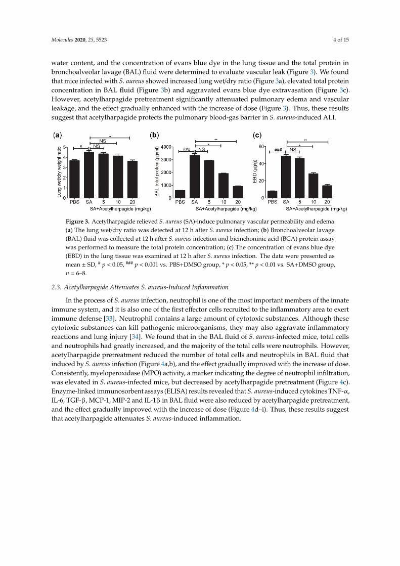
The pulmonary gas-blood barrier is a membrane that is used to exchange oxygen in the alveoli and carbon dioxide in the capillaries of the lungs. Its integrity is critical to gas exchange and to prevent the backflow of substances from the blood into the interstitial and alveolar cavities. The destruction of its integrity would result in increased permeability and pulmonary edema, which are the core pathological changes of ALI [32]. In our study, lung wet/dry ratio was examined to evaluate
Figure 2. Acetylharpagide protected mice from Staphylococcus aureus (SA)-induced acute lung injury(ALI). (a) Acetylharpagide was injected intraperitoneally daily from day 0 to 5. The survival ofS. aureus-infected mice with or without acetylharpagide was recorded. Kaplan-meier curves and logrank test were used to compare mortality rates, ## p < 0.01 vs. phosphate buffered saline (PBS)+DMSOgroup, * p < 0.05 vs. SA+DMSO group, n = 10; (b) The bacterial loads of the lung homogenate weremeasured at 12 h after S. aureus infection. The data were presented as mean ± SD, ### p < 0.001 vs.PBS+DMSO group, n = 6–8; (c) Hematoxylin and eosin staining of mouse lung tissues that infectedwith S. aureus for 12 h, ×200.
2.2. Acetylharpagide Protects the Pulmonary Blood-Gas Barrier in S. aureus-Induced ALI
The pulmonary gas-blood barrier is a membrane that is used to exchange oxygen in the alveoliand carbon dioxide in the capillaries of the lungs. Its integrity is critical to gas exchange and to preventthe backflow of substances from the blood into the interstitial and alveolar cavities. The destructionof its integrity would result in increased permeability and pulmonary edema, which are the corepathological changes of ALI [32]. In our study, lung wet/dry ratio was examined to evaluate lung
Molecules 2020, 25, 5523 4 of 15
water content, and the concentration of evans blue dye in the lung tissue and the total protein inbronchoalveolar lavage (BAL) fluid were determined to evaluate vascular leak (Figure 3). We foundthat mice infected with S. aureus showed increased lung wet/dry ratio (Figure 3a), elevated total proteinconcentration in BAL fluid (Figure 3b) and aggravated evans blue dye extravasation (Figure 3c).However, acetylharpagide pretreatment significantly attenuated pulmonary edema and vascularleakage, and the effect gradually enhanced with the increase of dose (Figure 3). Thus, these resultssuggest that acetylharpagide protects the pulmonary blood-gas barrier in S. aureus-induced ALI.
Molecules 2020, 25, x FOR PEER REVIEW 4 of 15
lung water content, and the concentration of evans blue dye in the lung tissue and the total protein in bronchoalveolar lavage (BAL) fluid were determined to evaluate vascular leak (Figure 3). We found that mice infected with S. aureus showed increased lung wet/dry ratio (Figure 3a), elevated total protein concentration in BAL fluid (Figure 3b) and aggravated evans blue dye extravasation (Figure 3c). However, acetylharpagide pretreatment significantly attenuated pulmonary edema and vascular leakage, and the effect gradually enhanced with the increase of dose (Figure 3). Thus, these results suggest that acetylharpagide protects the pulmonary blood-gas barrier in S. aureus-induced ALI.
Figure 3. Acetylharpagide relieved S. aureus (SA)-induce pulmonary vascular permeability and edema. (a) The lung wet/dry ratio was detected at 12 h after S. aureus infection; (b) Bronchoalveolar lavage (BAL) fluid was collected at 12 h after S. aureus infection and bicinchoninic acid (BCA) protein assay was performed to measure the total protein concentration; (c) The concentration of evans blue dye (EBD) in the lung tissue was examined at 12 h after S. aureus infection. The data were presented as mean ± SD, # p < 0.05, ### p < 0.001 vs. PBS+DMSO group, * p < 0.05, ** p < 0.01 vs. SA+DMSO group, n = 6–8.
2.3. Acetylharpagide Attenuates S. aureus-Induced Inflammation
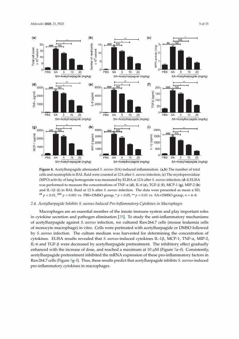
In the process of S. aureus infection, neutrophil is one of the most important members of the innate immune system, and it is also one of the first effector cells recruited to the inflammatory area to exert immune defense [33]. Neutrophil contains a large amount of cytotoxic substances. Although these cytotoxic substances can kill pathogenic microorganisms, they may also aggravate inflammatory reactions and lung injury [34]. We found that in the BAL fluid of S. aureus-infected mice, total cells and neutrophils had greatly increased, and the majority of the total cells were neutrophils. However, acetylharpagide pretreatment reduced the number of total cells and neutrophils in BAL fluid that induced by S. aureus infection (Figure 4a,b), and the effect gradually improved with the increase of dose. Consistently, myeloperoxidase (MPO) activity, a marker indicating the degree of neutrophil infiltration, was elevated in S. aureus-infected mice, but decreased by acetylharpagide pretreatment (Figure 4c). Enzyme-linked immunosorbent assays (ELISA) results revealed that S. aureus-induced cytokines TNF-α, IL-6, TGF-β, MCP-1, MIP-2 and IL-1β in BAL fluid were also reduced by acetylharpagide pretreatment, and the effect gradually improved with the increase of dose (Figure 4d–i). Thus, these results suggest that acetylharpagide attenuates S. aureus-induced inflammation.
Figure 3. Acetylharpagide relieved S. aureus (SA)-induce pulmonary vascular permeability and edema.(a) The lung wet/dry ratio was detected at 12 h after S. aureus infection; (b) Bronchoalveolar lavage(BAL) fluid was collected at 12 h after S. aureus infection and bicinchoninic acid (BCA) protein assaywas performed to measure the total protein concentration; (c) The concentration of evans blue dye(EBD) in the lung tissue was examined at 12 h after S. aureus infection. The data were presented asmean ± SD, # p < 0.05, ### p < 0.001 vs. PBS+DMSO group, * p < 0.05, ** p < 0.01 vs. SA+DMSO group,n = 6–8.
2.3. Acetylharpagide Attenuates S. aureus-Induced Inflammation
In the process of S. aureus infection, neutrophil is one of the most important members of the innateimmune system, and it is also one of the first effector cells recruited to the inflammatory area to exertimmune defense [33]. Neutrophil contains a large amount of cytotoxic substances. Although thesecytotoxic substances can kill pathogenic microorganisms, they may also aggravate inflammatoryreactions and lung injury [34]. We found that in the BAL fluid of S. aureus-infected mice, total cellsand neutrophils had greatly increased, and the majority of the total cells were neutrophils. However,acetylharpagide pretreatment reduced the number of total cells and neutrophils in BAL fluid thatinduced by S. aureus infection (Figure 4a,b), and the effect gradually improved with the increase of dose.Consistently, myeloperoxidase (MPO) activity, a marker indicating the degree of neutrophil infiltration,was elevated in S. aureus-infected mice, but decreased by acetylharpagide pretreatment (Figure 4c).Enzyme-linked immunosorbent assays (ELISA) results revealed that S. aureus-induced cytokines TNF-α,IL-6, TGF-β, MCP-1, MIP-2 and IL-1β in BAL fluid were also reduced by acetylharpagide pretreatment,and the effect gradually improved with the increase of dose (Figure 4d–i). Thus, these results suggestthat acetylharpagide attenuates S. aureus-induced inflammation.
Molecules 2020, 25, 5523 5 of 15Molecules 2020, 25, x FOR PEER REVIEW 5 of 15
Figure 4. Acetylharpagide attenuated S. aureus (SA)-induced inflammation. (a,b) The number of total cells and neutrophils in BAL fluid were counted at 12 h after S. aureus infection; (c) The myeloperoxidase (MPO) activity of lung homogenate was measured by ELISA at 12 h after S. aureus infection; (d–i) ELISA was performed to measure the concentrations of TNF-α (d), IL-6 (e), TGF-β (f), MCP-1 (g), MIP-2 (h) and IL-1β (i) in BAL fluid at 12 h after S. aureus infection. The data were presented as mean ± SD, ## p < 0.01, ### p < 0.001 vs. PBS+DMSO group, * p < 0.05, ** p < 0.01 vs. SA+DMSO group, n = 6–8.
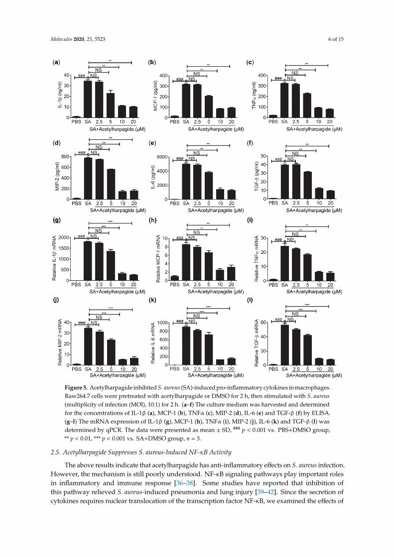
2.4. Acetylharpagide Inhibits S. aureus-Induced Pro-Inflammatory Cytokines in Macrophages
Macrophages are an essential member of the innate immune system and play important roles in cytokine secretion and pathogen elimination [35]. To study the anti-inflammatory mechanisms of acetylharpagide against S. aureus infection, we cultured Raw264.7 cells (mouse leukemia cells of monocyte macrophage) in vitro. Cells were pretreated with acetylharpagide or DMSO followed by S. aureus infection. The culture medium was harvested for determining the concentration of cytokines. ELISA results revealed that S. aureus-induced cytokines IL-1β, MCP-1, TNF-α, MIP-2, IL-6 and TGF-β were decreased by acetylharpagide pretreatment. The inhibitory effect gradually enhanced with the increase of dose, and reached a maximum at 10 μM (Figure 5a–f). Consistently, acetylharpagide pretreatment inhibited the mRNA expression of these pro-inflammatory factors in Raw264.7 cells (Figure 5g–l). Thus, these results predict that acetylharpagide inhibits S. aureus-induced pro-inflammatory cytokines in macrophages.
Figure 4. Acetylharpagide attenuated S. aureus (SA)-induced inflammation. (a,b) The number of totalcells and neutrophils in BAL fluid were counted at 12 h after S. aureus infection; (c) The myeloperoxidase(MPO) activity of lung homogenate was measured by ELISA at 12 h after S. aureus infection; (d–i) ELISAwas performed to measure the concentrations of TNF-α (d), IL-6 (e), TGF-β (f), MCP-1 (g), MIP-2 (h)and IL-1β (i) in BAL fluid at 12 h after S. aureus infection. The data were presented as mean ± SD,## p < 0.01, ### p < 0.001 vs. PBS+DMSO group, * p < 0.05, ** p < 0.01 vs. SA+DMSO group, n = 6–8.
2.4. Acetylharpagide Inhibits S. aureus-Induced Pro-Inflammatory Cytokines in Macrophages
Macrophages are an essential member of the innate immune system and play important rolesin cytokine secretion and pathogen elimination [35]. To study the anti-inflammatory mechanismsof acetylharpagide against S. aureus infection, we cultured Raw264.7 cells (mouse leukemia cellsof monocyte macrophage) in vitro. Cells were pretreated with acetylharpagide or DMSO followedby S. aureus infection. The culture medium was harvested for determining the concentration ofcytokines. ELISA results revealed that S. aureus-induced cytokines IL-1β, MCP-1, TNF-α, MIP-2,IL-6 and TGF-β were decreased by acetylharpagide pretreatment. The inhibitory effect graduallyenhanced with the increase of dose, and reached a maximum at 10 µM (Figure 5a–f). Consistently,acetylharpagide pretreatment inhibited the mRNA expression of these pro-inflammatory factors inRaw264.7 cells (Figure 5g–l). Thus, these results predict that acetylharpagide inhibits S. aureus-inducedpro-inflammatory cytokines in macrophages.
Molecules 2020, 25, 5523 6 of 15Molecules 2020, 25, x FOR PEER REVIEW 6 of 15
Figure 5. Acetylharpagide inhibited S. aureus (SA)-induced pro-inflammatory cytokines in macrophages. Raw264.7 cells were pretreated with acetylharpagide or DMSO for 2 h, then stimulated with S. aureus (multiplicity of infection (MOI), 10:1) for 2 h. (a–f) The culture medium was harvested and determined for the concentrations of IL-1β (a), MCP-1 (b), TNFα (c), MIP-2 (d), IL-6 (e) and TGF-β (f) by ELISA. (g–l) The mRNA expression of IL-1β (g), MCP-1 (h), TNFα (i), MIP-2 (j), IL-6 (k) and TGF-β (l) was determined by qPCR. The data were presented as mean ± SD, ### p < 0.001 vs. PBS+DMSO group, ** p < 0.01, *** p < 0.001 vs. SA+DMSO group, n = 3.
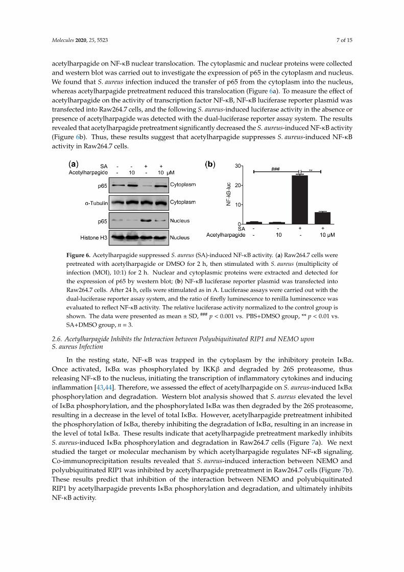
2.5. Acetylharpagide Suppresses S. aureus-Induced NF-κB Activity
The above results indicate that acetylharpagide has anti-inflammatory effects on S. aureus infection. However, the mechanism is still poorly understood. NF-κB signaling pathways play important roles in inflammatory and immune response [36–38]. Some studies have reported that inhibition of this pathway relieved S. aureus-induced pneumonia and lung injury [39–42]. Since the secretion of cytokines requires nuclear translocation of the transcription factor NF-κB, we examined the effects of acetylharpagide on NF-κB nuclear translocation. The cytoplasmic and nuclear proteins were collected and western blot was carried out to investigate the expression of p65 in the cytoplasm
Figure 5. Acetylharpagide inhibited S. aureus (SA)-induced pro-inflammatory cytokines in macrophages.Raw264.7 cells were pretreated with acetylharpagide or DMSO for 2 h, then stimulated with S. aureus(multiplicity of infection (MOI), 10:1) for 2 h. (a–f) The culture medium was harvested and determinedfor the concentrations of IL-1β (a), MCP-1 (b), TNFα (c), MIP-2 (d), IL-6 (e) and TGF-β (f) by ELISA.(g–l) The mRNA expression of IL-1β (g), MCP-1 (h), TNFα (i), MIP-2 (j), IL-6 (k) and TGF-β (l) wasdetermined by qPCR. The data were presented as mean ± SD, ### p < 0.001 vs. PBS+DMSO group,** p < 0.01, *** p < 0.001 vs. SA+DMSO group, n = 3.
2.5. Acetylharpagide Suppresses S. aureus-Induced NF-κB Activity
The above results indicate that acetylharpagide has anti-inflammatory effects on S. aureus infection.However, the mechanism is still poorly understood. NF-κB signaling pathways play important rolesin inflammatory and immune response [36–38]. Some studies have reported that inhibition ofthis pathway relieved S. aureus-induced pneumonia and lung injury [39–42]. Since the secretion ofcytokines requires nuclear translocation of the transcription factor NF-κB, we examined the effects of
Molecules 2020, 25, 5523 7 of 15
acetylharpagide on NF-κB nuclear translocation. The cytoplasmic and nuclear proteins were collectedand western blot was carried out to investigate the expression of p65 in the cytoplasm and nucleus.We found that S. aureus infection induced the transfer of p65 from the cytoplasm into the nucleus,whereas acetylharpagide pretreatment reduced this translocation (Figure 6a). To measure the effect ofacetylharpagide on the activity of transcription factor NF-κB, NF-κB luciferase reporter plasmid wastransfected into Raw264.7 cells, and the following S. aureus-induced luciferase activity in the absence orpresence of acetylharpagide was detected with the dual-luciferase reporter assay system. The resultsrevealed that acetylharpagide pretreatment significantly decreased the S. aureus-induced NF-κB activity(Figure 6b). Thus, these results suggest that acetylharpagide suppresses S. aureus-induced NF-κBactivity in Raw264.7 cells.
Molecules 2020, 25, x FOR PEER REVIEW 7 of 15
and nucleus. We found that S. aureus infection induced the transfer of p65 from the cytoplasm into the nucleus, whereas acetylharpagide pretreatment reduced this translocation (Figure 6a). To measure the effect of acetylharpagide on the activity of transcription factor NF-κB, NF-κB luciferase reporter plasmid was transfected into Raw264.7 cells, and the following S. aureus-induced luciferase activity in the absence or presence of acetylharpagide was detected with the dual-luciferase reporter assay system. The results revealed that acetylharpagide pretreatment significantly decreased the S. aureus-induced NF-κB activity (Figure 6b). Thus, these results suggest that acetylharpagide suppresses S. aureus-induced NF-κB activity in Raw264.7 cells.
Figure 6. Acetylharpagide suppressed S. aureus (SA)-induced NF-κB activity. (a) Raw264.7 cells were pretreated with acetylharpagide or DMSO for 2 h, then stimulated with S. aureus (multiplicity of infection (MOI), 10:1) for 2 h. Nuclear and cytoplasmic proteins were extracted and detected for the expression of p65 by western blot; (b) NF-κB luciferase reporter plasmid was transfected into Raw264.7 cells. After 24 h, cells were stimulated as in A. Luciferase assays were carried out with the dual-luciferase reporter assay system, and the ratio of firefly luminescence to renilla luminescence was evaluated to reflect NF-κB activity. The relative luciferase activity normalized to the control group is shown. The data were presented as mean ± SD, ### p < 0.001 vs. PBS+DMSO group, ** p < 0.01 vs. SA+DMSO group, n = 3.
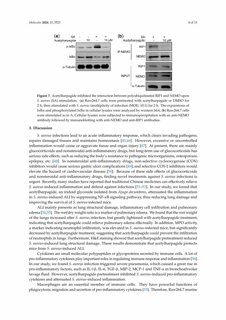
2.6. Acetylharpagide Inhibits the Interaction between Polyubiquitinated RIP1 and NEMO upon S. aureus Infection
In the resting state, NF-κB was trapped in the cytoplasm by the inhibitory protein IκBα. Once activated, IκBα was phosphorylated by IKKβ and degraded by 26S proteasome, thus releasing NF-κB to the nucleus, initiating the transcription of inflammatory cytokines and inducing inflammation [43,44]. Therefore, we assessed the effect of acetylharpagide on S. aureus-induced IκBα phosphorylation and degradation. Western blot analysis showed that S. aureus elevated the level of IκBα phosphorylation, and the phosphorylated IκBα was then degraded by the 26S proteasome, resulting in a decrease in the level of total IκBα. However, acetylharpagide pretreatment inhibited the phosphorylation of IκBα, thereby inhibiting the degradation of IκBα, resulting in an increase in the level of total IκBα. These results indicate that acetylharpagide pretreatment markedly inhibits S. aureus-induced IκBα phosphorylation and degradation in Raw264.7 cells (Figure 7a). We next studied the target or molecular mechanism by which acetylharpagide regulates NF-κB signaling. Co-immunoprecipitation results revealed that S. aureus-induced interaction between NEMO and polyubiquitinated RIP1 was inhibited by acetylharpagide pretreatment in Raw264.7 cells (Figure 7b). These results predict that inhibition of the interaction between NEMO and polyubiquitinated RIP1 by acetylharpagide prevents IκBα phosphorylation and degradation, and ultimately inhibits NF-κB activity.
Figure 6. Acetylharpagide suppressed S. aureus (SA)-induced NF-κB activity. (a) Raw264.7 cells werepretreated with acetylharpagide or DMSO for 2 h, then stimulated with S. aureus (multiplicity ofinfection (MOI), 10:1) for 2 h. Nuclear and cytoplasmic proteins were extracted and detected forthe expression of p65 by western blot; (b) NF-κB luciferase reporter plasmid was transfected intoRaw264.7 cells. After 24 h, cells were stimulated as in A. Luciferase assays were carried out with thedual-luciferase reporter assay system, and the ratio of firefly luminescence to renilla luminescence wasevaluated to reflect NF-κB activity. The relative luciferase activity normalized to the control group isshown. The data were presented as mean ± SD, ### p < 0.001 vs. PBS+DMSO group, ** p < 0.01 vs.SA+DMSO group, n = 3.
2.6. Acetylharpagide Inhibits the Interaction between Polyubiquitinated RIP1 and NEMO uponS. aureus Infection
In the resting state, NF-κB was trapped in the cytoplasm by the inhibitory protein IκBα.Once activated, IκBα was phosphorylated by IKKβ and degraded by 26S proteasome, thusreleasing NF-κB to the nucleus, initiating the transcription of inflammatory cytokines and inducinginflammation [43,44]. Therefore, we assessed the effect of acetylharpagide on S. aureus-induced IκBαphosphorylation and degradation. Western blot analysis showed that S. aureus elevated the levelof IκBα phosphorylation, and the phosphorylated IκBα was then degraded by the 26S proteasome,resulting in a decrease in the level of total IκBα. However, acetylharpagide pretreatment inhibitedthe phosphorylation of IκBα, thereby inhibiting the degradation of IκBα, resulting in an increase inthe level of total IκBα. These results indicate that acetylharpagide pretreatment markedly inhibitsS. aureus-induced IκBα phosphorylation and degradation in Raw264.7 cells (Figure 7a). We nextstudied the target or molecular mechanism by which acetylharpagide regulates NF-κB signaling.Co-immunoprecipitation results revealed that S. aureus-induced interaction between NEMO andpolyubiquitinated RIP1 was inhibited by acetylharpagide pretreatment in Raw264.7 cells (Figure 7b).These results predict that inhibition of the interaction between NEMO and polyubiquitinatedRIP1 by acetylharpagide prevents IκBα phosphorylation and degradation, and ultimately inhibitsNF-κB activity.
Molecules 2020, 25, 5523 8 of 15Molecules 2020, 25, x FOR PEER REVIEW 8 of 15
Figure 7. Acetylharpagide inhibited the interaction between polyubiquitinated RIP1 and NEMO upon S. aureus (SA) stimulation. (a) Raw264.7 cells were pretreated with acetylharpagide or DMSO for 2 h, then stimulated with S. aureus (multiplicity of infection (MOI), 10:1) for 2 h. The expressions of IκBα and phosphorylated IκBα in cellular lysates were analyzed by western blot; (b) Raw264.7 cells were stimulated as in A. Cellular lysates were subjected to immunoprecipitation with an anti-NEMO antibody followed by immunoblotting with anti-NEMO and anti-RIP1 antibodies.
3. Discussion
S. aureus infections lead to an acute inflammatory response, which clears invading pathogens, repairs damaged tissues and maintains homeostasis [45,46]. However, excessive or uncontrolled inflammation would cause or aggravate tissue and organ injury [47]. At present, there are mainly glucocorticoids and nonsteroidal anti-inflammatory drugs, but long-term use of glucocorticoids has serious side effects, such as reducing the body’s resistance to pathogenic microorganisms, osteoporosis, epilepsy, etc. [48]. In nonsteroidal anti-inflammatory drugs, non-selective cyclooxygenase (COX) inhibitors would cause serious gastric ulcer complications [49], and selective COX-2 inhibitors would elevate the hazard of cardiovascular disease [50]. Because of these side effects of glucocorticoids and nonsteroidal anti-inflammatory drugs, finding novel treatments against S. aureus infections is urgent. Recently, many studies have reported that traditional Chinese medicines can effectively relieve S. aureus-induced inflammation and defend against infections [51–53]. In our study, we found that acetylharpagide, an iridoid glycoside isolated from Ajuga decumbens, attenuated the inflammation in S. aureus-induced ALI by suppressing NF-κB signaling pathway, thus reducing lung damage and improving the survival of S. aureus-infected mice.
ALI mainly presents as lung structural damage, inflammatory cell infiltration and pulmonary edema [54,55]. The wet/dry weight ratio is a marker of pulmonary edema. We found that the wet weight of the lungs increased after S. aureus infection, but greatly lightened with acetylharpagide treatment, indicating that acetylharpagide could relieve pulmonary edema effectually. In addition, MPO activity, a marker indicating neutrophil infiltration, was elevated in S. aureus-infected mice, but significantly decreased by acetylharpagide treatment, suggesting that acetylharpagide could prevent the infiltration of neutrophils in lungs. Furthermore, H&E staining showed that acetylharpagide pretreatment reduced S. aureus-induced lung structural damage. These results demonstrate that acetylharpagide protects mice from S. aureus-induced ALI.
Cytokines are small molecular polypeptides or glycoproteins secreted by immune cells. A lot of pro-inflammatory cytokines play important roles in regulating immune response and inflammation [56]. In our study, we found S. aureus infection triggered severe pneumonia, which caused a great rise in pro-inflammatory factors, such as IL-1β, IL-6, TGF-β, MIP-2, MCP-1 and TNF-α in bronchoalveolar lavage fluid. However, acetylharpagide pretreatment inhibited S. aureus-induced pro-inflammatory cytokines and attenuated S. aureus-induced inflammation.
Macrophages are an essential member of immune cells. They have powerful functions of phagocytosis, migration and secretion of pro-inflammatory cytokines [35]. Therefore, Raw264.7 murine macrophages were used in vitro to investigate the activity and mechanism of acetylharpagide on S. aureus infection. In our study, Raw264.7 cells were pretreated with acetylharpagide or DMSO followed by S. aureus infection. The culture medium was harvested for determining the concentration of cytokines. ELISA results revealed that S. aureus-induced cytokines IL-1β, MCP-1, TNF-α, MIP-2,
Figure 7. Acetylharpagide inhibited the interaction between polyubiquitinated RIP1 and NEMO uponS. aureus (SA) stimulation. (a) Raw264.7 cells were pretreated with acetylharpagide or DMSO for2 h, then stimulated with S. aureus (multiplicity of infection (MOI), 10:1) for 2 h. The expressions ofIκBα and phosphorylated IκBα in cellular lysates were analyzed by western blot; (b) Raw264.7 cellswere stimulated as in A. Cellular lysates were subjected to immunoprecipitation with an anti-NEMOantibody followed by immunoblotting with anti-NEMO and anti-RIP1 antibodies.
3. Discussion
S. aureus infections lead to an acute inflammatory response, which clears invading pathogens,repairs damaged tissues and maintains homeostasis [45,46]. However, excessive or uncontrolledinflammation would cause or aggravate tissue and organ injury [47]. At present, there are mainlyglucocorticoids and nonsteroidal anti-inflammatory drugs, but long-term use of glucocorticoids hasserious side effects, such as reducing the body’s resistance to pathogenic microorganisms, osteoporosis,epilepsy, etc. [48]. In nonsteroidal anti-inflammatory drugs, non-selective cyclooxygenase (COX)inhibitors would cause serious gastric ulcer complications [49], and selective COX-2 inhibitors wouldelevate the hazard of cardiovascular disease [50]. Because of these side effects of glucocorticoidsand nonsteroidal anti-inflammatory drugs, finding novel treatments against S. aureus infections isurgent. Recently, many studies have reported that traditional Chinese medicines can effectively relieveS. aureus-induced inflammation and defend against infections [51–53]. In our study, we found thatacetylharpagide, an iridoid glycoside isolated from Ajuga decumbens, attenuated the inflammationin S. aureus-induced ALI by suppressing NF-κB signaling pathway, thus reducing lung damage andimproving the survival of S. aureus-infected mice.
ALI mainly presents as lung structural damage, inflammatory cell infiltration and pulmonaryedema [54,55]. The wet/dry weight ratio is a marker of pulmonary edema. We found that the wet weightof the lungs increased after S. aureus infection, but greatly lightened with acetylharpagide treatment,indicating that acetylharpagide could relieve pulmonary edema effectually. In addition, MPO activity,a marker indicating neutrophil infiltration, was elevated in S. aureus-infected mice, but significantlydecreased by acetylharpagide treatment, suggesting that acetylharpagide could prevent the infiltrationof neutrophils in lungs. Furthermore, H&E staining showed that acetylharpagide pretreatment reducedS. aureus-induced lung structural damage. These results demonstrate that acetylharpagide protectsmice from S. aureus-induced ALI.
Cytokines are small molecular polypeptides or glycoproteins secreted by immune cells. A lot ofpro-inflammatory cytokines play important roles in regulating immune response and inflammation [56].In our study, we found S. aureus infection triggered severe pneumonia, which caused a great rise inpro-inflammatory factors, such as IL-1β, IL-6, TGF-β, MIP-2, MCP-1 and TNF-α in bronchoalveolarlavage fluid. However, acetylharpagide pretreatment inhibited S. aureus-induced pro-inflammatorycytokines and attenuated S. aureus-induced inflammation.
Macrophages are an essential member of immune cells. They have powerful functions ofphagocytosis, migration and secretion of pro-inflammatory cytokines [35]. Therefore, Raw264.7 murine
Molecules 2020, 25, 5523 9 of 15
macrophages were used in vitro to investigate the activity and mechanism of acetylharpagide onS. aureus infection. In our study, Raw264.7 cells were pretreated with acetylharpagide or DMSOfollowed by S. aureus infection. The culture medium was harvested for determining the concentrationof cytokines. ELISA results revealed that S. aureus-induced cytokines IL-1β, MCP-1, TNF-α, MIP-2,IL-6 and TGF-β were decreased by acetylharpagide pretreatment. Consistently, acetylharpagidepretreatment inhibited the mRNA expression of these pro-inflammatory factors. These results predictthat acetylharpagide inhibits the secretion of pro-inflammatory factors that is induced by S. aureusin macrophages.
The canonical NF-κB signaling pathway, which plays important roles in inflammation and immuneresponse, has been considered a valuable target for drug design [36–38]. Because of the several levels,NF-κB signaling pathway can be targeted at multiple points including phosphatase, ubiquitination,nuclear translocation, DNA binding, protein acetyltransferase and methyltransferase [57]. Some studieshave shown that many drugs inhibit the NF-κB signaling pathway by inhibiting the phosphorylationof IκB or the nuclear transport of p65 [58,59]. In our study, we found S. aureus-induced IκBαphosphorylation and p65 translocation from the cytoplasm into the nucleus were reduced byacetylharpagide pretreatment. In addition, S. aureus-induced luciferase activity of NF-κB reporterplasmid was also reduced by acetylharpagide pretreatment.
In the canonical NF-κB signaling pathway, once the ligands, such as lipopolysaccharide orcytokines, bind to the related receptor, it will cause a configuration change in the latter, and inducethe polyubiquitination of RIP1 at Lys-377. This polyubiquitin chains act as a scaffold to recruit TAK1and IKK complexes via their adaptor proteins TAB2/3 and NEMO, respectively [60,61]. After that,TAK1 phosphorylates IKKβ and activates the IκB kinase IKK, which phosphorylates the inhibitor ofNF-κB (IκB) and makes its 26S proteasome degrade, thus releasing NF-κB to the nucleus to start upthe expression of target genes, such as inflammatory cytokines [43,44]. Therefore, the interaction ofNEMO and RIP1 is indispensable for the activation of the NF-κB signaling pathway. Blocking thebinding of NEMO to polyubiquited RIP1 could inhibit the activation of IKK [62–64]. In this study,we found the interaction between polyubiquitinated RIP1 and NEMO induced by S. aureus infectionwas inhibited by acetylharpagide pretreatment in Raw264.7 cells. Thus, acetylharpagide inhibits theactivity of NF-κB upon S. aureus infection by blocking polyubiquitinated RIP1 and NEMO interaction,but the mechanism needs further study.
4. Materials and Methods
4.1. Chemicals and Reagents
Six to eight week-old male C57BL6 mice (weight: 18–22 g) were obtained from the modelanimal research center of Nanjing University (Nanjing, China). Raw264.7 cell line (TIB-71) andUSA300 (BAA-1717) were purchased from American Type Culture Collection (Manassas, VA, USA).RPMI1640 medium and lipofectamine 3000 were purchased from Thermo Scientific (Waltham, MA,USA). Penicillin-streptomycin solution, Ethylene Diamine Tetraacetic Acid (EDTA)-trypsin, dimethylsulfoxide side (DMSO), wright stain, hematoxylin-eosin stain kit and nuclear protein extraction kitwere purchased from Solarbio (Beijing, China). Acetylharpagide (CAS No. 6926-14-3, Item No. S9474)was purchased from Selleck (Houston, TX, USA). Polyvinylidene difluoride (PVDF) membrane andprotein A/G agarose slurry were purchased from Millipore (Billerica, MA, USA). Evans blue dye waspurchased from Sigma-Aldrich (St. Louis, MO, USA). Enzyme-linked immunosorbent assays (ELISA)kits were purchased from R&D (Minneapolis, MN, USA). P65, IκBα, phospho IκBα, NEMO, RIP1,α-tubulin and Histone H3 antibodies were purchased from Proteintech Group (Chicago, IL, USA).Dual-luciferase reporter assay system was purchased from Promega (Madison, WI, USA). TransStartGreen qPCR SuperMix was purchased from TransGene Biotechnology (Beijing, China). Cell lysis bufferwas purchased from Beyotime Biotechnology (Shanghai, China). Trizol reagent was purchased fromInvitrogen (Carlsbad, CA, USA).
Molecules 2020, 25, 5523 10 of 15
4.2. Animals
C57BL6 mice were preserved in a sterilized standard cage at 25 ◦C under a regular 12/12 h lightand dark cycle, and supplied food and water randomly.
USA300 were cultivated in tryptic soy broth (TSB) medium overnight at 37 ◦C, and thenre-inoculated into new TSB medium at 10:1. When it reached the logarithmic phase, pelleted andresuspended in phosphate buffered saline (PBS) to 1 × 10⁹ colony forming units (CFU)/mL. For thelung infection, mice were injected with 4% chloral hydrate intraperitoneally at a dose of 0.1 mL/10gfor anesthesia. 2 × 10⁸ CFU of S. aureus or same volume of PBS was slowly dropped into the tracheathrough a self-made blunt-head syringe. Then kept the mouse upright for 1 min to distribute thebacterial solution evenly on both lungs. The experimental mice were grouped randomly as follows:(1) group PBS: mice were injected intraperitoneally with DMSO 2 h before PBS infection; (2) groupSA: mice were injected intraperitoneally with DMSO 2 h before S. aureus infection; (3) group 5 mg/kgacetylharpagide + SA: mice were injected intraperitoneally with 5 mg/kg of acetylharpagide 2 h beforeS. aureus infection; (4) group 10 mg/kg acetylharpagide + SA: mice were injected intraperitoneally with10 mg/kg of acetylharpagide 2 h before S. aureus infection; (5) group 20 mg/kg acetylharpagide + SA:mice were injected intraperitoneally with 20 mg/kg of acetylharpagide 2 h before S. aureus infection;(6) group 40 mg/kg acetylharpagide + SA: mice were injected intraperitoneally with 40 mg/kg ofacetylharpagide 2 h before S. aureus infection. Acetylharpagide was dissolved in DMSO as a stocksolution at a concentration of 200 mM. For in vivo/animal studies, the stock solution was suspended insterile saline at a total volume of 200 µL per mouse. Taking the maximum concentration of 40 mg/kginjected into mice (18–22 g) as an example, it took about 10 µL of the stock solution to be dilutedto 200 µL, so the final DMSO concentration did not exceed 5%. Control mice were injected with 5%DMSO. During the whole experiment, no mice showed obvious abnormal performance. For in vitroassays, the stock solution was suspended in cell culture medium in 2 mL for each well of a 6-wellplate. Taking the maximum concentration of 20 µM as an example, it took 0.2 µL of stock solution to bediluted to 2 mL for each well, so the final DMSO concentration did not exceed 0.01%. Control samplewas treated with 0.2 µL DMSO.
The survival of infected mice was monitored daily. The whole lung was harvested 12 h afterS. aureus infection and homogenized for evaluating bacterial loads and MPO activity. BAL fluidwas harvested for examining the concentration of cytokines and total protein, and the number ofneutrophils. For lung histomorphology analysis, the lungs were fixed with 4% paraformaldehyde,dehydrated gradually with gradient ethanol, and then embedded in paraffin. The wax block was slicedwith a thickness of 5 µm, and finally hematoxylin-eosin staining was performed.
The methods were conducted according to the approved guidelines and every effort was made tominimize suffering. All experimental protocols were approved by the Animal Experiment Committeeof Nanjing University (SYXK (SU) 2019-0056, 2019.12.16).
4.3. Western Blot
Cell culture medium was removed and the cells were washed three times with PBS. 100 µL celllysis buffer was added to each well of a 6-well plate. Cells were lysed on ice for 10 min and moved intoa 1.5 mL centrifuge tube and centrifuged at 12,000 rpm for 10 min, the supernatant were collected andseparated by sodium dodecylsulphate polyacrylamide gel electrophoresis. After that, the proteinson the gel were transferred to a PVDF membrane, which was then kept in 5% skimmed milk for60 min. Next, the blocked membrane was coated with primary antibodies for p65, IκBα and phosphoIκBα. It was then washed with phosphate buffered saline tween (PBST) buffer on a shaker for 10 min,and repeated twice. The washed membrane was further coated with a horseradish peroxidase-labeledantibody for 1 h. It was then washed with PBST buffer for 10 min, and repeated twice. Finally, the signalwas detected by Tanon chemiluminescence imaging system.
Molecules 2020, 25, 5523 11 of 15
4.4. Pulmonary Vascular Permeability and Edema
Evans blue dye can bind to albumin in plasma. When pulmonary capillaries leak, albumin thatis bound to evans blue dye will penetrate into the lung tissue. The amount of evans blue dye in thelung can be measured by chemical colorimetry to reflect the permeability of blood vessels. In brief,mice were injected with 30 mg/kg of evans blue dye through the tail vein 1 h before completing theexperiment. Once the mice were sacrificed at the indicated times, cardiac perfusion was performed untilthe outflow was completely clean. The lungs were then removed, immersed in dimethylformamideand kept at 60 ◦C for 24 h, and then centrifuged at 12,000 rpm for 30 min. The optical density value ofthe supernatant was detected at a wavelength of 620 nm with a spectrophotometer. The evans bluedye concentration is calculated based on its standard curve in dimethylformamide.
The wet/dry weight ratio is assessed to show the lung tissue water content. The lungs werecollected and weighed, and this weight was recorded as the wet weight. Then the lungs were placed in60 ◦C for 48 h and weighed again, and this weight was recorded as dry weight.
4.5. Cytokines and Neutrophils in BAL Fluid
To collect BAL fluid, mice were injected with 4% chloral hydrate intraperitoneally at a dose of0.1 mL/10 g for anesthesia. Inserted a self-made blunt-head syringe into the trachea of the mouseand ligated the trachea to prevent the backflow. Gently injected 0.3 mL of sterile saline, then slowlywithdrew. Three times in total. The collected liquid is BAL fluid. BAL fluid was treated with red bloodcell lysis buffer and centrifuged at 1000 rpm for 5 min, the supernatant were applied for ELISA to detectthe concentration of MIP-2, IL-1β, TNF-α, IL-6, MCP-1 and TGF-β. The pellet was resuspended in PBSand the total cell number was counted. The number of neutrophils were counted with wright stain.
4.6. Bacterial Counts
The lungs were removed aseptically, weighed and ground with a sterilized tissue grinder in1 mL of sterilized PBS. The homogenate was diluted by 10-fold gradient and serially diluted to 108.100 µL of the tissue suspension was smeared on tryptic soy agar medium and kept at 37 ◦C for 24 h.Bacteria were counted and calculated as CFU per g of lung.
4.7. Coimmunoprecipitation Assay
Cell culture medium was removed and the cells were washed three times with PBS. 500 µL celllysis buffer was added to each 60 mm dish. Cells were lysed on ice for 10 min and moved into a1.5 mL centrifuge tube and centrifuged at 12,000 rpm for 10 min, the supernatant were collected.For immunoprecipitation, 500 µL of the supernatant was matched with 0.5 µg of antibody plus 30 µLof a 50% slurry of protein A/G agarose at 4 ◦C overnight. The precipitation was washed three timeswith PBS, resuspended in 20 µL SDS loading buffer and underwent immunoblotting.
4.8. Dual Luciferase Reporter Assay
Raw264.7 cells were transfected with a NF-κB luciferase reporter plasmid and infected withS. aureus as described above. Each sample was co-transfected with 20 ng pRL-TK Renilla luciferasereporter plasmid as a control reporter gene. Dual-luciferase reporter assay system was used todetect the luminescence, and NF-κB activity is presented by the ratio of firefly luminescence torenilla luminescence.
4.9. Quantitative Real-Time PCR
This experiment was carried out as previously described [65]. 1 mL Trizol reagent was added toeach well of a 6-well plate. Cells were lysed at room temperature for 5 min, and then moved into a1.5 mL centrifuge tube. Then added 200 µL chloroform, shook vigorously for 15 s, placed the tube onice for 3 min, and centrifuged at 12,000 rpm for 10 min. Then aspirated the upper liquid into a new tube,
Molecules 2020, 25, 5523 12 of 15
added an equal volume of isopropanol, mixed well, placed the tube on ice for 10 min, and centrifugedat 12,000 rpm for 10 min. Discarded the supernatant, added 1 mL 75% alcohol to the tube, and thencentrifuged at 12,000 rpm for 5 min. The precipitation was RNA.
Reverse transcriptase was then used to reverse-transcribe the RNA into cDNA, which was thensubjected to quantitative PCR with the CFX Connect Real-Time PCR Detection System under thefollowing conditions: 94 ◦C for 30 s, then 40 cycles at 94 ◦C for 5 s and 60 ◦C for 30 s. The mRNA levelsof specific genes were normalized to β-Actin. The specific primer sequences for IL-1β, MCP-1, TNFα,MIP-2, IL-6 and TGF-β are listed in Supplementary Table S1.
4.10. Extraction of Cytoplasm and Nuclear Proteins
Nuclear protein extraction kit was used to separate nuclear and cytoplasmic proteins. Cells werefirst lysed by plasma protein extraction reagent. Added 100µL plasma protein extraction reagent to eachsample. Cells were lysed on ice for 10 min and centrifuged at 12,000 rpm for 10 min, the supernatantwas the cytoplasmic protein and the precipitation was the nucleus. Aspirated the supernatant ascompletely as possible, then the nucleus was lysed by the nuclear protein extraction reagent. Added50 µL nuclear protein extraction reagent to each sample. The nucleus was lysed on ice for 10 min andcentrifuged at 12,000 rpm for 10 min, the supernatant was the nuclear protein.
4.11. Statistical Analysis
Data are expressed as the mean ± SD, and Student’s t-test was used to determine the statisticallysignificant differences between mean values. Survival was analyzed using the Kaplan-Meier methodand statistical analyses were performed using the log-rank test. * p < 0.05, ** p < 0.01, *** p < 0.001,and not significant, NS > 0.05.
Supplementary Materials: The following are available online, Table S1: Primer sequences.
Author Contributions: Conceptualization, Z.Z. and W.Y.; methodology, Z.Z. and W.Y.; software, Z.Z.; validation,Y.S.; formal analysis, W.Y.; investigation, Z.Z.; resources, Y.W.; data curation, W.Y.; writing—original draftpreparation, Z.Z.; writing—review and editing, Y.W.; visualization, Y.S.; supervision, Y.W.; project administration,Y.W.; funding acquisition, Y.W. All authors have read and agreed to the published version of the manuscript.
Funding: This research was funded by National Natural Science Foundation of China, grant number 81473292,91540119, 81673462 and 81671939; Six talent peaks project in Jiangsu Province to Y.W., grant number YY-012;Key development project of Jiangsu Province, grant number BE2017712.
Acknowledgments: We thank Zhao Xiaojuan for valuable technical support.
Conflicts of Interest: The authors declare no conflict of interest.
References
1. Rubenfeld, G.D.; Caldwell, E.; Peabody, E.; Weaver, J.; Martin, D.P.; Neff, M.J.; Stern, E.J.; Hudson, L.D.Incidence and Outcomes of Acute Lung Injury. N. Engl. J. Med. 2005, 353, 1685–1693. [CrossRef] [PubMed]
2. Johnson, E.; Matthay, M.A. Acute Lung Injury: Epidemiology, Pathogenesis, and Treatment. J. Aerosol Med.Pulm. Drug Deliv. 2010, 23, 243–252. [CrossRef] [PubMed]
3. Matthay, M.A.; Ware, L.B.; Zimmerman, G.A. The Acute Respiratory Distress Syndrome. J. Clin. Investig.2012, 122, 2731–2740. [CrossRef] [PubMed]
4. Dreyfuss, D.; Ricard, J. Acute Lung Injury and Bacterial Infection. Clin. Chest Med. 2005, 26, 105–112.[CrossRef] [PubMed]
5. Archer, G.L. Staphylococcus aureus: A well-armed pathogen. Clin. Infect. Dis. 1998, 26, 1179–1181. [CrossRef][PubMed]
6. Tong, S.Y.; Davis, J.S.; Eichenberger, E.M.; Holland, T.L.; Fowler, V.G. Staphylococcus aureus infections:Epidemiology, pathophysiology, clinical manifestations, and management. Clin. Microbiol. Rev. 2015, 28,603–661. [CrossRef]
7. Lowy, F.D. Staphylococcus aureus Infections. N. Engl. J. Med. 2009, 339, 520–532. [CrossRef]
Molecules 2020, 25, 5523 13 of 15
8. Allen, T.C.; Kurdowska, A. Interleukin 8 and acute lung injury. Arch. Pathol. Lab. Med. 2014, 138, 266–269.[CrossRef]
9. Krzak, A.; Pleva, M.; Napolitano, L.M. Nutrition therapy for ALI and ARDS. Crit. Care Clin. 2011, 27, 647–659.[CrossRef]
10. Lin, X.; Dean, D.A. Gene therapy for ALI/ARDS. Crit. Care Clin. 2011, 27, 705–718. [CrossRef]11. Chen, J.; Huang, C.; Chang, H.; Li, P.; Liang, Y.; Deng, J.; Huang, S.; Huang, G. Scutellaria baicalensis
ameliorates acute lung injury by suppressing inflammation in vitro and in vivo. Am. J. Chin. Med. 2017, 45,137–157. [CrossRef] [PubMed]
12. Hsu, H.; Hsiao, P.; Kuo, T.; Chiang, S.; Chen, S.; Chiou, S.; Ling, X.; Liang, M.; Cheng, W.; Houng, J.Antioxidant and anti-inflammatory activities of Lonicera japonica Thunb. var. sempervillosa Hayata flowerbud extracts prepared by water, ethanol and supercritical fluid extraction techniques. Ind. Crop. Prod. 2016,89, 543–549. [CrossRef] [PubMed]
13. Wen, B.; He, R.; Li, P.; Xu, Q.; Lu, Y.; Peng, B.; Li, J. Pharmacokinetics of 8-O-acetylharpagide and harpagideafter oral administration of Ajuga decumbens Thunb extract in rats. J. Ethnopharmacol. 2013, 147, 503–508.[CrossRef] [PubMed]
14. Hwang, Y.; Kim, D.; Li, W.; Yang, H.J.; Yim, N.; Ma, J.Y. Anti-inflammatory effects of Forsythia suspensa indextran sulfate sodium-induced colitis. J. Ethnopharmacol. 2017, 206, 73–77. [CrossRef] [PubMed]
15. Nanjing University of Traditional Chinese Medicine. Dictionary of Chinese Materia Medica, 2nd ed.; ShanghaiScience and Technology Publishing House: Shanghai, China, 2006; pp. 1035–1037.
16. Ni, B.; Dong, X.; Fu, J.; Yin, X.; Lin, L.; Xia, Z.; Zhao, Y.; Xue, D.; Yang, C.; Ni, J. Phytochemical and biologicalproperties of Ajuga decumbens (Labiatae): A review. Trop. J. Pharm. Res. 2015, 14, 1525–1536. [CrossRef]
17. Olatunde, O.Z.; Yang, Y.; Yong, J.; Lu, C. Advance of the Chemical Components and Biological Activities ofAjuga Decumbens Thunb. Biomed. J. Sci. Tech. Res. 2019, 5, 1–8.
18. Breschi, M.C.; Martinotti, E.; Catalano, S.; Flamini, G.; Morelli, I.; Pagni, A.M. Vasoconstrictor activity of8-O-acetylharpagide from Ajuga reptans. J. Nat. Prod. 1992, 55, 1145–1148. [CrossRef]
19. Konoshima, T.; Takasaki, M.; Tokuda, H.; Nishino, H. Cancer chemopreventive activity of an iridoid glycoside,8-acetylharpagide, from Ajuga decumbens. Cancer Lett. 2000, 157, 87–92. [CrossRef]
20. You, Y.; Wang, J.; Tong, Y.; Hao, Q.; Li, Y.; Yang, H.; Huang, L.; Liao, F. Anti-inflammatory effect ofacetylharpagide demonstrated by its influence on leukocyte adhesion and transmigration in endothelial cellsunder controlled shear stress. Clin. Hemorheol. Microcirc. 2014, 56, 205–217. [CrossRef]
21. Xie, Z.Y.; Qin, M.Z.; Fang, Y.L. The pharmacological activity of 8-O-acetylharpagide. World Notes Plant Med.2005, 20, 56–58.
22. Chen, J.; Wu, H.; Li, H.; Hu, S.; Dai, M.; Chen, J. Anti-inflammatory effects and pharmacokinetics study ofgeniposide on rats with adjuvant arthritis. Int. Immunopharmacol. 2015, 24, 102–109. [CrossRef] [PubMed]
23. He, Y.; Zhu, S.; Ge, Y.; Kazuma, K.; Zou, K.; Cai, S.; Komatsu, K. The anti-inflammatory secoiridoid glycosidesfrom Gentianae Scabrae Radix: The root and rhizome of Gentiana scabra. J. Nat. Med. 2015, 69, 303–312.[CrossRef] [PubMed]
24. Wang, L.; Liu, X.H.; Chen, H.; Chen, Z.Y.; Weng, X.D.; Qiu, T.; Liu, L. Picroside ii protects rat kidney againstischemia/reperfusion-induced oxidative stress and inflammation by the tlr4/nf-κb pathway. Exp. Ther. Med.2015, 9, 1253–1258. [CrossRef] [PubMed]
25. Hu, H.; Wang, C.; Jin, Y.; Meng, Q.; Liu, Q.; Liu, Z.; Liu, K.; Liu, X.; Sun, H. Catalpol inhibitshomocysteine-induced oxidation and inflammation via inhibiting nox4/nf-κb and grp78/perk pathways inhuman aorta endothelial cells. Inflammation 2019, 42, 64–80. [CrossRef] [PubMed]
26. Zhang, L.; Zhu, T.; Qian, F.; Xu, J.; Dorje, G.; Zhao, Z.; Guo, F.; Li, Y. Iridoid glycosides isolated fromScrophularia dentata Royle ex Benth. and their anti-inflammatory activity. Fitoterapia 2014, 98, 84–90.[CrossRef]
27. Zhu, W.; Pang, M.; Dong, L.; Huang, X.; Wang, S.; Zhou, L. Anti-inflammatory and immunomodulatoryeffects of iridoid glycosides from Paederia scandens (LOUR.) MERRILL (Rubiaceae) on uric acid nephropathyrats. Life Sci. 2012, 91, 369–376. [CrossRef]
28. Li, M.; Shang, X.; Zhang, R.; Jia, Z.; Fan, P.; Ying, Q.; Wei, L. Antinociceptive and anti-inflammatory activitiesof iridoid glycosides extract of Lamiophlomis rotata (Benth.) Kudo. Fitoterapia 2010, 81, 167–172. [CrossRef]
29. Yu, B.; Shen, Y.; Qiao, J.; Cui, Q. Geniposide attenuates Staphylococcus aureus-induced pneumonia in miceby inhibiting NF-κB activation. Microb. Pathog. 2017, 112, 117–121. [CrossRef]
Molecules 2020, 25, 5523 14 of 15
30. He, J.; Li, J.; Liu, H.; Yang, Z.; Zhou, F.; Wei, T.; Dong, Y.; Xue, H.; Tang, L.; Liu, M. Scandoside exertsanti-inflammatory effect via suppressing NF-κB and MAPK signaling pathways in LPS-Induced RAW264.7 macrophages. Int. J. Mol. Sci. 2018, 19, 457. [CrossRef]
31. Liang, H.; Zhang, L.; Wang, H.; Tang, J.; Yang, J.; Wu, C.; Chen, S. Inhibitory Effect of Gardenoside on FreeFatty Acid-Induced Steatosis in HepG2 Hepatocytes. Int. J. Mol. Sci. 2015, 16, 27749–27756. [CrossRef]
32. West, J.B. Thoughts on the pulmonary blood-gas barrier. Am. J. Physiol.-Lung Cell. Mol. Physiol. 2003, 285,L501–L513. [CrossRef] [PubMed]
33. Mantovani, A.; Cassatella, M.A.; Costantini, C.; Jaillon, S. Neutrophils in the activation and regulation ofinnate and adaptive immunity. Nat. Rev. Immunol. 2011, 11, 519–531. [CrossRef] [PubMed]
34. Nathan, C. Neutrophils and immunity: Challenges and opportunities. Nat. Rev. Immunol. 2006, 6, 173–182.[CrossRef]
35. Fujiwara, N.; Kobayashi, K. Macrophages in inflammation. Curr. Drug Targets 2005, 4, 281–286. [CrossRef][PubMed]
36. Liu, T.; Zhang, L.; Joo, D.; Sun, S.C. NF-κB signaling in inflammation. Signal Transduct. Target. Ther. 2017, 2,1–9. [CrossRef]
37. Wullaert, A.; Bonnet, M.C.; Pasparakis, M. NF-κB in the regulation of epithelial homeostasis and inflammation.Cell Res. 2011, 21, 146–158. [CrossRef]
38. Silverman, N.; Maniatis, T. NF-κB signaling pathways in mammalian and insect innate immunity. Genes Dev.2001, 15, 2321–2342. [CrossRef]
39. Yu, B.; Qiao, J.; Shen, Y.; Li, L. Protective effects of tenuigenin on Staphylococcus aureus-induced pneumoniain mice. Microb. Pathog. 2017, 110, 385–389. [CrossRef]
40. Xu, F.; Diao, R.; Liu, J.; Kang, Y.; Wang, X.; Shi, L. Curcumin attenuates staphylococcus aureus-induced acutelung injury. Clin. Respir. J. 2015, 9, 87–97. [CrossRef]
41. Liu, Y.; Wu, H.; Nie, Y.; Chen, J.; Su, W.; Li, P. Naringin attenuates acute lung injury in LPS-treated mice byinhibiting NF-κB pathway. Int. Immunopharmacol. 2011, 11, 1606–1612. [CrossRef]
42. Zhu, T.; Wang, D.; Zhang, W.; Liao, X.; Guan, X.; Bo, H.; Sun, J.; Huang, N.; He, J.; Zhang, Y.; et al.Andrographolide protects against LPS-induced acute lung injury by inactivation of NF-κB. PLoS ONE 2013,8, e56407. [CrossRef]
43. Delhase, M.; Hayakawa, M.; Chen, Y.; Karin, M. Positive and negative regulation of IκB kinase activitythrough IKKβ subunit phosphorylation. Science 1999, 284, 309–313. [CrossRef] [PubMed]
44. Baldwin, A.S. The NF-κB and IκB proteins: New discoveries and insights. Annu. Rev. Immunol. 1996, 14,649–681. [CrossRef] [PubMed]
45. Pollitt, E.J.; Szkuta, P.; Burns, N.; Foster, S.J. Staphylococcus aureus infection dynamics. PLoS Pathog. 2018,14, e1007112. [CrossRef]
46. Newton, K.; Dixit, V.M. Signaling in Innate Immunity and Inflammation. Cold Spring Harbor Perspect. Biol.2012, 4, a006049. [CrossRef] [PubMed]
47. Lin, S.; Wu, H.; Wang, C.; Xiao, Z.; Xu, F. Regulatory T Cells and Acute Lung Injury: Cytokines, UncontrolledInflammation, and Therapeutic Implications. Front. Immunol. 2018, 9, 1545–1554. [CrossRef]
48. Oray, M.; Abu Samra, K.; Ebrahimiadib, N.; Meese, H.; Foster, C.S. Long-term side effects of glucocorticoids.Expert Opin. Drug Saf. 2016, 15, 457–465. [CrossRef]
49. Lanza, F.L. A review of gastric ulcer and gastroduodenal injury in normal volunteers receiving aspirin andother non-steroidal anti-inflammatory drugs. Scand. J. Gastroenterol. 1989, 163, 24–31. [CrossRef]
50. Wong, M.; Chowienczyk, P.; Kirkham, B. Cardiovascular issues of COX-2 inhibitors and NSAIDs. Aust. Fam.Physician 2005, 34, 945–948.
51. Chen, X.; Shang, F.; Meng, Y.; Li, L.; Cui, Y.; Zhang, M.; Qi, K.; Xue, T. Ethanol extract of Sanguisorbaofficinalis L. inhibits biofilm formation of methicillin-resistant Staphylococcus aureus in an ica-dependentmanner. J. Dairy Sci. 2015, 98, 8486–8491. [CrossRef]
52. Jiang, X.; Wang, Y.; Qin, Y.; He, W.; Benlahrech, A.; Zhang, Q.; Jiang, X.; Lu, Z.; Ji, G.; Zheng, Y. Micheliolideprovides protection of mice against Staphylococcus aureus and MRSA infection by down-regulatinginflammatory response. Sci. Rep. 2017, 7, 41964. [CrossRef] [PubMed]
53. Wang, J.; Zhou, X.; Li, W.; Deng, X.; Deng, Y.; Niu, X. Curcumin protects mice from Staphylococcus aureuspneumonia by interfering with the self-assembly process of α-hemolysin. Sci. Rep. 2016, 6, 28254. [CrossRef][PubMed]
Molecules 2020, 25, 5523 15 of 15
54. Mokra, D.; Kosutova, P. Biomarkers in acute lung injury. Respir. Physiol. Neurobiol. 2015, 209, 52–58.[CrossRef] [PubMed]
55. Abraham, E. Neutrophils and acute lung injury. Crit. Care Med. 2003, 31, S195. [CrossRef] [PubMed]56. Dinarello, C.A. Proinflammatory cytokines. Chest 2000, 118, 503–508. [CrossRef]57. Gupta, S.C.; Sundaram, C.; Reuter, S.; Aggarwal, B.B. Inhibiting NF-κB activation by small molecules as a
therapeutic strategy. BBA-Gene Regul. Mech. 2010, 1799, 775–787. [CrossRef]58. Koo, T.H.; Lee, J.H.; Park, Y.J.; Hong, Y.S.; Kim, H.S.; Kim, K.W.; Lee, J.J. A sesquiterpene lactone, costunolide,
from magnolia grandiflora inhibits nf-κb by targeting iκb phosphorylation. Planta Med. 2001, 67, 103–107.[CrossRef]
59. Greiner, J.F.W.; Müller, J.; Zeuner, M.T.; Hauser, S.; Seidel, T.; Klenke, C.; Grunwald, L.M.; Schomann, T.;Widera, D.; Sudhoff, H.; et al. 1,8-cineol inhibits nuclear translocation of nf-κb p65 and nf-κb-dependenttranscriptional activity. BBA Mol. Cell Res. 2013, 1833, 2866–2878. [CrossRef]
60. Wertz, I.E.; Dixit, V.M. Signaling to NF-κB: Regulation by Ubiquitination. Cold Spring Harbor Perspect. Biol.2010, 2, a003350. [CrossRef]
61. Ea, C.K.; Deng, L.; Xia, Z.P.; Pineda, G.; Chen, Z.J. Activation of ikk by tnfalpha requires site-specificubiquitination of rip1 and polyubiquitin binding by nemo. Mol. Cell 2006, 22, 245–257. [CrossRef]
62. Wu, C.; Conze, D.B.; Li, T.; Srinivasula, S.M.; Ashwell, J.D. Sensing of Lys 63-linked polyubiquitination byNEMO is a key event in NF-κB activation. Nat. Cell Biol. 2006, 8, 398–406. [CrossRef]
63. Bist, P.; Leow, S.C.; Phua, Q.H.; Shu, S.; Zhuang, Q.; Loh, W.T.; Nguyen, T.H.; Zhou, J.; Hooi, S.C.; Lim, L.Annexin-1 interacts with NEMO and RIP1 to constitutively activate IKK complex and NF-κB: Implication inbreast cancer metastasis. Oncogene 2011, 30, 3174–3185. [CrossRef] [PubMed]
64. Odonnell, M.A.; Hase, H.; Legarda, D.; Ting, A.T. NEMO inhibits programmed necrosis in anNFκB-independent manner by restraining RIP1. PLoS ONE 2012, 7, e41238. [CrossRef] [PubMed]
65. Guo, W.; Sun, Y.; Liu, W.; Wu, X.S.; Guo, L.; Cai, P.; Shen, Y.; Shu, Y.; Gu, Y.; Xu, Q. Small molecule-drivenmitophagy-mediated NLRP3 inflammasome inhibition is responsible for the prevention of colitis-associatedcancer. Autophagy 2014, 10, 972–985. [CrossRef] [PubMed]
Sample Availability: Samples are not available from the authors.
Publisher’s Note: MDPI stays neutral with regard to jurisdictional claims in published maps and institutionalaffiliations.
© 2020 by the authors. Licensee MDPI, Basel, Switzerland. This article is an open accessarticle distributed under the terms and conditions of the Creative Commons Attribution(CC BY) license (http://creativecommons.org/licenses/by/4.0/).