Embed Size (px)
Citation preview
P TICLE ART RAPY HE
CO- O ATIVE PERGROUP
Chair Secretary Gudrun Goitein M. D. Janet Sisterson Ph. D. Paul Scherrer Institute Northeast Proton Therapy CenterDivision of Radiation Medicine Massachusetts General Hospital Villigen PSI CH-5232 30 Fruit Street Switzerland Boston MA 02114 +41 56 310 35 12 617 724 1942 +41 56 310 35 15 Fax 617 724 9532 Fax [email protected] [email protected]
ABSTRACTS
of the
39th PTCOG meeting
San Francisco, California, USA
October 26 – 29 2003
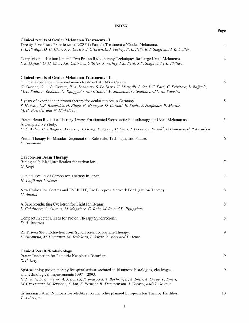
INDEX Page Clinical results of Ocular Melanoma Treatments - I Twenty-Five Years Experience at UCSF in Particle Treatment of Ocular Melanoma. 4 T. L. Phillips, D. H. Char, J. R. Castro, J. O’Brien, L. J. Verhey, P. L. Petti, R. P Singh and I. K. Daftari Comparison of Helium Ion and Two Proton Radiotherapy Techniques for Large Uveal Melanoma. 4 I. K. Daftari, D. H. Char, J.R. Castro, J. O’Brien, J. Verhey, P.L. Petti, R.P. Singh and T.L. Phillips Clinical results of Ocular Melanoma Treatments - II Clinical experience in eye melanoma treatment at LNS – Catania. 5 G. Cuttone, G. A. P. Cirrone, P. A. Lojacono, S. Lo Nigro, V. Mongelli, J. Ott, I. V. Patti, G. Privitera, L. Raffaele, M. L. Rallo, A. Reibaldi, D. Rifuggiato, M. G. Sabini, V. Salamone, C. Spatola and L. M. Valastro 5 years of experience in proton therapy for ocular tumors in Germany. 5 S. Hoecht , N.E. Bechrakis, H. Kluge, H. Homeyer, D. Cordini, H. Fuchs, J. Heufelder, P. Martus, M. H. Foerster and W. Hinkelbein Proton Beam Radiation Therapy Versus Fractionated Stereotactic Radiotherapy for Uveal Melanomas: 5 A Comparative Study. D. C Weber, C. J Bogner, A Lomax, D. Georg, E. Egger, M. Caro, J. Verwey, L Escudé•,.G Goitein and .R Miralbell. Proton Therapy for Macular Degeneration: Rationale, Technique, and Future. 6 L. Yonemoto Carbon-Ion Beam Therapy Biological/clinical justification for carbon ion. 7 G. Kraft Clinical Results of Carbon Ion Therapy in Japan. 7 H. Tsujii and J. Mizoe New Carbon Ion Centres and ENLIGHT, The European Network For Light Ion Therapy. 8 U. Amaldi A Superconducting Cyclotron for Light Ion Beams. 8 L. Calabretta, G. Cuttone, M. Maggiore, G. Raia, M. Re and D. Rifuggiato Compact Injector Linacs for Proton Therapy Synchrotrons. 8 D. A. Swenson RF Driven Slow Extraction from Synchrotron for Particle Therapy. 9 K. Hiramoto, M. Umezawa, M. Tadokoro, T. Sakae, Y. Mori and Y. Akine Clinical Results/Radiobiology Proton Irradiation for Pediatric Neoplastic Disorders. 9 R. P. Levy Spot-scanning proton therapy for spinal axis-associated solid tumors: histologies, challenges, 9 and technological improvements 1997 – 2003. H. P. Rutz, D. C. Weber, A. J. Lomax, R. Bearpark, T. Boehringer, A. Bolsi, A. Coray, F. Emert, M. Grossmann, M. Jermann, S. Lin, E. Pedroni, B. Timmermann, J. Verwey, and G. Goitein. Estimating Patient Numbers for MedAustron and other planned European Ion Therapy Facilities. 10 T. Auberger
1
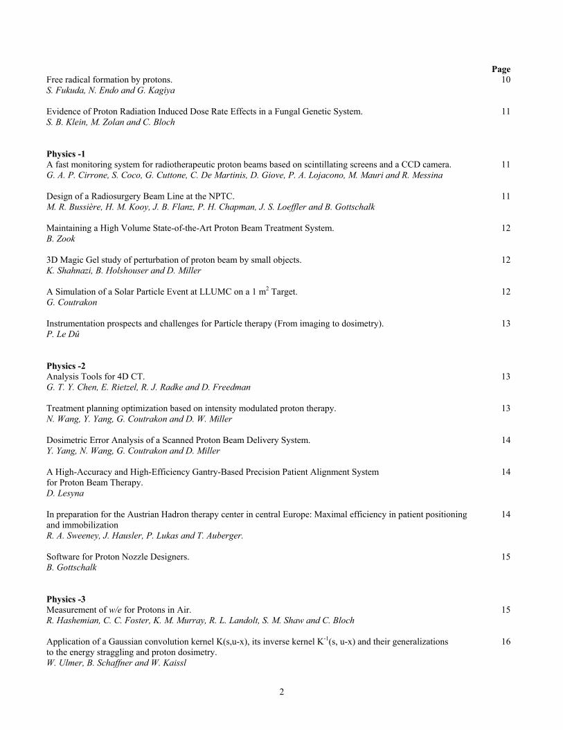
Page Free radical formation by protons. 10 S. Fukuda, N. Endo and G. Kagiya Evidence of Proton Radiation Induced Dose Rate Effects in a Fungal Genetic System. 11 S. B. Klein, M. Zolan and C. Bloch Physics -1 A fast monitoring system for radiotherapeutic proton beams based on scintillating screens and a CCD camera. 11 G. A. P. Cirrone, S. Coco, G. Cuttone, C. De Martinis, D. Giove, P. A. Lojacono, M. Mauri and R. Messina Design of a Radiosurgery Beam Line at the NPTC. 11 M. R. Bussière, H. M. Kooy, J. B. Flanz, P. H. Chapman, J. S. Loeffler and B. Gottschalk Maintaining a High Volume State-of-the-Art Proton Beam Treatment System. 12 B. Zook 3D Magic Gel study of perturbation of proton beam by small objects. 12 K. Shahnazi, B. Holshouser and D. Miller A Simulation of a Solar Particle Event at LLUMC on a 1 m2 Target. 12 G. Coutrakon Instrumentation prospects and challenges for Particle therapy (From imaging to dosimetry). 13 P. Le Dû Physics -2 Analysis Tools for 4D CT. 13 G. T. Y. Chen, E. Rietzel, R. J. Radke and D. Freedman Treatment planning optimization based on intensity modulated proton therapy. 13 N. Wang, Y. Yang, G. Coutrakon and D. W. Miller Dosimetric Error Analysis of a Scanned Proton Beam Delivery System. 14 Y. Yang, N. Wang, G. Coutrakon and D. Miller A High-Accuracy and High-Efficiency Gantry-Based Precision Patient Alignment System 14 for Proton Beam Therapy. D. Lesyna In preparation for the Austrian Hadron therapy center in central Europe: Maximal efficiency in patient positioning 14 and immobilization R. A. Sweeney, J. Hausler, P. Lukas and T. Auberger. Software for Proton Nozzle Designers. 15 B. Gottschalk Physics -3 Measurement of w/e for Protons in Air. 15 R. Hashemian, C. C. Foster, K. M. Murray, R. L. Landolt, S. M. Shaw and C. Bloch
Application of a Gaussian convolution kernel K(s,u-x), its inverse kernel K-1(s, u-x) and their generalizations 16 to the energy straggling and proton dosimetry. W. Ulmer, B. Schaffner and W. Kaissl
2
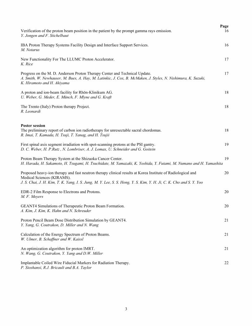
Page Verification of the proton beam position in the patient by the prompt gamma rays emission. 16 Y. Jongen and F. Stichelbaut IBA Proton Therapy Systems Facility Design and Interface Support Services. 16 M. Notarus New Functionality For The LLUMC Proton Accelerator. 17 K. Rice Progress on the M. D. Anderson Proton Therapy Center and Technical Update. 17 A. Smith, W. Newhauser, M. Bues, A. Hay, M. Latinkic, J. Cox, B. McMaken, J. Styles, N. Nishimura, K. Suzuki, K. Hiramoto and H. Akiyama A proton and ion-beam facility for Rhön-Klinikum AG. 18 U. Weber, G. Meder, E. Münch, F. Mlyne and G. Kraft The Trento (Italy) Proton therapy Project. 18 R. Leonardi Poster session The preliminary report of carbon ion radiotherapy for unresectable sacral chordomas. 18 R. Imai, T. Kamada, H. Tsuji, T. Yanag, and H. Tsujii First spinal axis segment irradiation with spot-scanning protons at the PSI gantry. 19 D. C. Weber, H. P.Rutz , N. Lombriser, A. J. Lomax, U. Schneider and G. Goitein Proton Beam Therapy System at the Shizuoka Cancer Center. 19 H. Harada, H. Sakamoto, H. Tsugami, H. Tsuchidate, M. Yamazaki, K. Yoshida, Y. Futami, M. Numano and H. Yamashita Proposed heavy-ion therapy and fast neutron therapy clinical results at Korea Institute of Radiological and 20 Medical Sciences (KIRAMS). J. S. Chai, J. H. Kim, T. K. Yang, I. S. Jung, M. Y. Lee, S. S. Hong, Y. S. Kim, Y. H. Ji, C. K. Cho and S. Y. Yoo EDR-2 Film Response to Electrons and Protons. 20 M. F. Moyers GEANT4 Simulations of Therapeutic Proton Beam Formation. 20 A. Kim, J. Kim, K. Hahn and N. Schreuder
Proton Pencil Beam Dose Distribution Simulation by GEANT4. 21 Y. Yang, G. Coutrakon, D. Miller and N. Wang Calculation of the Energy Spectrum of Proton Beams. 21 W. Ulmer, B. Schaffner and W. Kaissl An optimization algorithm for proton IMRT. 21 N. Wang, G. Coutrakon, Y. Yang and D.W. Miller Implantable Coiled Wire Fiducial Markers for Radiation Therapy. 22 P. Sioshansi, R.J. Bricault and B.A. Taylor
3
Twenty-Five Years Experience at UCSF in Particle Treatment of Ocular Melanoma. T. L. Phillips1, D. H. Char2, J. R. Castro1, J. O’Brien3, L. J. Verhey1, P. L. Petti1, R. P. Singh4 and I. K. Daftari1,
1Department of Radiation Oncology, University of California San Francisco, San Francisco, CA 94143. 2Tumori Foundation, 35 Castro Street, San Francisco, 3Department of Ophthalmology, UC San Francisco, CA 94143, 4Lawrence
Berkeley National Lab; Berkeley, CA In a joint effort between UCSF, Lawrence Berkeley Laboratory and more recently the Crocker Nuclear Laboratory at
U.C. Davis a total of 868 patients with uveal melanoma have been treated with charged particles. Between 1978 and 1992 347 patients were treated with helium ions at LBL and between 1992 and 2003 521 patients have been treated with protons at CNL. The techniques used were continuously improved and were based originally on those used at the Harvard Cyclotron. Originally a dose of 70 GyE was used with helium, finally reduced to 48 GyE. The dose used at CNL was initially 48 GyE but after 47 patients was increased to 56 GyE. At the same time treatment plans were designed to minimize the dose to the ciliary body and lens. At this time 347 helium patients and 356 proton patients are evaluable with follow-up ranging from 3 to 101 months for the proton patients. The rate of neovascular glaucoma (NVG) was 35% for helium and 7% for proton patients. Enucleation (ENU) was required in 19% of the helium patients and 8% of the proton patients. Local Failure (LFR) occurred in 4% of the helium patients as defined by requirement for ENU and in 2.5% of the proton patients, 1.6% of those treated to 56GyE. The rate of distant metastases (DM) was 24% for the helium patients and 4% for the proton patients. The change in technique and the change in particle has led to improved results although care must be taken in interpretation because of the large differences in follow up time and changes in surgical treatment.
References: [1] J.R. Castro et al. “15 years experience with helium ion radiotherapy for uveal melanoma”, Int. J. Rad. Onc. Biol. Phys. 39, 989-996 (1997). [2] I.K. Daftari et al, “Anterior segment sparing to reduce charged particle radiotherapy complications in uveal melanoma”, Int. J. Rad. Onc. Biol. Phys., 39,997-1010 (1997)
**************************************************************
Comparison of Helium Ion and Two Proton Radiotherapy Techniques for Large Uveal Melanoma. I. K. Daftari1, D. H. Char2, J. R. Castro1, J. O’Brien3, L. J. Verhey1, P. L. Petti1, R. P. Singh4 and T. L. Phillips1,
1Department of Radiation Oncology, University of California San Francisco, San Francisco, CA 94143. 2Tumori Foundation, 35 Castro Street, San Francisco, 3Department of Ophthalmology,UC San Francisco, CA 94143, 4Lawrence
Berkeley National Lab; Berkeley, CA Purpose: This study was undertaken to compare the incidence of local failure (LFR), neovascular glaucoma (NVG), enucleation (ENU) and distant metastases (DM) as a function of treatment technique with helium ions and with protons for large and extra-large tumors. Methods and Materials: 142 evaluable patients with large and extra large uveal melanoma were treated with the proton beam from the Crocker Cyclotron at U.C. Davis between 1994 and 2003 with mean Follow up of 23 ± 19.4 months ( range 3 to 96.3 months). Two different techniques were employed: for the first 21 patients a standard technique was used to deliver 48 GyE (Gray equivalent) in 4 fractions over 4 days, for the subsequent 121 patients a dose of 56 GyE was delivered in 4 fractions using techniques which spared the anterior segment as proposed in a previous publication [1]. The results of these treatments were compared to results for 215 patients with large and extra-large tumors treated by us with helium ions [1,2]. The ion helium patients were treated at dose levels from 48 to 80 GyE given initially in five fractions over 6-7 days with a mean follow-up of 89.6 ± 51 months [range 3 to 194.5 months] Results: Among the 21 patients treated with 48 GyE the LFR was 3/21, NVG 8/21 and DM 4/21. ENU was required in 2/3 with LFR while 2 had DM. ENU was required in 3/8 patients who had NVG without LFR. The overall enucleation rate was 6/21. Among the 121 patients treated with 56GyE and sparing of the anterior chamber, the LFR was 5/121; 3 requiring ENU and 2 others received 1 additional laser treatment and no ENU. 13/121 patients developed NVG and 7/13 required enucleation. The overall enucleation rate was 17/121 (14%). The DM rate was 3/121(2.5%). In contrast, the patients treated with helium ions had LFR of 7/215 (3.2%), NVG in 93/215 (43%) patients with 44/93(47.3%) requiring enucleation. The overall enucleation rate was 24%. The DM rate was 33%. Conclusions: The present technique of 56GyE using protons and anterior chamber sparing plans appears superior to helium ion results in terms of decreased NVG and a decrease in the need for enucleation for NVG symptoms. The LFR was similar between 56 GyE of protons (4 %) and Helium treatment (3%) but much higher in the 48 GyE proton group (14 %), a finding previously seen in a small group of helium ion patients treated with 48 GyE in 4 fractions. The DM rates appear to be falling in the more recent groups, but follow up time is not equal. References:[1] J.R. Castro et al. “15 years experience with helium ion radiotherapy for uveal melanoma”, Int. J. Rad. Onc. Biol. Phys. 39, 989-996 (1997). [2] I.K. Daftari et al, “Anterior segment sparing to reduce charged particle radiotherapy complications in uveal melanoma”, Int. J. Rad. Onc. Biol. Phys., 39,997-1010 (1997)
**************************************************************
4
Clinical experience in eye melanoma treatment at LNS – Catania. G. Cuttone1, G. A. P. Cirrone1,2, P. A. Lojacono1,5, S. Lo Nigro2, V. Mongelli2, J. Ott3, I. V. Patti2, G. Privitera4, L. Raffaele1, M. L. Rallo3, A. Reibaldi3, D. Rifuggiato1, M. G. Sabini1, V. Salamone1, C. Spatola4, L. M. Valastro1,2,
1Laboratori Nazionali del Sud (INFN), Catania, Italy, 2Physics Department, University of Catania, Italy, 3Ophthalmology Institute, University of Catania, Italy, 4Radiology Institute, University of Catania, Italy, 5C.S.F.N.S.M., Corso Italia 57,
95100 Catania, Italy
At the INFN Laboratori Nazionali del Sud in Catania (Italy) the first Italian proton therapy facility, named CATANA (Centro di AdroTerapia e Applicazioni Nucleari Avanzate) has been realized in collaboration with the University of Catania.
It is based on the use of the 62 MeV proton beam delivered by the K=800 Superconducting Cyclotron installed and working at LNS since 1995. The facility is mainly devoted for the treatment of ocular diseases like uveal melanoma. A beam treatment line in air has been realized together with a dedicated positioning patient system. The facility is in operation since the beginning of 2002 and 52 patients have been successfully treated up to now.
The main features of CATANA together with the clinical and dosimetric features will be extensively reported.
**************************************************************
5 years of experience in proton therapy for ocular tumors in Germany. S. Hoecht, N. E. Bechrakis, H. Kluge, H. Homeyer, D. Cordini, H. Fuchs, J. Heufelder, P. Martus, M. H. Foerster W.
Hinkelbein., Charité Medical School, Camp. B. Franklin, Berlin, Germany Background: In June 1998 proton beam therapy of ocular tumors started at the Hahn-Meitner-Institut Berlin, Germany. Purpose of this study is to evaluate treatment outcome for uveal melanomas and to describe the new strategy of endoresection following proton beam irradiation for larger tumors. Material and Methods: 245 consecutive patients with primary melanoma of the uvea were treated from June 1998 to April 2003 with a 68 MeV – proton beam. In 96.2% of all pts. a uniform fractionation scheme was applied: single dose 15 CGE (Cobalt Gray Equivalent), total dose 60 CGE on 4 consecutive days. Follow-up is available in 229 patients. Since May 2000 45 patients with large tumors of at least 5 mm prominence have been treated with an endoresection following proton beam treatment. Results: At the time of median follow up (18,4 months) local control is 96.4% and 95.5% at 3 years. Eye retention rate is 92.6% at 20 months (median follow-up) and 87.5% at 3 years. The subset of patients treated with an endoresection did fairly well: Although those tumors were larger in volume their outcome does compare favourably with the others. Local control is still 100% at 18,1 months at the time of median follow-up and calculated eye-retention rate is 88,6% after 2 years. Conclusions: Proton beam irradiation of uveal melanomas at the Hahn-Meitner-Institut after the first 5 years of its initiation reveals local tumor control and eye- retention rates in the range of other centers with larger experience. Delivering high treatment quality in hadron therapy from the beginning has been achieved. The concept of endoresection after proton beam irradiation seems promising but needs further evaluation with more patients and longer follow-up.
**************************************************************
Proton Beam Radiation Therapy Versus Fractionated Stereotactic Radiotherapy for Uveal Melanomas: A Comparative Study.
D. C. Weber1,2, J. Bogner3, A. Lomax1, D. Georg3, E. Egger1, M. Caro4, J. Verwey1, L. Escudé4, G. Goitein1, and R. Miralbell2,4, Departments of Radiation Oncology,1Paul Scherrer Institut, Villigen, and 2University Hospital, Geneva,
Switzerland, 3University Hospital, Vienna, Austria, and 4Centro Médico Teknon, Barcelona, Spain. Purpose: A comparative treatment planning study was undertaken between proton and photon therapy in uveal melanoma (UM), as to assess the potential benefits and limitations of these treatment modalities. A fixed proton horizontal beam, as used at the Paul Scherrer Institute (PSI), and intensity modulated (IM) spot-scanning proton therapy, with multiple non-coplanar beam arrangements, was compared to linear accelerator-based stereotactic radiotherapy with micromultileaf collimation (Novalis®, BrainLAB AG): static conformal, dynamic arc plan and IM radiotherapy (IMRT). Method and Materials: A planning brain CT scan was performed on a brain metastasis patient, with a slice spacing of 3 mm acquisition and the eyeballs in 3 different (neutral, 250 right and left) positions (patient looking a luminous spot). Four different gross tumor volumes (GTVs) were defined for each treatment technique. These target scenarios represented
5
different location (involving vs. not involving the macula; temporal vs. nasal vs. posterior) and volumes (10x6 vs. 16x10 mm), as to both challenge proton and photon treatment techniques. Target volumes and organs at risk (OARs) were delineated on the CT data set by the same radiation oncologist (DCW) and physicist (EE). Planning target volume (PTV) was defined as GTV + 1mm for protons and GTV + 2mm for photons. For a given dose prescription of 50 CGE-Gy in 10 CGE-Gy (daily fractions), a minimum of 95% to 95% of the PTV volume was used as planning ‘goal’ for all techniques. OAR’s dose-constraints (maximum) for both proton and photon plans were 27.5, 22.5, 20 and 9 CGE-Gy for the optic apparatus, retina, lacrimal gland and lens, respectively. Plans for proton and photon therapy were computed using the PSI (PSIPlan) and the BrainSCAN® V5.2 (BrainLAB, Heimstetten, Germany) treatment planning system, respectively. Results: Target coverage was optimal and equally conformal with all proton and photon treatment techniques. For conventional radiotherapy, tumor dose homogeneity in IMRT plans was always better than no-intensity modulation. Complete results of this comparison will be available at the time of presentation. Conclusion: These results suggest that the use of IM and SRT photon therapy, when compared to protons, can result in similar levels of tumor conformation for the treatment of UM.
**************************************************************
Proton Therapy for Macular Degeneration: Rationale, Technique, and Future.
L. Yonemoto, Loma Linda University Medical Center (LLUMC)
Age-Related Macular Degeneration (AMD) is a leading cause of legal blindness in the Western World. The Framingham Study estimates that 8.5 to 11 million Americans have this irreversible disease. The prevalence is 11% for ages 65–74 and 28% for ages 75-85. The wet type of AMD accounts for about 10% of cases but cause 88% of the legal blindness. This type is called wet due to the leakage from neovascularization at or near the macula, distorting, clouding and eventually destroying the central vision.
In the 1980’s, the Macular Photocoagulation Study (MPS) demonstrated a statically significant improvement with laser therapy. However, only the classic types were eligible and this accounts for only 10-20 % of cases. The improvement was an average decline of vision of only 3 lines on the MPS eye chart vs. 5 lines without laser therapy at 2 years. The recurrence rate was 46% at 1 year and 79% at 5 years. The thermal laser permanently damages the retina near the targeted blood vessel and sometimes damages the macula.
Radiation therapy was used in the 1990’s with the rationale that endothelial cells, especially rapidly proliferation ones were more sensitive to radiation than non-proliferation retinal cells. Most radiation therapy studies used fractionation regiments and had mixed results. Animal studies at LLUMC demonstrated a lack of benefit with fractionation. LLUMC modeled their regiment after the single fraction AVM treatment. The Phase I/II study in 1995 demonstrated a dose response of 8 Gy and 14 Gy with 44% and 89% lesion control at 1 year respectively. The recurrence rate was less than 10% at 2 years and study included the non-classic types. The average vision loss was zero at 3 years.
The LLUMC technique is similar to the ocular melanoma, employing the eye beam, chair, mask and eyelid retractors. The difference is that the set-up is a clinical, and treatment planning used standard apertures. Patients were instructed to look at the center of an aperture with a cross hairs pattern. The eye beam light projected the cross hairs onto the eye and the patient chair is moved until the cross hairs was centered towards the macula. Then the patient was instructed to look at a blinking light that was placed 30 degrees nasally which displace the macula laterally 6 mm. The patient is move 6 mm in the nasal direction to place the beam line back onto the macula. A standard circular aperture at 10, 13 or 15 mm in diameter was place on the eye nozzle. The light field through the aperture should project onto the sclera just lateral of the limbus and avoid the eyelids. This technique spares the anterior and posterior chambers from the beam.
In 2000, a photosensitizer, verteporfin demonstrated a modest but statically significant benefit of a non-thermal laser. At 2 years, 53% of patients treated with Photo Dynamic Therapy (PDT) had less than 3 lines of visually acuity loss and compared to 38% of the placebo group. However, the retreatment with PDT due to more leakage was an average of 3.4 times the first year and 2.1 times the second year.
There is potential for synergy by combining proton therapy and PDT. Proton therapy has a low recurrence rate but takes 2 to 6 months for lesion control. PDT has a rapid control but a short recurrence interval. Thus, treating with PDT prior to referral to proton therapy could be the ideal mix of therapies.
Currently we are developing a model for diabetic retinopathy, the leading cause of blindness in the working age Americans. This disease is also results from leakage from blood vessels and is treated with thermal laser. The laser also destroys the retina near the targeted leaking vessels.
With an estimated worldwide incidence of wet AMD at 500,000 per year, and the potential for treatment of diabetic retinopathy, this effective proton therapy technique is also capable of high volume.
**************************************************************
6
Biological / clinical justification for carbon ion. G. Kraft, Biophysik GSI Darmstadt
The primary reason to use ions, heavier than protons, first at Berkeley, was the decrease of the OER and the increase in
RBE. For carbon ions the RBE changes are restricted to the last two centimetres of the particle range. In order to protect the normal tissue outside of the target volume from high LET effects, a strict tumor conform irradiation technique should be used, like the intensity modulated scanning system of GSI. There a perfect agreement between planned and irradiated volume can be achieved. Using online Positron Emission Tomography PET, the precision of the dose delivery can be controlled inside the patient for the first time.
The major challenge of the Intensity Modulated Particle Therapy IMPT is the complex distribution of the RBE over the treatment area. Each treatment voxel can have a different particle composition, mainly in energy, but also in atomic number and consequently an RBE differing from the next voxel.
In order to solve the RBE problem, a Local Effect Model LEM is used at GSI to calculate the local RBE values. In this calculation, the track structure and the x-ray sensitivity of the individual tissue are used for the calculation of the individual RBE values. As endpoint the RBE for inactivation of the tumor and the RBE for late effects of the normal tissue in the entrance channel are used for treatment planning.
Using the methods of intensity modulated beam delivery and biological treatment planning an optimal clinical gain factor can be achieved in carbon therapy.
**************************************************************
Clinical Results of Carbon Ion Therapy in Japan. H. Tsujii and J. Mizoe, Research Center for Charged Particle Therapy, National Institute of Radiological Sciences(NIRS),
Chiba, Japan
Currently in Japan there are two facilities for carbon ion therapy including National Institute of Radiological Sciences (NIRS) in Chiba and Hyogo Ion Beam Medical Center (HIBMC) in Hyogo. In 1992 the project for heavy ion therapy was initiated as part of the Japanese government’s 10-year plan to combat cancer, and at NIRS the Heavy Ion Medical Accelerator in Chiba (HIMAC) was constructed. In June 1994, clinical research for the treatment of malignant tumors was begun using carbon ions generated by the HIMAC. Since then more than 1,600 patients have been treated with carbon ions. The Hyogo Prefecture decided to initiate a challenging program for cancer control in 1992, followed by a construction of accelerator for both protons and carbon ions. Proton therapy was first started in April 2001, followed by carbon ion therapy in the next year.
As of August 2003, a total of 1,601 patients were enrolled in the study at NIRS. In the very first dose escalating study, complications of the recto-sigmoid colon were observed in <3%. Such adverse effects, however, did disappeared as a result of determining the safe dose level and improvement in irradiation method. Carbon ion RT has shown improvement of outcome for head and neck tumor (adenocarcinoma, adenoid cystic carcinoma, malignant melanoma), early-stage NSCLC, bone/soft tissue sarcoma, hepatoma, and locally advanced prostate carcinoma. There is a rationale to justify the use of short-course RT due to the superior dose localization and the unique biological property of high-LET radiations. This has been proven in treatment for NSCLC and hepatoma, where the fraction number has been shortened to 1~4 fractions given in 1~4 days. Even in prostate cancer and bone/ soft tissue tumor, treatment can be performed using 16 fractions in 4 weeks with acceptable morbidity. As of September 2003 at Hyogo, a total of 182 patients were treated with proton therapy for head and neck tumors, hepatoma, lung cancer and prostate cancer, and 30 patients with carbon ion therapy for head and neck tumors, lung cancer, hepatoma, and bone/soft tissue tumors. Although it is too early to assess the treatment results, each treatment appears to be safe and effective for those tumors treated.
**************************************************************
7
New Carbon Ion Centres and ENLIGHT, The European Network For Light Ion Therapy. U. Amaldi, University of Milano Bicocca and TERA Foundation, Via Puccini 11, 28100 Novara, Italy
Carbon ions have a larger biological effectiveness than X-rays and protons and are considered to be particularly suited
to treat radioresistant tumours, as shown by the encouraging results obtained on about 1'600 patients in HIMAC (Chiba, Japan) and on about 200 patients at GSI (Darmstadt). A second Japanese centre is running in Hyogo (HIBMC) and two centres are under construction in Europe: one in Heidelberg (HICAT) and the other in Pavia (CNA). The first part of the talk will be devoted to these four centres. The second part will discuss the other European projects, that are at various stages of advancements in Wiener Neustadt (Austria), Lyon (France) and Stockholm (Sweden). Europe is proceeding coherently towards the new frontiers of carbon therapy, as also proven by the fact that the five European projects are collaborating in the framework of ENLIGHT, the European Network for LIGHt-ion Therapy. Also ESTRO, EORTC, CERN and GSI are part of the network that works on six themes: epidemiology and patient selection; design and conduct of clinical trials; preparation, delivery and dosimetry; radiation biology; in-situ monitoring with PET; health economics aspects.
**************************************************************
A Superconducting Cyclotron for Light Ion Beams.
L. Calabretta, G. Cuttone, M. Maggiore, G. Raia, M. Re, D. Rifuggiato, Laboratori Nazionali del Sud – I.N.F.N., Via S. Sofia 44, 95123 Catania, Italy
Abstract: A four sector compact superconducting cyclotron able to deliver light ion beams and protons with a maximum energy of 250 A MeV has been studied. The range of Carbon ions at this energy is of 128 mm in water, so it is enough for therapy of a large amount of the lung, head, neck and brain tumors. While the range of Lithium ion at 250 A MeV is 250 mm in water, which allow to treats deeper tumors. This cyclotron is able to accelerate ions with charge to mass ratio 0.5. The mean magnetic field at the extraction radius is 3.9 T. The magnetic field is supplied by a twin coils with 2.5 MA⋅turn. To accelerate different ions, like 12C6+, 6Li3+ and H2
+, the main coil current must be adjusted of 0.2%. The 4 RF cavities are driven at the fixed frequency of 91 MHz, in 4th harmonic. The H2
+ beam can be extracted by stripping which allow to reach 100% of extraction efficiency so reducing the accelerator activation, this extraction allow to delivers also high proton current for ancillary research and activities. The extraction of other fully stripped ions is performed by electrostatic deflectors. On the two electrostatic deflectors voltages lower than 50 kV are required. The proton beam and the other ion could be delivered at the same external position or at two different locations if required. The design model of the magnet circuit has been accomplished with the 3D electromagnetic code OPERA. The main coils, the cryostat, the RF cavities and vacuum system characteristics have been studied and will be presented. The expected performance of the accelerator will be also described.
**************************************************************
Compact Injector Linacs for Proton Therapy Synchrotrons. D.A. Swenson, Linac Systems, Albuquerque, NM.
Synchrotron injector linacs, based on the new Rf Focused Interdigital (RFI) linac structure will be smaller and more
efficient than ever before. The RFI linac structure, under development at Linac Systems, is an effective combination of the interdigital (Wideröe) linac structure and the rf electric quadrupole focusing used in the Radio Frequency Quadrupole (RFQ) linac structure. The RFI structure is three times smaller in diameter and four-to-six times more efficient than the conventional Drift Tube Linac (DTL) structure, and ten-to-twenty times more efficient than the RFQ linac structure, in the 0.75-to-10 MeV range. A prototype of the RFI structure is currently under construction at Linac Systems, with operation scheduled for the fall of 2004. Synchrotron injector linac designs will be presented for 4, 7, and 10 MeV models. Estimates of the size, power requirement, performance, and cost will be presented for each model. System availability and methods of procurement will be outlined.
**************************************************************
8
RF Driven Slow Extraction from Synchrotron for Particle Therapy. K. Hiramoto1, M. Umezawa1 and M. Tadokoro1 T. Sakae2, Y. Mori2,3 and Y. Akine2, 1Hitachi, Ltd., 2PMRC, University of
Tsukuba, 3KEK, High Energy Accelerator Research Organization
Slow extraction method from synchrotron dedicated for particle therapy and its application results are presented. In the present method, the beam can be extracted only by applying an RF signal with keeping all kind of magnetic fields constant, while the previous extraction method needed changing several kinds of magnetic fields during the extraction. The present method has been applied to the proton therapy system of PMRC, Univ. of Tsukuba and other facilities. In these applications, the present simple method has shown excellent beam stability and reproducibility of smaller than 0.5 mm without beam position feedback. Also, it has been verified that the beam extraction and stop can be switched within a few hundred micro seconds by turning on and off the RF signal for the extraction. These capabilities have been successfully utilized in the respiration gating irradiation with passive scattering in Tsukuba system. Furthermore, the above verified features show that the present method can realize the pencil beam scanning in which especially excellent stability and reproducibility are needed.
**************************************************************
Proton Irradiation for Pediatric Neoplastic Disorders. R. P. Levy, Loma Linda University Medical Center, Loma Linda, CA, USA
The treatment of malignant and benign neoplastic disorders in children with X-irradiation is associated with significant
normal tissue toxicity. Sites of particular concern are the brain, cochlea, musculoskeletal structures, thyroid, thorax, abdomen, and pelvis. Acute sequelae may include neutropenia, especially if chemotherapy is used, as well as pharyngitis, esophagitis, nausea, and vomiting. Delayed sequelae may include secondary tumors, neurocognitive impairment, hearing loss, musculoskeletal growth deformities, cardiopulmonary toxicity, and sterility.
Fractionated proton beam therapy (PBT) has been advocated for use in pediatric neoplastic disorders, due to its ability to place a highly conformal dose of radiation about a multitude of target volumes throughout the body, while also providing a much lower integral dose to normal tissues than X-ray-based techniques. At Loma Linda University Medical Center (LLUMC), the world’s first hospital-based proton treatment center was built as part of the Children’s Hospital. This proximity has made it possible to deliver PBT in a variety of clinical settings, including for the treatment of young children who have required daily anesthesia.
In the case of posterior fossa tumors, PBT has been used to lessen the dose to the cochlea as compared to both standard and intensity modulated radiation therapy. PBT also provides excellent sparing of uninvolved adjacent brain tissues when used for supratentorial tumors. Cardiopulmonary dose can be minimized with PBT for mediastinal lymphoma. Craniospinal PBT for medulloblastoma has been accomplished without resultant acute neutropenia, while minimizing the dose to skeletal structures and providing nearly complete sparing of thoracic, abdominal and pelvic organs.
Delayed sequlae of X-irradiation in children will be briefly described. Examples will then be presented of the range of PBT applications used at LLUMC to mitigate these sequelae.
**************************************************************
Spot-scanning proton therapy for spinal axis-associated solid tumors: histologies, challenges, and technological improvements 1997 – 2003.
H. P. Rutz, D. C. Weber, A. J. Lomax, R. Bearpark, T. Boehringer, A. Bolsi, A. Coray, F. Emert, M. Grossmann, M. Jermann, S. Lin, E. Pedroni, B. Timmermann, J. Verwey, and G. Goitein, Proton Therapy Program, Paul Scherrer Institute,
CH-5232 Villigen PSI, Switzerland Purpose: Critical structures within and along the spinal axis restrict high-dose radiation therapy for malignant tumors. Spot-scanning technology, on the other hand, allows for an organ and tissue sparing dose conformation. Several challenges had to be overcome. Patients and Methods: Among the first 155 patients treated at the PSI gantry, 42 had a spinal axis-associated solid tumor. All but one of the 42 patients were treated with a locally curative intent. Four targets were single metastasis, three in potentially curative situations. Here, we analyze histologies, problems and solutions regarding spot-scanning treatments. Results: Histologies were chordoma (24), sarcoma (12), carcinoma (2), ependymoma (1), meningeoma (1), schwannoma (1) and primitive neuroectodermal tumor (1). Several challenges had to be dealt with, such as CT artifacts from implants, PTV wrapping around the myelon, and the need for irradiation of a paraspinal GTV along with the entire craniospinal axis.
9
Homogeneous PTV coverage for each beam direction (conventional spot-scanning PT) was used to treat 36 of the tumors. The other treatments required the delivery of inhomogeneous dose within beam directions (intensity modulated PT, IMPT), which requires spot-scanning technology, to limit dose to the myelon and for depth ranges from skin to deeper than 200 mm. Patient/PTV movement is a difficulty that we deal with through multiple applications of the same field. Implants are a major problem: we must assign soft tissue density manually; they reduce accuracy in target definition; and they cause hotspots due to nuclear interactions and high proton energy required to reach PTV behind the metal. To treat patients with implants, we decided to limit dose per fraction to 1.8 CGE, as it is our current policy to adhere to conventional fractionation schemes. Delivering a donut-shaped high-dose configuration around the myelon, which requires IMPT, has solved the problem of PTVs wrapping around the myelon. Finally, we made a first step towards another important goal of our program: the treatment of the entire craniospinal axis with spot-scanning PT. However, we are not yet able to treat the entire axis by PT. Therefore, we applied a combined PT and photon radiotherapy of the axis. Discussion: Spot-scanning PT is a good technological approach to treat tumors that arise along the spinal axis. While we were able to solve a number of technological problems over the last several years, these are the issues that we currently work at: implementing a method to eliminate implant-dependent CT artifacts; adapting IMPT programs for routine use; development of a sliding mechanism for the cast, so as to soon become able to treat the entire craniospinal axis with spot-scanning PT. This will simplify therapy and eliminate dose uncertainties at matching lines. Clinical follow-up will be addressed at PTCOG XL.
**************************************************************
Estimating Patient Numbers for MedAustron and other planned European Ion Therapy Facilities. T. Auberger, Dept. of Radiotherapy, Univ. Medical School of Innsbruck, Austria
Background: In the frame of the Med-AUSTRON project it is planned to establish a hadron therapy facility in Wiener Neustadt/Austria. Precise epidemiological data are required to most accurately define the number of potential patients. Materials and Methods: In a prospective study, disease and treatment related data of all patients starting radiotherapy at three Austrian University hospitals during May, 15 to Aug, 15 2002 were assessed by questionnaires. Results: In total, this pilot study investigated the data of 1.592 patients; 45.7 % of the patients suffered from tumors, which were possibly suitable for heavy charged particle therapy. These tumors are defined as Priority I or Priority II tumours in the priority list. Priority I tumours are rare entities, in which hadron therapy clearly has shown improved clinical results. Priority II tumours include frequent tumour entities, in which hadrons showed potential advantages in relation to conventional radiotherapy, whereby, however, further studies are necessary. Conclusion: As pointed out by other European Groups, about 50% of the patients with Priority I tumours and approximately 9% of Priority II tumours might be referred for hadron therapy. At present, data of all Austrian irradiation treatment facilities were investigated; from these nationwide data, the number of patients suitable for hadron therapy in Austria will be estimated.
**************************************************************
Free radical formation by protons.
S. Fukuda1, N. Endo2, and G. Kagiya1, 1Medical division, The Wakasa Wan Energy Research Center, 2Biology and material group, The Wakasa Wan Energy Research Center64-52-1 Nagatani, Tsuruga City, Fukui 914-0192 JAPAN The interaction between cells including tumors and radiation can be classified into the direct interaction and the indirect
one, the former is dominant for the low LET radiation such as X-rays and the latter is dominant for the high LET radiation such as heavy ions.
Generally, proton is classified as the low LET radiation and can have the same RBE as the X-rays, and many experiments in vivo and in vitro indicate RBE is almost equal to one. Meanwhile it was reported that the RBE of protons becomes 1.2 beyond Bragg peak where the protons has a higher LET like heavy ions. Therefore we aimed at the free radicals that play an important role in the indirect interaction and measured the LET dependence of the production of the free radicals induced by the proton radiation. As a preliminary result, the yield of the free radicals per unit dose around the Bragg peak was smaller than that at the proximal region and increases beyond the Bragg peak. We will report the details of this experiment and discuss the mechanism of the RBE of protons.
**************************************************************
10
Evidence of Proton Radiation Induced Dose Rate Effects in a Fungal Genetic System. S. B. Klein1, M. Zolan2, and C. Bloch3, 1Indiana University Cyclotron Facility, 2Indiana University, 3University of Texas,
M. D. Anderson Medical Center The delivery of scanned particle beams requires high dose rate administration. Scanning technology and development
has rekindled an interest in the possibility of a biological dose rate effect for particle beams. This effect is particularly pertinent to the case of proton irradiation, which most closely resembles photon radiation biology. During a series of experiments designed to discover the enzyme complexes responsible for high LET chromosomal damage repair, we identified a proton radiation dose rate effect experienced by a fungal genetic model system: Coprinus cinereus. This effect was apparent in the wild-type fungus as well as the radiation sensitive mutant strains developed for the ongoing project. This paper will present the preliminary results including cell cycling, genetic and radiation quality impact upon a well-defined proton radiation dose rate effect.
**************************************************************
A fast monitoring system for radiotherapeutic proton beams based on scintillating screens and a CCD camera. G. A. P. Cirrone1, S. Coco2, G. Cuttone1, C. De Martinis3, D. Giove3, P. A. Lojacono1, M. Mauri3, R. Messina2,
1Laboratori Nazionali del Sud – I.N.F.N., Catania, Italy, 2Electrical, Electronic and Systemistic Department (DIEES) of Catania University, Catania, Italy, 3Physics Department of Milano University, Milano, Italy
A facility in which a 62 MeV proton beam is employed in the radiotherapeutic treatment of several ocular diseases is
active since March 2002 at “Laboratori Nazionali del Sud”. A fast and accurate quality control system of such beam has been designed and tested. The system consists of a scintillating screen mounted perpendicularly to the beam axis at a fixed distance and observed by a highly sensitive CCD camera. The conventional way to perform this quality control exploits several kinds of radiation detectors like ion chambers, silicon diodes and radiochromic films. All these methods, though very accurate and reliable, are quite slow. The purpose of the application is the on-line reconstruction of the lateral dose distribution, released to biological tissue, and the evaluation of beam quality parameters like flatness, homogeneity, symmetry and penumbras. The basic idea is the possibility to obtain informations about the relative dose delivered to tissue through the measurement of the light emission in the scintillating screen. The linearity of the yield (light output) with the beam current, the noise in the signal and the spatial resolution have been investigated. Comparisons with existing radiation detectors have been carried out. The good capabilities of the system make it worthy of further investigations on its applicability in proton beams monitoring for radiotherapeutic treatments (i.e. reconstruction of depth dose distribution and beam monitoring during patient irradiation).
**************************************************************
Design of a Radiosurgery Beam Line at the NPTC. M. R. Bussière1, H. M. Kooy1, J. B. Flanz1, P. H. Chapman2, J. S. Loeffler1, and B. Gottschalk3, 1Department of Radiation Oncology2Department of Neurosurgery Massachusetts General Hospital, Boston, MA 02114, 3Harvard University High
Energy Physics Lab, Cambridge, MA 02138 In 1961, MGH (Massachusetts General Hospital) physicians began treating patients with the proton beam at the
Harvard Cyclotron Laboratory (HCL), Harvard University. Up to 1993, about 3,000 patients with a variety of brain tumors and vascular malformations had been treated. At that time an improved system of radiosurgical treatment known as STAR (Stereotactic Alignment for Radiosurgery) was implemented. By the time the HCL closed it’s doors in 2002, almost 800 patients had been treated using the STAR device. Soon after the opening of the MGH’s NPTC (Northeast Proton Therapy Center), in November 2001, the first gantry based proton radiosurgery case was treated. However, because of the high demand and capacity limitations in the two gantry rooms a fixed beam line is being designed and constructed to treat radiosurgery patients, allowing more fractionated patients to be treated on the gantries. The fixed beam line will incorporate the proven technology of the STAR device and improve upon what was available at the HCL. The necessary components of the new beamline include bending, quadrupole and trim magnets, energy degrader and scatterer for the purpose of range and modulation control, ion chambers for beam profile monitoring, x-ray imaging system, and collimation and beam shaping system, and a control system. A Radionics MMLC is included in the design to provide maximum flexibility in treating fractionated SRT patients. The integration of the beam line components is discussed. The improved design is compared to what was available at the HCL and the treatment scenario detailed.
**************************************************************
11
Maintaining a High Volume State-of-the-Art Proton Beam Treatment System. B. Zook, Optivus Technology, Inc., 1475 S. Victoria Ct., San Bernardino, CA. 92408
This talk will share some of the insight gained over the years at the LLUMC Proton Beam Treatment System (PBTS).
Since 1990 more than 8500 patients have been treated with a current treatment rate of approximately 40,000 fields per year and better than 97% uptime in it’s thirteenth year of continuous operation.
To be successful, a high volume PBTS must remain economically viable over the 40 years of expected life. That means sustaining a continuous operation with limited time and support staff to perform preventive and corrective maintenance, highly reliable subsystems and equipment, high operational availability design criteria, and an industrial class maintenance program which includes: detailed procedures and schedules, extensive diagnostics, automated sensory data collection and trending capabilities and a well developed logistics support infrastructure.
The PBTS must be capable of delivering high-quality, safe and reliable beam on demand 24 hours a day, 7 days a week. Currently, facility access time for preventative maintenance in the LLUMC interlock area is limited to 6 hours per week on average. In 1993, after LLUMC had been operational for 3 years, we enjoyed an average of 46 hours per week for maintenance activities. That is an 87% reduction in maintenance access time over the last 10 years. We believe this trend will continue as treatment protocols expand with the commissioning of our precision patient positioning systems and active beam capabilities. That challenge has been met by establishing design constraints for a 40-year system life, 99% system level availability and minimal maintenance requirements for subsystems and equipment. In addition, the PBTS maintenance program has been refined to include clear criteria for identifying and processing high-risk subsystems for upgrade or replacement.
**************************************************************
3D Magic Gel study of perturbation of proton beam by small objects. K. Shahnazi, B. Holshouser, and D. Miller, Loma Linda University Medical Center, Department of Radiation Medicine,
Loma Linda CA Purpose: Treatment-planning system, Opti-Rad, is used for designing the proton beams for a phantom. Magic Gel provides a medium where the dose is applied and can be calibrated and verified. Using CT of the phantom, and the treatment planning, beam perturbation by small objects can be visualized and verified. Verification of the delivered dose around the odd object surfaces is done by MRI of the Magic Gel. 3D-gel dosimetry may be able to verify proper study in phantom test cases. Method: Several cylindrical phantoms were filled with MAGIC gel, XCT scanned, and the target volumes are drawn, and treatment plans were developed. The first phantom was used to study patch fields; the second phantom was used to study air cavities, water cavities, bone, and contrast materials. The third phantom filled with gel and Titanium screw imbedded in it. Proton beams were designed, apertures and boluses manufactured, and the phantoms treated. A MRI scan was performed on each of the phantoms, and the signal compared with the treatment plan. Results: A MRI of the MAGIC gel clearly illustrated the treated area. The positions of the MRI isodose curves were less than 1 mm of the curves calculated by the treatment planning system as referenced to phantom fiducials. Conclusion: This work shows the use Magic Gel in understanding of the dose delivery to a phantom.
**************************************************************
A Simulation of a Solar Particle Event at LLUMC on a 1 m2 Target. G. Coutrakon, Loma Linda University Medical Center
Using the 250 MeV proton synchrotron at Loma Linda University, we have developed a 36 hour simulation of a Solar
Particle Event (SPE) for use in NASA radiobiology experiments. A magnetic scanning system is used to spread the beam over a 1 m2 target so that a large population of animals may be irradiated at a single time. A variable range shifter is used to produce the required proton energy spectrum from 30 to 210 MeV. The beam intensity and the computer-controlled range shifter are varied as a function of time in order to simulate the distinctive time profile of an SPE. The first test of the simulation was carried out in September 2003, as part of Proton ICCHIBAN, an international intercomparison of space radiation dosimeters. Dosimetric instruments from over nine countries were exposed simultaneously at both low and high dose rates within the large field. Preliminary dosimetry results are presented as well as plans for future activities.
**************************************************************
12
Instrumentation prospects and challenges for Particletherapy (From imaging to dosimetry). P. Le Dû, CEA - DAPNIA Saclay
The scope of this presentation is to summarise the «state of the art» of technological developments imposed by the various needs and constraints associated with the patient dose optimization, delivery and monitoring. It will be illustrated by some R&D projects presented in recent related workshops like the 9th HCBM/ENLIGHT (Lyon, France) and 2003 IEEE NSS-MIC (Portland, USA) satellite workshop on hadrontherapy. Details will cover various topics like:
• Moving target: modelling of motion and organ deformation, detection and tracking organ motion • Dose quantification from in-beam PET • Advanced technologies for a dedicated hadron PET • Proton Computed Tomography developments • Dosimetry issues and Nanodosimetry
**************************************************************
Analysis Tools for 4D CT. G. T. Y. Chen1, E. Rietzel1, R. J. Radke2 and D. Freedman2, 1Massachusetts General Hospital, 2Rensselaer Polytechnic
Institute Four dimensional CT data provides important data on target and normal tissue motion during respiration. To fully
utilize these data for treatment planning, appropriate software analysis tools are needed. Specific tools include those that are utilized to extract and quantify the target/organ shape and trajectory during respiration, deformation maps that provide essential information on voxel motion, and automated segmentation tools that deal with an increase of an order of magnitude in image data. Machine vision approaches to segmentation that are being developed in collaboration with CenSSIS (http://www.censsis.neu.edu) are also applicable to image guided therapy. We briefly describe the current status and works in progress at MGH that deal with these issues.
**************************************************************
Treatment planning optimization based on intensity modulated proton therapy. N. Wang, Y. Yang, G. Coutrakon, and D. W. Miller, Loma Linda University Medical Center, Loma Linda, CA
An iterative optimization algorithm for three dimensional intensity modulated proton dose planning is under
development at LLUMC. The goal of this optimization is to deliver the prescribed dose to the target by a dynamic spot scanning technique using small pencil beams. Treatments are to be given by a stepped sequence of accelerated beam energies. Each energy step treats a layer of the target by magnetically deflecting a small scanning beam over a matrix of aiming points. The scan beam is held stationary at each aiming point until a pre-specified dose for that point is delivered. In order to meet specified target and normal tissue constraints, the optimization algorithm must determine the number of target layers, the acceleration energy for each layer, the spacing of aiming points within each layer, and the dose to be delivered to each aiming point. Once the optimization is completed, the intensity, location and energy for each pencil beam are transferred to another program. This program will simulate the actual beam delivery and convert the dose prescription into an instruction set which the beam delivery system in the treatment room can execute.
Dose distributions are computed by superposition of pencil beams ranging in energy from 70 - 250 MeV. Pencil beam dose distributions have been computed by Monte Carlo simulations and have been confirmed by physical measurements in a water phantom. The optimization is accomplished in two stages. First, a method called “structure optimization” is used to optimize the layer intervals, number of aiming points, and initial intensities of each aiming spot. The structure optimization is based on full coverage of the target along each beam direction without considering normal tissue constraints. Next, the “patient grid optimization” is performed based on tissue dose constraints and importance factors for the multiple beam configuration. Also, we add a neighborhood smoothing factor for each pixel of targets and normal tissues in the patient grid optimization in order to eliminate cold and hot spots during optimization. Phantom tests and clinical cases are shown here and demonstrate that the optimization is working well. Early clinical treatment planning comparisons indicate that the intensity optimized spot scanning technique can produce superior dose distributions compared with those produced by passive spreading of proton beams. However, some indication of broader penumbra by scanning pencil beams and potentially less exact correction for tissue inhomogeneities requires further study.
**************************************************************
13
Dosimetric Error Analysis of a Scanned Proton Beam Delivery System. Y. Yang, N. Wang, G. Coutrakon, and D. Miller, Radiation Medicine Department, Medical Center, Loma Linda University
Any treatment delivery system has dose errors associated with hardware limitations. In this paper, simulations of a spot
scanning proton beam delivery system are presented. A three dimensional target volume is prescribed to a uniform dose in a uniform phantom. Using GEANT4 simulated dose distributions of small proton pencil beams and optimized spot intensities and positions in the target; an ideal dose volume histogram was generated. By modeling the magnets’ scanning speed, the measured time structure of the beam intensity, the proton beam energy and spot position accuracy, additional dose errors can be studied. The resulting dose errors in the target are then analyzed by examining distortions in the dose volume histogram (DVH) relative to the ideal, error-free DVH. By allocating different errors to the individual components of the beam delivery system, one can determine which hardware components contribute most to the dose errors. The benefits of multiple “passes” to the distal regions of the target are discussed as well as the effects of changing the spot spacing.
**************************************************************
A High-Accuracy and High-Efficiency Gantry-Based Precision Patient Alignment System for Proton Beam
Therapy. D. Lesyna, Optivus Technology Inc.
A gantry-based precision patient alignment system (PPAS) is being developed that combines high accuracy alignment
and high patient throughput. A requirement of positioning and delivering proton beam within 1 mm relative to a target identified in the planning CT data is established. A requirement for setup time from immobilization to initiation of beam delivery within 4 minutes is also established. A Systems Engineering approach is used to develop a comprehensive understanding of all contributing factors to these core requirements. A detailed error analysis has been performed and has identified contributions from all sources affecting the beam delivery accuracy requirement. Trade-off studies have yielded a selected concept with associated detailed implementation requirements necessary to achieve the stringent performance requirements. A state of the art gantry-based robotic PPAS is currently in the detailed design phase that combines advanced patient positioning, immobilization, radiographic imaging, automatic registration, automated motion, and collision avoidance. The system under development is expected to fully meet the high accuracy and high efficiency requirements, with installation at LLUMC planned in 2004.
**************************************************************
In preparation for the Austrian Hadron therapy center in central Europe: Maximal efficiency in patient positioning and immobilization.
R. A. Sweeney, J. Hausler, P. Lukas, T. Auberger, Dept. of Radiooncology, University Hospital Innsbruck, Austria Optimal patient positioning and immobilization is paramount to fully exploit the advantages offered by the Bragg-Peak.
The prerequisites for positioning systems in proton and ion therapy are similar to those of photon treatments, being optimal repositioning precision, maximal fixation of the body part receiving treatment, minimal strain on the patient, and cost effective and therefore as simple and fast as possible.
In the scope of the MedAustron preparations, the initial results of an inter-institutional study currently being conducted, comparing two commercially available stereotactic head-fixation systems, are presented. Stereotactic extracranial systems are also discussed. Furthermore in planning for MedAustron are infrared tracking, stereoscopic fluoroscopy and ultrasound systems, possibly allowing treatments to be expedited and delivered more accurately. In the near future, automated patient scanning/detection and automatic (on-line) patient positioning correction corrections will do their part to increase precision, safety and efficacy.
In conclusion, patient positioning and immobilization should be an integral aspect of quality hadron therapy, requiring specifically dedicated medical, physical and technical staff to enable this promising technique to show its full potential.
**************************************************************
14
Software for Proton Nozzle Designers. B. Gottschalk, Harvard University High Energy Physics Lab, 42 Oxford St., Cambridge, MA 02138, USA
Three useful programs are available at www.hepl.harvard.edu/~gottschalk. They should run on any high end Intel PC
under any recent version of Windows. The downloadable ZIP files include executables, supporting files, examples, documentation, Fortran source files and a disclaimer of warranty. To create new executables you will need Compaq Visual Fortran Standard Edition V6.6A or the equivalent and a few routines from Numerical Recipes.
LOOKUP is a proton desk calculator which finds simple things like the range of a 158.6 Mev proton in polyethylene, more complicated ones like the combination of lead and lexan that will reduce proton energy from 160 to 100 MeV while introducing 30 mRad of multiple scattering, and still more complicated ones like the lateral penumbra obtained in a beamline with a specified range shifter and modulator (and many other parameters). It is very useful for by-hand calculations.
NEU is a program that designs double scattering systems with the modulator (first scatterer) upstream and a contoured second scatterer further downstream. It will also design simpler systems (e.g. single scattering). Variants of NEU have been used to design components of several treatment centers including NPTC. Sample problem: given 175MeV protons and a throw of 250cm, design a modulator and second scatterer that will yield a flat dose out to a radius of 10cm, a treatment depth d100 = 16cmW, and a modulation m100 = 10cmW.
FITSCAN analyzes a measured (water tank) SOBP by fitting it with three polynomials, and computes the treatment depth d100, the modulation m100 (or d90, m90 ... as desired) and other parameters such as the entrance dose and the slope and smoothness of the SOBP. It provides a rapid, automatic and unbiased assessment of QA and calibration measurements. It is currently used at NPTC.
**************************************************************
Measurement of w/e for Protons in Air.
R. Hashemian1, C. C. Foster2, K. M. Murray3, R. L. Landolt1, S. M. Shaw1 and C. Bloch4, 1Purdue University School of Health Sciences, W. Layfayette, IN, 2Indiana University Cyclotron Facility, Bloomington, IN, 3K M. Sciences,
Bloomington, IN, 4U. T. M. D. Anderson Cancer Center, Houston, TX The average energy required to produce an ion pair, w/e, is an important quantity in dosimetry. For proton beams,
there are only a dozen or so measurements of w/e reported over the last 50 years, and until recently, uncertainties as large as 4% have been stated. Various analyses have been used to extract a best value to recommend as a standard for clinical use.[1,2] However, it has been noted that more data of high precision would provide the best means of reducing the uncertainty. Toward that end, measurements were made in the proton beam from the Indiana University cyclotron with both an ionization chamber and a water calorimeter. The calorimeter design was similar to the Schulz system [1]. A method was developed and implemented to correct for heat loss, which improved the accuracy of the calorimeter measurements. Three separate experiments were performed at proton energies of 133, 162 and 164 MeV, and several measurements were done at each energy. A precise value of w/e was determined, and is consistent with the recent recommendations. Incorporation of the additional data should reduce the overall uncertainty of the best value. References: [1] International Commission on Radiation Units and Measurements, Report 59, Bethesda, MD (1998). [2] International Atomic Energy Agency, Technical Report Series no. 398, IAEA, Vienna (2000). [3] R. J. Schulz, C. S. Wuu and M. S. Weinhous, Med. Phys. 14, 790-796 (1987).
**************************************************************
15
Application of a Gaussian convolution kernel K(s,u-x), its inverse kernel K-1(s, u-x) and their generalizations to the energy straggling and proton dosimetry.
W.Ulmer, B. Schaffner and W. Kaissl, Varian Medical Systems International AG, Neuenhoferstrasse 107, CH 5400 BADEN, Switzerland
Since Bohr’s proposal, the energy straggling of proton beams (degrader of beam line, water tank) is described by a
Gaussian convolution kernel K(s, u-x). This kernel is derived as a Greens function of a nonrelativistic quantum statistical energy operator exp(-H/Eex). Eex is the local energy exchange of the protons with the environmental medium by fluctuation, H is the kinetic energy operator –h’2∆/2M that provides exp(0.25s2∆) and h’ = h/2π. The inverse operator exp(-0.25s2∆) yields the Greens function K-1(s, u-x) [1]. They both satisfy exp(-0.25s2∆)⋅ exp(0.25s2∆) = 1 (unit operator). The effect of detector size in dosimetry is taken into account by K, and the removal of this influence requires a deconvolution with K-1. The downgrading of a proton beam provides a spectral distribution at the surface of a water tank. A comparison with monoenergetic Monte Carlo calculations can also be performed by deconvolution of a measured Bragg curve with K-
1. By inclusion of the Dirac equation a generalization of the kernel K is obtained that provides the Landau and Vavrilov distribution functions as integral operator kernel. Since therapeutic proton energies are E ≤ 250 MeV, the practical application is only the downgrading of proton beams with E > 250 MeV. References: [1] W. Ulmer and W. Kaissl. The inverse problem of a Gaussian convolution and its application to the finite size of measurement chambers/detectors in photon and proton dosimetry. Phys. Med. Biol. 48 (2003) 707-727.
**************************************************************
Verification of the proton beam position in the patient by the prompt gamma rays emission. Y. Jongen and F. Stichelbaut, IBA
Several authors have studied the production of PET isotopes by therapeutic proton beams. The goal is to use a PET
scanner to verify the location of the proton beam in the patient body immediately after the treatment. But, when protons are stopped in the patient body, they produce also copious amounts of prompt gamma rays, which could be imaged during the irradiation using a classical gamma camera. This would allow visualizing the proton energy deposition in the patient. We have conducted Monte Carlo simulations of this problem using the GEANT Code. These simulations indicate that this method could offer a real potential in proton therapy treatment quality assurance.
**************************************************************
IBA Proton Therapy Systems Facility Design and Interface Support Services. M. Notarus, IBA
IBA Proton Therapy Systems Facility Design and Interface Support Services for the Development and Construction of
Treatment Facilities Worldwide. IBA working with Clients, their Design Teams and their General Contractors. IBA provides extremely valuable experience and expertise in the development of the Facility Design both in the Proton
Equipment and Clinical Areas. The process and method of the IBA Team approach of extensive support in the Development and Construction of 7 Proton Facilities Worldwide currently in operation, Integrating Equipment or in the Construction Phase.
**************************************************************
16
New Functionality for The LLUMC Proton Accelerator. K. Rice, Optivus Technology Inc.
Recently Optivus delivered a new proton accelerator control system to Loma Linda University Medical Center. This
control system featured several advanced capabilities such as electronically variable energy, spill to spill energy changes, and a closed loop beam energy monitoring system.
The electronically variable energy feature permits the automatic creation of any energy between 70 and 250 MeV with a 0.1 MeV tolerance. When the treatment room control system is complete this will permit physicians to prescribe up to 1800 energies for patient treatments.
The spill to spill energy change capability has been demonstrated on the control system currently installed at LLUMC. This capability will be validated in the near future. Optivus has demonstrated the accelerator control system’s ability to change energies within one accelerator cycle (2.2 seconds). Current Optivus efforts are focused on installing, integrating and testing the beam transport control system which will be integrated to the accelerator control system. With the delivery of the beam transport control system, any energy between 70 and 250 MeV (0.1 MeV tolerance) will be transported and automatically centered from one accelerator cycle to the next.
The closed loop beam energy monitoring feature is currently undergoing commissioning exercises at LLUMC. This system monitors beam parameters to calculate beam energy. This system is capable of detecting beam energy within the ring to 1 KeV and if the accelerated beam’s energy is out of tolerance beam extraction does not occur. Once the QA and acceptance tests are complete, the accelerator at Loma Linda will have the capability of automatically servicing variable energy requests from beam creation through beam delivery from multiple treatment rooms.
**************************************************************
Progress on the M. D. Anderson Proton Therapy Center and Technical Update. A. Smith1, W. Newhauser1, M. Bues1, A. Hay1, M. Latinkic1, J. Cox1, B. McMaken2, J. Styles3, N. Nishimura4, K. Suzuki4,
K. Hiramoto4 and H. Akiyama4, 1University of Texas M. D. Anderson Cancer Center, Houston, Texas; 2Sanders Morris Harris, Houston, Texas; 3The Styles Company, Houston, Texas; 4Hitachi, Ltd., Hitachi City, Japan
Construction of the M. D. Anderson Proton Therapy Center building is on schedule. The concrete for the accelerator,
beam line, treatment room and experimental room vaults has been poured and the building is rising above surface grade. The building is scheduled for completion in June 2004 and installation of the equipment will begin in July 2004. At that time we will move the administrative and physics groups into the building. We will present a time sequence of the construction process.
At ten months after signing an agreement with Hitachi, Ltd. to furnish the equipment, we have completed 95% of the design reviews and have issued Notices to Proceed for the construction/fabrication/procurement of that equipment. We will briefly discuss the design review process and describe those components that are new designs, i.e., different from the equipment installed at Tsukuba University.
The new double scattering nozzle will be briefly discussed. This nozzle will have manually-inserted modulation wheels which also serve as the first scatterer in the double scattering system. Beam gating will be used to achieve variable modulation widths.
We are considering an innovation to the technology and schedule for beginning treatments with the pencil beam scanning nozzle. This concept includes an interim period when treatments will be delivered using broad, uniforms beams developed from pencil beam scanning with depth modulation accomplished through energy changes in the synchrotron. This will be followed by full-blown IMPT carried out with a dynamic edge shaping mechanism. This new concept will be discussed.
**************************************************************
17
A proton and ion-beam facility for Rhön-Klinikum AG. U. Weber, G. Meder, E. Münch, F. Mlynek, and G. Kraft, Rhön-Klinikum AG, Heidelberg, Biophysik, GSI Darmstadt
In order to offer a highly effective radiotherapy for its patient, Rhoen-Klinikum AG (RKA) plans to establish several
centres of combined proton and ion therapy. RKA does focus on the ion therapy because in the long run a significantly higher quality of the therapy and lower costs per treatment (due to a shorter fractionation scheme) are expected.
RKA wants to assign Siemens MED for the technical implementation of a cost-efficient facility, to get the special know-how for the accelerator technology, the irradiation technology and medical engineering. RKA will introduce its experiences in the organisation of an effective work flow in its therapy centres and has executed extensive simulation, based on practical exercises, to obtain a smooth patient flow with minimal stress. The layout of the proposed facility has been optimized, according to these studies. RKA plans to develop a country wide network, in order to make the particle-beam therapy centres available for university hospitals too.
RKA is the biggest German hospital company quoted on the stock exchange. Thirty hospitals with 8000 beds in total belong to RKA. In the past year 473,000 patients were treated in the hospitals of RKA. Most of the patients are members of the public (not private) health insurances. The intension of RKA is to ensure a nationwide medical supply at a high quality level. Under these guidelines, the Rhoen-Klinikum AG can reduce its costs to a level of 20 - 40% under the German average. This outstanding cost-efficiency is realized by an optimised organisation and an effective work flow in its hospitals.
By means of a cost-efficient particle therapy unit with 3-4 treatment rooms and an operating concept with an optimised patient throughput, the load to the health insurance companies should be in order of 10000-15000 Euros per patient for a complete proton/ion therapy.
**************************************************************
The Trento (Italy) Proton therapy Project. R. Leonardi, ATreP (Agenzia Provinciale Per la Protonterapia), Trento, Italy
The Autonomous Province of Trento (Italy), on the basis of a preliminary project started in 2001, has financed a
Protontherapy Centre to be built within the Trento Hospital system. A special public body has been created (Agenzia Provinciale Per la Protonterapia ( ) to carry out the final
project, up to the facility commissioning. ATreP is planned as a five years project, matched to the needs of a medium-sized department, rather than a huge
stand-alone “proton centre”; with the goal, however, of setting up a “State of the Art” Machine based on a single gantry (eventually extendable to a second one) with beam scanning, high safety, reliability, maintainability and efficiency.
The project relies primarily on a commercial solution implemented, however, with specific innovations.
**************************************************************
The preliminary report of carbon ion radiotherapy for unresectable sacral chordomas. R. Imai, T. Kamada, H. Tsuji, T. Yanagi, and H. Tsujii, National Institute Radiological Sciences, Research Center Hospital
for Charged Particle Therapy, Chiba, Japan Background: It has been considered that radical resection is the treatment of choice for patients with chordoma of the sacrum. However, surgical intervention often resulted in high mortality and severe functional impairment in those patients. Carbon Ion radiotherapy (CIRT) has superior depth dose distribution and unique radiological effectiveness compared to conventional radiotherapy. We analyzed the outcome of patients with unresectable sacral chordoma treated with CIRT. Method: We performed a retrospective analysis of 30 patients with unresectable sacral chordoma treated between June 1996 to February 2003 with CIRT at the Heavy Ion Medical Accelerator in Chiba, Japan. Twenty- three patients presented with unresectable disease, and remaining seven patients with locally recurrent disease after previous surgical resection. The median clinical target volume (CTV) was 546 cm3. The applied carbon ion dose was ranging 52.8 to 73.6 GyE (Gray eqiuivalent, median 70.4) in 16 fixed fractions over 4 weeks. Findings: A median follow-up of 30 months (range 9~87 months) showed 26 patients were still alive and 24 patients were continuous disease free. The overall and the cause-specific survival rates at 5 years were 52 % and 94% respectively. The overall local control rate at 5 years was 97 %. Severe skin/soft tissue complication required skin graft was experienced in 2 18
patients. No other treatment related surgical intervention including colostomy or urinary diversion was carried out. All patients have been in home ambulatory after CIRT. Conclusion: This analysis showed that CIRT was effective and safe in the management of the patients with unresectable sacral chordoma and offering a promising alternative to surgery.
**************************************************************
First spinal axis segment irradiation with spot-scanning protons at the PSI gantry. D. C. Weber, H. P. Rutz , N. Lombriser1, A. J. Lomax, U. Schneider1 and G. Goitein, Proton Therapy Program, Paul
Scherrer Institute (PSI), CH-5232 Villigen PSI, Switzerland, 1Division of Radiation Oncology, Triemli Hospital, CH-8063 Zurich, Switzerland
Purpose: To describe clinical and technical aspects concerning the first partial spinal axis irradiation using spot-scanning protons within the context of a craniospinal axis treatment. Patient and Methods: A 26 yr old patient required treatment of tumor bed and craniospinal axis for the treatment of a primitive neuroectodermal tumor of the adult, which was located in the right lumbar region. The tumor had responded favorably to four cycles of chemotherapy (vincristine, ifosfamide, doxorubicin, etoposide). Spot-scanning proton therapy was used to treat the tumor bed and the adjacent distal section of the spinal axis. The whole brain and the proximal section of the spinal axis were treated conventionally with photons. The same whole body cast was used to plan and deliver both treatments. Results: The use of spot-scanning technology allowed a major reduction of dose to the left kidney and intestines, and a seamless matching of tumor bed and spinal axis volumes. In a first series, only the tumor bed was treated (PTV1), in the second and third series both tumor bed and distal spinal axis were treated (PTV2 and PTV3). This part of the treatment was delivered in two series for security reasons concerning matching with the photon beam. Accordingly, the tumor bed was treated to 63 CGE, and the axis to 36 CGE/Gy. Treatment was tolerated very well. In particular, no gastrointestinal symptoms occurred. Discussion: The flexibility of spot-scanning technology is ideally suited to conform dose to a most complex volume consisting of a tumor bed adjacent to and including a section of the spinal axis. The matching with the photon fields was no problem as the same cast was used and both treatments were planned in 3D. If an appropriate sliding system were available to move the cast between two reproducible positions on the proton gantry table, the whole treatment could be delivered with protons – this would be a great simplification. As the current range within which protons can be delivered measures 84 cm in length, irradiation of the entire craniospinal axis in children is clearly possible without a slider.
**************************************************************
Proton Beam Therapy System at the Shizuoka Cancer Center. H. Harada1, H. Sakamoto1, H. Tsugami1, H. Tsuchidate1, M. Yamazaki1, K. Yoshida1, Y. Futami2, M. Numano2, H.
Yamashita2, 1Energy & Public Infrastructure Systems Center, Mitsubishi Electric Corporation, Japan, 2Proton Therapy Division, Shizuoka Cancer Center, Japan
The proton beam therapy (PBT) facility at Shizuoka Cancer Center consists of a synchrotron-based accelerator system,
two rotating gantries, and a fixed horizontal beam line. Mitsubishi Electric Corporation was the contractor for the PBT system. The approval of the Japanese Ministry of Health, Labour and Welfare was obtained in July 2003.
The accelerator features a compact 250 MeV proton synchrotron, which is injected at 3 MeV and has a 19.7 m circumference. The synchrotron can be adjusted to extract beam at any energy between 70 MeV and 250 MeV, but the current operation is at 115, 150, 190, 220 and 235 MeV, providing very stable beam to the treatment rooms.
The beam delivery nozzle consists of a pair of wobbler magnets and scatterer foils which spread the beam in the transverse direction. Bar-type ridge filters are used to provide the SOBP in the longitudinal direction. The Multi-leaf collimator (MLC) has a leaf pitch of 3.75 mm (gantries) and 2.25 mm/3.75 mm mixed pitch (fixed horizontal). The treatment system is configured to make the treatment flow smooth. The wobbler method of broad beam treatment provides a robust design against beam misalignment. The use of MLC eliminates the need to fabricate patient-specific collimators when it is applicable. All treatment parameters are automatically set and verified according to the results from the treatment planning system based on FOCUS-M, which was jointly developed by CMS and Mitsubishi Electric.
**************************************************************
19
Proposed heavy-ion therapy and fast neutron therapy clinical results at Korea Institute of Radiological and Medical Sciences (KIRAMS).
J. S. Chai, J. H. Kim, T. K. Yang, I. S. Jung, M. Y. Lee, S. S. Hong, Y. S. Kim, Y. H. Ji1, C. K. Cho2, and S. Y. Yoo2, Cyclotron Application Laboratory, 1Radiation Physics and Biology Laboratory, 2Therapeutic Radiology Department,
Korea Institute of Radiological and Medical Sciences (KIRAMS), 215-4 Gongreung-dong, Nowon-Gu, Seoul, Korea 139-706
Korea Institute of Radiological and Medical Sciences (KIRAMS) have proposed to construct Heavy-ion accelerators for cancer treatment from the experience of improved treatment techniques of fast neutron therapy and an accomplishment of cyclotrons. At 1986 the initial neutron therapy had started at KIRAMS in Korea. During the first three years, 48 cancer patients were treated by fast neutron beam generated from the Be target by 50 MeV protons and reported its high local control rate in prostate, salivary gland, bone and soft tissue malignances. Therefore, the neutron therapy was considered as an effective treatment with the advantages of radiation effect of neutron beam, such as a high Linear Energy Transfer (LET) and a high Relative Biological Effectiveness (RBE). The accurate measurements of the dose distribution and the total adsorbed dose of fast neutron were carried out with water phantom to apply for patients. At the central axis the maximum dose depth and 50 % dose depth were 1.32 cm and 1.48 cm, respectively. The surface dose rate was 41.6 % ~ 54.1 % throughout the entire irradiated fields and increased as the entire irradiated fields were increased. Unfortunately, the neutron therapy was stopped at 1998 and MC50 cyclotron has been dedicated to produce Radioisotopes until now. Recently, cyclotrons were accomplished at KIRAMS and proposed to heavy-ion therapy using carbon ions. In this poster, we will present the new plan for heavy-ion therapy and clinical results of fast neutron therapy at KIRAMS.
**************************************************************
EDR-2 Film Response to Electrons and Protons. M. F. Moyers, Department of Radiation Medicine, Loma Linda University Medical Center
A useful tool for verifying segmental or dynamic treatments with multiple multi-leaf collimator positions, spinning
range modulator propellors, or magnetically scanned beams would be a film with a linear dose response up to several hundred centiGray, as typical for delivered treatments. Kodak has released an extended range film (EDR-2) that may satisfy this desire. In this study, dose response curves were obtained for several electron and proton beams of different energies to determine the utility of this new film.
**************************************************************
GEANT4 Simulations of Therapeutic Proton Beam Formation. A. Kim, J. Kim1, K. Hahn2, and N. Schreuder3, Dept. of Physics, Ewha Womans Univ., Seoul Korea, 1National Cancer
Center, Ilsan, Kyonggi, Korea, 2Dept. of Science Education, Ewha Womans Univ., Seoul Korea, 3Midwest Proton Radiotherapy Institute, IN, USA
Cancer therapy using proton beams has been used worldwide recently. As the pre-therapeutic work, a Monte Carlo
program GEANT4 was used to simulate the therapeutic beam formation of a horizontal beam line at the Midwest Proton Radiotherapy Institute in Indiana. The GEANT4 code, made originally for high-energy physics, has been tested extensively and known to be reliable even for low-energy applications including particle therapy, as the code covers a wide energy range (from 250 eV to ~ TeV). In this study, depth dose distributions were calculated using GEANT4 for proton beams at three different energies; 103 MeV, 153 MeV and 205 MeV. Formation of Spread-Out Bragg Peaks using a rotating lucite wheel in a water phantom was tested with varying relative intensities of Bragg curves for different beam energies. To ensure the GEANT calculation on multiple scattering, angular distributions of scattered beams for two different thicknesses of copper plates were compared to analytical and experimental values published in the literature. The lateral penumbra was investigated for the horizontal beam line. The first scatterer was a 4-mm –thick lead plate and the second one consists of stacks of different radial lead disks and lexan rings. Some of the GEANT4 calculations were compared to experimental results, which were measured at the MPRI. The Monte Carlo results were generally in agreement with the experimental measurements. The GEANT calculations were found to be a useful tool for understanding therapeutic beams, and they can be used to optimize the beam line for proton therapy.
************************************************************** 20
Proton Pencil Beam Dose Distribution Simulation by GEANT4. Y. Yang, G. Coutrakon, D. Miller, and N. Wang, Medicine Radiation Department, Medical Center, Loma Linda University
In developing a proton scanning beam treatment system, accurate dose distributions of small proton pencil beams of different energies are needed. However, it is very difficult to measure these beams’ 3D dose distributions accurately. For this paper, we used the GEANT4 toolkit to simulate the proton transmission in water and obtained the pencil beam 3D dose distributions for proton energies ranging from 70 MeV-250 MeV in 10 MeV steps. We compare the simulation results with our limited experimental results for 70 MeV, 155 MeV, 210 MeV and 250 MeV. They are found to be in good agreement with each other.
The simulated 3D dose distributions were then normalized into dose/peak-dose vs. depth/peak-depth ‘dose kernel’. From these normalized simulated dose kernels, any other energy’s dose kernel can be linearly interpolated. A few interpolated kernels and simulated kernels are compared and good agreement was achieved. We also compared the simulation results in different GEANT4 versions (version 4.0, 4.1, 5.0) and no significant differences were found.
**************************************************************
Calculation of the Energy Spectrum of Proton Beams. W. Ulmer, B. Schaffner and W. Kaissl, Varian Medical Systems International AG, Neuenhoferstrasse 107, CH 5400
BADEN, Switzerland
The energy deposition of therapeutic protons (ca. 50 MeV up to 250 MeV) in matter can be theoretically modeled by some different methods. The numerical integration of the Bethe-Bloch equation and some simplified versions provide the very successful stopping-power description of energy loss of mono-energetic protons (csda-approach). A further powerful tool of the proton energy deposition is the application of Monte-Carlo (MC) codes. We have made use of the MC-code GEANT-Fluka Version (CERN). While the stopping-power description does not take account of energy straggling and inelastic nuclear scattering, which have to be included by a further convolution and corrections, the elaborated MC-codes provide the computation of all these contributions. On the other side, MC-calculations of mono-energetic proton beams are only partially sufficient, since the complicated system of head scatter implies a spectral distribution of the proton beam passing the field collimators. These influences can be included by a further convolution. In the case of the Harvard beam a Gaussian convolution is adequate. The proton beams of IBA machines are subjected to an additional deflector coil and a collimator. The spectral distribution is nonGaussian, but a combination of error-functions. With the help of the shift of the Bragg peak of mono-energetic protons the spectral energy distribution can be determined.
**************************************************************
An optimization algorithm for proton IMRT. N. Wang, G. Coutrakon, Y. Yang, and D.W. Miller, Loma Linda University Medical CenterLoma Linda, CA 92354
An optimization algorithm is under development for treatment planning of proton beams delivered by a dynamic spot
scanning technique. Treatments are to be given by a stepped sequence of accelerated beam energies. Each energy step treats a layer of the target by magnetically deflecting a small scanning beam over a matrix of aiming points. The scan beam is held stationary at each aiming point until a pre-specified dose for that point is delivered. In order to meet specified target and normal tissue constraints, the optimization algorithm must determine the number of target layers, the acceleration energy for each layer, the spacing of aiming points within each layer, and the dose to be delivered to each aiming point.
Dose distributions are computed by superposition of pencil beams ranging in energy from 70 - 250 MeV. Pencil beam dose kernels are computed by Monte Carlo simulations that have been confirmed by physical measurement. The optimization is accomplished in two stages. First, the energy layers, point spacing and aiming point dose values are optimized to deliver uniform prescribed dose to the target by each individual treatment beam. In the second stage, aiming point dose values are optimized to meet both target and normal tissue constraints for the multiple beam configuration. Examples show the method gives a practical approach for three-dimensional proton IMRT.
**************************************************************
21
Implantable Coiled Wire Fiducial Markers for Radiation Therapy. P. Sioshansi, R. J. Bricault,and B. A. Taylor, RadioMed Corporation, Tyngsboro, MA
The concept of image-guided radiation therapy has been promoted in recent years for improving patient survival and
reducing unintentional exposure of neighboring healthy tissue. Image-guided targeting of radiation by using implantable fiducial markers is a powerful tool when using proton therapy, IMRT, or other focused field radiotherapy procedures.
Fine coiled wire (0.35 mm diameter) gold line markers (VISICOILTM) have been developed and are now being introduced for implantation into soft tissue as an alignment aid for external beam radiotherapy. The design of the coiled wire marker is such that it reflects the sound isotropically and, thus, visible at any angle, making it ideal for ultrasound-guided implantation. Subsequent to implantation, the marker is visualized by diagnostic x-rays, ultrasounds, CT, and/or MRI for patient positioning. A slightly thicker version of the coil (0.75 mm diameter) has been developed for visualization by high-energy photon portal imaging. The extended length and the coiled wire structure enhance stability in soft tissue. Animal studies and early clinical evaluation have shown the markers to be stable, non-migratory, and well tolerated by host tissue. The extended length and high flexibility of the markers may prove useful in porous, highly vascularized or mobile organs, such as the lungs and liver. Additional studies will be required to establish the efficacy of VISICOIL in these applications.
**************************************************************
22