Embed Size (px)
Citation preview

Protons for Head and Neck CancerWilliam M Mendenhall, M.D.
Protons for Head and Neck Cancer
Potential Advantages:• Reduce late complications via more conformal
dose distributions
• Likely to be the major advantage of protonsfor sites where they would be advantageous
Protons for Head and Neck Cancer
Potential Advantages:• Dose-escalation to improve local-regional
control– Unlikely to occur because dose-escalation,
even with more conformal treatmentvolumes, will likely result in increased latecomplications
Protons for Head and Neck Cancer
Potential Disadvantages:• Depending on daily variability, air cavities
may cause dose distributions to be lesspredictable compared with photons
• Increased skin reactions
• Overly conformal dose distributions mayresult in marginal misses that would likelynot be salvaged

Protons for Head and Neck Cancer
Reduce Late Complications :• Paranasal sinuses, nasal cavity, nasopharynx,
minor salivary gland carcinomas involving skullbase, skin cancer with clinical perineural invasion
• Protons alone or combined with IMRT to reducerisk of visual and CNS complications

Protons for Head and Neck Cancer
Reduce Late Xerostomia :• Oropharynx
• IMRT plus proton boost
• Reduce dose to salivary gland(s) to ≤ 26 Gy
Protons for Head and Neck Cancer
Protons Unlikely to be Beneficial :• Oral Cavity
• Larynx
• Hypopharynx
• Thyroid

Ca Oropharynx – Concomitant Boost72 Gy

Ca Oropharynx – Concomitant Boost72 Gy
95% PTV receives prescription dose, 99% PTVreceives 93% ofprescription dose, and 20% PTV receives <110% of prescription dose
7243 (106%)7722 (107.3%)20% of PTV 5400/7200
6975 (96.7%)7221 (100.3%)99% of PTV 5400/72007178 (99.7%)7320 (101.6%)95% of PTV 5400/7200
ProtonsPhoton IMRTTumor coverage
61486928Contralateralsubmandibular gland(mean dose ≤ 2600)
14822529Contralateral parotid(mean dose ≤ 2600)
5464400Spinal cord (0.1 c.c.)
26855020Brain stem (0.1 c.c.)

Protons for Head and Neck Cancer
“Where’s the Beef?”• Supposition that protons will be advantageous
based on comparative dosimetry• Limited long-term outcome data including
variable primary sites, histologies, de novo vs.recurrent, etc…
• There’s not much “beef”!

Nasal Cavity and Paranasal Sinus Ca
University of Florida•1964 – 2005•109 patients
•Definitive RT, 56 patients•Surgery and RT, 53 patients•Altered fractionation, 96 patients (88%)
•Median follow-up on living patients, 9.4 years(range, 2.0 to 35.9 years)•5 NED patients (5%) lost to follow-up from 4.9years to 16.6 years

Nasal Cavity and Paranasal Sinus Ca
University of Florida 5-yr Outcomes
55%45%71%OS62%52%81%CSS81%75%91%DMFS
OverallIVI-III
63%50%82%Local control
OverallT4T1 – T3Outcome
Mendenhall et al, unpublished

Nasal Cavity and Paranasal Sinus CaUniversity of Florida – Severe Complications
Mendenhall et al, unpublished
• Definitive RT – 9 (16%) of 56 patients:– Ipsilateral blindness (6)– Bilateral blindness (1)– Maxillary ORN (1)– Fatal post-op meningitis after salvage CFR (1)
• Surgery and RT – 13 (25%) of 53 patients:– Ipsilateral blindness (3)– Post-op infection (1)– Graft failure (1)– Frontal bone ORN (1)– Frontal lobe necrosis (1)– Intracranial bleed (1)– Post-op meningitis (1)– Bilateral blindness (1)– Fatal infected bone flap (1)

Melanoma Maxillary Sinus

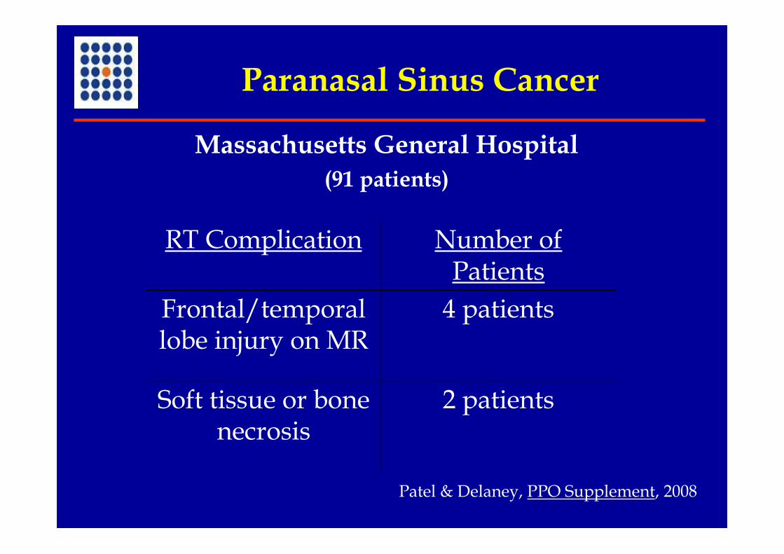
Paranasal Sinus Cancer
Patel & Delaney, PPO Supplement, 2008
Massachusetts General Hospital• 91 patients – carcinoma, 82 patients; sarcoma, 9
patients– Median dose – 73.6 Gy (range, 59.4 and 77.8 Gy)– Median proportion of proton dose – 49% (range,
23% to 84%)– 87% treated with accelerated hyperfractionated RT– 35% received adjuvant chemotherapy– Median follow-up, 45 months

Paranasal Sinus Cancer
Massachusetts General Hospital5-yr Outcomes (91 patients)
Patel & Delaney, PPO Supplement, 2008
58%OS52%DFS75%DMFS86%Ultimate local control82%Local control
PercentageOutcome
Paranasal Sinus Cancer
Massachusetts General Hospital(91 patients)
Patel & Delaney, PPO Supplement, 2008
2 patientsSoft tissue or bonenecrosis
4 patientsFrontal/temporallobe injury on MR
Number ofPatients
RT Complication
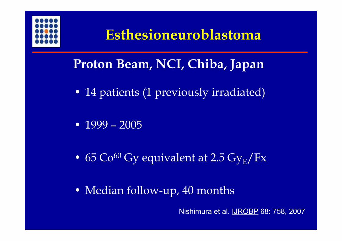
Esthesioneuroblastoma
Proton Beam, NCI, Chiba, Japan
• 14 patients (1 previously irradiated)
• 1999 – 2005
• 65 Co60 Gy equivalent at 2.5 GyE/Fx
• Median follow-up, 40 months
Nishimura et al. IJROBP 68: 758, 2007
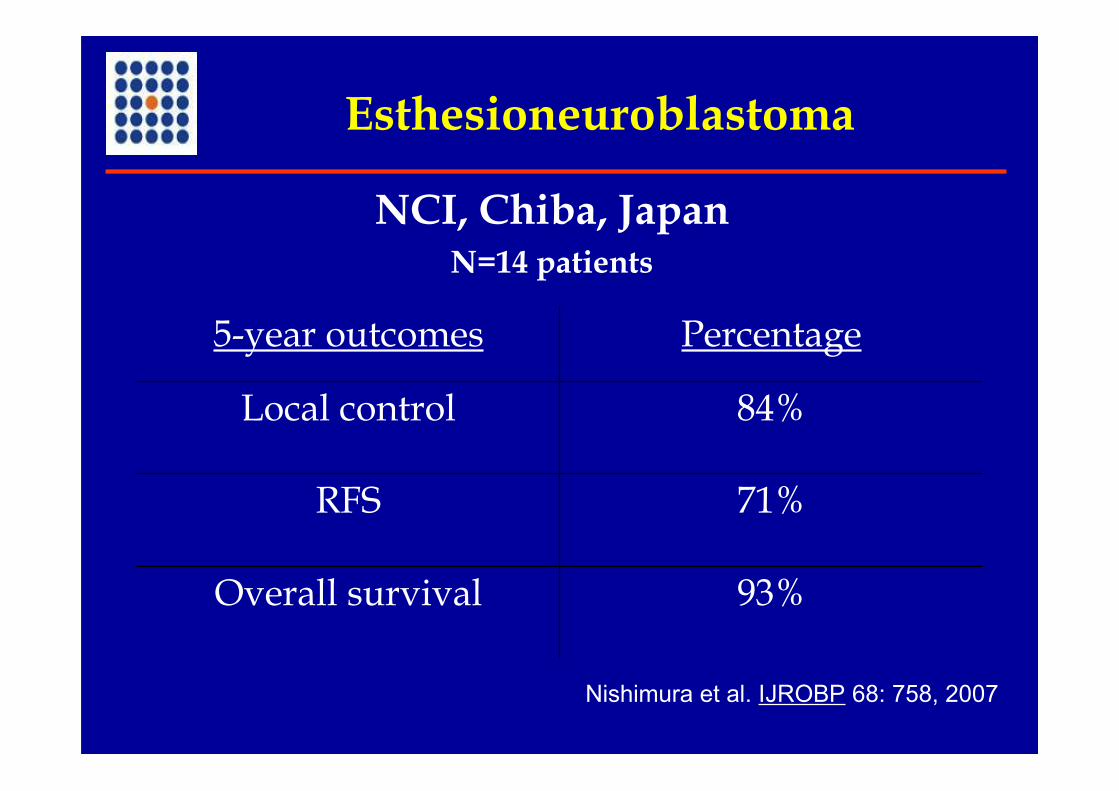
Esthesioneuroblastoma
NCI, Chiba, JapanN=14 patients
Nishimura et al. IJROBP 68: 758, 2007
93%Overall survival
71%RFS
84%Local control
Percentage5-year outcomes
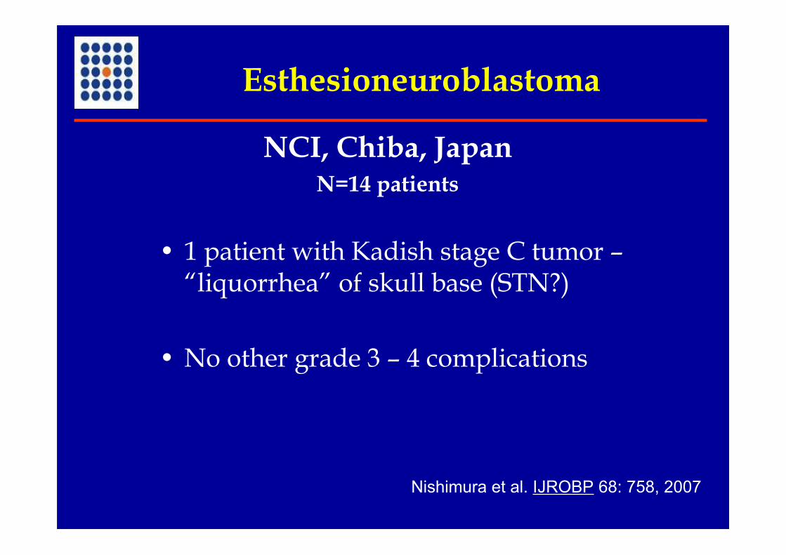
Esthesioneuroblastoma
NCI, Chiba, JapanN=14 patients
Nishimura et al. IJROBP 68: 758, 2007
• 1 patient with Kadish stage C tumor –“liquorrhea” of skull base (STN?)
• No other grade 3 – 4 complications
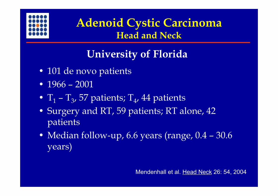
Adenoid Cystic CarcinomaHead and Neck
University of Florida
• 101 de novo patients• 1966 – 2001• T1 – T3, 57 patients; T4, 44 patients• Surgery and RT, 59 patients; RT alone, 42
patients• Median follow-up, 6.6 years (range, 0.4 – 30.6
years)
Mendenhall et al. Head Neck 26: 54, 2004
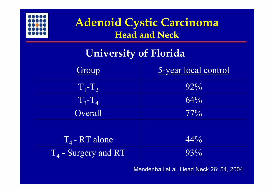
Adenoid Cystic CarcinomaHead and Neck
University of Florida
Mendenhall et al. Head Neck 26: 54, 2004
93%T4 - Surgery and RT44%T4 - RT alone
77%Overall64%T3-T4
92%T1-T2
5-year local controlGroup

Adenoid Cystic CarcinomaHead and Neck
University of Florida (N=110 patients)
Mendenhall et al. Head Neck 26: 54, 2004
1Fatal hemorrhage afterreconstructive surgery for tracheal
stenosis
1Fatal meningitis after salvagesurgery
1Oral antral fistula1Permanent PEG
3ORN requiring surgery
0Bilateral blindness6Ipsilateral blindness
Number of PatientsComplications
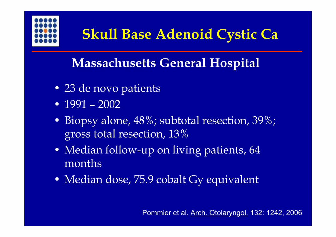
Skull Base Adenoid Cystic Ca
Massachusetts General Hospital
• 23 de novo patients• 1991 – 2002• Biopsy alone, 48%; subtotal resection, 39%;
gross total resection, 13%• Median follow-up on living patients, 64
months• Median dose, 75.9 cobalt Gy equivalent
Pommier et al. Arch. Otolaryngol. 132: 1242, 2006
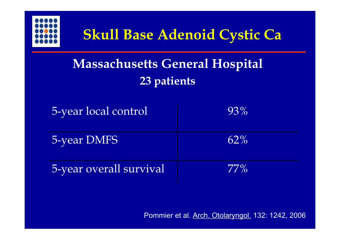
Skull Base Adenoid Cystic Ca
Massachusetts General Hospital23 patients
Pommier et al. Arch. Otolaryngol. 132: 1242, 2006
77%5-year overall survival
62%5-year DMFS
93%5-year local control

Skull Base Adenoid Cystic Ca
Massachusetts General Hospital23 patients
Pommier et al. Arch. Otolaryngol. 132: 1242, 2006
• No grade 5 visual complications; 1 grade 4retinopathy
• 7 chronic seizure disorders controlled with meds
• One fistula with CSF leak and meningitis

Oropharyngeal SCCA
Loma Linda
• 29 patients, stage II – IV
• 1991 – 2002
• 75.9 GyE / 45 FX / 5.5 weeks
• Follow-up, 2 to 90 months
Slater et al. IJROBP 62: 494, 2005
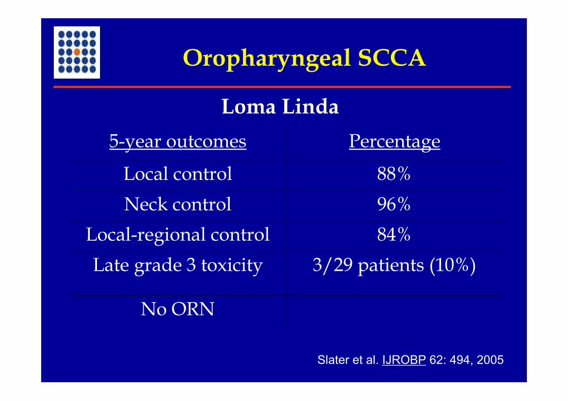
Oropharyngeal SCCA
Loma Linda
Slater et al. IJROBP 62: 494, 2005
No ORN
3/29 patients (10%)Late grade 3 toxicity
84%Local-regional control
96%Neck control
88%Local control
Percentage5-year outcomes

Oropharyngeal SCCA
16%77%82%75%21%333UF – BOT
12%73%79%61%17%503UF –Tonsil
10%84%88%62%21%29LomaLinda
Latecomplications
5-yearlocal
regionalcontrol
5-year local
control% StIV
%T4
No. ofpatien
tsSeries
Slater et al. IJROBP 62: 494, 2005Mendenhall et al. AJCO 29: 32, 2006Mendenhall et al. AJCO 29: 290, 2006

Conclusions
• Protons probably most useful for tumorsinvolving skull base to reduce CNS and visualcomplications and possibly improve localcontrol
• Hyperfractionated to reduce visualcomplications
• May be useful in oropharygeal cancer to reducelate effects, particularly xerostomia – decreaseparotid dose to less than median 26 Gy

Caution
Do not be too conformal!
If you can miss with IMRT,you can miss with protons!