Embed Size (px)
Citation preview
Abstracts Posters
1. Computer aided detection of ureteral stones in thin slice computed tomography volumes using 3D convolutional neural networks
2. Focal MEG: A new window into the brain?
3. Third Eye: An Intelligent Assisting Aid for Visually Impaired Elderly
4. Hybrid Haptic Stimulation for Prosthetic Hands
5. Andningspool - resultat av regionalisering
6. Turbulent wall shear stress assessment using 4D flow MRI
7. Towards automatic analysis of 4D flow MRI: Time-resolved visualization and flow analysis
8. Medicinteknik. Om regelv(ä)rk
9. Framtidens Akademiska, byggprojekt
10. A Piezoelectric Smart Textile Sock for Gait Analysis – A Feasibility Study
11. E-care @ home: Inititial Usability Considerations
Computer aided detection of ureteral stones in thin slice computed tomography volumes using3D convolutional neural networks [email protected] Antai Llaquet Bayo1, Martin Längkvist1, Mats Lidén2, Johan Jendeberg2, Per Thunberg3 andAmy Loutfi1 1Örebro University, Center For Applied Autonomous Sensor Systems, Örebro 2Faculty Of Medicine And Health, Örebro University, Department Of Radiology, Örebro 3Faculty Of Medicine And Health, Örebro University, Department Of Medical Physics, Örebro Background Computed tomography (CT) is the method of choice for diagnosing kidney stones that obstructsthe ureter. The purpose of the present study was to develop a computer aided detection (CAD)algorithm for identifying an obstructing ureteral stone in a thin slice CT volume, takingadvantage of the 3D CT data. The challenge in CAD for urinary stones lies in the similarity inshape and intensity of stones with non-stone structures and how to efficiently deal with largehigh-resolution CT volumes. We address these challenges by developing a 3D Convolutional Neural Network (CNN) thatworks directly on the high resolution CT volumes and present a process for selecting volumes-of-interest (VOI). This work also gives an analysis of the choice of hyperparameters for thealgorithm. Material and Methods The dataset consisted of 600 unenhanced CT scans (460 patients with stone in the ureter, 140without stone). A subset, consisting of 20% randomly extracted CT scans with stone, was usedfor testing and the remaining 80% was used for training. Data augmentation was used to increasethe number of training examples. The VOI were extracted by thresholding and selection of connected components and fed to a 3DCNN and a softmax classifier for the final classification. Results After tuning the hyperparameters of the CNN for optimal performance, the best CNN achieved98.3% accuracy for identifying ureteral stones. The CNN was 20 times faster than traditionalneural networks. The best identified previous work using 2D feature extraction for identifyingkidney stones achieved 88% accuracy. Conclusion A 3D CNN is used for detecting kidney stones from thin slice CT scans. The proposed methoddoes not require any design of features and is faster and more reliable than previous approaches.

Focal MEG: A new window into the brain? [email protected] Justin Schneiderman1, Bushra Riaz1, Christoph Pfeiffer2, Silvia Ruffieux2, Minshu Xie2, MaximChukharkin2, Alexey Kalabukhov2, Mikael Elam1, Daniel Lundqvist3 and Dag Winkler2 1Medtech West And The University Of Gothenburg, Institute Of Neuroscience And Physiology,Göteborg 2Chalmers University Of Technology, Department Of Microtechnology And Nanoscience - MC2,Göteborg 3Karolinska Institute, Natmeg, Stockholm Background Brain disorders cost the EU ~€800 billion in 2010 [1]; this economic burden will increase as thepopulation ages. Neuroimaging is critical for diagnosis, understanding, and treatment of braindisorders. Magnetoencephalography (MEG) is a neuroimaging approach with a uniquecombination of spatial and temporal resolutions [2]. Our approach We develop "Focal MEG" that is based on high critical-temperature superconducting quantuminterference devices (high-Tc SQUIDs). Because our sensors operate at less extremetemperatures than conventional MEG technology, our approach promises improved signal-to-noise ratios and spatial resolution at a lower cost than the state-of-the-art [3]. Results We benchmarked our sensors vs. the state-of-the-art MEG system at NatMEG [4] and observedimprovement in signal magnitudes [5]. Recordings on relatively deep neural sources (25-30 mmdepth) match standard models of brain activity whereas more shallow sources (15-20 mm depth)generate unexpected features. We are constructing a seven-channel system for more refinedstudies of the spatial characteristics of the neuromagnetic field at the head surface. Conclusion The benchmarking results indicate high-Tc SQUID-based MEG can provide more informationabout brain activity that can be used for improved diagnosis, understanding, and treatment ofbrain disorders. As such, the Focal MEG system we aim to construct can lead to improvedhealthcare outcomes while reducing the economic burden of brain disease. We gratefully acknowledge support from the Knut and Alice Wallenberg Foundation,Vetenskapsrådet, and Barncancerfonden. [1] Oleson, J. et al. (2012), Euro. J. of Neurol. 19(1):155-162. [2] Hari R., et al. (2011), NeuroImage 61:386-396. [3] Schneiderman, J., (2014), J. of Neuro. Methods 222:42-46 [4] The Swedish National Facility for Magnetoencephalography at Karolinska Institutet,www.natmeg.se. [5] Xie, M., et al. (2015), IEEE Trans. Appl. Supercon. 25(3):1601905.
●
●
●
●
●
Third Eye: An Intelligent Assisting Aid for Visually Impaired Elderly [email protected] Mobyen Uddin Ahmed1, Shahina Begum1, Birgitta Kerstis2, Nikola Petrovic1 and MariaSandborgh3 1Mälardalen University, School Of Innovation, Design And Engineering, Västerås 2Mälardalen University, School Of Health, Care And Social Welfare Share With Others, Västerås 3Mälardalen University, School Of Health, Care And Social Welfare, Västeås Visually impaired older persons need support in daily activities, e.g. moving around inside thehouse; making and eating food and taking medicine. A system that simulates the environmentbased on both dynamic and static objects, identify obstacles, navigates and translates sensoryinformation in voice would be valuable. Today sensors and camera-based systems are popular asambient-assisted living tools for older adults. However, intelligent assisting aid (IAA) to supportolder individuals with a recently acquired visual impairment is limited. The proposed system‘Third Eye’ focuses on the advanced research and development of an IAA to support olderindividuals with a recently acquired visual impairment. The main goal in this system is toprovide a usable, feasible and cost-effective solution for older persons to support their dailyactivities using intelligent sensor based system. The system consists of the following five phasesto meet several central challenges in developing IAA in such domain.
User-perspective, focuses on user-driven technical development, investigating needs ofpotential users. The study will have a participatory design with focus group interviews oflead users. Sensor-based system, focuses on the identification obstacles based on ultrasounds and/orradio frequencies embedded in white-cane or walker.Camera-based system, focuses on image based information translation into voice embeddedin white-cane or walker or glasses.System of systems, focuses on integration of above systems where knowledge is engineeredand suitable representations are learned and reasoning for decisions are made.Experimental, focuses on usability and feasibility of the IAA, with idiographic and groupstudies.
The initial results have shown the necessity of the proposed AAI systems for older individualswith a recently acquired visual impairment. However, more extension work e.g., process andanalyze the information and synthesize it with existing literature for developing the system isongoing.
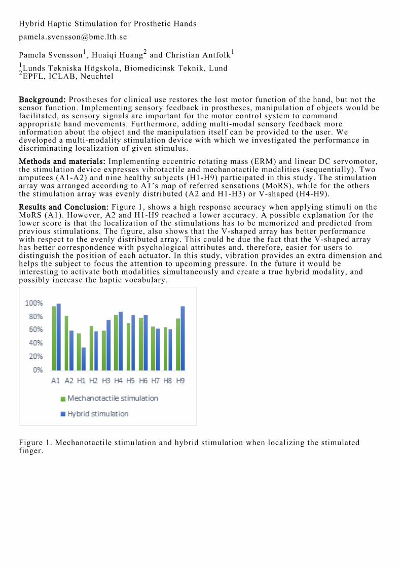
Hybrid Haptic Stimulation for Prosthetic Hands [email protected] Pamela Svensson1, Huaiqi Huang2 and Christian Antfolk1 1Lunds Tekniska Högskola, Biomedicinsk Teknik, Lund 2EPFL, ICLAB, Neuchtel Background: Prostheses for clinical use restores the lost motor function of the hand, but not thesensor function. Implementing sensory feedback in prostheses, manipulation of objects would befacilitated, as sensory signals are important for the motor control system to commandappropriate hand movements. Furthermore, adding multi-modal sensory feedback moreinformation about the object and the manipulation itself can be provided to the user. Wedeveloped a multi-modality stimulation device with which we investigated the performance indiscriminating localization of given stimulus. Methods and materials: Implementing eccentric rotating mass (ERM) and linear DC servomotor,the stimulation device expresses vibrotactile and mechanotactile modalities (sequentially). Twoamputees (A1-A2) and nine healthy subjects (H1-H9) participated in this study. The stimulationarray was arranged according to A1’s map of referred sensations (MoRS), while for the othersthe stimulation array was evenly distributed (A2 and H1-H3) or V-shaped (H4-H9). Results and Conclusion: Figure 1, shows a high response accuracy when applying stimuli on theMoRS (A1). However, A2 and H1-H9 reached a lower accuracy. A possible explanation for thelower score is that the localization of the stimulations has to be memorized and predicted fromprevious stimulations. The figure, also shows that the V-shaped array has better performancewith respect to the evenly distributed array. This could be due the fact that the V-shaped arrayhas better correspondence with psychological attributes and, therefore, easier for users todistinguish the position of each actuator. In this study, vibration provides an extra dimension andhelps the subject to focus the attention to upcoming pressure. In the future it would beinteresting to activate both modalities simultaneously and create a true hybrid modality, andpossibly increase the haptic vocabulary.
Figure 1. Mechanotactile stimulation and hybrid stimulation when localizing the stimulatedfinger.
Andningspool - resultat av regionalisering [email protected] Kennet Larsson1 and Bodil Ivarsson2 1IT/MT Divisionen, Medicinsk Serviceförvaltningen, Region Skåne, IT/MT Service Kryh, Skåne 2Medicinsk Serviceförvaltningen, Region Skåne, Ledningsstaben, Skåne Bakgrund Medicinsk service startade 2014 ett projekt för att bilda en regional andningspool(RA) för medicintekniska andningshjälpmedel såsom portabla oxygenkoncentratorer, syrgas,ventilatorer, hostmaskiner och CPAP med syfte att få enhetligt arbetssätt, apparatsortiment ochekonomisk modell inom Region Skåne. Bärbara syrgasflaskor har bytts mot portablaoxygenkoncentratorer och utlämningsförråd för utrustning har införts. Införandet av RA harinneburit en kraftig besparing för Region Skåne. Syfte Utvärdera bildandet av regional pool för medicintekniska andningshjälpmedel inom RegionSkåne avseende patientsäkerhet, service och spårbarhet. Material och metod En expertvaliderad webbenkät besvarades av 17 sjuksköterskor, 5 läkare, 3fysioterapeuter, 14 medicintekniker från olika verksamheter inom Region Skåne, samtliga mederfarenhet av andningshjälpmedel. Data analyserades med deskriptiv statistik. Resultat Enkäten besvarades av 73,6 % varav 59 % kvinnor och erfarenheten inom yrket var imedeltal, 20, 9 år. Både tillhandahållande av färre fabrikat och modeller av medicintekniskaandningshjälpmedel och övergång från syrgasflaskor till portabla oxygenkoncentratorerupplevdes av merparten (97,4 respektive 100 %) ha ökat patientsäkerheten. Merparten av desvarande upplevde att den totala servicenivån för sjukvårdsförvaltningarna (92,1 %),kontaktvägarna till IT/MT service (81,5 %) samt ekonomisk modell (89,4 %) har förbättrats ochförenklats avseende medicintekniska andningshjälpmedel sedan införandet av regionalandningspool. Däremot upplevde 38,8 % att de inte hade tillräcklig utbildning för att registrerapatientavtal i utrustningsdatabasen för att kunna upprätthålla spårbarheten av medicintekniskaandningshjälpmedel. Slutsats Regional andningspool upplevdes öka patientsäkerheten vilket gagnar bådepatienter/brukare, organisation och ekonomi. Dessutom är det en fördel med likartat arbetssättoch enhetligt apparatsortiment med tanke på upplärning och den allt mer ökandepersonalrörligheten inom både sjukhus- och hemsjukvården. För att kunna koppla sammanpatienter med utrustning och därmed öka spårbarheten behöver utbildningsinsatserna öka, vilketmöjligen kan göras i form av en webbaserad utbildning. Resultatet kan ligga till grund förliknande apparatpooler för annan medicinskteknisk utrustning.

Turbulent wall shear stress assessment using 4D flow MRI [email protected] Magnus Ziegler1, Jonas Lantz1, Tino Ebbers1 and Petter Dyverfeldt1 1Linköpings Universitet, Institutionen För Medicin Och Hälsa, Kardiovaskulär Medicin,Linköping Background Chaotic velocity fluctuations caused by turbulent blood flow create fluctuations in the shearstress acting on the vascular wall. This is referred to as turbulent wall shear stress (tWSS) andmay cause vascular remodelling and increased endothelial cell turnover. The purpose of thiswork was to explore MR-estimated near-wall turbulent kinetic energy (nwTKE) for theassessment of tWSS. Methods Numerical velocity data was obtained using computational fluid dynamics (CFD) for twophysiological flow rates in realistic models of aortic coarctation and aortic stenosis. 4D FlowMRI measurements were simulated at three different spatial resolutions and used to investigatethe estimation of tWSS using nwTKE. tWSS describes the variation of WSS due to fast chaoticvariations in velocity. nwTKE estimation was performed for wall voxels by calculating the meanTKE from voxels inside a predefined range. In order to avoid partial volume effects, any voxeladjacent to the wall was excluded. Linear regression analysis was used to assess agreementbetween the MR-estimated nwTKE and the true nwTKE from CFD, and the relationship betweennwTKE and tWSS. Discussion Linear regression showed a strong correlation (mean R2 = 0.90 +/- 0.01) between MR-estimatednwTKE values and the true nwTKE from CFD. Visual inspection of the estimated nwTKEagainst CFD tWSS shows that the regions of high nwTKE correspond to regions of high tWSS inboth the coarctation and stenosis. Analysis showed strong correlations between nwTKE andtWSS (mean R2 = 0.84+/- 0.04). The possibility to identify regions of elevated tWSS opens newpathways for understanding pathologically driven vascular remodelling, damage to endothelialcells, and plaque rupture.
Figure 1: Volume rendering of TKE in an aortic coarctation with sampling region shown (A).tWSS from CFD (B). Estimated nwTKE (C) and relationship between nwTKE and tWSS (D).

Towards automatic analysis of 4D flow MRI: Time-resolved visualization and flow analysis [email protected] Mariana Bustamante1, Vikas Gupta1, Petter Dyverfeldt1, Carl-Johan Carlhäll1 and Tino Ebbers1 1Linköping University, Division Of Cardiovascular Medicine, 58185 Background: 4D flow MRI is an MRI acquisition technique used to evaluate blood flow in theheart and great thoracic vessels over the cardiac cycle. Analysis of this data is a time-consumingcumbersome process, hampered by the inherent poor contrast between blood and surroundingtissue, as well as by the size and complexity of the data. Using image analysis approaches suchas image registration and atlas-based segmentation, we have developed a technique to improveand automate visualization and assessment of 4D flow MRI acquisitions over the entire cardiaccycle. Materials and Methods: First, a new method for PC-MRA data generation from 4D flow MRI wasdeveloped. It uses registration between the timeframes of the 4D acquisition to create a "4D PC-MR CardioAngiography (4D PC-MRCA)" that retains vascular and cardiac blood flowinformation over the entire cardiac cycle. Second, the generated PC-MRCA was used to performatlas-based segmentation of the great thoracic vessels over time. Subsequently, net flow volumesat different locations in the great thoracic vessels could be automatically obtained (Figure1). The approach was evaluated using data from ten healthy volunteers. Results: Qualitative evaluation showed that 4D PC-MRCA out performed other methods toderive angiographic information form 4D flow MRI, especially when cardiac or vessel motionwas present. High correlation was obtained when comparing automatically obtained flowvolumes that should be closely related, such as those in the ascending aorta and the pulmonaryartery root. Conclusions: The proposed PC-MRCA visualization method represents an improvement overprevious 4D flow MRI analysis techniques, particularly when the type of problem assessed isinfluenced by vessel motion. Additionally, the proposed method permits fully automaticsegmentation and analysis of flow in the great thoracic vessels, and it can be used to produce ageneral view of the function of the cardiovascular system without any user interaction.

Medicinteknik. Om regelv(ä)rk [email protected] Malin Hollmark1, Mia Engman Hyrén2, Jan Heidebrandt2, Anna Lefevre Skjöldebrand2 and PeterLöwendahl3 1Uppsala Universitet, Industriell Teknik, Uppsala 2Swedish Medtech, Swedish Medtech, Stockholm 3Elekta AB, Quality &Regulatory Affairs, Stockholm Medicinteknikfältet är brett och brokigt. Produkter såsom blodtrycksmätare, rullatorer, höftleder,aktiva implantat och röntgenutrustningar kan alla förse vård och patienter med möjligheter tilldiagnostik, monitorering och terapi. Och produkterna ska alla rymmas inom samma regelverk. ISverige sköter bl.a. Läkemedelsverket, Socialstyrelsen och Inspektionen för vård och omsorgtillsyn för att övervaka att lagar och föreskrifter efterlevs. De följer upp och rapporterar olyckoroch tillbud, inspekterar tillverkare, granskar planerade kliniska prövningar, m.m. Trots dettasaknar forskare och utvecklare ofta tillräcklig kännedom om kraven på medicintekniskaprodukter - för att genomföra kliniska studier och för att ta medicintekniska produkter i bruk ivården. Som ett svar till röster som hävdar att det medicintekniska regelverket är svagt kommer underåret den revidering som kommer att bli ännu bättre än dagens redan starka regelverk. Processenför hur en medicinteknisk produkt ska CE-märkas innan försäljning och användning, regleras avlagen om medicintekniska produkter och de föreskrifter som Läkemedelsverket utfärdat. Dettabaseras på EU direktiv och finns tydligt beskrivet på Läkemedelsverkets hemsida. Däremot föranleder olyckor, missbruk och missförstånd en diskussion om att kunskapen behöverstärkas om de medicintekniska regelverken och lagen om etikprövning. Och att det finns delar iutvecklingsprocessen som inte täcks av det medicintekniska direktivet. Vi har forskningsetiskaoch yrkesetiska kodexar, föreskrifter om patientdata, Helsingforsdeklarationen, GCP för kliniskastudier av medicintekniska produkter, etc. Och vi har en medicinteknisk sektor där krav ochutvärderingsmodeller skapade för läkemedelsutveckling inte matchar behoven. Frågor vi här villförsöka besvara, eller åtminstone belysa, är exempelvis vad som måste göras innan CE-märkning. Vad gäller till exempel vid tidiga tester av icke CE-märkta produkter? Hur vet manom en befintlig CE-märkt produkt modifieras på ett sådant sätt att den måste CE-märkas på nytt?Och vad gäller för utveckling och användning/provning av mjukvara?
●
●
●
●
●
●
●
●
●
●
Framtidens Akademiska, byggprojekt [email protected] Anders Fernlöf1 1Akademiska Sjukhuset, Programkontoret För Framtidens Akademiska Sjukhus, FAS, Uppsala Välkommen till Framtidens Akademiska Denna poster riktar sig till dig som vill veta mer om projektet Framtidens Akademiska i Uppsala. På sju år ska vi bygga ett halvt nytt Akademiska Projektet Framtidens Akademiska är ett av de största och mest genomgripande ny- ochombyggnadsprojekt som någonsin har genomförts på Akademiska sjukhuset. Fram till år 2022ska vi bygga 60 000 kvadratmeter och bygga om och uppgradera ytterligare 80 000 kvadratmeter.Det innebär en omfattande förnyelse av ingång 85, ingång 70, operations- och röntgenvåningarnasamt en ny vård- och behandlingsbyggnad. Men framtidens Akademiska sjukhus innehåller dessutom byggandet av ett nytt parkeringshus,ett nytt centralkök och en ny stor miljöstation som gör det möjligt att ta hand om sjukhusetsavfall även i framtiden. Varför bygger vi? Ombyggnationerna beror delvis på ett normalt behov av renovering, eftersom husen är omkring40 år gamla. Det finns även myndighetskrav på ombyggnad, med tanke på bland annatbrandsäkerhet, hygienutrymmen och arbetsmiljö. Efter ombyggnad kommer Akademiskasjukhuset ha moderna lokaler som kan möta vårdens krav, idag och i framtiden. Vad kännetecknar Framtidens Akademiska?
Patientens behov står i centrum.Patientens säkerhet och integritet ska värnas.Patienten ska erbjudas läkande miljöer.Vårdprocesserna ska vara effektiva så att resurserna utnyttjas på bästa möjliga sätt.Sjukhuset och dess olika enheter ska ha god samverkan och logistik.Verksamhetens vision (det ledande universitetssjukhuset som skapar störst värde förpatienterna) och sjukhusets kärnvärden (ödmjukhet, skicklighet, långsiktighet) ska beaktas.Uppgradering och modernisering av befintliga lokaler.Långsiktighet och hållbar planering med minst 40 års perspektiv.Byggnaderna ska vara generella och flexibla för att möta framtidens krav på förändring.Byggnaderna ska vara energisnåla och hållbara.
A Piezoelectric Smart Textile Sock for Gait Analysis – A Feasibility Study [email protected] Leif Sandsjö1, Anna Ragnerius2, Frida Widelund2, Stefan Candefjord3, Karin Rundqvist4, ErikNilsson5 and Nils-Krister Persson6 1Högskolan I Borås, Medtech West/Akademin För Vård, Arbetsliv Och Välfärd, Borås 2Chalmers, Signaler Och System, Göteborg 3Chalmers, Medtech West/Signaler Och System, Göteborg 4Högskolan I Borås, Akademin För Textil, Teknik Och Ekonomi, Borås 5Swerea/IVF, Swerea, Mölndal 6Högksolan I Borås, Akademin För Textil, Teknik Och Ekonomi, Borås Gait analysis focusing on mobility parameters such as cadence, stance and swing time, and jointangles of hip, knee and ankle, has become a common tool in sports and rehabilitation medicine.Traditionally, gait analysis has been confined to the laboratory as it makes use of the combinedinputs from e.g. treadmill, force plates, accelerometers and (3D) video systems. Recentdevelopments in materials science have produced textile based sensors that can be integrated inregular clothing which opens up for ambulatory monitoring outside the lab on a 24-7 basis. Theaim of this study was to investigate the feasibility of extracting gait information from signalsrecorded by a smartphone from a newly developed piezoelectric textile sensor integrated in theheel and toe of a smart textile sock. Signals from the heel and toe of an instrumented sock were recorded from five subjects. Aprotocol for treadmill walking and running at pre-defined and self-chosen speeds was followedusing the three foot-strike types; heel-, mid- and toe-strike, resulting in a database with well-defined walking/running sequences. A software system for gait analysis based on thepiezoelectric textile sensor signals was developed, focusing on the timing of heel and toe contactto the ground and foot-strike type identification. A neural network based classification of eachstep as a heel-, mid-, or toe-strike, was implemented. Apart from presenting basic gait parameters (cadence, stance and swing time) the developedsoftware system identified foot strike patterns with an accuracy of up to 98%. This demonstratesthe feasibility to use the piezoelectric textile sensor for direct measurement of gait relatedparameters. Further development of the sensor is needed to overcome the signal fade off within5–20 minutes, which currently limits the smart textile sock’s general applicability.
E-care@home: Initial Usability Considerations [email protected] Erik Prytz1, Annica Kristoffersson2, Maria Lindén3, Nikola Petrovic3, Mobyen Uddin Ahmed3 and Leili Lind1 1Swedish ICT, SICS East, Linköping 2Örebro University, School Of Science And Technology, Örebro 3Mälardalen University, Embedded Sensor Systems For Health, Västerås Background The E-care@home project aims to develop a smart sensor and communication infrastructure withsemantic interoperability capable of monitoring patients in their home. The system will use awide array of sensors, both medical and environmental, and an advanced computer reasoninglayer capable of interpreting sensor data and deliver natural language summaries of the patienthealth status. One critical issue is that of the usability of such a system to the users, i.e. patientsand care providers. Method The project’s usability work focuses on user-centered design and the overall usability of thefuture system. There are three identified user groups: care providers, elderly patients withchronic obstructive pulmonary disease and co-morbidity, and elderlies in better health but thatare afraid of or at risk for falls. Interviews, observations, and literature reviews will be used in afirst phase to construct personas of prototypical users, goal-oriented user stories outlining howand why the different user groups want to use the E-care@home system, user expectations on thefeatures of the system, as well as other system design constraints and opportunities. Results To date, interviews and observations have been carried out with representatives from the usergroups at three different sites: SICS East/Linköping University, Örebro University, andMälardalen University. Work is ongoing to process and analyze this data and synthesize it withexisting literature. Some examples of important user expectations that have been identified thusfar are simplified and personal communication between care providers and caretakers, increasedfrequency of health data collection, increased security through automatic alarms, and usablesystem interfaces. Future work The work on usability will continue throughout the E-care@home project. The next phaseincludes controlled user tests of prototype technology in test beds and, pending results, potentiallongitudinal studies in real home environments.