Embed Size (px)
Citation preview

ABSTRACT Title of thesis: AN INVESTIGATION OF RESPIRATORY RESISTANCE
DURING RESTING BREATHING IN TEENAGE FEMALE ATHLETES WITH AND WITHOUT PARADOXICAL VOCAL FOLD MOTION DISORDER
Andrea Pham, Master of Arts, 2017 Co-Directors of thesis: Professor Yasmeen Faroqi-Shah Adjunct Professor Nancy Pearl Solomon Department of Hearing and Speech Sciences
Paradoxical vocal fold motion disorder (PVFMD) is a laryngeal disorder that is typically
triggered by vigorous exercise and primarily affects female athletes in their teenage
years. Previous research demonstrated that measures of inspiratory (Ri) and expiratory
(Re) resistance reflect laryngeal constriction associated with PFVMD following exercise,
but that baseline differences between PVFMD and normal function may also exist. This
study explored Ri and Re as measured by an Airflow Perturbation Device (APD) during
resting tidal breathing (RTB) in 16 teenage female athletes with PVFMD and 16 healthy
matched controls; half were 12-15 and half were 16-18 years old. Ri and Re during RTB
did not differ significantly between experimental and control groups nor between
younger and older age groups. These findings failed to replicate previous findings of
baseline differences between groups and across age, although trends in the data suggest
that sampling error may account for the difference in results.

AN INVESTIGATION OF RESPIRATORY RESISTANCE DURING RESTING
BREATHING IN TEENAGE FEMALE ATHLETES WITH AND WITHOUT PARADOXICAL VOCAL FOLD MOTION DISORDER
by
Andrea Pham
Thesis submitted to the Faculty of the Graduate School of the University of Maryland, College Park in partial fulfillment
of the requirements for the degree of Master of Arts in Speech-Language Pathology
2017
Advisory Committee: Associate Professor Yasmeen Faroqi-Shah (Co-Chair)
Adjunct Professor Nancy Pearl Solomon (Co-Chair) Professor Jan Edwards Assistant Professor Sally Gallena Adjunct Professor Jafar Vossoughi

©Copyright by
Andrea Pham
2017

ii
Acknowledgement I would first like to thank my thesis co-advisors Dr. Yasmeen Faroqi-Shah, Associate Professor in Department of Hearing and Speech Sciences at the University of Maryland, and Dr. Nancy Solomon, Research Speech Pathologist in the Audiology and Speech Pathology Center at Walter Reed National Military Medical Center and Adjunct Professor in the Department of Hearing and Speech Sciences at the University of Maryland. The door to Dr. Shah’s and Dr. Solomon’s office was always open whenever I ran into a trouble spot or had a question about my research or writing. They consistently allowed this paper to be my own work, but steered me in the right the direction whenever they thought I needed it. I would also like to thank the expert who provided the retrospective data for participants with paradoxical vocal fold motion disorder in this research project: Assistant Professor Sally Gallena. Additionally, I would also like to thank the expert who provided the instrumental device, the Airflow Perturbation Device, used for this research project: Adjunct Professor Jafar Vossoughi. Without their passionate participation and input, this work could not have been successfully conducted. I would also like to acknowledge Professor Jan Edwards of the Department of Hearing and Speech Sciences at the University of Maryland, Assistant Professor Sally Gallena of the Loyola Clinical Center at Loyola University Maryland, and Adjunct Professor Jafar Vossoughi at the Biomed Research Foundation and the Fischell Department of Bioengineering at the University of Maryland as second readers of this thesis, and I am gratefully indebted to them for their very valuable comments on this thesis. Finally, I must express my very profound gratitude to my friends, family, and to my spouse, John Davenport, for providing me with unfailing support and continuous encouragement throughout my years of study and through the process of researching and writing this thesis. This accomplishment would not have been possible without them. Thank you. Author Andrea Pham

iii
Table of Contents
Acknowledgement………………………………………………………………..………ii Table of Contents ………………………………………………………………..………iii List of Tables……………………………………………………………………..………iv List of Figures……………………………………………………………………..………v Introduction……………………………………………………………………………......1 PVMFD and Populations of Interest……………………………………….….…..3 Diagnosis of PVFMD……………………………………………………………..5
Respiratory Resistance and PVFMD…….………………………………………..9 Summary and Statement of Problem…………………………………………….12 Methods…………………………………………………………………………………..14 Experimental Design……………………………………………………….…….14 Participants………….……………………………………………………………14 Procedure…………………………………………..…………………………….15
Statistical Analysis...……………………………………………………….…….18 Results……………………………………………………………………………………19 Discussion………………………………………………………………………………..21 Limitations and Future Directions………………………….………………........27 Conclusions………………………………………………………………………………28 Appendix A………………………………………………………………………………31 References……………………………………………………………………….………33
iv
List of Tables
Table 1. Results of the Repeated Measures Analysis Comparing Ri and Re Across Trials
for Each Group……………………………………..…………………..……………...…19
Table 2. Descriptive Statistics (M (SD)) for Ri and Re During RTB……………….……20

v
List of Figures
Figure 1. - Schematic of the APD (left) and an Athlete Using the APD (right)………...8
Figure 2. Mean Inspiratory (circles) and Expiratory (squares) Resistances for 12 Athletes
With (black) and 12 Without (grey) PVFMD for Resting Tidal Breathing (RTB), Post-
Exercise Breathing (PEB), and After One Minute of Rest (RB).……………………….11
Figure 3. Results of the Between Group Analysis Comparing Diagnostic Group and Age
Group on Ri in RTB With Error Bars Indicating SD………………...…………………..20
Figure 4. Results of the Between Group Analysis Comparing Diagnostic Group and Age
Group on Ri in RTB With Error Bars Indicating SD…….……………………………..21

AN INVESTIGATION IN RESPIRATORY RESISTANCE
1
Introduction
The respiratory system provides aerodynamic power to support phonation for
speech, but its primary function is respiration. During typical inspiration, the vocal folds
are abducted to allow air to flow into the lungs. During typical expiration, the vocal folds
are also abducted but move in a slightly adductory direction as air flows out from the
lungs. In paradoxical vocal fold motion disorder (PVFMD), also known as vocal cord
dysfunction, the vocal folds partially adduct during inspiration and sometimes remain
partially adducted during expiration, thereby restricting the airway opening and impeding
breathing (Matthers-Schmidt, 2001). This laryngeal disorder can be triggered by
vigorous exercise and primarily affects female athletes in their teenage years. Since
PVFMD affects breathing, an essential requirement for life, it has serious implications,
and is within the scope of practice for diagnosis and treatment by speech-language
pathologists (SLP; Matthers-Schmidt, 2001).
The anatomy of the respiratory system includes the upper respiratory and lower
respiratory tracts as well as the supporting structures of the chest wall. The upper
respiratory tract is composed of the nasal cavities, oral cavity, pharynx, and larynx. The
lower respiratory tract is composed of the trachea, bronchi, and lungs. Resting
inspiration is an active process that involves the diaphragm. When the diaphragm
contracts, lung volume increases. Thus, the air pressure inside the lungs decreases
relative to air pressure outside of the body, and air flows into the lungs allowing air
pressure to equalize. Resting expiration is accomplished passively via gravity, lung
tissue recoil, and rib torque. These passive processes result in decreasing lung volume,
increasing air pressure relative to air outside the lungs, and air flowing out of the lungs.

2
The impedance to airflow in the respiratory system results from resistances offered by the
chest wall, lungs, bronchi, trachea, and larynx.
The inward and outward flow of air is the result of the physical properties of
gases: flow, pressure, and resistance. Aerodynamic evaluation of voice measures airflow
with a pneumotachometer, and air pressure with a pressure transducer. The
pneumotachograph operates on the aerodynamic analog of Ohm’s law to effectively
measure flow by determining the drop in pressure across a known resistance over time
(Miller & Daniloff, 1993). Once flow and pressure are determined, respiratory resistance
can be calculated by dividing pressure (in cm H2O) by flow (in liters per second) at the
airway opening (i.e., mouth). Tools that introduce periodic perturbations to airflow
during breathing can determine resistance by calculating modulations in pressure and
flow during the perturbations. Instruments based on this principle include whole-body
plethysmography, impulse oscillometry, and a custom-designed device called the Airflow
Perturbation Device (APD) (Haque et al., 2013). The APD has been demonstrated to be
feasible and valid for the assessment of inspiratory (Ri), expiratory (Re), and average
respiratory (Rr) resistance during resting tidal breathing (RTB) as well as during a
PVFMD episode (Gallena, Solomon, Johnson, Vossoughi, & Tian, 2015; Gallena,
Solomon, Johnson, Vossoughi, & Tian, 2014; Haque et al., 2013; Johnson et al., 2007).
This study aims to compare RTB in inspiratory and expiratory resistance values
(i.e., Ri and Re) measured by the APD between young teenage girls (i.e., 12-14 years of
age) and older teenage girls (i.e., 16-18 years of age) in good health, who deny
experiencing exercise-related dyspnea. Additionally, this study will investigate if teenage
girls with PVFMD and those without PVFMD differ in Ri and Re during RTB. The

3
following sections of this thesis will initially provide a brief background on PVFMD and
the population of people who are susceptible to this disorder. Secondly, differences will
be explored that may explain why teenage, female athletes are at risk for this disorder.
Subsequently, conflicting evidence on who is at risk for this disorder will be addressed
and discussed to show the need for this research. A critical overview of how this disorder
is diagnosed will then be explored by elucidating other ways to supplement physicians’,
laryngologists’ and SLPs’ diagnosis with the use of the APD. Finally, this review will
highlight existing research that used this tool to assess PVFMD, and show future
extensions on research to measure Ri and Re with this tool in the PVFMD population.
PVFMD and Populations of Interest
PVFMD can be triggered by vigorous exercise (Kayani & Shannon, 1998;
Landwehr, Wood, & Milgrom, 1996; Mather-Schmidt, 2001; Rundell & Spiering, 2003;
Sandage & Zelazny 2004). This type of PVFMD is referred to as exercise-induced
PVFMD, and typically affects athletes (Kayani & Shannon, 1998; Landwehr et al., 1996;
Mather-Schmidt, 2001; Newman & Dubester, 1994; Rundell & Spiering, 2003; Sandage
& Zelazny, 2004; Selner, Staudenmayer, Koepke, Harvey, & Christopher, 1987; Wood &
Milgrom, 1996). One study found that PVFMD occurred in 5% of 370 elite athletes, ages
16-37 years, at one Olympic training facility (Rundell & Spiering, 2003). Another study
found similar evidence: 10% of athletes seen for dyspnea were later diagnosed with
PVFMD (Maturo et al., 2011). The types of activities that athletes with PVFMD engage
include a range of sports: track, cross country, skiing, swimming, volleyball, softball,
football, soccer, Tae Kwon Do, cheerleading, and basketball (Landwehr et al., 1996;
Selner et al., 1987; Wood & Milgrom, 1996). Rundell & Spiering (2003) found the same

4
sport activities among people with PVFMD already listed, but also included bobsledding,
kayaking, badminton, figure skating, and biathlons. Overall, aerobic activities seem to
put athletes at risk for PVFMD.
Studies show that PVFMD affects female athletes more than male athletes
(Kayani & Shannon, 1998; Landwehr et al., 1996; Maturo et al., 2011; Newman &
Dubester, 1994; Sandage & Zelazny, 2004). Furthermore, teenagers, typically 11 to 15
years of age, are diagnosed with PVFMD more than any other age group (Kayani &
Shannon, 1998; Kuppersmith, Rosen, & Wiatrak, 1993; Landwehr et al., 1996; Powell et
al., 2000; Sandage & Zelazny, 2004), although it may occur in older women as well
(Newman & Dubester, 1994; Rundell & Spiering, 2003).
Evidence on the etiology of PVFMD in teenage girls is sparse, but the most
common trigger is exercise. However, several additional triggers for PVFMD exist.
Possible links with upper airway sensitivity to laryngeal irritants, laryngopharyngeal
reflux (LPR), laryngeal dystonia, psychological factors, and neurological abnormalities
have all been associated with PVFMD (Koufman & Block, 2008; Mathers-Schmidt,
2001). Competitive teenage athletes have very busy schedules, often have more than one
activity in a day, and frequently report not eating a meal, eating late at night, or eating
immediately before a competitive activity, which are all risk factors for LPR (Sandage &
Zelazny, 2004). Some studies have found an association with psychological disorders
including sexual abuse, depression, and anxiety (Landwehr et al., 1996; Selner, et al.,
1987), whereas other studies have found a psychological link to PVFMD unlikely
(Christopher et al., 1983; Hayes, Nolan, Brennan, & Fitzgerald, 1993; Newman &
Dubester, 1994). Any conclusions regarding the psychological link should be guarded

5
for a disorder that is functional in nature (Koufman & Block, 2008; Mathers-Schmidt,
2001). Furthermore, the relationship between the changes in the larynx and what
constitutes PVFMD should not be confused with other disorders that cause airway
obstruction, such as laryngomalacia, papillomatosis, vocal fold paralysis, and laryngeal
webbing (Sandage & Zelazny, 2004).
Differences in the larynx between the sexes may explain why girls are more at
risk for PVFMD. Titze (1989) reported significant differences between the male and
female larynx. Specifically, Titze (1989) found that the larynx increases in size by 62%
in boys between the ages of 10 and 16 years and by 34% in girls from 12-16 years.
Additionally, vocal fold length increases to 16 mm on average in males and about 10 mm
on average in females, and the thyroid cartilage is 20% larger in males versus females in
both the angle of the thyroid and from its anterior to posterior dimension between the
specified ages. Thus, females have a smaller larynx and glottal area than males.
Interestingly, the timing of these pubertal, laryngeal changes in girls matches evidence
that PVFMD is most prevalent in girls from 11-15 years of age (Kayni & Shannon, 1998;
Kuppersmith et al., 1993; Maturo et al. 2011). Overall, physiological differences may
interact in a way that puts girls, especially those in their early teens, at risk for PVFMD.
Diagnosis of PVFMD
Differential diagnosis of PVFMD is difficult because it is commonly
misdiagnosed as asthma (Brugman & Simons; Franca, 2014; Koufman & Block, 2008;
Kuppersmith et al., 1993; Newman & Dubester, 1994; Sandage & Zelanzy, 2004). While
similar, these two disorders have distinct diagnostic criteria. Asthma presents with chest
tightness, wheezing (i.e., audible breathing on expiration), is triggered by exercise with a

6
longer onset (i.e., typically 5-10 minutes) and recovery period (i.e., 15-60 minutes to
several weeks), and has a positive response to treatment with bronchodilators (Brugman
& Simons, 1998; Mathers-Schmidt, 2001; Sandage & Zelazny, 2004). Alternatively,
PVFMD presents with throat tightness, stridor (i.e., audible breathing on inspiration), is
triggered by exercise with a short onset (i.e., typically under 5 minutes) and recovery
period (i.e., 5-10 minutes), and has a negative response to treatment with bronchodilators
(Brugman & Simons; Mathers-Schmidt, 2001; Sandage & Zelazny, 2004). Overall, both
asthma and PVFMD affect both phases of respiration, but asthma is characterized
primarily by audible expiration, whereas PVFMD is characterized primarily by audible
inspiration. However, misdiagnosis of PVFMD occurs and leads to inappropriate
treatment with corticosteroids, intubation, and even tracheostomy in rare cases (Newman
& Dubester, 1994). Furthermore, asthma and PVFMD can co-occur, further
complicating matters (Landwehr et al., 1996; Newman & Dubester, 1994). One study
found that an appropriate diagnosis of PVFMD was delayed by an average of four years
because of misdiagnosis and unnecessary treatment (Maturo et al., 2011). Therefore,
proper techniques to diagnose this disorder are essential.
To aid in differential diagnosis, authors have defined PVFMD’s diagnostic
criteria to include inappropriate vocal fold adduction during inspiration, inability to
abduct the vocal folds upon command, and the presence of a posterior glottal chink
during a symptomatic episode (Koufman & Block, 2008; Landwehr et al., 1996; Newman
& Dubester, 1994). Given these laryngeal findings to diagnose PVFMD, this condition is
typically diagnosed by a SLP and/or otolaryngologist with laryngoscopy (Matthers-
Schmidt, 2001). However, laryngoscopic examination at the time of a PVFMD episode
7
is not guaranteed because the occurrence of PVFMD is episodic and evidence of PVFMD
may not be seen. Moreover, this procedure cannot be tolerated by everyone because it
involves insertion of a flexible laryngoscope through the nasal cavity, pharynx, and
positioning its tip just above the vocal folds for examination.
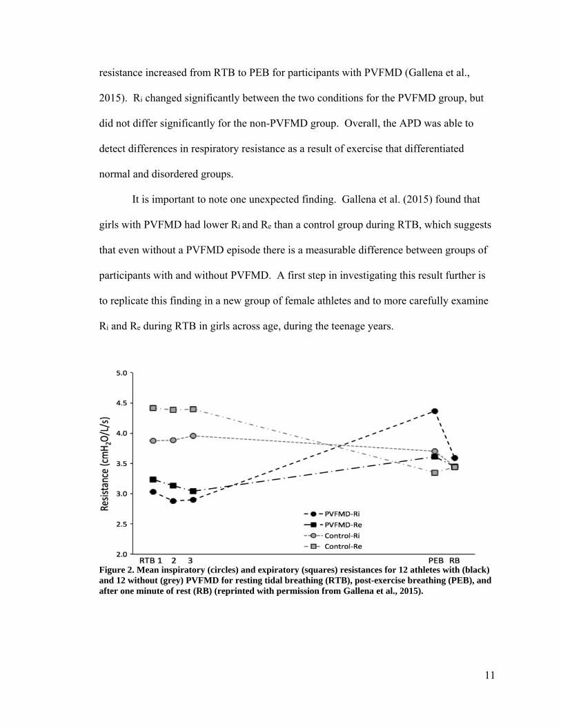
The APD could supplement laryngoscopy and could aid in evaluation of PVFMD.
Lausted and Johnson (1999) invented the APD, a device that measures Ri, Re, and Rr
using an airflow-perturbation technique. During exercise, the airways offer even lower
resistance than at rest because of efficient air exchange. Efficient air exchange involves
increased respiratory rate, greater air volume exchange, and a wider glottis during
breathing (England & Bartlett, 1982). This means that airway resistance is typically
lower during aerobic exercise than during RTB in healthy individuals. However, when a
person with exercise-induced PVFMD is symptomatic, resistance is abnormally elevated
because the glottis narrows, especially during inspiration. The APD device is shown in
Figure 1. In brief, the APD provides a rotating wheel that offers periodic resistance
modulation. While breathing through a pneumotachometer, the periodic perturbations
cause the airflow from the mouth to vary. This affects the air pressure within the airways
and the airflow at the airway opening. Although the APD provides a measure of
resistance across the entire respiratory system, specific changes at the level of the larynx
can be detected. This principle was demonstrated and validated by Gallena, Tian,
Johnson, Vossoughi, Sarles, and Solomon (2013).

8
Figure 1. - Schematic of the APD (left) and an athlete using the APD (right) (Reprinted with permission from Gallena et al., 2013).
Haque et al. (2013) compared two devices that measured Ri and Re: the APD and
whole-body plethysmography. Whole-body plethysmography measures respiratory
resistance indirectly using spirometric indices: forced expiratory volume (FEV) and peak
expiratory flow (PEF). Haque et al. (2013) found that the APD and plethysmography
measures were highly correlated, but the APD was advantageous for two reasons. First,
the APD only requires spontaneous breathing (unlike plethysmography that requires
trained breathing with the support of a respiratory therapist) and secondly, the APD is a
portable device suitable for use outside a healthcare facility (unlike plethysmography
which needs to be performed in a pulmonary-function lab). Overall, Haque et al. (2013)
showed that the APD has concurrent validity with other instruments that measure
respiratory resistance.
Physical characteristics such as age, height, weight, and sex can lead to variations
in respiratory resistance values across individuals. In a sample size of over 900
participants from 2-88 years old, Ri and Re were higher in those who were shorter,
younger, and heavier (Johnson et al., 2007). This means that, on average, children will

9
have higher Ri and Re values than women, and women will have higher values than men.
Additionally, Ri and Re decreased from 12 to 18 years of age with a steeper decrease
beginning at 14 to 15 years of age. This is consistent with research that showed that
teenage girls from 11-15 years of age were typically diagnosed with PVFMD and that
changes in the larynx occur at approximately the same time (Kayani & Shannon, 1998;
Kuppersmith, et al., 1993; Landwehr et al., 1996; Powell et al., 2000; Sandage &
Zelazny, 2004; Titze, 1989). Given the findings from Johnson et al. (2007) on
differences in Rr between young teenage girls and older teenage girls, future research
should examine differences in Ri and Re between young teenage girls and older teenage
girls.
A limitation of the study by Johnson et al. (2007) is that it did not control for
medical history, so it is unknown if the sample contained people with respiratory
disorders such as PVFMD or asthma. Therefore, future studies should control for
respiratory disorders so that results can be generalized to normal or disordered
populations.
Respiratory Resistance and PVFMD
Very little research on the quantitative assessment of PVFMD exists. A series of
investigations by Gallena et al. (2013, 2014, 2015) utilized the APD to determine
respiratory resistance during inspiration and expiration. Gallena et al. (2013) investigated
the construct validity of the APD by demonstrating that measurements from the APD
corresponded to concurrent changes in glottal area (GA). Using simultaneous laryngeal
imaging with laryngoscopy and resistance measurement by the APD with a participant in
a seated position, Gallena et al. (2013) found a strong negative correlation between

10
glottal area and respiratory resistance such that decreased glottal area was associated with
increased respiratory resistance and vice versa.
In a follow-up study, Gallena et al. (2014) investigated the test-retest reliability of
the APD to measure respiratory resistance before exercise (RTB), after exercising (post-
exercise breathing, PEB), and after ~2-min of recovery from exercise (recovery
breathing, RB) while seated. Gallena et al. (2014) included 24 teenage female athletes:
12 without PVFMD and 12 with PVFMD. The study matched participants for sex, age,
weight, and athletic performance. An exercise challenge was introduced as a provocation
activity to induce a PVFMD episode. Additionally, reliability was examined within one
session and across three sessions. This supported data for test-retest reliability of the
APD from Lausted and Johnson (1999) who found that inter-trial variability of 4-7% was
acceptable and non-significant. Results from Gallena et al. (2014) revealed strong test-
retest reliability for Ri and Re during RTB within one session for participants with and
without PVFMD. Due to ethical concerns over delaying treatment for PVFMD,
participants with PVFMD could not be examined across sessions, but only across trials
for one session. The main effect across sessions was not significant for participants
without PVFMD demonstrating that Ri and Re did not change and were reproducible
within the same session (i.e., trials) and across sessions.
Given good construct validity and reliability of the APD to measure Ri and Re in
the PVFMD population, Gallena et al. (2015) compared the effect of exercise on the same
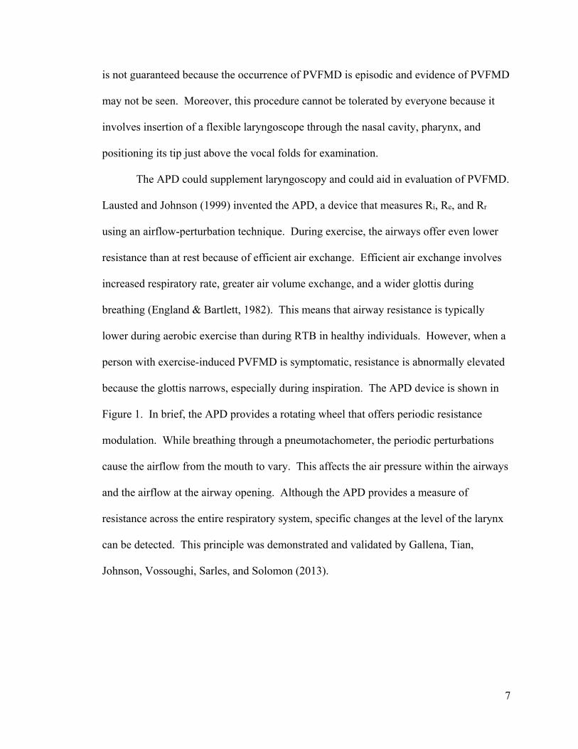
participants with and without PVFMD during RTB and PEB (See Figure 2). Measuring
breathing before and after exercise in a seated position with the APD, respiratory
resistance decreased from RTB to PEB for participants without PVFMD, but respiratory
11
resistance increased from RTB to PEB for participants with PVFMD (Gallena et al.,
2015). Ri changed significantly between the two conditions for the PVFMD group, but
did not differ significantly for the non-PVFMD group. Overall, the APD was able to
detect differences in respiratory resistance as a result of exercise that differentiated
normal and disordered groups.
It is important to note one unexpected finding. Gallena et al. (2015) found that
girls with PVFMD had lower Ri and Re than a control group during RTB, which suggests
that even without a PVFMD episode there is a measurable difference between groups of
participants with and without PVFMD. A first step in investigating this result further is
to replicate this finding in a new group of female athletes and to more carefully examine
Ri and Re during RTB in girls across age, during the teenage years.
Figure 2. Mean inspiratory (circles) and expiratory (squares) resistances for 12 athletes with (black) and 12 without (grey) PVFMD for resting tidal breathing (RTB), post-exercise breathing (PEB), and after one minute of rest (RB) (reprinted with permission from Gallena et al., 2015).

12
Summary and Statement of Problem
The review of literature outlines some key points related to PVFMD. If there is a
constriction that impedes respiration due to adductory motion of the vocal folds, then
flow is reduced and resistance is increased, consistent with a PVFMD episode that makes
it difficult to breathe especially during inspiration. Much of the evidence suggests that
young teenage female athletes are at particular risk for PVFMD, but that the disorder also
occurs in older teenagers (Kayani & Shannon, 1998; Kuppersmith et al., 1993; Landwehr
et al., 1996; Maturo et al., 2011; Newman & Dubester, 1994; Powell et al., 2000; Rundell
& Spiering, 2003; Sandage & Zelazny, 2004). The APD has been shown to be a reliable
tool to measure Ri and Re, and could possibly be used to supplement laryngological
examination for the diagnosis of PVFMD (Gallena et al., 2013, 2014, 2015). Studies
have examined overall respiratory resistance across age as measured by the APD
(Johnson et al., 2007), but not specifically related to changes in laryngeal resistance.
What is currently known about inspiratory and expiratory resistances in people
with PVFMD is based on a group of 12 teenage female athletes (Gallena et al., 2014,
2015). PVFMD can be triggered by exercise, causing laryngeal resistance to increase
(Gallena et al., 2014, 2015; Kayani & Shannon, 1998; Landwehr et al., 1996; Mather-
Schmidt, 2001; Rundell & Spiering, 2003; Sandage & Zelazny, 2004). An unexpected
finding by Gallena et al. (2015) was that athletes with PVFMD demonstrated lower
respiratory resistance values during RTB than teenage girls without PVFMD. This
finding is in stark contrast to higher than normal resistance values in participants during
episodes of PVFMD. Thus, this study aimed to further investigate resting breathing in
13
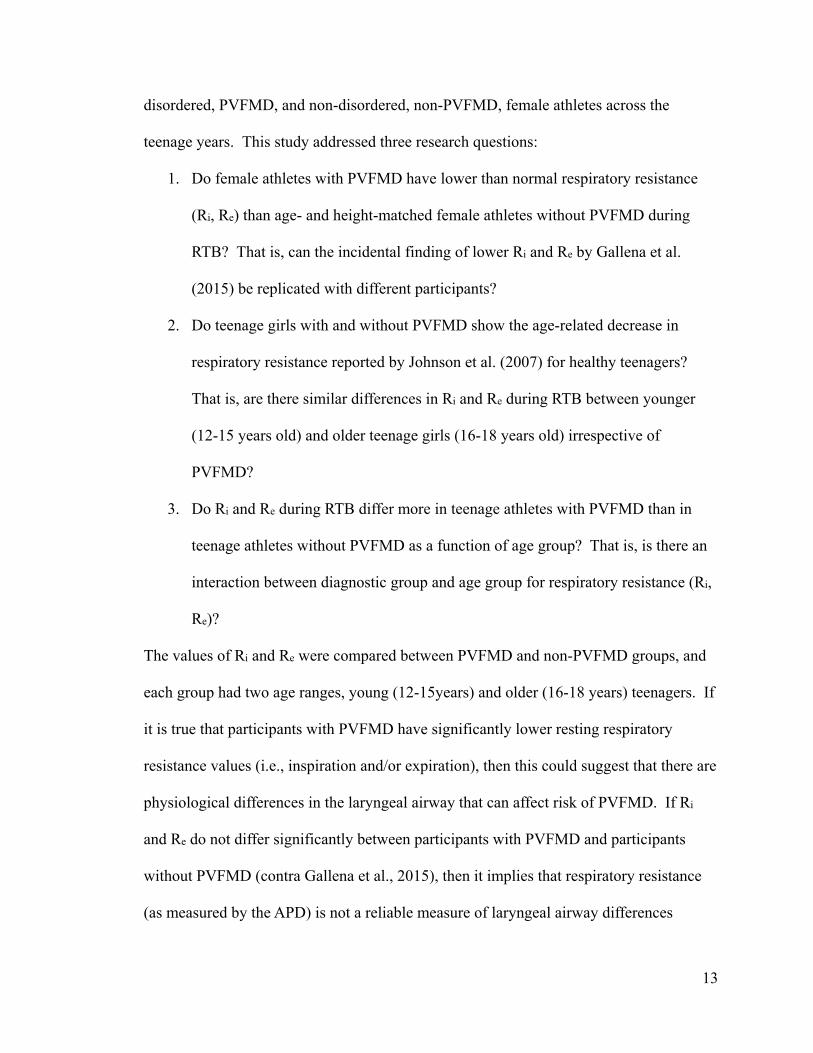
disordered, PVFMD, and non-disordered, non-PVFMD, female athletes across the
teenage years. This study addressed three research questions:
1. Do female athletes with PVFMD have lower than normal respiratory resistance
(Ri, Re) than age- and height-matched female athletes without PVFMD during
RTB? That is, can the incidental finding of lower Ri and Re by Gallena et al.
(2015) be replicated with different participants?
2. Do teenage girls with and without PVFMD show the age-related decrease in
respiratory resistance reported by Johnson et al. (2007) for healthy teenagers?
That is, are there similar differences in Ri and Re during RTB between younger
(12-15 years old) and older teenage girls (16-18 years old) irrespective of
PVFMD?
3. Do Ri and Re during RTB differ more in teenage athletes with PVFMD than in
teenage athletes without PVFMD as a function of age group? That is, is there an
interaction between diagnostic group and age group for respiratory resistance (Ri,
Re)?
The values of Ri and Re were compared between PVFMD and non-PVFMD groups, and
each group had two age ranges, young (12-15years) and older (16-18 years) teenagers. If
it is true that participants with PVFMD have significantly lower resting respiratory
resistance values (i.e., inspiration and/or expiration), then this could suggest that there are
physiological differences in the laryngeal airway that can affect risk of PVFMD. If Ri
and Re do not differ significantly between participants with PVFMD and participants
without PVFMD (contra Gallena et al., 2015), then it implies that respiratory resistance
(as measured by the APD) is not a reliable measure of laryngeal airway differences

14
between individuals with and without PVFMD. Conversely, it implies that individuals
with PVFMD are heterogeneous in their laryngeal airway physiology during RTB.
If Ri and Re differ significantly across age groups, then it shows that respiratory
resistance changes with maturation, as demonstrated by Johnson et al. (2007).
Furthermore, if Ri and Re differ significantly between younger and older teenage girls,
then this indicates consistent maturational changes in respiratory resistance, expanding
the findings of age-related differences in respiratory resistance by Johnson et al. (2007).
If there is an interaction between group and age, then this could indicate the normal
physiologic maturation pattern is not found or is accelerated in PVFMD.
Methods
Experimental Design
This study used a between-groups experimental design. The independent
variables were diagnostic group (PVFMD, non-PVFMD) and age group (12-15, 16-18
years), yielding four experimental groups. The dependent variables were mean Ri and Re
during resting breathing.
Participants
This study included 16 participants with PVFMD and 16 without PVFMD. Half
of each group was between the ages of 12-15 years and the other half was 16-18 years
old. A power analysis, based on data from Gallena et al. (2015), indicated that a sample
size of 8 for Ri and 12 for Re would be adequate to achieve power of 0.80 (two-tailed)
with a Bonferroni corrected α value of 0.025. Inclusionary criteria for both groups
included aerobic activity at least two seasons out of the year and/or an average of aerobic
activity three times per week over the past two months. Exclusionary medical criteria for

15
the healthy controls included past and/or present conditions of asthma, allergies,
respiratory disorders, voice disorders, neurological disorders, or cardiovascular disorders.
Exclusionary criteria for the PVFMD group included a history of a respiratory or
laryngeal disorder other than PVFMD, a neurological disorder, and/or cardiovascular
disease.
Participants without PFVMD were recruited from University of Maryland and the
community (e.g., gyms, youth centers) using flyers, emails, and referrals from clinicians
by the primary investigator (PI). De-identified data of participants with PVFMD
(different from those participants used in the original Gallena et al. (2015) study were
obtained retrospectively from the Loyola Clinical Centers (LCC) in the Speech-
Language-Hearing Sciences Department at Loyola University Maryland because the
facility diagnoses and treats patients with PVFMD.
Healthy (non-PVFMD) participants were matched 1:1 to PVFMD participants for
age (within 6 months) and height (within 7.62 cm). An independent t-test was conducted
to compare height and age in both groups. There was no significant difference in the
height of participants without PVFMD (M = 166.37 cm, SD = 7.16 cm) and participants
with PVFMD (M = 166.62 cm, SD = 6.99 cm), t (16) = -0.13, p = .90. There was no
significant difference in age between participants with PVFMD (M = 15.6, SD=1.57) and
participants without PVFMD (M=15.6, SD= 1.57), t (16) = 0.01, p = .99.
Procedure
Consent and participant questionnaire
Participants with PVFMD had previously consented to the use of their de-
identified data for research purposes, which was obtained from the LCC at Loyola
16
University Maryland as part of standard protocol for PVFMD evaluation. The diagnosis
of PVFMD was established by an otolaryngologist and SLP. Asthma was ruled out by a
pulmonologist and allergies were ruled out or well controlled by an allergist. Informed
consent (and participant assent for those under 18 years of age) was obtained prior to any
testing for the non-PVFMD group.
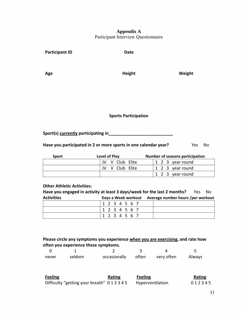
Both groups, with and without PVMFD, filled out a similar questionnaire, which
was based on a questionnaire used by Gallena et al. (2015) (Appendix A) to describe type
and level of aerobic activity and medical history but was amended by the PI to query
caffeine intake, exposure to second-hand smoke, history of playing a wind or brass
instrument, and medical history. Medical history included past and present conditions of
asthma, allergies, respiratory disorders, voice disorders, neurological disorders, or
cardiovascular disorders. Additionally, participants listed the weekly duration and
intensity of physical activity, the level of play (e.g., junior varsity, varsity), and number
of seasons they were involved in a sport.
Data Collection
Testing procedures for the non-PVFMD participants closely followed those used
by Gallena et al. (2014, 2015) for the PVFMD participants obtained retrospectively for
the current study. Testing occurred in the Hearing and Speech Department at University
of Maryland or a mutually agreeable public location that included gyms and recreation
centers before engaging in exercise or more than 30 minutes after exercising. All
screening and procedures were administered in one session of about 30 minutes. The
equipment in the test room included use of the same chair for each participant. The APD
procedure included (see also Figure 1):

17
1. Seating the participant;
2. Placing a disposable nose clip to eliminate nasal breathing;
3. Placing a disposable filtered mouthpiece securely to the lips to ensure
a good seal;
4. Participants holding their cheeks to reduce movement that can distort
resistance values;
5. Placing their tongue below the mouthpiece to avoid airflow obstruction;
6. Breathing into the device as naturally as possible via the mouthpiece for
approximately one minute.
Three acceptable trials that varied by no more than 10% were collected, each trial was
separated by at least 10 seconds, and collected during a single session; breaks were
provided as requested. Participant instructions included: a description of the APD
procedure, an illustration of an athlete correctly using the APD as a visual cue to perform
the task correctly, and an explanation of the number of trials to be collected.
Instrumentation
The APD unit self-calibrated each time it was turned on for use. During the
course of data collection, the device did not pass calibration three times and required
equipment maintenance and replacement by a qualified technician (i.e., the inventor of
the device). As a result, three different APDs were used to collect data. A single device
was used for the PVFMD group (N=16 with APD N25) and, after the repair, for the
majority of the non-PVFMD group (N= 9 with APD N25). The remaining data were
collected with two other APDs (N=1 with APD N29, N=6 with APD N6). To assess
consistency in measurements across instruments, physiological calibrations were
18
conducted on the experimenter’s values of Ri and Re during RTB before each data-
collection session. Results varied by < 10% throughout the duration of the study.
Furthermore, no device was used at any time that did not pass self-calibration. A single
examiner (SG) collected all PVFMD data and a second examiner (AP) collected all non-
PVFMD data after being trained by and deemed reliable with the first examiner.
Each perturbation resulted in a pressure and flow measure used to calculate
respiratory resistance (i.e., quotient of air pressure in cm H2O divided by airflow in liters
per second (L/s)). A trial consisted of approximately 1 minute of breathing or ~500
perturbations of airflow by the wheel on the APD. The mean resistances during the
inspiratory (Ri) and expiratory (Re) phases of breathing were obtained for each participant
per trial from a digital display on the APD. The following measures are displayed on the
APD screen, and were recorded manually by the tester:
• Mean respiratory resistance (Rr) (in cm H2O/L/s)
• Mean inspiratory resistance (Ri) (in cm H2O/L/s)
• Mean expiratory resistance (Re) (in cm H2O/L/s)
Statistical Analysis
Statistical analyses were performed with SPSS software (SPSS, International
Business Machines, version 22, Chicago, Illinois) with a α value for significance set at
.025. A conservative α value was used in order to account for the two dependent
variables of interest, Ri and Re. The data met the requirement of homogeneity of variance
for parametric statistical tests based on Mauchley’s Test of Sphericity or Levene’s Test of
Equality of Error Variances. Therefore, parametric tests were used.
19
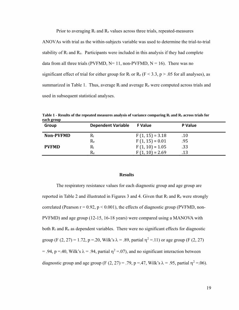
Prior to averaging Ri and Re values across three trials, repeated-measures
ANOVAs with trial as the within-subjects variable was used to determine the trial-to-trial
stability of Ri and Re. Participants were included in this analysis if they had complete
data from all three trials (PVFMD, N= 11, non-PVFMD, N = 16). There was no
significant effect of trial for either group for Ri or Re (F < 3.3, p > .05 for all analyses), as
summarized in Table 1. Thus, average Ri and average Re were computed across trials and
used in subsequent statistical analyses.
Table 1 - Results of the repeated measures analysis of variance comparing Ri and Re across trials for each group
Group Dependent Variable F Value P Value
Non‐PVFMD Ri F(1,15)=3.18 .10 Re F(1,15)=0.01 .95PVFMD Ri F(1,10)=1.05 .33 Re F(1,10)=2.69 .13
Results
The respiratory resistance values for each diagnostic group and age group are
reported in Table 2 and illustrated in Figures 3 and 4. Given that Ri and Re were strongly
correlated (Pearson r = 0.92, p < 0.001), the effects of diagnostic group (PVFMD, non-
PVFMD) and age group (12-15, 16-18 years) were compared using a MANOVA with
both Ri and Re as dependent variables. There were no significant effects for diagnostic
group (F (2, 27) = 1.72, p =.20, Wilk’s = .89, partial 2 =.11) or age group (F (2, 27)
= .94, p =.40, Wilk’s = .94, partial 2 =.07), and no significant interaction between
diagnostic group and age group (F (2, 27) = .79, p =.47, Wilk’s = .95, partial 2 =.06).

20
Table 2- Descriptive Statistics (M (SD)) for Ri and Re during RTB
Group Age (years) Ri (SD) Re (SD)
Non‐PVFMD(N=16)
12‐15(N=8) 4.02 (1.09) 4.42 (1.34)
16‐18(N=8) 3.50 (0.99) 4.08 (1.09) PVFMD(N=16)
12‐15(N=8) 3.31 (0.75) 3.74 (1.32)
16‐18(N=8) 3.05 (0.54) 3.34 (0.65)
Figure 3 - Results of the between group analysis comparing diagnostic group and age group on Ri in RTB with error bars indicating SD.
0
1
2
3
4
5
6
Non‐PVFMD PVFMD
Ri(incmH
2O/L/s)
InspiratoryResistance
younger
older

21
Figure 4 - Results of the between group analysis comparing diagnostic group and age group on Re in RTB with error bars indicating SD.
Individuals were matched closely across groups for age; therefore, optimizer
statistics were conducted using one ANOVA with a covariate of age for Ri and a second
ANOVA with a covariate of age for Re. There was no significant effect of diagnostic
group, but the difference in Ri and Re values between non-PVFMD and PVFMD groups
trended toward significance (Ri: F (1,29) = 4.12 p =.052; Re: F (1,29) = 3.67, p =.065).
Discussion
The present study examined respiratory resistance in female teenage athletes with
PVFMD compared to individuals without PVMFD by measuring Ri and Re during RTB
using an APD. The purpose of the study was to determine if measures of Ri and Re could
differentiate participants with PVFMD from healthy controls matched for sex, age, and
height. The findings indicated that, although there are differences in Ri and Re on average
between the two groups in the expected direction, these differences did not meet the
criterion for statistical significance. Therefore, these results do not support
0
1
2
3
4
5
6
Non‐PVFMD PVFMD
Re(incmH
2O/L/s)
ExpiratoryResistance
younger
older

22
differentiating the two diagnostic groups based on resting breathing measures alone. The
second goal of this study was to determine if respiratory resistance in athletes differed
between younger and older teenage girls. Although there were differences in Ri and Re
on average between the two age groups such that Ri and Re values decrease with age, the
differences were not statistically significant. Based on these findings, it is unclear if there
are maturational differences in teenage years that affect resting respiratory resistance.
Additionally, there were no interactions between diagnostic group and age group. These
findings will be discussed in the following sections.
The first research question was designed to replicate the findings of Gallena et al.
(2015) that Ri and Re were lower during RTB in female teenage athletes with PVFMD
than in those without PVFMD. A statistically significant difference between groups
would have supported the hypothesis of physiologically different breathing patterns in
participants with PVFMD, prior to changes that occur during physical exertion. In this
new group of teenagers, Ri and Re values were generally lower in the group of
participants with PVFMD than those without PVFMD, but the differences did not meet
criterion for statistical significance due to the large within-group variability (error bars in
Figures 3 and 4). When the analysis was repeated using age as a covariate and
comparing the data between diagnostic groups, there was a trend toward significance for
diagnostic group compared to the MANOVA with two between-subjects factors and
without covariates, but was still not significant, when the α was Bonferroni-adjusted for
multiple comparisons.
Gallena et al. (2015) proposed that differences in RTB between PVFMD and
healthy groups found previously might be the outcome of neural adaptation to breathing

23
patterns as a result of PVFMD. That is, respiration is regulated by central pattern
generators that involve a feed-forward mechanism and chemoreceptor feedback system
that regulates, senses, and adjusts respiration based on the level of carbon dioxide (CO2)
and other factors in a complex system to maintain homeostasis (Mitchell & Babb, 2006).
This shows that sensory input can drive motor aspects of respiration, due to the cyclic
relationship between the feed-forward and chemoreceptor feedback system. Mitchell and
Babb (2006) proposed that this system is affected by exercise for within-trial alterations
(i.e., one exercise session) called modulation and across-trial alterations (i.e., multiple
exercise sessions) called plasticity that results in motor learning that affects all aspects of
respiration (i.e., RTB, PEB, RB). Moreover, Mitchell and Babb (2006) suggested neural
adaptation can result in short-term or long-term changes in respiration and be influenced
by physiological conditions associated with impaired pulmonary mechanics (for example
PVFMD). Although Ri and Re during RTB was not statistically significantly lower for
the PVFMD than the non-PVFMD participants in the current study, some of these factors
may have contributed to the trend in these data in the expected direction.
This study’s findings that Ri and Re in RTB were not significantly different
between participants with and without PVFMD may also be explained by gender
differences in neural adaptation, given that females have decreased neural adaptation with
exercise during hypoxic states (Mitchell & Babb, 2006). Furthermore, hypoxia is
affected by the terrain and altitude in which athletes perform (Czuba et al., 2011). Since
neural adaptation affects all aspects of respiration (i.e., RTB, PEB, RB), it is possible that
this study included female participants that exercise in environments that cause hypoxia,
thus affecting neural adaptation and RTB. The study by Gallena et al. (2015) and this

24
study recruited participants from the same geographical area. Thus, the role of hypoxia
may be minimal given that athletes perform generally in the same exercise terrain and
altitude. The types of athletic activities that athletes with PVFMD engaged in included a
range of sports involving aerobic activity (Landwehr et al., 1996; Rundell & Spiering,
2003; Selner et al., 1987; Wood & Milgrom, 1996). Gallena et al. (2015) and the current
study included participants that engaged in a range of sports including: track, cross-
country, swimming, volleyball, field hockey, and soccer. The variety of sports increases
the likelihood of different terrain and exercise conditions. However, participants were
not matched on specific sports and the questionnaire did not include a question about the
exercise environments, so these issues cannot be explored.
The second research question was designed to replicate the findings of Johnson et
al. (2007) that Ri and Re during RTB decrease with age. If Ri and Re differed significantly
between younger and older teenage girls, then this would have further expanded Johnson
et al.’s (2007) findings that children, teenagers, and adults represent different values of
respiratory resistance. Although there was an overall decreasing trend with age (Figures
3 & 4), Ri and Re during RTB did not differ significantly between the two age groups
irrespective of PVFMD, indicating that these measures should be considered similar for
teenage girls as a single group.
Gallena et al. (2015) matched participants for sex, age, height, weight, and
athletic performance. This study matched for sex, age, height, and athletic performance,
but participants were not matched for weight. Johnson et al. (2007) found that weight
was a significant factor in determining Rr, which is an average of Ri and Re. Given the
increased significance in the statistical analyses with the covariate of age, results may

25
have reached statistical significance for group if participants were matched on age,
height, and weight.
The third research question was to determine if age group interacted with
diagnostic group when examining data for Ri and Re during RTB. There was a trend of
decreasing respiratory resistance values with increasing age in the absence of PVFMD.
However, this trend was not statistically significant, as shown by the lack of a significant
interaction between age group and PVFMD diagnosis. These findings indicated that
participants were similar enough that it may make it difficult to explore the compound
effect of diagnostic group and age group on Ri and Re in RTB.
Since there was no significant difference between groups and no interaction
between disorder group and age group, these findings fail to provide physiological
evidence for the increased susceptibility of PVFMD in teenage girls, based upon RTB
alone (Kayani & Shannon, 1998; Landwehr et al., 1996; Maturo et al., 2011; Newman &
Dubester, 1994; Sandage & Zelazny, 2004). An alternative explanation, as described
previously, involves neural adaptation that could vary between males and females for a
variety of factors (Mitchell & Babb, 2006). It should also be noted that neural adaption is
more robust with increased exercise (i.e., neural adaptation was not noted for one
exercise session, but was for repeated exercise sessions) (Mitchell & Babb, 2006). This
might explain why PVFMD affects elite athletes who engage in vigorous exercise
(Kayani & Shannon, 1998; Landwehr et al., 1996; Mather-Schmidt, 2001; Newman &
Dubester, 1994; Rundell & Spiering, 2003; Sandage & Zelazny 2004; Selner et al., 1987;
Wood & Milgrom, 1996).

26
An interesting finding by Mitchell and Babb (2006) regards the relationship
between respiration, neural adaptation, and serotonin. The relationship between
respiration and puberty could explain why young teenage girls, typically 11 to 15 years of
age, are diagnosed with PVFMD more than any other age group, and match laryngeal
changes that occur in puberty between the ages of 12-16 years of age in girls (Kayani &
Shannon, 1998; Kuppersmith et al., 1993; Landwehr et al., 1996; Powell et al., 2000;
Sandage & Zelazny, 2004; Titze, 1989). The onset and changes that occur during
puberty are derived from a complex interplay between neuropeptides, neurotransmitters
(e.g., serotonin), and neurosteroids observed in animal and human models (Genazzani,
Bernardi, Monteleone, Luisi, & Luisi, 2000). Furthermore, serotonin is linked with the
reproductive cycle and hormonal events (Genazzani et. al., 2000). Given the relationship
between respiration and puberty that serotonin seems to play a role in mediating, this
could explain the diagnosis of young teenage girls with PVFMD at the same time that
pubertal changes are co-occurring.
To summarize, this study failed to find significant differences in RTB between
diagnostic groups (PVFMD, non-PVFMD) or age groups (11-15, 16-18). The possible
reasons for this are large within-group variability in RTB values, not controlling for
certain confounds (i.e., weight, sport, exercise environment), minor age differences
between younger and older groups, tendency of smaller neural adaptation effects in
females, and use of different APDs across participants. Therefore, it may be that Ri and
Re in RTB differ between diagnostic groups and across age groups, but that it was
difficult to find these differences based on this sample of participants and/or the methods
used.

27
Limitations and Future Directions
The sample of participants in the study by Gallena et al. (2015) and this study
were different. It is difficult to state which sample group is more representative of the
entire population of patients with PVFMD and, therefore, which results have more
external validity in representation of true Ri and Re during RTB. Although the present
sample size was larger than Gallena et al.’s, it is possible that it was not large enough.
However, based on data from Gallena et al. (2015), a sample size of 8 for Ri and 12 for Re
should have been adequate to achieve power of 0.80 (two-tailed) with a Bonferroni
corrected α value of .025.
Future studies should match for sex, age, height, weight, and sport to control for
confounding factors based upon earlier research (Czuba et al., 2011; Gallena et al., 2015;
Johnson et al., 2007; Mitchell & Babb, 2006). Additionally, future studies should include
a questionnaire on puberty, exercise terrain, and geographical area of sports activity.
Given the results of some studies (Genazzani et. al., 2000; Mitchell and Babb, 2006),
understanding the relationship between puberty, serotonin, and respiration may be a
valuable comparison. Therefore, future studies should compare pre-pubescent and post-
pubescent teenage girls.
Johnson et al. (2007) examined age across the lifespan to include the teenage
years for Rr by year (e.g., 12-year-olds versus 13-year-olds). Therefore, age with two
levels, 12-15 years of age and 16-18 years of age, may represent an arbitrary group that
does not accurately reflect the difference in RTB for Ri and Re. Additionally, teenage
girls diagnosed with PVFMD are typically between 11-15 years of age (Kayani &
Shannon, 1998; Kuppersmith, et al., 1993; Landwehr et al., 1996; Powell et al., 2000;

28
Sandage & Zelazny, 2004; Titze, 1989). Therefore, 11-year-old girls should be included
in the future.
Some studies showed that males and adults are diagnosed with PVFMD as well
(Gurevich-Uvena et al., 2010; Newman & Dubester, 1994; Powell et al., 2000; Rundell &
Spiering, 2003; Sandage & Zelazny, 2004). Since people with PVFMD may represent a
heterogeneous group, comparing sex (i.e., males versus females) and age across the
lifespan (i.e., teenagers versus adults) could yield useful information on Ri and Re in RTB
that is more representative of the entire group of people diagnosed with PVFMD.
In the present study, procedures matched those used by Gallena et al. (2015) to control
for different methods that could affect Ri and Re values. While the examiners and testing
location differed for the PVFMD and non-PVFMD participants, measures were taken to
ensure consistency across testing conditions. Although testing included the use of
different instrumentation (i.e., three different APDs were used), physiological
calibrations were conducted, and the reliability and validity of the devices are assumed to
have low instrumentation error and to provide comparable data.
Conclusions
Overall, the findings of this study do not support the hypothesis that measures of
Ri and Re during RTB differentiate individuals with PVFMD from healthy individuals
with the use of the small, portable APD. A small body of literature suggested that the
APD is a useful non-invasive instrumental measure in a diagnostic protocol including an
exercise provocation (Gallena et al. 2015). The current diagnostic protocol includes
laryngoscopy to diagnose this disorder, but this procedure may not provide a conclusive
29
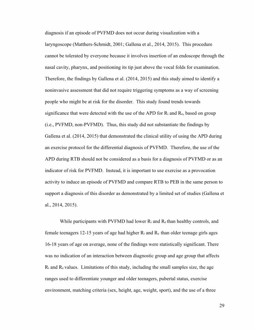
diagnosis if an episode of PVFMD does not occur during visualization with a
laryngoscope (Matthers-Schmidt, 2001; Gallena et al., 2014, 2015). This procedure
cannot be tolerated by everyone because it involves insertion of an endoscope through the
nasal cavity, pharynx, and positioning its tip just above the vocal folds for examination.
Therefore, the findings by Gallena et al. (2014, 2015) and this study aimed to identify a
noninvasive assessment that did not require triggering symptoms as a way of screening
people who might be at risk for the disorder. This study found trends towards
significance that were detected with the use of the APD for Ri and Re, based on group
(i.e., PVFMD, non-PVFMD). Thus, this study did not substantiate the findings by
Gallena et al. (2014, 2015) that demonstrated the clinical utility of using the APD during
an exercise protocol for the differential diagnosis of PVFMD. Therefore, the use of the
APD during RTB should not be considered as a basis for a diagnosis of PVFMD or as an
indicator of risk for PVFMD. Instead, it is important to use exercise as a provocation
activity to induce an episode of PVFMD and compare RTB to PEB in the same person to
support a diagnosis of this disorder as demonstrated by a limited set of studies (Gallena et
al., 2014, 2015).
While participants with PVFMD had lower Ri and Re than healthy controls, and
female teenagers 12-15 years of age had higher Ri and Re than older teenage girls ages
16-18 years of age on average, none of the findings were statistically significant. There
was no indication of an interaction between diagnostic group and age group that affects
Ri and Re values. Limitations of this study, including the small samples size, the age
ranges used to differentiate younger and older teenagers, pubertal status, exercise
environment, matching criteria (sex, height, age, weight, sport), and the use of a three

30
different APDs to collect data could have affected the findings. Further research is
needed to determine if Ri and Re differ based on sex (males versus females) and age
(teenagers versus adults). Further study of these measures is necessary to evaluate their
clinical utility in the differential diagnosis of PVFMD with the APD.
31
Appendix A Participant Interview Questionnaire
Participant ID Date
Age Height Weight
Sports Participation
Sport(s) currently participating in___________________________ Have you participated in 2 or more sports in one calendar year? Yes No
Sport Level of Play Number of seasons participation
JV V Club Elite 1 2 3 year round
JV V Club Elite 1 2 3 year round
1 2 3 year round
Other Athletic Activities: Have you engaged in activity at least 3 days/week for the last 2 months? Yes No Activities Days a Week workout Average number hours /per workout
1 2 3 4 5 6 7
1 2 3 4 5 6 7
1 2 3 4 5 6 7
Please circle any symptoms you experience when you are exercising, and rate how often you experience these symptoms. 0 never
1 seldom
2 occasionally
3 often
4 very often
5 Always
Feeling Rating Feeling Rating Difficulty “getting your breath” 0 1 2 3 4 5 Hyperventilation 0 1 2 3 4 5

32
Feeling of throat closing 0 1 2 3 4 5
Chest tightness 0 1 2 3 4 5
Making a noise in your throat 0 1 2 3 4 5
Noise in your chest 0 1 2 3 4 5
Other feelings
Medical/Psychological History
Please circle past or current conditions that have been diagnosed by a medical doctor:
Asthma ‐If yes, do you use an inhaler? Y N
Allergies
Voice/Respiratory/Cardiovascular/Neurological Disorder: _____________________________
Other Medical (Please specify): _____________________________
Rate your health today (please circle)
Fine OK Not feeling well Sick (e.g., cold) Signs/Symptoms (please describe):
Are you exposed to second hand smoke? Yes No Do you drink caffeinated beverages? Yes No If so, how often _______________/ how many cups per day? _____________________ Do you play a wind or brass instrument (if yes, please specify)? Yes No ________________________________________________________________________

33
References
Brugman, S. M., & Simons, S. M. (1998). Vocal cord dysfunction: Don’t mistake it for asthma. The Physician and Sports Medicine, 26(5), 63–74.
Christopher, K., Wood, R., Eckert, C., Blager, F., Raney, R., & Souhrada, J. (1983). Vocal-cord dysfunction presenting as asthma. The New England Journal of Medicine, 308(26), 1566-1570.
Czuba, M., Waskiewicz, Z., Zajac, A., Poprzecki, S., Cholewa, J., & Rocznickok, R. (2011). The effects of intermittent hypoxic training on aerobic capacity and endurance in cyclists. Journal of Sports Science and Medicine, 10, 175-183.
England, S. & Bartlett, D. (1982). Changes in respiratory movements of the human vocal cords during hyperpnea. Journal of Applied Physiology, 52(4), 780-785.
Franca, M. (2014). Differential diagnosis in paradoxical vocal fold movement (PVFM): An interdisciplinary task. International Journal of Pediatric Otorhinolaryngology, 78, 2169-2173.
Gallena, S., Solomon, N., Johnson, A., Vossoughi, J., & Tian, W. (2015). The effects of exercise on respiratory resistance in athletes with and without paradoxical vocal fold motion disorder. American Journal of Speech-Language Pathology, 24, 470-479.
Gallena, S., Solomon, N.P., Johnson, A., Vossoughi, J., & Tian, W. (2014). Test-retest reliability of respiratory resistance measured with an airflow perturbation device. Journal of Speech, Language, and Hearing Research, 57, 1323-1329.
Gallena, S., Tian, W., Johnson, A., Vossoughi, J., Sarles, S. & Solomon, N. (2013). Validitiy of a new respiratory resistance measurement device to detect glottal area change. Journal of Voice, 27(3), 299-304.
Genazzani, A., Bernardi, F., Monteleone, P., Luisi, S., & Luisi, M. (2000). Neuropeptides, neurotransmitters, neurosteroids, and the onset of puberty. Annuals of the New York Academy of Sciences, 900(1), 1-9.
Gurevich-Uvena, J., Parker, J., Fitzpatrick, T., Makashay, M., Perello, M., Blair, E., & Solomon, N. (2010) Medical comorbidities for paradoxical vocal fold motion (vocal cord dysfunction) in the military population. Journal of Voice, (24)6, 728-731.
Haque, T., Vossoughi, J., Johnson, A., Farrell, W., Fitzgerald, T., & Scharf, S. (2013). Resistance measured by airflow perturbation compared with standard pulmonary function measures. Open Journal of Respiratory Diseases, 3, 63-67.
Hayes, J., Nolan, M., Brennan, N., & Fitzgerald, M. (1993). Three cases of paradoxical vocal cord adduction followed up over a 10 year period. Chest, 104(3), 678-680.
34
Johnson, A., Scott, W., Russek-Cohen, E., Koh, F., Silverman, N. & Coyne, K. (2007). Resistance values obtained with the airflow perturbation device. International Journal of Medical Implants and Devices, 2(2), 45-58.
Kayani, S. & Shannon, D. (1998). Vocal cord dysfunction associated with exercise in adolescent girls. Chest, 113(2), 140-141.
Koufman J. & Block C. (2008). Differential diagnosis of paradoxical vocal fold movement. American Journal of Speech-Language Pathology, 17, 327-334.
Kuppersmith, R., Rosen, D. & Wiatrak, B. (1993). Functional stridor in athletes. Journal of Adoloscent Health, 14, 166-171.
Landwehr, L, Wood, R., Blager, F. & Milgrom, H. (1996). Vocal cord dysfunction mimicking exercise-induced bronchospasm in adolescents. Pediatrics, 98, 971-974.
Lausted C. & Johnson, A. (1999). Respiratory resistance measured by an airflow perturbation device. Physiological Measures, 20, 21-35.
Matthers-Schmidt, B. (2001). Paradoxical vocal fold motion: A tutorial on a complex disorder and the speech language pathologist's role. American Journal of Speech-Language Pathology,10(2), 111-126.
Maturo, S., Hill, C., Bunting, M., Baliff, C., Ramakrishna, J., Scirica, C., Fracchia, S., Donovan, A., & Hartnick, C. (2011). Pediatric paradoxical vocal fold motion: Presentation and natural history. Pediatrics, 128(6), 1443-1449.
Miller, C., & Daniloff, R. (1993). Airflow measurement: theory and utility of findings. Journal of Voice, 7(1), 38-46.
Mitchell, G., & Babb, T. (2006). Layers of exercise hyperpnea: Modulation and plasticity. Respiratory Physiology & Neurobiology, 151, 251-256.
Newman, K. & Dubester, S. (1994). Vocal cord dysfunction: Masquerader of asthma. Seminars in Respiratory and Critical Care Medicine, 15(2), 161-167.
Powell, D., Karanfilov, B., Beechler, K., Treole, K., Trudeau, M., & Forrest, A. (2000). Paradoxical vocal cord dysfunction in juveniles. Arch Otolaryngol Head Neck Surgery,126, 29-34.
Rundell, K., & Spiering, B. (2003). Respiratory stridor in elite athletes. Chest, 123(2), 468-474.
Sandage, M. & Zelazny, S. (2004). Paradoxical vocal fold motion in children and adolescents. Language, Speech, and Hearing Services in Schools, 35, 353-362.

35
Selner, J., Staudenmayer, H., Koepke, J., Harvey, R., & Christopher, K. (1987). Vocal cord dysfunction: The importance of psychogenic factors and provocation challenge testing. The Journal of Allergy and Clinical Immunology, 79(5), 726-733.
Titze, I. (1989). Physiological and acoustic differences between male and female voices. Journal of Acoustical Society of America, 85(4), 1699-1707.
Wood, R., & Milgrom, H. (1996). Vocal cord dysfunction mimicking exercise-induced bronchospasm in adolescents. Pediatrics. 98 (5), 971-974.