Embed Size (px)
Citation preview

ABSTRACT BOOK
Black Sea Ophthalmological Society - BSOS
Tbilisi International Ophthalmology Conference - TIOC 2020
JOINT ONLINE MEETING
December 19 - 20Tbilisi, Georgia
2020


3
ROLE OF AHMED GLAUCOMA VALVE IN REFRACTORY GLAUCOMA SURGERYVytautas Jasinskas, Lithuanian University Health Sciences
Implantation of Ahmed glaucoma valve is an effective surgical technique to reduce intraocular pressure in glaucoma patients. Earlier this device was used in refractory glaucoma cases after previous surgical proce-dures have failed. Nowadays, collected data and mutual experience suggest a broader appliance of this device. It could not only be beneficial for refractory glaucoma but also as a primary surgery in selected cases. Implantation of Ahmed glaucoma valve can be challenging for the surgeon, especially in patients who already underwent previous multiple surgeries. Several specific skills have to be acquired by the sur-geon. Consequently implantation success reveals a steep learning curve. The device has a valve mechanism to decrease the risk of postoperative hypotony-related complications. Unfortunately a careful follow-up is needed as risk of complications associated with surgery remains. The aim of this presentation is to describe current surgical technique for Ahmed glaucoma valve implantation, postoperative results and related com-plications.
MY 10 TIPS ON SUCCESSFUL DMEK SURGERYProf. Dr. Tarek Katamish, Egypt
DMEK operation is indicated for endothelial decompensation mainly; Fuchs’ endothelial dystrophy and pseudophakic bullous keratopathy. It has great advantages over penetrating keratoplasty as well as over DSAEK. On the other hand it is a difficult technique and needs a tough learning curve. In my presentation I will try to demonstrate a lot of surgical tips that help surgeons pass the learning curve smoothly. In a step-wise approach I will go throughout the technique as follows:1- DMEK Case Selection2- DMEK Tissue Preparation3- DMEK Graft Loading4- Improving visualization5- Wound Construction & Marking6- Descematorhexsis7- Inferior iridotomy8- Injection of DMEK Graft in AC9- DMEK Roll Unfolding10- Avoid Upside Graft
CONTROVERSIES IN CATARACT SURGERY IN DIABETIC MACULAR EDEMA CASESSüleyman Kaynak M.D.FEBO
In daily practice we see frequently senile or pathologic cataracts in diabetic cases. Some of them may have diabetic retinopathy in any stage or diabetic macular edema in different severity. In cataract surgery popu-lation, almost 34 % of cases have diabetes and 5% and 1% of cases have non-proliferative and proliferative DRP respectively. In recent years we have a lot of approaches for treatment of DRP and DME according to many multi-centric studies either with Anti VEGFs and steroid implants or laser applications. Of course surgical trauma induces the progression of these pathologic changes and therefore, we need some special considerations on these cases preoperative, operative and postoperative stages. From systemic problems to severity of DRP or DME are very important issues for these cases for making decision of cataract surgery regarding technics, timing and follow up considerations. So timing, preparation of the patients and ocular diabetic pathologies before operation, tricks for surgery, IOL selection and preoperative and postoperative treatment modalities and follow up tips will be reviewed in this talk.

4
BINOCULARITY: ITS RELEVANCE TO STRABISMUS!!Sameera Irfan
Abstract: In this 25 min presentation, the following concepts will be discussed:i:The significance of Binocular Single Vision.i: The concept of Pannum’s Fusion Area.iii: The Relevance of Convergence & Accommodation in Strabismus.iv The Laws of Ocular Muscle Physiology & their relevance to Unilateral versus Bilateral Rectus muscle surgery.A thorough knowledge and a clear understanding of all these concepts is mandatory by an ophthalmologist to ensure ocular alignment in a strabismus patient for the rest of his/her life.
TITLE: DEEP ANTERIOR LAMELLAR KERATOPLASTY - OVERVIEW OF SURGICAL TECH-NIQUES AND CLINICAL OUTCOMES.Rajesh Fogla
Purpose: To describe various surgical approaches for performing DALK surgery with optical visual out-comes.Setting: Cornea Clinic, Apollo Hospitals, Hyderabad, INDIAMethods: Surgical techniques for DALK surgery were reviewed for the 15 years. Successful surgery, intra-operative complications, and post operative outcomes were analysed. Corrected Distance Visual Acuity, refraction, corneal topography, endothelial cell counts were noted at each follow up visits to assess out-comes. Results: Manual near Descemet Membrane dissection, Big Bubble technique, and Viscobubble dissection were the main techniques employed for DALK surgery. Keratoconus and other ectatic disorders were the main indication of DALK surgery, besides corneal scars, corneal dystrophy, and non healing infections. There were no conversions to full thickness PKP. Postoperatively the average CDVA was noted to be 20/40, <1% of cases required a repeat DALK surgery. Conclusion: DALK surgery is the preferred technique of corneal replacement surgery for corneal patholo-gies not involving the corneal endothelium. Financial disclosure: None
INFECTIOUS KERATITIS: CHALLENGES IN DIAGNOSIS AND MANAGEMENTPetja VassilevaUniversity Eye Hospital “Prof. Pashev”- Sofia, Bulgaria
It is demonstrated that infectious keratitis has an increasing incidence in recent years. Various predisposing factors, including contact lens wear, trauma, ocular surface disease, systemic diseases and immunosup-pression may alter the defense mechanism of the ocular surface, and permit bacteria to invade the cor-nea. Bacterial and fungal keratitis have higher incidence in patients of lower social economic status. Most common pathogenic organisms identified in bacterial keratitis include Staphylococci and Psuodomonas Species. Untreated or severe bacterial keratits may lead to corneal perforation, necessitating emergency in-terventions. Corneal scarring, subsequent to keratitis, is associated with loss of vision. Most frequent cause of infectious keratitis is ocular herpes. It is a recurrent disease, and its complications may lead to blindness. In last years the incidence of ocular herpes is increasing for unknown reasons. Recent epidemic of VZV is observed worldwide, and more than 50% of patients with ocular and systemic manifestation are immuno-competent and younger than 60 years. In recent series of 159 patients with herpetic keratitis we demonstrate typical signs and symptoms of the disease, and emphasize on pathognomonic characteristics of this infection. Detailed history for previous herpetic attacks was gathered, and comprehensive eye exam with specialized serologic and immune methods were applied. The therapeutic approach depended on the clinical form and stage of the disease, and included new generation antiviral drugs, resurfacing and surgical interventions: amniotic membrane transplantations (AMT) and penetrating keratoplasty (PK). Mean age of these patients was 42 years (16-82). Our etiological search demonstrated that VZV was the cause of the infection in 64 patients (40%). We observed a great variety of clinical manifestations – con-
5
junctivitis, keratitis, scleritis, uveitis, neuroretinitis. Referral cases represented over 70% of patients with VZV. Misdiagnosis and delayed appropriate treatment with antiviral drugs was common observation. Wide use of corticosteroids elsewhere had worsened the course and prognosis of viral infection in our patients. Most cases were in advanced stage of corneal and intraocular inflammation with visual impairment and severe structural damages. The phenomenon of latency and life-long coexistence in individuals with herpetic infection leads to high morbidity and variety in the severity of the process, depending on accompanying diseases, lifestyle, envi-ronmental influences, etc. Differentiating HSV/VZV is very important for appropriate treatment. Antiviral therapy is very challenging and there is limited evidence-based data on recommended management strate-gies. Early start – at latest 72 hours of first symptoms is of key importance. Prolonged use of antibiotics and steroids can provoke development of secondary bacterial and fungal infection. We have to keep in mind that more antiviral drugs and fewer corticosteroids are needed recently. Infectious keratitis is a disastrous disease causing suffering to millions of people, threatening vision, and also represents an important economic burden.
CANALOPLASTY AB INTERNO AND AB EXTERNO - TECHNIQUE AND RESULTSNorbert Koerber, MD,PhD, FEBOAugencentrum Koeln , Germany / University Eye Hospital, Padova, Italy
Canaloplasty ab externo ( CP ) was introduced in Germany ,the US, and UK in 2003. In this year, a multi-center study was started to evaluate the results with a three year follow up. All 4 european surgeons in this study had been performing Stegmann`s viscocanalostomy since 1996. The author was part of this group.In the lecture, the basic surgical steps, the underlying principle and the results will be presented. Since Dec. 2014 we are also performing canaloplasty ab interno using two different technical systems. These techniques and results will also be presented.In the presentation also the results of combined operations and the technique of canaloplasty after failed trabeculectomies will be discussed.
Possible Topic for the breakfast with experts meeting:Evaluation of the aequeous outflow in vivo
SURGICAL TECHNIQUE FOR CONGENITAL CATARACTS AND LENS ABNORMALITIESProf. Bobrova N.F.Odessa, Ukraine
Background. Congenital cataracts are different by its etiology, mono-binocular forms, morphological mod-ifications, concomitant pathology etc. Various modern technique and terms of surgery described in litera-ture.Purpose. Analysis of personal surgical experience for creation a clinico-surgical classification of congenital cataract with determination of the individual surgical techniques and terms surgery.Material and methods. More than 3 thousand surgeries in pediatric different ages (from 1 mo - 18 y/o old) with various congenital cataracts were performed by one surgeon.Results. Clinico-surgical congenital cataracts classification of different cases consists of 3 big groups:Cluster I - “lamellar” binocular cataracts; Cluster II - “total” binocular cataracts; Cluster III - “atypical” cata-racts (mainly monocular).Basic congenital cataract surgery principles are: anterior approach (mainly limbal); small tunnel incision; viscoelastics usage; anterior capsulorexis; phacoaspiration-irrigation; foldable cartridge in the bag IOL implantation, (preferably from hydrophobic acrylic); incisions suturing. Posterior capsulorexis and “dry” vitrectomy should be performed after IOL implantation in cases of posterior capsule opacifications.Conclusions. Clinico-surgical systematization made possible to unify congenital cataracts variety for choos-ing individual surgical technology and terms for achieving higher functional results and avoid complica-tions.
6
SAVE THE EPITHELIUM. A PARADIGM SHIFT IN CXLMiltos BALIDIS MD, PhD, FEBOphth
An update on Epi On Collagen cross linking in patient with keratoconus. Theory and the latest long=term results on Customised epi on CXL procedures with Oxygen supplement. The largest series worldwide with at least 1 year follow up. Also an treatment algorithm , featuring Epi on O2 procedure as treatment of choice in stage 1 and 2 keratoconus Keratoconus new diagnosticsNew theoriesThe rub-mechanical hypothesis, and the inflammatory theories in keratoconus pathogenesis and progres-sion Bio Markers in Tears and corneal stroma. Their role in the pathogenesis of keratoconus, and the potential of new targeted treamentNew DiagnosticsNew development in Anterior OCT technology Epithelial and Bowman thickness mapping, collagen layers mappingCorneal Biomechanics. Ocular Response Analyzer (ORA). Corneal Hysteresis (CH) and Corneal Resistance Factor (CRF)Stiffness Parameter (SP)by the CorVis. CBI and TBI. Stress strain indexBrillouin microscopyGeneticsLinkage studiesGenome-Wide Association Analysis (GWAS)Whole Exome Sequencing (WES)Candidate Genes Family studies Central Corneal ThicknessCorneal Curvature
PATIENT SELECTION AND MANAGEMENT: MULTIFOCAL INTRAOCULAR LENSESMahmut Kaskaloglu
Cataract surgery has evolved from merely restoring sight to restoring sight with better visual acuity then before the cataract and justly called refractive cataract surgery. Because with proper surgical technique and intraocular lens selection preexisting refractive errors can be corrected. For this purpose, multifocal intra-ocular lenses have found wide use. Optimal results for satisfied patients with multifocal lenses depend on patient selection, preoperative examination, postoperative follow-up and care. Patient dependent selec-tion criteria are patients age, occupation, preexisting systemic disease and motivation. Preoperative exam-ination requires careful evaluation of the ocular surface, corneal astigmatism, aberrations, pupil diameter, lens and the retina. Detailed information on the expected result should also be given in a manner without causing doubt and anxiety to the patients. Postoperatively patients should be closely counseled, assured and treated for ocular surface disorders and residual refractive errors. While many patients experience ear-ly postoperative haloes and loss of contrast sensitivity, by time most of these symptoms fade. In this presen-tation I will elaborate the steps crucial for optimal results after cataract surgery.
ABSTRACT FOR DIABETIC VITRECTOMY COMPLICATIONSMahmoud Soliman
Aim: To cover the potential complications facing the vitreoretinal surgeons during and following vitrectomy for proliferative diabetic eye disease This video-assisted presentation will demonstrate how to deal with complex situations and how to avoid them.Pre-, intra- and postoperative measures will be discussed

7
BLEPHAROPTOSIS DIAGNOSIS AND MANAGEMENTImran Jarullazada, MD. Avrasiya Hospital, Baku Azerbaijan
Blepharoptosis defined as an abnormally low position of the upper eyelid, which can be congenital or acquired. Main reason most of the times is inadequate muscle function. There is multiple etiologic factors that can lead to pathological position. Ptosis can be as a stand alone disease and also can be a part of the syndrome such as Marcus Gunn, Horner syndrome, Blepharophimosis, CPEO etc. It is of outmost impor-tance to be familiar with diseases such as Myasthenia Gravis, Kearns-Sayre, neoplasms, etc. that can pres-ent to ophthalmic practice as blepharoptosis and can be life threatening and when prompt diagnosis may help initiate timely management. Two main reasons that can influence patients to seek a medical advice are cosmetic and functional. Decrease in superior visual field, headaches due to frontal muscle overreac-tion in adults and amblyopia as well as an attractive appearance makes this pathology an important part of ophthalmic practice. There are multiple tests and evaluations that can shed light on the etyology of the disease. We peresent step by step approach, its medical management type and timing of surgical interven-tion for the patient seeking medical support from the point of view of functional as well as cosmetically oriented patient.
CORNEAL CROSS-LINKING UVA PROCEDURESAssoc.Prof.Dr.Cristina NiculaRomania, Cluj-Napoca
SummaryThe topic takes into consideration all types of corneal crosslinking UVA procedures, indications and con-traindications, surgical steps of the procedure, advantages, safety and efficacy, adverse effects and results regarding keratometry, cylinder, spherical equivalent and visual acuity.
MACULA HOLE SURGERY A STEP BY STEP (VIDEO PRESENTATION)Athanasios Nikolakopoulos
On a small PP and a VIDEO presentation transferred to A 2D Projection from a 3D Ngenuity and EIBOS 2 wide angle Observation System Of A macula Hole 27g Surgery Showing the Benefits of 3D macula surgery It is shown the extended depth of Vision, and the High Magnification that can be achieved using 3D with-out loosing any resolution details as in a normal microscope magnification
It shows also that with this High Resolution and use of BriliantBlue, there is no need to go back to macula lens and we can use the same wide angle EIBOS 2 even for the hole Macula surgery and the importance of the finesse loop, including a detailed view on air exchange, and macula hole drainage. 27g Macula surgery is also a great help for a complete fill without post surgical Hypotony, and this complete fill that is needed for the successful closing of the macula hole.
CAN HORISONTAL RECTUS MUSCLE RESECTION IN STRABISMUS SURGERY AFFECT PROPRIORECEPTION?Ala Paduca, PhD student, University of South-Eastern Norway | USN · Faculty of health sciences; PhD ,Associate Professor, State University of Medicine and Pharmacy “ Nicolae Testemitanu” of Republic of Moldova
J-R Bruenech , PhD, Professor, University of South-Eastern Norway | Faculty of health sciences
Per Lundmark, PhD, Associate Professor, University of South-Eastern Norway | Faculty of health sciencesE Bendelic, PhD, Professor, State University of Medicine and Pharmacy “ Nicolae Testemitanu” of Republic of Moldova
AbstractIncreasing evidence from recent morphological, and physiological studies on humans supports the opinion that proprioception from extraocular muscles play a significant role in retaining cortical binocular inte-gration, oculomotor control and spatial localization. In human strabismus it has been demonstrated that the extraocular muscles (EOMs) have ultrastructural alterations on the distal myotendinous junction, the

8
so-called innervated myotendinous cylinders (IMCs), which have been considered to be the principal pro-prioceptors of human EOMs. These morphologic data support the hypothesis that a disturbance of ocular proprioception in the myotendinous junction may play a role in the pathogenesis of concomitant strabis-mus and strabismus surgery outcome. Purpose This study was undertaken to analyze the morphology of the resected area of the horizontal ex-traocular muscles and the possible proprioception disroption during strabismus surgery Methods The lateral and medial rectus muscles of 48 patients with manifest strabismus were collected during strabismus surgery correction. Distal myotendinous specimens were obtained from 25 (52.1%) patients with convergent strabismus and 23 (47.9%) with divergent strabismus during strabismus surgery. 43 muscle samples were processed and studied by using light microscopy.ResultsThe median age of patients was 19 years old (ranged 2-68 years). The analysis of complete serial cross sections of EOMs samples revealed that 17 (39.5%) out of 43 samples were included part of a distal myo-tendinous junction. Muscle fibers and small nerves enclosed by a loose capsule of connective tissue cells terminated in the myotendinous region were found at the distal myotendinous junction of these 17 EOMs, several IMCs were observed near the muscle fibers. All muscle samples were from patients with divergent manifest squint, respectively all were medial rectus muscle samples. The mean resection amount in cases were the morphological examination revealed that surgery was performed within the muscle tendon was 5.8mm (SD=1.1), and within myotendinous junction 6.6mm (SD=1.3). A lower surgical outcome was noticed in case of proprioreception disroption with a mean of postoperative deviation angle of 15PD vs 10PD in cases when surgery was performed within the muscle tendon in children cases and 18PD vs 9PD in adult cases.ConclusionsIMCs in human EOMs are affected mostly during divergent strabismus surgery due to the shorter tendon of MRM muscle compared to LRM. Proprioceptors disroption could lead to lower surgical motor outcome.
MENINGIOMA AND OPHTHALMOLOGISTZurab Glonti MD; Ina Malinouskaya MD PhD; Giorgi Mekvabishvili MD;
Purpose: To report a case presentation of patient with extra axial meningioma and secondary papilledema. Method: 25 years old Caucasian man referred with symptoms of transient visual obscurations and mild photophobia. BCVA was 1.0 (decimal) in both eyes. Patient complained severe headaches for last 2 weeks; He also noted nausea, vomiting and tinnitus, which lasted for a year prior to referral. Posterior segment examination showed elevated optic nerve heads with major blood vessel obscuration and peripapillary lame-shaped hemorrhages. Optic disc edges were severely blurred, generalized retinal vessel tortuosity and retinal vein engorgement was seen during posterior segment examination. Automated Visual field test showed enlarged blind spots and incongruous visual field defects bilaterally. (1 month prior to presentation, only retinal vessel tortuosity and engorged retinal veins appeared without optic nerve edema was ob-served.) Patient was referred to neurology department for further evaluation.Result: During neuroimaging (MRI) lobular, extra axial mass with well-circumscribed margins was discov-ered. Total resection of extra axial mass was successfully performed and gross specimen was sent to Pathol-ogy department for histopathological evaluation. Histopathology confirmed Grade I meningioma. Papill-edema Frisen Grade 4 completely resolved ten weeks after successful craniotomy and tumorectomy. Conclusion: Comprehensive neuro-ophthalmic evaluation in conjunction with neuro and multimodal im-aging is essential in patients with signs and symptoms of elevated intracranial pressure. Fincial Disclosure: None
СLINICAL COURSE AND SURGICAL TREATMENT OF ORBITAL DERMOID CYSTS IN CHILDREN Tronina S.A., Bobrova N.F. SI “The Filatov Insitutte of Eye Diseases and Tissue Therapy of AMS of Ukraine”. Odessa Ukraine
Dermoid cysts are common benign orbital lesions in pediatric patients.Purpose: To analyse the clinical course and surgical treatment results of orbital dermoid cysts in children.Matherial and methods. 42 children aged 3-14 years with congenital orbital dermoid cysts were operated
9
on at the pediatric ophthalmopathology department of the Filatov Institute. Superficial lesions of preseptal localization were observed in 76,1 %, cysts ocupied deep parts of the orbit - in 23,9 %. The clinical course of superficial and deep dermoid cysts was essentially differ. Superficial lesions characterized by rather small size (12-25 mm), sometimes eyelid deformation development, no influence to visual functions. Deep orbital dermoid cysts distinguished by proptosis, eye fissure displacement, significantly bigger size, reached 35-45 mm, visual impairment. CT and MRI scan allowed to specify cyst topography and surrounding tissues changes. The indications to surgical removal were: progressive growth of superficial cysts; visual functions worsening, expressed dis-placement of eye fissure, proptosis - in deep localization. The technique of operation consists in total cyst removal with capsule integrity preservation. Deep localization required extensive orbital intervention. Results. Good functional and cosmetic outcomes were achieved in all cases, especially demonstrative in group of deep localized cysts: the proptosis elimination, normalisation of eye fissure position and complete asymmetry reduction were marked. Improvement of visual functions was noted as a result of orbital struc-tures compression removal.Conclusion. Superficial dermoid cyst is a congenital orbital pathology, requiring surgical removal in cases of progressive growth and sizes which cause eyelids deformation. The principal approach is maintaining high cosmetic requirements and total removal within healthy tissues. Deep orbital dermoid cysts characterized by features of the space-occupied orbital lesion, demanding more thorough preoperative examination for differential diagnosis with enother orbital formations, in distinction from superficial cysts, require “big” orbital surgery with individual approach for every case.
RUNNING TREATMENT IN ACUTE CENTRAL SEROUS CHORIORETINOPATHYSuleyman Korhan Karaman1, Ali Keles2, Mehmet Kadri Akboga31Department of Ophthalmology, Ulucanlar Eye Training and Research Hospital, Ankara, Turkey. 2Department of Ophthalmolo-gy, Gerede State Hospital, Bolu, Turkey. 3Department of Cardiology, Gazi University, Ankara, Turkey.
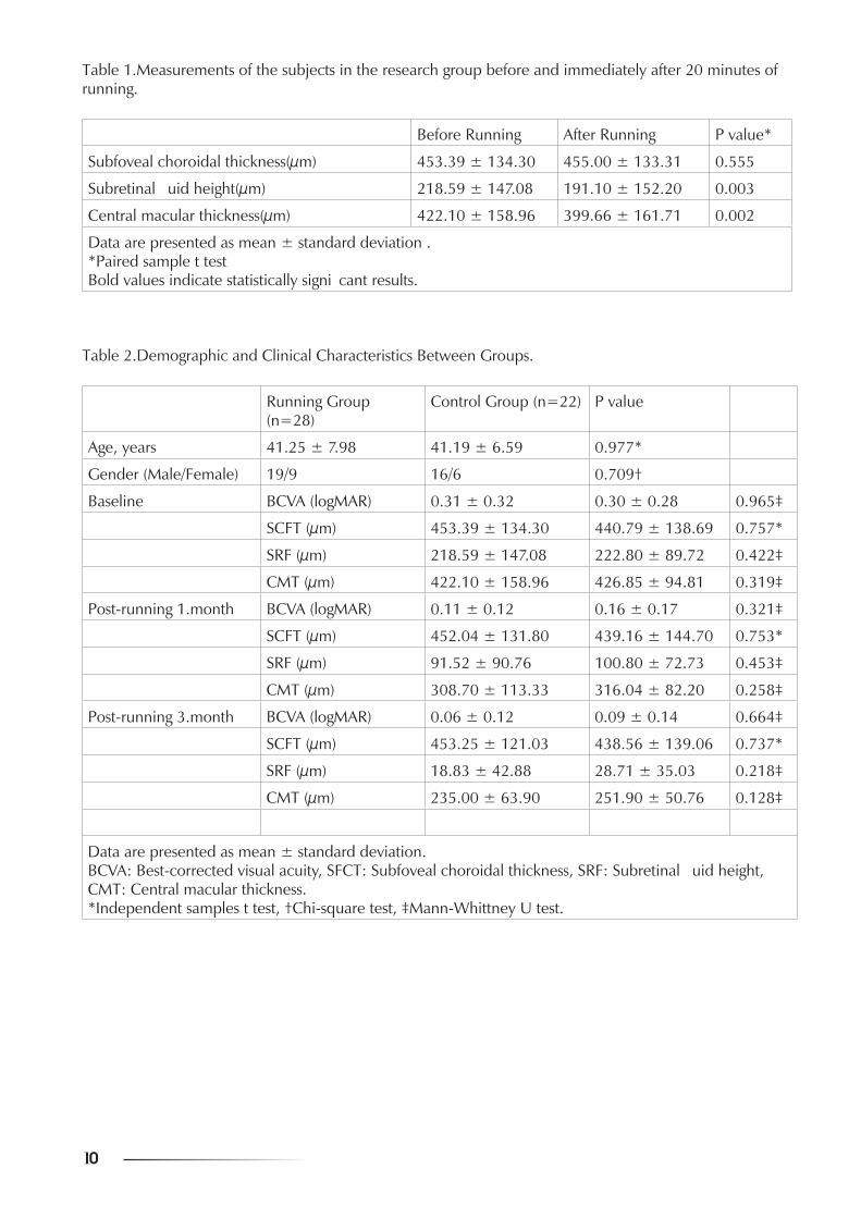
Purpose: To investigate the effects of 1-month running treatment on the anatomical and functional results of patients with acute central serous chorioretinopathy (CSC).Setting: This prospectivecomparative study was performed in the Eye Clinic of the University of Health Sciences Ulucanlar Eye Training and Research Hospital. Methods: Fifty eyes of 50 patients with acute CSCwere divided into 2 groups. Individuals of 28 patients were asked to run 3 kilometers on the treadmill for 20 minutes every day during 1 month (researchgroup), and 22cases were only observed without any treatment or intervention (control group). Subfoveal cho-roidal thickness (SFCT), subretinal fluid height (SRF), andcentral macular thickness (CMT) wereassessed byoptical coherence tomography (Spectralis SD-OCT, Heidelberg Engineering, Heidelberg, Germany). The best-corrected visual acuity (BCVA), SFCT, SRF, and CMT valuesof the two groups at the time of diagnosis, 1 month, and 3 months were compared. Also, pairwise comparisons ofthe research cases were evaluated-concerningpre- and post-runningvalues.Results: Both groups were similar with regard to age and gender (P=0.977 and P=0.709, respectively).The mean SCFT was similar at pre- and post-running20 minutes (P=0.555), but the mean SRF and CMT decreased significantly after 20 minutes of running(P=0.003 and P=0.002, respectively).There was no significant difference between the mean BCVA, SFCT, SRF, and CMT valuesof the two groups at the time of diagnosis, 1 month, and 3 months (for each, P> 0.05).Conclusion: Although running therapy had an immediate benefit on the anatomical and functional out-comes for acute CSC, long-term positive effects were not observed.Acknowledgments: Financial Disclosure(s): The study was not funded. The authors have no proprietary or commercial interest in any materials discussed in this article.Conflict of Interest: The authors declared no conflict of interest.
10
Table 1.Measurements of the subjects in the research group before and immediately after 20 minutes of running.
Before Running After Running P value*
Subfoveal choroidal thickness(μm) 453.39 ± 134.30 455.00 ± 133.31 0.555
Subretinal fluid height(μm) 218.59 ± 147.08 191.10 ± 152.20 0.003
Central macular thickness(μm) 422.10 ± 158.96 399.66 ± 161.71 0.002
Data are presented as mean ± standard deviation .*Paired sample t testBold values indicate statistically significant results.
Table 2.Demographic and Clinical Characteristics Between Groups.
Running Group (n=28)
Control Group (n=22) P value
Age, years 41.25 ± 7.98 41.19 ± 6.59 0.977*
Gender (Male/Female) 19/9 16/6 0.709†
Baseline BCVA (logMAR) 0.31 ± 0.32 0.30 ± 0.28 0.965‡
SCFT (μm) 453.39 ± 134.30 440.79 ± 138.69 0.757*
SRF (μm) 218.59 ± 147.08 222.80 ± 89.72 0.422‡
CMT (μm) 422.10 ± 158.96 426.85 ± 94.81 0.319‡
Post-running 1.month BCVA (logMAR) 0.11 ± 0.12 0.16 ± 0.17 0.321‡
SCFT (μm) 452.04 ± 131.80 439.16 ± 144.70 0.753*
SRF (μm) 91.52 ± 90.76 100.80 ± 72.73 0.453‡
CMT (μm) 308.70 ± 113.33 316.04 ± 82.20 0.258‡
Post-running 3.month BCVA (logMAR) 0.06 ± 0.12 0.09 ± 0.14 0.664‡
SCFT (μm) 453.25 ± 121.03 438.56 ± 139.06 0.737*
SRF (μm) 18.83 ± 42.88 28.71 ± 35.03 0.218‡
CMT (μm) 235.00 ± 63.90 251.90 ± 50.76 0.128‡
Data are presented as mean ± standard deviation.BCVA: Best-corrected visual acuity, SFCT: Subfoveal choroidal thickness, SRF: Subretinal fluid height, CMT: Central macular thickness.*Independent samples t test, †Chi-square test, ‡Mann-Whittney U test.
11
FOLDABLE CAPSULAR VITREOUS BODY: SURGICAL SOLUTION FOR PREPHTHISIS EYEShalva Skhirtladze MD; Onise Tsertsvadze MD; Nino Tavberidze MD, PhD; Giorgi Mekvabishvili MD;
Purpose: To report a surgical procedure and the outcome of foldable capsular vitreous body implantation in patient with prephthisical eye with chronic retinal and uveal detachment.Methods: 35 years old Caucasian male referred to our department with the painful blind eye. The patient had the history of chronic retinal detachment since childhood. On routine ophthalmic examination, the left eye had no light perception and sensory exotropia. The IOP was 0 mmHg. On slit lamp examination 360 de-grees of posterior synechia, iridodonesis and crystalline lens opacification was discovered. B-scan showed total retinal detachment and sectorial choroidal effusion. Ultrasound biomicroscopy demonstrated choroi-dal effusion with partial ciliary body detachment. In order to support and maintain the eyeball, total retinectomy and FCVB implantation was planned. Surgical synechialysis was performed and iris was expended using iris hooks. AC maintainer was utilzed to support phacoemulsification procedure. 23 gauge chandelier assisted pars plana vitrectomy was done. After performing core vitrectomy and posterior hyaloid dissection, 360 endodiathermy was performed and was proceeded with total retinectomy. Fluid-air exchange was done. 6 mm scleral incision was made 3.4 mm from the limbus and prefolded FCVB device was introduced to the vitreous cavity through scleral incision. 8.0 Nylon was used to suture scleral wound and to ligate FCVB tube. After wound closure, 23 G VFC silicone oil injection system was utilized to facilitate oil delivery to the FCVB device. After FCVB became sufficiently filled with a silicone oil, the tube was placed in sub-Tenon’s pocket superotemporaly. Conjunctiva and Tenon’s capsule was closed with 8.0 Vycril sutures. Surgical iridectomy was made at 12 o clock position. Corneal wounds were hydrated, subconjunctival antibiotic was administered at end of the surgery.
The treated eye Results:Immediate postoperative complication was hyphema (“8 ball”) and was managed in first postoperative week. At the 6-months follow-up, choroidal reatechment and stable (9-10 mmHg) intraocular pressure was achieved. FCVB showed excellent biocompatibility and stable positioning within the eye.Conclusion: Silicone oil-filled FCVB has shown to be effective and safe in our case as a vitreous substitute over a 6 month observation period. Financial Disclosure: None
EFFECT OF LASER ENERGY AND PHOTOCOAGULATED RETINAL AREA ON REFRACTIVE ERROR IN PATIENTS WITH TYPE 1 RETINOPATHY OF PREMATURITY.Serdar Ozates, Emrah Utku Kabatas
Purpose: Knowledge regarding how retinopathy of prematurity (ROP) and its treatment strategies affect the emmetropization process remains limited. We hypothesize that the photocoagulated retinal area and laser photocoagulation (LP) parameters may affect the emmetropization process in patients with type 1 ROP. In this study, we sought to investigate the influence of photocoagulated retinal area and laser energy on emmetropization in patients with type 1 ROP.Setting: This retrospective and cross-sectional study included 230 eyes of 115 patients with type 1 ROP.Methods: Patients who received LP formed the LP group and patients who were screened without treat-ment formed the control group. Gestational age, birth weight, stage and zone of the ROP, laser shoot count, and total laser energy were noted. The size of the estimated photocoagulated retina was deter-mined by multiplying the area of a single laser spot by laser shoot count. The magnification effect of the lens on the laser beam and the influence of the duration of photocoagulation on the laser spot were con-sidered in the calculations. At 24 months of corrected age, refractive errors of patients were evaluated. Results: No significant difference was observed between groups regarding mean gesatational age, mean birth weight, and gender (p>0.05). Mean cylindrical refractive error and spherical equivalent were signifi-cantly higher in the LP group (p<0.001). Regression analysis revealed that total laser energy was associated with myopic refraction in the LP group (p=0.003). Total laser energy equal to or higher than 254,700 mW
12
had 88% sensitivity and 82% specificity (p<0.001), while ablated retinal area equal to or greater than 9.7 mm2 had 85% sensitivity and 81% specificity (p<0.001) in predicting myopic refraction at the end of 24 months of follow-up. Conclusions: The present study revealed that the size of photocoagulated retinal area and total laser ener-gy may affect the emmetropization process, leading to higher myopia and astigmatism. The present study results also provided more proof that the peripheral retina may have an influence on ocular development and the emmetropization process.Financial Disclosure: The presenting author (Serdar Ozates) and the co-author (Emrah Utku Kabatas) did not have a financial interest in the subject matter and did not receive money from any mentioned company.
ACUTE GLAUCOMA ATTACK ASSOCIATED WITH GENERAL ANESTHESIAP. Manolova Y. Kirilova P. Vassileva
Introduction: Some medicines used in the general anesthesia (atropine, fentanyl, propofol, adrenalin, do-pamin ) may cause mydriasis and provoke acute glaucoma attack. The patients who have shallow anterior chamber, exfoliative syndrome, narrow/closed angle are predisposed. Setting: Specialised eye hospital “Proff Pashev”, Sofia Purpose: To present two clinical cases of female patients with acute glaucoma attack after surgery under general anesthesia, our management and methods of treatment.Methods: Two female patients with acute closure glaucoma attack. Both have positive family history for glaucoma. We performed gonioscopy, anterior and posterior segment OCT and YAG- laser treatment. Results: Case 1: V.Y. caucasian female patient 69 y.o. Three days after gynecological surgery the patient started complaining of blurred vision and pain of the left eye. At that time the intraocular pressure of the left eye was up to 50mmHg and therapy with Brinzolamid and Timolol was started. She was admitted to our clinic two weeks later. Both eyes were diagnosed with closed angle glaucoma, YAG iridotomies were performed. After the procedure ТОD=15mmHg ТОS=12mmHgCase 2: P.M. caucasian female patient 52 y.o. The day after gynecological operation she complained of acute reduction of vision of the right eye. At the time she was treated for iridocyclitis. Two months later high intraocular pressure was measured of the left eye up to 42mmHg and an iridotomy of the left eye has been performed. A treatment with Travoprost has been started. She was admitted to our hospital six months later with blind right eye and ТОD= 50mmHg. An iridotomy of the right eye was performed, we changed anti-glaucoma-tous therapy and the result was TOD= 22mmHg ТОS=14mmHg Conclusion: In order to prevent vision loss of patients, it is necessary to consult an ophthalmologist before sur-gery under general anesthesia for all patients with family history for glaucoma. None of the authors have financial interest concerning the pharmaceutical products discussed in this abstract.
İN VITRO FERTILIZATION AND PREMATURE RETINOPATHYOnur Gokmen, MD*, Ozlem Beyazyildiz, MD** Health Sciences University Samsun Training and Research Hospital, Department of Ophthalmology Samsun, Turkey.
Aim: To evaluate the characteristics of premature retinopathy (ROP) in premature babies born by in vitro fertilization (IVF) method.Methods: 152 eyes of 152 infants screened for ROP were included in this study. Infants born by IVF were included in group 1 (n=74) and infants born by normal fertilization method were included in group 2 (n=78). Groups were compared according to demograpic data, birth weight, birth weeks, stages of ROP and retinal vascularization time. Results: The mean birth weeks and weights of the babies in Group1 were 32.54 ± 2.93 weeks and 1866.3 ± 559.4 g, respectively, whereas in Group 2 they were 32.24 ± 2.29 weeks, 1775.5 ± 499.0 g, respec-tively. There was no statistically significant difference between the groups in terms of birth weight and birth week. (p> 0.05). ROP development was detected in 18 (64%) cases in Group 1, while ROP development was observed in 10 (36%) cases in Group 2. When the groups were compared according to the ROP stag-es, in Group 1, 10 patients (55.6) had stage 1, 4 patients (22.2%) had stage 2, and 4 patients (22.2%) had stage 3 ROP. In group 2, 9 patients (90%) had stage 1 ROP, while only 1 patient (10%) had stage 2 ROP, and stage 3 ROP was not observed. Mean retinal complete vascularization time was found as 49.6 ± 6.8 weeks in Group 1, while it was 43.4 ± 3.3 weeks in Group 2. Retinal vascularization completion time was

13
statistically significantly higher in Group 2 patients (p <0.05). In Group 1, 5 patients (27.8%) were treated with intravitreal injection, while the remaining 13 patients (72.2%) had spontaneous regression. In Group 2, spontaneous regression was observed in all patients.Conclusion: IVF can be a risk factor for the development of ROP. ROP is observed more frequently in these babies, the frequency of advanced stage ROP requiring treatment increases, and retinal vascularization completion time is prolonged in these cases.
VISUAL AND REFRACTIVE OUTCOMES IN TORIC INTRAOCULAR LENS IMPLANTATIONDr. Ömer Takeş
ABSTRACT Purpose: Evaluate the visual results, refractive correction and intraocular lens stability of the eyes with cataract and greater than and equal to 1.25 diopters (D) astigmatism after phacoemulsification with toric intraocular lens implantation (TIOL) surgeryMethods: 24 eyes of 15 patients were included in this retrospective study. Cataract surgery and implanta-tion of TIOL was performed to the patients with cataract and greater than and equal to 1.25 diopters (D) astigmatism. Patients were followed for minimum 6 months. The uncorrected (UDVA) distance visual acu-ities, residual refraction status and IOL rotation degrees after one day, one week, one month, three months and siz months were evaluated.Results: 24 eyes of 15 patients (10 women, 5 men) with mean age of 65.95 ± 9.40 (50-81) were included. Preoperative mean best corrected visual acuity (BCVA) was 0.38 ± 0.19 logMAR. 6 months after surgery, UDVA was greater than and equal to 0.10 logMAR and mean UDVA was 0.03 ± 0.04 logMAR (0.00-0.15). Also, BCVA was 0.01 ± 0.04 logMAR (-0.10-0.05). Mean preoperative cylindirical refraction value, mean corneal astigmatism and mean residual astigmatism was 2.58 ± 1.07 D (0.75-4.75), 2.34 ± 0.68 D (1.25–3.85) and 0.44 ± 0.18 D (0.25-0.75) respectively. Mean IOL rotation value was measured as 4.16 ± 2.01 º (2-8) at 6 months. Surgically induced astigtmatism (SIA) was 0.39 (0.10 - 1.05). None of our patients were operated for correction of residual astigmatism.Conclusions: TIOL implantation to the eyes with cataract and greater than and equal to 1.25 diopters (D) astigmatism is a convenient option for rotation stability and refractive gain in patients with vision expecta-tion independent of glasses.Key words: Toric intraocular lens, astigmatism
THE QUANTITATIVE EVALUATION OF RETINAL LAYERS AFTER RESOLUTION OF SUB-RETINAL FLUID IN ACUTE CENTRAL SEROUS CHORIORETINOPATHY Merve Inanc1,2, Kemal Tekin1,21M.D., Ercis State Hospital, Ophthalmology Department, Van, Turkey.2M.D., Ankara Ulucanlar Eye Training and Research Hospital, Ankara, Turkey.Purpose: The aim of this study was to evaluate the average retinal layer thicknesses in eyes with unilateral acute central serous chorioretinopathy (CSC) (with subretinal fluid (SRF)) at the time of diagnosis and after complete resolution of SRF and to compare these results with those of unaffected fellow eyes and healthy control eyes. Setting: Ulucanlar Eye Training and Research Hospital, Ophthalmology Department, Ankara, Turkey.Methods: The medical records of the patients with unilateral acute CSC were consecutively recruited. Each participant had a documented episode of acute CSC with serous retinal detachment and complete resolution of SRF at follow up visits in one eye and no history of CSC diagnosed or suggested in the fellow eye. 54 eyes of 27 patients with unilateral acute CSC and 25 eyes of 25 healthy control subjects enrolled in the study. The average thicknesses of the retinal layers were measured by segmentation analysis of optical coherence tomography at baseline and 6 months after complete resolution of SRF.Results: The mean outer nuclear layer (ONL) thickness was significantly lower in eyes with CSC than in fellow eyes (p<0.001). The mean ONL thickness was 82.2 ± 19.8 μm at the time of the diagnosis and in-creased to 85.3 ± 20.8 μm 6 months after the complete resolution of SRF, but still low compared to unaf-fected fellow eye and the increment was not statistically significant (p>0.05) There were significant correla-tions between the best corrected visual acuity (BCVA) and ONL thicknesses at baseline and 6 months after complete resolution of SRF (p<0.001, r= -0.810; p<0.001, r= -0.705, respectively).Conclusion: The ONL thickness is thinned in cases with acute CSC, and although there is some increment

14
in ONL thickness 6 months after complete resolution of SRF, it is still thinner compared to unaffected fellow eyes. Additionally, the ONL thickness is correlated with the BCVA in eyes with CSC before and after resolution of SRF.Financial disclosureAll authors report no conflicts of interest. This research received no specific grant from any funding agency in the public, commercial, or not-for-profit sectors.
CONJUNCTIVAL AUTOGRAFT TECHNIQUE WITH FIBRIN ADHESIVE IN PTERYGIUM SURGERYMehmet Ozbas, Health Science University, Bakirkoy Dr. Sadi Konuk Training and Research Hospital
Aim: To evaluate the efficacy and stability of using fibrin glue in conjunctival limbal autograft transplanta-tion during pterygium surgery.Method: 25 eyes of 25 patients were included to the study. After pterygium excision, Limbal-based con-junctival autograft transplantation from superior temporal conjunctiva in the same eye was performed using fibrin glue. Operation times were recorded. Patients were called for postoperative control on the 1st day, 1st week and 1st month after surgery. At each visit, patients’ subjective complaints, postoperative compli-cations and recurrence rates were recorded. Result: The mean age of the patients was 50.64 ±10.79 (32 -75). The mean follow-up time was 24.44 ± 6.53 weeks. Mean surgery time was 17.2 ± 2.72 minutes. It was observed that in 15 of 25 eyes (60%), there was a significant decrease in subjective complaints (pain, foreign body sensation, epiphora, irritation) in the first postoperative week, and all patients’ subjective complaints disappeared in the first month. In one patient (4%), a partial folding was observed in the upper temporal of the graft on postoperative day 1, the graft was reposed under biomicroscope and the eye was closed again. The next day, it was observed that the graft was in place. During the follow-up period, no complications in the pterygium and fibrin glue were observed in any patient.Conclusion: Fibrin glue is an effective and reliable method in the placement of conjunctival autograft in pterygium surgery. The use of fibrin glue enables the subjective complaints of the patients to be reduced and eliminated in a shorter time and shortens the duration of autografted pterygium surgery.
EARLY EFFECT OF VITAMIN D DEFICIENCY ON ANTI-VEGF TREATMENT OF MACULAR EDEMA SECONDARY TO CENTRAL RETINAL VEIN OCCLUSIONMehmet Murat Uzel, Melek Köroğlu Balıkesir University School of Medicine Department of Ophthalmology
Purpose: It has been shown that vitamin D deficiency (VDD) is more common in patients with central retinal vein occlusion (CRVO). In the case of VDD, changes occur in vascular structures, inflammation response and platelet activities. The aim of our study was to investigate the effect of VDE on treatment re-sponse in patients who received anti-vascular endothelial growth factor (ranibizumab) therapy for macular edema secondary to CRVO.Setting: Retrospective studyMethods: Thirty-two eyes of 32 patients who underwent a single dose ranibizumab injection were evaluated. At baseline all eyes underwent a complete ophthalmological examinations, including best corrected visual acuity, slitlamp biomicroscopy, intraocular pressure evaluation, dilated fundus examination, optical coherence tomography and fluorescein angiography. Systemic diseases and vitamin D levels of patients were recorded. Potential prognostic factors for outcomes were evaluated using multivariate logistic regression analysis.Results: VDD was present in 14 (58.3%) patients. Preoperative central macular thickness (CMT) was 485.29 ± 99.58 μm, postoperative 1st month CMT was 259.70 ± 50.27 μm and was statistically signifi-cant (p <0.001). According to the multiple regression analysis, absence of DM, absence of VDE and high preoperative CMT level were found to be positive predictive factors for CMT reduction after firts anti-vegf treatment. (OR 0.22, 95% CI 6.99-71.52, p = 0.020; OR 5.29, 95% CI 65.60-153.21, p <0.001; OR 0.36, 95% CI 0.10-0.65, p = 0.009; respectively).Conclusion: According to the results of our study, VDD reduces the effectiveness of anti-vegf treatment. This may be due to the activation of the proinflammatory process in VDD. Consideration of vitamin D levels in patients with CRVO before anti-vegf therapy may be beneficial for treatment success.Financial Disclosure: None
15
EVALUATION OF ANTERIOR SEGMENT PARAMETERS IN PSEUDOEXFOLIATION SYN-DROME.Dr. Mehmet Gokhan Asian.
PURPOSE: We aimed to assess the anterior segment parameters of pseudoexfoliation syndrome (PXFS) cases and to compare the values with cataract cases in the same age group.SETTING: Recep Tayyip Erdogan University Medicine Faculty Ophthalmology DepartmentMETHODS: Forty-four eyes of 44 patients with PXFS and 50 eyes of 50 patients who were diagnosed cataract for surgery were recruited. All participants were evaluated by autorefractokeratometer, specular microscope for endothelial cell count, optic biometry for anterior chamber depth, lens thickness measure-ment and air-puff tonometer for intraocular pressure (IOP) measurements. RESULTS: The mean corrected IOP of the eyes in the study group was 18.04±5.03 mmHg, while those were 16.06±2.57 mmHg in the control group. The mean K1 and K2 values were 43.07±1.59D and 43.86±1.48D in the study group. Those were 43.13±1.49D and 44±1.44D in the control group, respec-tively. The mean endothelial cell count was 2445.43±333.43 in the study group and 2438.46±289.63 in the control group. The anterior chamber depth and lens thickness values were 3.12±0.38mm and 4.63±0.35mm in the study group and 3.21±0.55mm and 4.39±0.4mm in the control group, respectively. The difference in lens thickness was statistically significant between two groups.(p<0.05)CONCLUSION: The lens thickness was found to be significantly higher in PXFS cases of the same age group compared to cataract cases with no comorbidity. However, there was no significant difference be-tween the two groups in terms of endothelial cell count, anterior chamber depth and intraocular pressure values. Our results indicate that the iris-lens dynamics of the PXFS patients are different than routine cata-ract cases and these patients should be carefully examined before cataract surgery to avoid complications. FINANCIAL DISCLOSURE: The author has no financial conflicts of interest to disclose concerning this pre-sentation.
VARICELLA ZOSTER VIRUS- THE SLEEPING DANGER TARGETING THE EYEM.Taneva, K.Racheva, P.Vassileva
Purpose: To present two cases of ocular involvement associated with Varicella Zoster Virus /VZV/ infec-tion, our clinical approach and the follow-up of the patients. To study the risk factors that can provoke the activation of the “sleeping” virus. Methods: Case 1: K.B. caucasian male 68 y.o., with redness and grittiness of the left eye; history of shingles and recent treatment with Metotrexate for psoriasis. At admission: left eye BCVA = 0.3-0.4, TOS=16mmHg, ciliary injection, corneal ulcer at the lower half of the cornea, precipitates and normal posterior segment. Blood serology: VZV IgG 1222 mIU/ml. Case 2: I.P. caucasian female 46 y.o., with blurred vision of both eyes; had recently undergone total hysterectomy and appendectomy. At presentation: BCVA OD= 0.4, OS=0.3, TOD= 19mmHg, TOS= 19mmHg, diffuse opacification of the vitreous and tortuous retinal vessels. The fluorescent angiography shows leakage around the optic nerve head and retinal vessels. Blood serology: VZV IgG 2286 mIU/ml.Setting: Specialized Eye Hospital “Acad. Pashev”Results: Immediate antiviral treatment with Valaciclovir was implemented for both patients, starting with 3g/day and gradual reduction to 1g/day. Case 1: mydriatics, epithelizing corneal gel, antibiotics and ad-ditional corneal cross- linking, amniotic membrane transplantation and parabulbar steroid after 24 days of antiviral treatment. Case 2: topical and systemic non steroidal anti- inflammatory drugs and subtenon injection of steroid after 4 months of antiviral treatment. Both patients showed improvement in clinical symptoms and vision during follow-up of 6 months. Conclusion: The VZV infection can be activated due to immune suppression, superinfection, psychological or physical stress. The VZV can affect different eye structures so it is important to intentionally search for it. The antiviral medications are the first line of treatment and the strict follow up is of major importance.
None of the authors have financial interest concerning the pharmaceutical products discussed in this ab-stract.
16
ART OF STRABISMUS SURGERY (SURGICAL TIPS FOR YOUNG OPHTHALMOLOGISTS/CLINICAL CASE).Lana Datuashvili MD. Georgia, Kutaisi. Rotterdam Eye Hospital/Clinic LJ
Purpose: The secret of successful strabismus surgery: understanding of the anatomy of the extraocular mus-cles, the mechanics of access to the operative site.History of Strabismus Surgery: Different techniques of suturing, Techniques of muscle transposition in dif-ferent types of squint. Tenotomy, Preparation for surgery. Pre- and post-operative care. Methods: Clinical Case: Patient: Female 13y Old Race - CaucasianVis OD= 0.8 – 0.5ax125=0.9-1.0Vis OS=0.5-0.6 -0.5 -0.5 ax 65=0.9-1.0Fundus – without abnormality; Pupil Reaction – N; Lang II – Neg; Convergence OU - 25PD in the dis-tance. 30 PD – near. Vertical - Hyper deviation OD=25PD. Diagnosis: Esotropia. OD – IV Nerve Palsy. OS Hypotropia Plan for Operation: OU – Medial Rectus Recession – 4mm. OD – Inferior Oblique Tenotomy. OS – Inferi-or Rectus Recession – 3mmIntra-operative Complication: Hemorrhage – during inferior oblique tenotomy.Results: Postoperative care: Anti-inflammatory drops, lubricant – OU. Cold Compresses - after the opera-tion – OD. Heparin gel 1000 IU/g – topically after 3 days during 2-3 weeks – OD. Total resorption after 3 weeks. Conclusion: Understanding and knowledge of different techniques of suturing, muscle transposition and tenotomy is crucial in squint surgery. Damage to vortex vein and orbital hemorrhage during strabismus sur-gery is one of the surgical complications. If a vortex vein is torn, it bleeds profusely and if it cannot cauter-ize successfully it should be tied off using a 7.0 vicryl suture. Financial Disclosure of authors: None
MEİBOMİAN GLAND DYSFUNCTİON AND DEMODEX İNFESTATION EVALUATION IN PATIENTS WITH DRY EYE SYMPTOMS.Dr. Lale Geribeyoglu
PURPOSE: To evaluate demodex infestation and meibomian gland dysfunction in patients with dry eye symptoms.METHODS: 16 patients complaing about dry eye smptoms were included to the study and evaluated. All enrolled subjects were tested in the following, OSDI, slit lamp examination, NIBUT, schirmer I test, meibo-mian quality, meibomian expressibiltiy, lid margin abnormality and Demodex excistence.RESULTS: 16 patients between 31-68 years old were evaluated, 10 were Demodex(+) and 6 were Demo-dex(-) , mean OSDI values were 42,51 and 47,73 respectively in the positive and negative groups. NIBUT values were 9,52/11,28 and 9,78/9,03 seconds in right/left eyes. Schirmer I test values were 10,6/11,4 and 13,8/15,6mm for the same groups. Meibomian quality and meibomian expressibility were also assesed and lid irregularity was rated during the slit lamp examination.CONCLUSIONS: Ocular demodex infeststion may be associated with ocular dyscomfort and ocular surface damage in MGD causing dry eye.
COMING SOON ?: INTRAOCULAR IMPLANTS WITH METAMATERIALS, ESPECIALLY GRAPHENE.Kazim Hilmi Or
Background: In ophthalmology, the use and range of intraocular implants has multiplied by advances in nanotechnology. Metamaterials are in nature non-existent molecules or crystals. Graphene is a modifica-tion of carbon with a two-dimensional structure, in which each carbon atom is surrounded by three more at an angle of 120 °. The clinical use of metamaterials, especially graphene, in many diseases in oph-thalmology will take place in these years. This lecture will report on what can change in ophthalmology through the use of graphene implants in the anterior and posterior segments of the eye.Methods: The properties of graphene are compared with the current state of the implanted intraocular lenses and other uses in ophthalmology.

17
Results: Graphene is a metamaterial made up of carbon atoms which makes it harder than steel but still flexible. Because graphene is one atom thick, it is called a 2D material. In addition to low water and tem-perature permeability, graphene has an important property as a “lens” (optically transparent): Because it has practically no thickness, it has special optical properties. One of them is that it has no chromatic aber-ation. This gives you sharp images (without optical modulation on the graphene lens). For higher diopters, you do not need thicker lenses as with conventional implants. Graphene is mechanically relatively flexible. But it is also more resistant than steel. As an implant in the eye, graphene would have many advantages. However, the thinness of the material means that it will need a supporting frame.Conclusions: Graphene will soon be used as an implant material in ophthalmology. It has many advantages visually, physically and clinically. With a suitable support structure, graphene and other metamaterials can very quickly reduce or even eliminate the optical disadvantages of today’s implants.
COMPARISON OF THE EFFECT OF BRIMONIDINE ON PUPIL SIZE IN GLAUCOMA PA-TIENTS AND HEALTHY SUBJECTSIbrahim TuncerAlfa Medical Center, Izmir, Turkey
Abstract: Objectives: To compare the effect of brimonidine on scotopic pupil size in patients using brimo-nidine for the treatment of glaucoma and healthy individuals.Materials and Methods: In this study, two groups of 30 patients with early stage glaucoma using 0.15% brimonidine tartrate drop and 30 healthy individuals were created. In the glaucoma group, pupil size measurements were made in a scotopic condition (1 cd/m2) using an infrared pupillometer before and 30 minutes after a drop of 0.15% brimonidine tartrate, in accordance with the patient’s drop instillation time. Pupil size measurements were made before and after brimonidine in the right eyes of the healthy group. Data of the right eye were used for statistical analysis.Results: The mean age was 44.16 ± 8.87 in the glaucoma group and 43.06 ± 8.48 in the healthy group. The mean scotopic pupil size before brimonidine was 6.12 ± 0.99 mm in the glaucoma group and 6.15 ± 1.02 mm in the healthy group. The mean scotopic pupil size at the 30th minute after brimonidine was 4.54 ± 1.10 mm in the glaucoma group and 4.49 ± 1.07 mm in the healthy group. The mean scoto-pic pupil size decreased by 1.58 mm in the glaucoma group and 1.66 mm in the healthy group. In both groups, the mean scotopic pupil size after brimonidine was significantly lower than before brimonidine (p <0.001 for both).Conclusion: A single dose of 0.15% brimonidine tartrate drop produced significant miosis in early stage glaucoma, similar to that in normal eyes. A single dose of brimonidine drop can be effective in reducing night vision complaints after laser refractive and premium intraocular lens surgery in early stage glaucoma patients.
THE EFFECT OF INFERIOR OBLIQUE MUSCLE Z-MYOTOMY ON PATIENTS WITH PRI-MARY INFERIOR OBLIQUE OVERACTIONHasan Kızıltoprak1, Hakan Halit Yaşar2, Kemal Tekin31MD, FEBO, FICO, Bingol Women’s Health and Children’s Hospital, Ophthalmology Department, Bingol, Turkey 2 MD, Ulucanlar Eye Training And Research Hospital, Ophthalmology Department, Ankara, Turkey. 3MD, Ercis State Hospital, Ophthalmology Department, Van, Turkey
Purpose: To investigate the surgical results of the inferior oblique muscle Z-myotomy in patients with inferi-or oblique muscle overaction (IOOA).Setting: Retrospective case series. Material and Method: Medical records of patients who had undergone inferior oblique muscle Z-myotomy with the diagnosis of primary IOOA in a single center between 2017-2018 were retrospectively analyzed. All patients had mild IOOA (+1 and between +1 and +2). Preoperative and postoperative IOOA degrees and ocular motility examinations were evaluated. The infeior oblique muscle Z-myotomy is performed at 6 mm along the physiological muscle line after defining the lower oblique muscle through the inferotemporal fornix incision. Results: 47 eyes of 44 patients were included in the study. The patients were divided into two groups as +1 group and + 1-2. In 37 (78.7%) of the cases, IOOA was +1, and in 10 (21.3%) of them, it was be-

18
tween + 1-2. The mean age of the +1 group was 14.18 ± 11.8, and the mean age of the 1-2 group was 13.40 ± 7.45. The mean follow-up period of the patients was 10.56 ± 8.7 (minimum: 6, maximum: 17) months. Bilateral Z-myotomy was performed in 3 (6.3%) and unilateral in 44 (93.7%) of the cases. While 43 (91.4%) of 47 eyes underwent Z-myotomy, AOHF improved after surgery, and 4 (8.6%) eyes had pre-operative levels of IOOA. There was no statistically significant difference between two groups after surgery (p = 0.849). When preoperative and postoperative IOOA values were compared, there was a statistically significant decrease in IOOA values in the postoperative period (p = 0.001). No intraoperative and post-operative complications were observed. Conclusion: Inferior oblique Z-myotomy is a simple, rapid, sutureless surgical procedure in which the orig-inal muscle insertion is preserved. Z-myotomy of the inferior oblique muscle can be used as a successful attenuation method in patients with minimal IOOA.Keywords: Inferior oblique muscle overaction, surgical results, Z-myotomy.Conflict of interestAll authors indicate no financial support or financial conflict of interest.
OPTIMIZING IMAGE QUALITY WITH EYEMAX MONO LENS IN DRY AGE-RELATED MACULAR DEGENERATIONHamidu Hamisi GOBEKA Asst. Prof., M.D.; Tansu ERAKGÜN Prof., M.D
ABSTRACT: Purpose: Investigation of clinical outcomes in dry AMD patients after intracapsular implanta-tion of a novel EyeMax MonoTM macular lens (IOL) (London Eye Hospital Pharma, London, UK), a fold-able and injectable acrylic hydrophobic IOL implanted similar to standard IOLs.Design: A single-centered prospective interventional studyPatients: Twenty-two phakic eyes of 19 moderate or advanced dry AMD patients with a postoperative follow-up of at least 3 months were investigated. Stable dry AMD was approved once free of any active choroidal neovascularization for ≥3 months without the need for intravitreal ant-VEGF therapy.Methods: A comprehensive preoperative ophthalmological assessment was conducted in all patients prior to small-incision intracapsular implantation of Eyemax mono IOL designed to improve the quality of the retinal image in all areas of the macula ≤10º from fixation and to produce mild hypermetropic correction for magnification.Main Outcome Measures: Optimization of visual acuity (VA) (logMAR), safety as determined by intraop-erative and postoperative complications, high intraocular pressure (IOP) demanding medical or surgical intervention, postoperative diplopia or dysphotopsia.Results: Mean age of the patients at surgery was 68.55 ± 9.53 years, with 73% male and 28% female. Mean duration of postoperative follow-up was 7 months. Preoperative VA (1.05 ± 0.44 logMAR) improved significantly to 0.72 ± 0.43 logMAR (P=0.001), equivalent to postoperative mean ETDR of 49.55 ± 20.05 (P=0.001). There was no statistically significant change in IOP (P=0.277). The mean postoperative refrac-tive spherical equivalent improved to +2.31 ± 1.55 D with substantial visual improvement as early as 3 months after surgery. No major surgical complications were identified either intraoperatively or postoper-atively, except for 2 patients (9%) who experienced intraoperative haptic rupture. Furthermore, no symp-toms of dysphotopsia or diplopia were identified.Conclusions: Extended macular vision IOL, intended to improve quality of the retinal image in the eyes with moderate or severe AMD, has a safety profile equivalent to standard IOLs in the medium term and may be the preferred lens for optimizing and preserving visual acuity in dry AMD patients with varying degrees of center-involving maculopathy. While major safety issues were not revealed over the entire follow-up period, a larger series with a longer follow-up period is required to assess the full potential of this technology.Key Words: Cataract; Dry Age-Related Macular Degeneration; Eyemax MonoTM Macular Lens; Spherical Equivalent
19
PHOTOPHOBIA TREATMENT IN PATIENTS WITH MIGRAINECristina Scerbatiuc, Eugen Bendelic; Republic of Moldova
Purpose: to highlight the neuro-ophthalmic features in patients with migraine, including determination of the photosensitivity threshold and the light spectrum causing photophobia, and to lay down the directions for increasing the efficiency of treatment.Objectives of the study: Evaluation of photophobia threshold in migraine patients, and determine the effectiveness of some spectral filters in reducing of photophobia.Determining correlations between photophobia threshold and the discomfort caused by visualization of trigger - figures in migraine patients.Methods: In study were included 128 patients with migraine and photophobia, whom it was proposed for use glasses with spectral filters for 2 months. The mean number of days with headache, before treatment was 13.5 per month. It was decreased to 4.1 days with headache per month. Headache intensity during the migraine attack was reduced from 0.31 to 3.0 points SVA. Also, the intensity of the photophobia was reduced from 4.1 to 1.8 pointsConclusions: Some spectral filters are successfully used in migraine. They reduce the frequency of migraine attacks and reduce the intensity of photophobia during headache. The filters with low light transmission can cause the eyes disadaptation to light; spectral filters that block blue spectrum of light can reduce pho-tophobia in patients with migraine, test with trigger - figures can be useful in the diagnosis of migraine. We have no Financial Disclosure for this article.
INTRAOCULAR PRESSURE AND CENTRAL CORNEAL THICKNESS ALTERATIONS AFTER PUPILLARY DILATION IN SUBJECTS WITH SENILE CATARACTDr. Cagri Ilhan, MD, FICO, Ophthalmology, Hatay State Hospital, Turkey
The author declares no financial interest. Purpose: To compare intraocular pressure (IOP) and central corneal thickness (CCT) alterations 1-hour after pupillary dilation with 1% tropicamide, between subjects with senile cataract and healthy control. Setting: Prospective, controlled study was conducted in Ophthalmology Department in Hatay State Hospi-tal, Turkey.Methods: 112 healthy subjects with senile cataract were included in the study group. Subjects had any ocular diseases, history of previous ocular trauma or surgery, any systemic condition or drug use associat-ed with cataract were excluded. The control group were constructed with 106 age and gender matched healthy subjects.The IOP measured with Goldmann applanation tonometer and CCT measured with ultrasonic pachymetry, were recorded before and 1-hour after pupillary dilation with 1% tropicamide. The differences between two measurements were calculated for IOP and CCT. Only one eye of each subject was included for the statistical analyses and the results were compared. Results:The demographic characteristics were similar (p>0.05).The mean IOPs were 18.45±3.5mmHg (13 to 24mmHg) and 18.24±2.9mmHg (12 to 23mmHg) in the study and control groups (p=0.835), before pupillary dilation.At the same time, the mean CCTs were 543.93±31.2mm (484 to 628mm) and 538.15±36.4mm (470 to 642mm) (p=0.266).After pupillary dilation, the mean changes were 0.49±2.8mmHg (-6 to 7mmHg) in IOP and -6.86±12.9mm (-38 to 11mm) in CCT for the study group, and 0.44±2.4mmHg (-6 to 8mmHg) in IOP and -1.54±6.6mm (-15 to 11mm) in CCT for the control group.The change in IOP was not significant (p=0.802), while it was significant in CCT (p=0.042). Conclusion: 1-hour after pupillary dilation with topical 1% tropicamide, CCT decreases more in subjects with senile cataract then in healthy subjects.

20
THE EFFECT OF LATERAL CANTHAL SLING PERFORMED IN THE SURGICAL TREATMENT OF INVOLUTIONAL ENTROPION OF THE LOWER EYELID ON SURGICAL OUTCOMEBurcu Dirim, MD
Introduction and Purpose: Evaluating as to how lateral canthal sling (LCS) performed on patients undergo-ing Jones retractor plication (JRP) in involutional entropion surgery affects surgical outcome. Method: This retrospective study included 40 eyes of 37 patients who had involutional entropion of the lower eyelid between June 2014 and March 2019. On 27 eyes of 25 patients, only Jones retractor plication was performed (Group 1) whereas on 13 eyes of 12 patients, lateral canthal sling in addition to retractor plication was performed (Group 2). Clinical success was evaluated on the basis of anatomical and func-tional recovery. Functional success was evaluated in terms of reduction in complaints such as epiphora and ocular irritation due to eyelid malposition.Results: The patients consisted of 23 males and 14 females with an average age of 79.84 in Group 1 and 74.66 in Group 2. 4 patients (16%) in Group 1 and 1 patient (8.3%) in Group 2 developed ectropion and recurrent entropion. Discussion and conclusion: The data obtained as a result of 6 months of follow-up revealed that the suc-cess rate was higher in retractor plication combined with lateral canthal sling than retractor surgery alone. Keywords: Involutional entropion, lateral canthal sling, Jones retractor plication.
LONG TERM RESULTS OF SCLEROPLASTY SURGERY IN HIGH MYOPIC PATIENTSBulent Kose. Department of Ophtalmology, Aritmi Osmangazi Hospital, Bursa, Turkey
AbstractPurpose: To investigate the safety and efficiency of scleral reinforcement with lyophilised human durama-ter graft in high myopia.Methods: In this retrospective study, medical records of 210 eyes of 121 patients with high myopia were reviewed. Study group included 156 eyes of 85 patients who undergone scleroplasty surgery in 1990 to 1998, in Beyoglu Eye Hospital, Istanbul ,Turkey and followed for 3 years. The control group included 64 eyes of 36 patients and followed for 1 year. The inclusion criteria were ages between 20 and 40 years old, increase of myopia more than 2.00 diopter in previous year, having axial length more than 25.00 mm. The exclusion criteria were the patients with systemic and ocular disorders such as cataract, glaucoma and corneal diseases. Detailed ophthalmologic examination including best corrected visual acuity measure-ment, slit lamp examination, biometry measurement with 3M Echonule Ultrasonic Biometer, tonometry with Goldman applanation and schiotz tonometer was made. The patients were fully informed about the surgery. During scleroplasty surgery, a 10 mm length limbus parallel conjunctival incision was made at four quadrants between the rectus muscles at a point 15-17 mm away from the limbus and subtennon space was bluntly dissected towards the optic nerve. A lyophilised human dura mater with 10x17 mm size was spreaded on the surface of bare sclera at these four quadrant without suturing. Conjunctiva was closed with a suture. All patients except 3 patients had general anesthesia for this surgery. In one patients who had local anesthesia had scleral perforation secondary to retrobulbar injection. Postoperatively chemosis and eyelid edema was observed in 8 patients. Late anterior migration of graft occurred in 7 eyes. There was no complication in the rest of the patients. Results:In the scleroplasty group, the mean axial length before surgery was 27.17 mm . Three years after the surgery the mean axial length 27.22 mm. The mean average increase in axial length was 0.05 mm. In the control group, at the time when this study was started, the mean axial length was 26.72 mm and 1 year later, the mean axial length was 27.27 mm. The mean average increase in axial length was 0.55 mm. There was statistically significant difference between the axial length progression in the scleroplasty group and the control group. (P<0.05)Conclusion:Our results showed that scleroplasty surgery with lyophilised human duramater graft was safe and effective way to stabilise the progression of high myopia.
21
AMNIOTIC MEMBRANE TRANSPLANTATION IN OCULAR SURFACE DISORDERSBilgehan Sezgin Asena 1, Bora Yüksel 2, Mahmut Kaskaloglu 3Kaskaloglu Eye Hospital, Izmir, Turkey
AIMS: Amniotic membrane has been widely used as a temporary or permanent graft in the treatment of various ocular surface diseases. It has a unique combination of properties, including the facilitation of mi-gration of epithelial cells, the reinforcement of basal cellular adhesion and the encouragement of epithelial differentiation. Its ability to modulate stromal scarring and its antiinflammatory activity has led to its use in the treatment of ocular surface pathologies. In this study, we evaluated the usefulness and effectiveness of amnionic membrane transplantation (AMT) in the different ocular surface disorders.METHODS: 45 consecutive patients who underwent AMT were included. Mean follow-up period was 9.9±6.6 months. Ocular surface disorders 16 (35.6%) cases of corneal ulcer, 6 (13.3%) bullous keratopa-thy, 6 (13.3%) persistent epithelial defect, 5 (11.1%) desmatocele, 4 (8.9%) recurrent pterygium, 4 (8.9%) impending or recent corneal perforation, 3 (6.7%) chemical burn and 1 (2.2%) case of limbal stem cell deficiency. Under sterile conditions amniotic membrane was prepared from a fresh placenta of a seronega-tive pregnant woman and stored at −70°C.RESULTS: Twenty-five of the 45 patients (55.6%) were men and 20 (44.4%) were women. The mean age was 53.27±15 years. In 41 eyes (91.1%), the amniotic membranes were applied with the graft technique. In 4 eyes (8.9%), the graft technique was combined with corneal patch technique. Keratolimbal allograft in 1 patient (2.2%) and limbal autograft in 1 patient (2.2%) were required as additional procedures. The ap-plication was not effective in 6 patients (13.3%) due to premature separation of the membrane. AMT was successful in 18 eyes (40.0%) and partially successful in 10 eyes (22.2%). In 11 eyes (24.4%), healing could not been provided with AMT. Penetrating keratoplasty was required in 6 eyes (13.3%).CONCLUSION: The amniotic membrane transplantation has advantages such as easy preparation and cost-effectiveness. It is a safe and effective procedure in ocular surface disease. However, although its well known clinical efficiancy, there are still many uncertainities regarding the fate of grafted amniotic mem-brane and its long-term effects on different ocular surface disorders. Further studies are required to deter-mine which ocular surface conditions are going to benefit from AMT.
PAPILLOMACULAR BUNDLE SPEARING VS CONVENTIONAL ILM PEELING FOR MEDI-UM SIZE IDIOPHATIC MACULAR HOLES: A COMPARATIVE STUDYAna Vachiberidze MD, Nikoloz Labauri MD.FVRS. Davinci Eye Clinic. Tbilisi, Georgia
PURPOSE: To compare outcomes of two different ILM peeling pattern in the treatment of medium size idio-pathic macular holes (MH).METHODS: Prospective, interventional case series study. Forty eyes of 40 patients having medium size (200-400μm) idiophatic macular holes were enrolled and divided into two main groups. Group I received vitrectomy and pupillomacular boundle (PMB) sparing ILM peeling with semi-oval pattern where convex edge is facing to-wards the temporal side and Group II conventional ILM peeling with round shape extended between arcades and touching the edge of Optic nerve disc on its horizontal plane. All patient underwent 25G vitrectomy, BBG staining, ILM peel and nonexpansile gas (SF6 20%) tamponade. Stand-alone vitrectomy was performed in all cases unless the lens opacification was significantly obscuring intraoperative view. To assess structural and functional status of the posterior retina SD-OCT, fundus stereoscopic photography and macular microperimetry (MP) were used pre-operatively and postoperatively at 1, 3 and 6 months. All patient accomplished 6 month of follow-up.RESULTS: Macular hole closure was achieved in all cases. Vision was improved in both groups, where mean preoperative BCVA was 0.2 in both, but mean postoperative BCVA was 0.4 in Group 1 and 0.7 in Group II. It showed slightly better visual outcomes in Group II was but the difference was not statistically significant (P> 0.05). OCT revealed type I closure in all cases from both groups, but neurosensory retinal (NSR) thickness at the ILM peeling projection compere to adjacent areas with intact ILM was relatively thinned out (mean NRS thickness 223μm vs 282μm). Mean Retinal sensitivity (6dB) and central fixation 80 by MP was remain un-changed on the ILM intact zone and slightly reduced (MS-4dB) postoperatively in the peeling area. Postoper-ative Optic head pallor was more remarkable in group II to compere to group I.CONCLUSIONS: This study concludes that limited ILM peeling shows superior anatomical and functional outcome as well as less iatrogenic damage on pupillomacular bundle to compare to conventional ILM peel-ing pattern. Thus, PMB sparing ILM peeling can be recommended for medium size idiophatic macular holes.No Financial Disclosure.
22
CHOROIDAL CHANGES IN PATIENTS WITH PSORIASIS ACCORDING TO SEVERITY. Dr. Alpastan Alkan.
Purposes: To investigate choroidal changes in patients with psoriasisMethods: Thirty-four eyes of 34 patients with psoriasis and 34 eyes of 34 healty controls with age-gender match were evaluated with the Optical Coherence Tomography (OCT). The patients were divided into two groups according to the Psoriasis Area and Severity Index (PASI) score. Twenty-one patients with a PASI score 12 and below were classified as mild severity. Eleven patients with a PASI score above 12 were classified as severe. Results: Choroidal thicknesses in the subfoveal and parafoveal areas did not differ between the control group and mild group. However, in the severe group, choroidal thickness was statistically higher than the control group in subfoveal area (p<0.05).Conclusions: These findings suggest that inflammation, which is increased in the severe group compared to the other two groups, causes an increase in choroidal thickness.Financial Disclosure : There are no financial conflicts of interest to disclose.
COMPARISON OF OPTIC DISC PARAMETERS WITH OCT ANGIOGRAPHY IN PATIENTS FOLLOWED UP FOR KERATOCONUS. Sibel Zirtiloglu, Health Science University, Bakirkoy Dr. Sadi Konuk Training and Research Hospital
Aim - Our aim of the study to evaluate optic disc circulation in keratoconus patients with OCT angiography.Method - Patients followed in the cornea clinic of Health Sciences University, Bakirkoy Dr. Sadi Konuk Training and Research Hospital were included in the study. After visual acuity, intraocular pressure, ante-rior and posterior segment examination, corneal parameters with Sirius Tomography, and RNFL and optic disc, peripapillary vessel density values with Optuvue RTVue were evaluated with OCT angiography.Results:Mean age of keratoconus patients was 31± 9,08. 18 female and 24 male patients were included in the study. 10 (23%) patients had a history of cross-linking, 32 (76%) patients had no history of cross-linking. 26 patients were staged according to Amsler-Krumeich system as stage 1 (37.1%), 8 patients as stage 2 (11.4%), 4 patients as stage 3 (5.7%) keratoconus. RNFL inferior, RNFL nasal peripapillary (RPC) vessel density (VD) whole, RPC VD inferior values were statistically significantly lower than the control group (p = 0.26, p <0.001, p = 0.006, p <0.001, respec-tively). RPC VD inferior value was found to be statistically significantly lower in those with a history of cross-linking.Conclusion:Choroidal and retinal circulation changes have been observed in patients with keratoconus in previous studies. In addition, the measured intraocular pressure values are not always reliable because corneal thicknesses differ from the normal population. Optic disc changes can be overlooked when focusing on anterior segment examination. According to this study, keratoconus does not seem to be a disease that only affects the anterior segment.
ORGANIZATION OF PLANNED OPHTHALMIC SURGICAL CARE IN THE CONTEXT OF THE COVID-19 PANDEMIC.Zabolotniy A.G.1,2, Mirgorodskaya M.E.2, Afonina E.V.1Russia, Krasnodar (e-mail: [email protected])
1The S.Fyodorov Eye Microsurgery Federal State Institution, Krasnodar branch 2FSBEI of High Education “Kuban State Medical University” of the Ministry of Health of Russia
For the period of the increased risk of the spread of COVID-19 in April-May 2020. in Russia, the provi-sion of routine ophthalmologic care (ROC) was suspended. Since June 2020, on behalf of the President of the Russian Federation V.V. Putin, the planned work of medical organizations to provide medical care to the population of the Russian Federation was resumed. The Ministry of Health of the Russian Federation approved a temporary procedure for admitting patients (TPAP) to clinics in the context of persisting risk of the new COVID spreading.
23
The S. Fyodorov Eye Microsurgery Federal State Institution, Krasnodar branch has resumed the providing of the planned ophthalmologic surgical care from June 8, 2020, TPAP has been adapted for the clinic and is being observed to date. Particular attention is paid to the organization of the work of the diagnostic, ad-mission departments and the hospital. The operating unit works almost normally. In cases of determination with thermometry T≥37 ° C and / or COVID +, the operation is postponed.Up to 250 patients are examined daily at the diagnostics department; more than 100-120 operations are performed, of which, on average, cataract and glaucoma surgery - 50%, refractive and vitreoretinal opera-tions - 20-25% each, the other 5%. No cases of nosocomial spread of COVID-19 have been reported.Conclusion. Compliance with the TPAP in the clinic has ensured the availability of the routine ophthal-mological care for the population of the South of Russia, while the risk of the spread of the new COVID remains.
COMPARING THE EFFICACY AND SAFETY OF MICROPULSE TRANSSCLERAL CYCLO-PHOTOCOAGULATION (MP-TSCPC) AND CONTINUOUS WAVE TRANSSCLERAL CY-CLOPHOTOCOAGULATION (CW-TSCPC)Chachanidze M.,Klug E., David Solá-Del Valle D., Chichua A., Chichua G.
Chichua medical center ‘‘Mzera’’, Tbilisi, GeorgiaMassachusetts Eye and Ear Infirmary, Department of Ophthalmology, Harvard Medical School, Boston, MA,USA;
Purpose: The purpose of this retrospective study was to compare the safety and effectiveness of MP-TSCPC to that of CW-TSCPC using the IRIDEX’s Generation-1 (G1) MP3 probe and G-probe devices (IRIDEX, Mountain View, CA), respectively. Methods: The retrospective analyses of patient’s medical records were performed. Data were collected from one provider who performed CW-TSCPC using G-probe, and another who performed MP-TSCPC using MP3 probe. 34 eyes (17-17 per group) were included. Patients were matched by glaucoma type and stage, age, race, and sex. Data was collected from the most recent preoperative visit, and postoperatively from previously selected time frames, which included patient demographics, diagnosis, cup-to-disk ratios, intraocular pressure (IOP), visual acuity (VA), number of glaucoma medications, and postoperative compli-cations. Results and conclusions: We achieved great reduction on IOP in both groups. A greater proportion of MP-TSCPC patients achieved a ≥20% reduction in IOP at all timepoints, however these differences were not significant . There were no differences in other rates of complications. We can conclude that This retrospective study highlights the efficacy and safety of both CW-TSCPC and MP-TSCPC. Both pro-cedures effectively lowered IOP and reduced patients’ medication burden with few complications, and no sight-threatening complications. We plan another bigger study to confirm or deny this trend and see if these findings will be maintained for long lasting period.
RELATIONSHIP BETWEEN OSDI, TEAR BREAK UP TIME, SCHIRMER TEST, AND PTERYG-IUM GRADES IN PTERYGIUM PATIENTS.Keywords: Pterygium, Dry Eye, OSDI, Schirmer Test, Break Up TimeOnur Gökmen* MD, FEBO. * Yuzuncu Yil University Medical Faculty, Ophthalmology Department, Van Turkey.
Corresponding author: *Assistant professor Dr. Onur Gokmen. Yuzuncu Yil University Medical Faculty, Ophthalmology Department, Van Turkey. Bardakçı, 65090 Van Merkez/ Turkey. E-mail: [email protected]. GSM: +90 507 467 07 67. Fax: +90 444 5 065Relationship Between OSDI, Tear Break Up Time, Schirmer Test, And Pterygium Grades In Pterygium PatientsAbstract:AIM: To investigate the relationship between OSDI (Ocular Surface Disease Index), the Schirmer test, and tear break up time (BUT) measurements in patients with pterygium and compare this according to pterygi-um grades.Methods: The BUT measurements and the Schirmer tests were evaluated in patients with pterygium, and OSDI questionnaires were applied. OSDI scores were calculated, and the patients were grouped according to their OSDI scores. The Pterygium grades were also determined, and the Anova test was used to inves-

24
tigate the relationship between the OSDI scores and the Schirmer tests/BUT measurements, and between the OSDI scores and pterygium grades. Results: Fifty-two pterygium patients were included in this study. Fourteen (26.9%) of the patients were in the normal OSDI group (Group 1), 17 (32.6%) patients in the mild-moderate OSDI group (Group 2), and 21 (40.3%) of the patients were in the severe OSDI group (Group 3). The difference between the BUT and Schirmer results were statistically significant between Group 1 and Group 2 (p=0.02, p=0.01, respective-ly). Also, the BUT measurement differences between Group 1 and Group 3 were significant (p=0.02). The OSDI scores were found to be higher in the Grade 3 pterygiums than in the Grade 1 pterygiums. When we considered pterygium grades, BUT was negatively correlated to OSDI (p=0.022), whereas Schirmer was not correlated with OSDI (p=0.325). Conclusion: Most of the pterygium patients showed ocular discomfort signs according to their OSDI scores. BUT measurements are correlated with pterygium grades, whereas Schirmer test results did not correlate with the pterygium grades. Keywords: Pterygium, Dry Eye, OSDI, Schirmer Test, Break Up Time
Group1 (n=14)
Group2 (n=17)
Group3 (n=21)
P value P value (Group 1 vs Group 2)
P value (Group 1 vs Group 3)
P value (Group 2 vs Group 3)
Agemean±SD (years)
38.3±10.9 42±11.5 45.9±12.4 0.181 0.658 0.159 0.576
Gender(F/M) n, (%)
F: 6, (42.9%) M: 8, (57.1%)
F:9, (52.9%) M:8, (47.1%)
F: 10, (47.6%) M: 11, (52.4%)
0.861 0.850 0.961 0.946
BUT mean±SD (seconds) 10.5±3.1 6.4±2.4 7.6±3.4 0.002* 0.002* 0.025* 0.457
Schirmer Test mean±SD (mm)
20.6±10.3 10.1±8.8 16.6±10.3 0.016* 0.013* 0.471 0.119
OSDI scoresmean±SD
12.9±4.8 28.6±5.7 52.3±9.4 NA NA NA NA
Table 1. Age, Gender, BUT, Schirmer Test and OSDI results according to groups. (Group 1: Normal OSDI group, Group 2: Mild-moderate OSDI group, Group 3: Severe OSDI group. 1vs2: Comparison between group 1 and 2; 1vs3: compar-ison between group 1 and 3, 2vs3: comparison between group 2 and 3. P Value: Post Hoc Tukey test results between groups, *: significant difference)
Grade1 (n=16) Grade2 (n=29)
Grade3 (n=7)
P value P value (Group 1 vs Group 2)
P value (Group 1 vs Group 3)
P value (Group 2 vs Group 3)
Agemean±SD (years)
40.2±10.3 43.6±13.2 43.7±10.3 0.642 0.640 0.796 0.894
Gender(F/M) n, (%)
F: 7, (43.8%) M: 9, (56.3%)
F: 15, (48.3%) M: 14, (51.7%)
F: 3, (42.9%)M: 4, (57.1%)
0.847 0.872 0.990 0.911
BUT mean±SD (seconds)
9.1±4.4 7.1±2.4 9.0±3.4 0.115 0.133 0.991 0.394
Schirmer Test mean±SD (mm)
17.8±11.3 13.4±9.5 19.1±12.1 0.259 0.371 0.961 0.407
OSDI scoresmean±SD
28.6±17.2 33.3±17.8 48.7±12.9 0.065 0.771 0.055 0.112
Table 2. Age, gender, OSDI, BUT and Schirmer test results according to pterygium grades. (P values: ANOVA test results according to pterygium grades, *: significant difference) Group 1: Pterygium grade 1, Group 2: Pterygium grade 2, Group 3: Pterygium grade 3. 1vs2: Comparison between group 1 and 2; 1vs3: comparison between group 1 and 3, 2vs3: com-parison between group 2 and 3. P Value: Post Hoc Tukey test results between groups, *: significant difference)
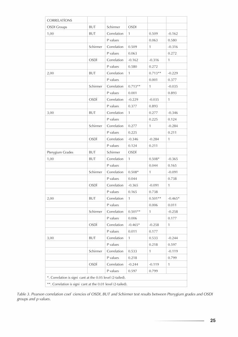
25
CORRELATİONS
OSDI Groups BUT Schirmer OSDI
1,00 BUT Correlation 1 0.509 -0.162
P values 0.063 0.580
Schirmer Correlation 0.509 1 -0.316
P values 0.063 0.272
OSDİ Correlation -0.162 -0.316 1
P values 0.580 0.272
2,00 BUT Correlation 1 0.713** -0.229
P values 0.001 0.377
Schirmer Correlation 0.713** 1 -0.035
P values 0.001 0.893
OSDİ Correlation -0.229 -0.035 1
P values 0.377 0.893
3,00 BUT Correlation 1 0.277 -0.346
P values 0.225 0.124
Schirmer Correlation 0.277 1 -0.284
P values 0.225 0.211
OSDİ Correlation -0.346 -0.284 1
P values 0.124 0.211
Pterygium Grades BUT Schirmer OSDİ
1,00 BUT Correlation 1 0.508* -0.365
P values 0.044 0.165
Schirmer Correlation 0.508* 1 -0.091
P values 0.044 0.738
OSDİ Correlation -0.365 -0.091 1
P values 0.165 0.738
2,00 BUT Correlation 1 0.501** -0.465*
P values 0.006 0.011
Schirmer Correlation 0.501** 1 -0.258
P values 0.006 0.177
OSDİ Correlation -0.465* -0.258 1
P values 0.011 0.177
3,00 BUT Correlation 1 0.533 -0.244
P values 0.218 0.597
Schirmer Correlation 0.533 1 -0.119
P values 0.218 0.799
OSDİ Correlation -0.244 -0.119 1
P values 0.597 0.799
*. Correlation is significant at the 0.05 level (2-tailed).
**. Correlation is significant at the 0.01 level (2-tailed).
Table 3. Pearson correlation coefficiencies of OSDI, BUT and Schirmer test results between Pterygium grades and OSDI groups and p values.
26
PHACOEMULSIFICATION WITH COEXISTING CORNEAL VASCULARISATION AND OPACITIESChichua G , Pargalava N. Chichua medical center ‘‘Mzera’’. Tbilisi, Georgia
Purpose: To present the surgical video of 54- year-old man with corneal opacities and a nuclear cataract who underwent cataract extraction with phacoemulsification and IOL implantation in his right eye.Settings: This video presentation is based on surgical procedure perfomed at Chichua Medical Center Mzera , Tbilisi,Georgia.Methods: After anesthesia the feeding vessels of corneal neovascularisation were coagulated to prevent intraoperative superficial bleeding we have initiated the continuous curvilinear capsulorhexis with the help of single-used cystotome. This is the most critical step of the surgery, because the visualisation of central part of lens capsule is impaired. So it is important to perform in the maneuver where the edges of rhexis go outside of corneal oppacification. In our case it was successfully done and we were able to start the next step of surgery. We mobilized the lens nucleus with hydrodissection and hydrodelineation technique. The pearls of this stage is to perform emulsification of the nucleous always directing the phaco tip not to the center, but to the periphery to avoid the damage of capsular edge. The rest of the cortical matherial was removed with bimanual I/A technique under constant rotation of eye globe to control rhexis edge and to see the red reflex. The surgery was finished with in-the bag implantation of foldable lens. Results:The procedure resulted without any intraoperative complications. After a month follow- up vis-it the lens is still in the central position and his visual acuity improved from paracentral hand motions to paracentral 20/25.Conclusion: In every non-standart case, care should be taken to avoid potential risks of intraoperative complications. Our plan was to performe minimaly invasive surgery to refrain from complications of com-bine surgical procedures.Financial Disclosure :None
IDIOPATHIC INTERMEDIATE UVEITIS: CLINICAL FEATURES IN CHILDREN Chichua G. , Chichua A. , Jikurashvili T. , Pargalava N., Chichua medical center ‘‘Mzera’’, Tbilisi , Georgia
Purpose :The aim of our study was to describe the clinical characteristics of intermediate uveitis (IU) in children.Methods:We have done retrospective analysis of medical records, evaluated clinical course of diseases, as-sessed different variables , including age and gender , laboratory data, presence of systemic diseases, clinical features, their complications ,treatment strategies with their outcomes, remission, final visual acuity. Results and conclusion : According to the results we can conclude that although the disease is sight-threaten-ing timely diagnostics , correct treatment tactics and close monitoring are the important factors for prolonga-tion of remission, prevention of serious complications and therefore maintaining of existing visual acuity.
MEIBOMIAN GLAND DYSFUNCTIONPhD. Medea Gobejishvili
The First University Clinic (Tbilisi)Academician I.P.Pavlov First St. Petersburg Medical University, Ophthalmology Department
PhD. Riks I. A.Introduction. Meibomian gland dysfunction (MGD) is very common disease. Meibomian gland dysfunc-tion is a chronic pathology of eyelids, characterized by blockage of its ducts and qualitative or quantitative changes of gland secret. It can cause dry eye and eyelid inflammation.The structural unit of the meibomian gland is the acinus, which contains meibocytes that produce fat. The meibocytes can express receptors activated by peroxisome proliferators PPARγ. These receptors are re-sponsible for the normal functioning of the glands and the quality of it’s secret.Enviralmental factors: dehydration, monitors and screens, use contact lenses, low calorie diet. Risk factors of MGD are Sjegren’s syndrome, atopy, psoriasis, rozacea, androgen deficiency, bening pros-tatic hyperplasia. Main role plays taking certain drugs – antihistamines, antiandrogens, antidepressants, postmenopausal hormone therapy by estrogens, much retinoids, drugs used to treat prostate adenoma
27
have influence on the development of MGD. Symptoms of MGD are all symptoms of dry eye. 65% patients with dry eye have MGD. Some times it will be associated with acute inflammatory disease of the ocular surface tissues, chalazion, trichiasis, anterior blepharities and etc. Diagnostic of MGD: visible blockage of the gland ducts, teleangiectasia on the posterior surface of the eyelid, loss of the eyelashes and etc.Treatment of MGD must be complex: eyelid hygiene, heating eyelid margin, reducing working time with computer, food rich with vit E, omega 3 fatty acid, eyelid massage, blink training, eye lubricant. Some times use antibiotics, steroids, antiseptics and anti- inflammatories.Relevance. MGD occurs 38-68% in persons over 40 years old, more often in the Asian race, than in Euro-peans. The relevance of the disease and its importance increased recently.Purpose: Early diagnosis and complex treatment of meibomian gland dysfunction. Evaluate the effective-ness of the eyelid massage. Evaluate the effectiveness of treatment.Material and methods: 20 eyes of 10 patients, aged 26 years to 75. Everyone did dry eye tests –Schirmer’s and Norn’s tests, biomicroscopy of eyelids.All patients was recommended eyelid hygiene, diet, warming and eye lubricants. 5 patients got local thera-py - steroid with antibiotic, 3 patients additionally got antibiotics per os: tetracycline 50 mg. 2 times daily duration 10 days and omega 3 fatty acid – 30days. 1 patient did long-acting steroid injection triamcinolone acetonide in the chalazion area. Only 1 patient gets Restasis 2 times daily. All patients have done procedures on the eyelid 10 days time frame: 1. Warming 2. After anesthesia with tetracaine 1 % we did eyelid massage with glass stick.Results. After 2 months each patients had significant improvement. The number of normally functioning glands and the stability of the tear film increased. Improving the quantity and quality of the meibum.Conclusion. Only such a comprehensive treatment is possible to obtain good results in patients with MGD.
PLATELET RICH GROWTH FACTOR IN THE TREATMENT OF COMPLEX CORNEAL DIS-ORDERSPalarie Natalia¹², Palii Natalia¹, Palarie Victor¹²
¹International Clinic, Orhei, Moldova²State University of Medicine and Pharmacy “Nicolae Testemitanu”, Chisinau, Moldova
Background: Platelet rich growth factor (PRGF) is an autologous blood product rich in proteins and growth factors which can be rapidly obtained from patient blood. Clinically, it is an affordable treatment with po-tentially broad spectrum of applications in ophthalmology especially in the treatment of complex or refrac-tory corneal wounds. Purpose: The aim of this study was to evaluate the efficiency of autologous platelet-rich factor in the treat-ment of complex corneal disorders. Methods: There were 17 patients with corneal surface disorders, among which 5 patients with chemical burns, 8 patients with corneal ulcers and 4 with neurotrophic keratopathy. Visual acuity varied from hand motion to 0.1. Solid PRGF was either just placed on the corneal surface or sutured with 2 nodes of 10-00 nylon suture at conjunctiva. If necessary, the procedure was repeated. All patients had corneal OCT scan before and after the treatment. Results: Improved visual acuity and less subjective symptoms were observed in all patients. Complete healing of cornea was observed in all patients with chemical burns. As well considerable improvement experienced 7 of 8 patients with corneal ulcers (reduced size and depth of the ulceration, improved visual acuity, smaller conjunctival injection). Also an improvement was seen in 2 of 4 patients with neurotrophic keratopathy. None of the patients reported general or local side effects of the treatment. Conclusions: PRGF is a reliable and effective therapeutic tool to promote wound healing in complex cor-neal disorders.
28
COMPARISON OF THE EFFECT OF BRIMONIDINE ON PUPIL SIZE IN GLAUCOMA PA-TIENTS AND HEALTHY SUBJECTSIbrahim TuncerAlfa Medical Center, Izmir, Turkey
AbstractObjectives: To compare the effect of brimonidine on scotopic pupil size in patients using brimonidine for the treatment of glaucoma and healthy individuals.Materials and Methods: In this study, two groups of 30 patients with early stage glaucoma using 0.15% brimonidine tartrate drop and 30 healthy individuals were created. In the glaucoma group, pupil size measurements were made in a scotopic condition (1 cd/m2) using an infrared pupillometer before and 30 minutes after a drop of 0.15% brimonidine tartrate, in accordance with the patient’s drop instillation time. Pupil size measurements were made before and after brimonidine in the right eyes of the healthy group. Data of the right eye were used for statistical analysis.Results: The mean age was 44.16 ± 8.87 in the glaucoma group and 43.06 ± 8.48 in the healthy group. The mean scotopic pupil size before brimonidine was 6.12 ± 0.99 mm in the glaucoma group and 6.15 ± 1.02 mm in the healthy group. The mean scotopic pupil size at the 30th minute after brimonidine was 4.54 ± 1.10 mm in the glaucoma group and 4.49 ± 1.07 mm in the healthy group. The mean scoto-pic pupil size decreased by 1.58 mm in the glaucoma group and 1.66 mm in the healthy group. In both groups, the mean scotopic pupil size after brimonidine was significantly lower than before brimonidine (p <0.001 for both).Conclusion: A single dose of 0.15% brimonidine tartrate drop produced significant miosis in early stage glaucoma, similar to that in normal eyes. A single dose of brimonidine drop can be effective in reducing night vision complaints after laser refractive and premium intraocular lens surgery in early stage glaucoma patients.
QUANTITATIVE ASSESSMENT OF RETINAL CHANGES IN COVID-19 PATIENTSDr. Gözde AKSOY AYDEMİR1, Dr. Emre AYDEMİR11: Ophthalmology Department, Adıyaman University Training and Research Hospital, ADIYAMAN, TURKEY
AbstractPurpose: We investigated the impact of COVID-19 on the retinal layers parameters in previously con-firmed COVID-19 patients by taking advantage of spectral domain optical coherence tomography (SD-OCT) and compared these results with those obtained from controls.Design: Prospective cross-sectional studyMethods: All subjects underwent a thorough ophthalmic examination, including best corrected visual acuity testing using the Snellen chart (6 m), intraocular pressure measurements using a pneumotonometer, ocular surface and anterior segment assessment by slit lamp biomicroscopy, and dilated fundus examina-tion. All the macular thickness measurements were performed using SD-OCT by the same technician. Results: This study involved 52 eyes from 52 subjects: 31 subjects comprised the COVID-19 group (15 females, 16 males), and the remaining 21 were the control group (11 females, 10 males). Statistically sig-nificant differences were seen in the mean central macular thickness values between the COVID-19 group and the control group (p = 0.02). The ganglion cell layer thickness values, and those of the inner nuclear layer of the COVID-19 group, were both significantly thinner compared to the control group (p = 0.04 and p = 0.04, respectively). Conclusion: We investigated possible pathological COVID-19 effects on the posterior segment of the eye by examining the macula and each retinal layer in COVID-19 patients. The present results demonstrate that some macular measurements were changed in the COVID-19 group compared to age-matched con-trol subjects, suggesting possible retinal involvement as part of COVID-19. These changes in the macula, ganglion cell layer, and inner nuclear layer could be seen in the early recovery phase, and these patients should be followed closely to identify any new pathologies that may develop in the late recovery phase.
29
COMPARISON OF THE EFFICACY OF AFLIBERCEPT AND RANIBIZUMAB IN MACULAR EDEMA WITH SEROUS RETINAL DETACHMENT SECONDARY TO BRANCH RETINAL VEIN OCCLUSIONAlper Halil BAYAT11 Department of Ophthalmology, İstanbul Medipol University, İstanbul, Turkey
Purpose: To compare the efficacy of intravitreal aflibercept (IVA) and ranibizumab (IVR) in macular edema (ME) with serous retinal detachment (SRD) secondary to branch retinal vein occlusion (BRVO).Materials and methods: Thirty-eight eyes of 38 patients who underwent IVA (n = 20) and IVR (n = 18) with macular edema secondary to BRVO were investigated retrospectively. All patients had SRD at base-line. After 3 consecutive IVA or IVR treatments, patients received additional injections in case of need. Best corrected visual acuity (BCVA), central macular thickness (CMT) and SRD heights were compared after 6 months of follow-up.Results: The patients were similar in terms of age, gender, baseline BCVA, CMT and SRD heights (p> 0.05). Although there was no statistical difference between the groups in BCVA changes over the 6-month period, the IVA group was found to have a better vision (p = 0.058). In terms of CMT gains, the IVA group achieved significantly more gain in the first month (p = 0.014). 6-month CMT gains were found to be sim-ilar (p = 0.548). No difference was found in terms of SRD changes (p = 0.202). At the end of 6 months, SRD was observed in 6 patients (33%) in the IVR group and 3 patients (15%) in the IVR group (p = 0.001).Figure-1: Comparison of best-corrected visual acuity changes between groupsFigure-2: Comparison of central macular thickness changes between groupsConclusion: Both drugs have been found to be effective in ME with SRD due to BRVO. IVA provided rapid anatomical gain. IVA was more effective in regression of SRD.
LONG-TERM RESULTS OF SURGICAL TREATMENT OF EYELIDS SKIN BASAL CELL CARCI-NOMA - 9 YEARS OF OBSERVATIONKardava T. PhD., Bregvadze E. “New Hospitals”, Tbilisi, Georgia.
Basal cell carcinoma(BCC) is one of the most common malignancies of the skin. According to the Inter-national Cancer Registry, there are 14.3-19.6 cases per 100 thousand. Population and 11.6% are on the eyelid and periorbital area, accounting for 90% of malignant tumors of the eyelid. And mortality on aver-age - 0.1%. Its main treatment is surgery. At the same time, recurrence is frequent, requiring additional intervention - repeat surgery, radiation therapy, and consequently leading to more serious cosmetic and functional prob-lems. It should be noted that in many cases the postoperative defect is quite large and requires immediate reconstruction to maintain eyelid function, which further determines the patient’s quality of life. PURPOSE: 2012-2020 Evaluation of the effect of radical surgical treatment of basal cell carcinoma of the eyelid skin.METHODS: The 116 patients were operated with histologically confirmed eyelid BCC T2bN0M0 size (tumor diameter 10-20 mm, mean 14.2 ± 4.5 mm) with instant reconstruction of the defect 2012-2020 in the ophthalmology department of the “New Hospitals” clinic. Males 61 (52.6%), females -55 (47.4%). The average age is 69 ± 10.2 years. Clinical forms: nodular 79 (68.1%), ulcerative-nodular-31 (26.7%), infil-trative - 6 (5.2%). In the nodular form, the formation is excised not less than 3 mm from the visible edges, in the ulcerative and infiltrative variants - not less than 4 mm, with ablation of the base. According to the histological examination of the postoperative material, all surgeries were radical - surgical resection edges free from tumor cells. Nevertheless, in the case of the infiltrative type, patients still underwent local radia-tion therapy to prevent relapse.RESULTS: During the observation period 01.2012-11.2020. (107 months) No continuous increase or re-currence was detected.CONCLUSIONS : In case of radical surgical treatment of BCC of the eyelid skin, the possibility of recur-rence is minimal and reconstructive surgery is effective, which determines the patient’s life expectancy and quality of life.
30
ANTI-VEGF DRUGS USAGE IN COMPLEX TREATMENT OF NEOVASCULAR GLAUCOMA IN PATIENTS WITH DIABETIC RETINOPATHYIryna Pastukh, Nataliya Goncharova, Uliana PastukhOphthalmology Department, Kharkiv Medical Academy of Postgraduate Education, Kharkiv, Ukraine
Purpose: To present and analyze results of Aflibercept usage in complex treatment of neovascular glau-coma (NG) in patients with non-proliferative diabetic retinopathy (nPDR) and diabetic macular oedema (DMO).Methods: Eight adult patients (8 eyes) with NG and nPDR with DMO were assigned to receive 2 monthly intravitreal (IVT) injections of Aflibercept. One week after first injection Trabeculectomy was performed and intraocular pressure (IOP) decreased. One week after second injection Phacoemulsification with intraocular lens (IOL) implantation was performed. After 3-4 weeks retinal laser coagulation performed for all eyes. Assessment of visual acuity changes was the percentage of patients gaining of best-corrected visual acuity (BCVA); the central retinal thickness (CRT) measured by optical coherence tomography (OCT); tonometry, biomicroscopy, fundus photographs, and fluorescein angiography were evaluated. Results: One week after first IVT Aflibercept injection iris neovascularization decreased and Trabeculecto-my was performed. IOP becomes 14-17 mm Hg. One week after second Aflibercept injection Phacoemul-sification with IOL implantation was performed. After 2 months of follow-up all patients gained BCVA; mean BCVA improved on 10-15%. CRT showed a mean decrease of 45-55%. IOP was normal. These gave the opportunity to start retinal laser coagulation for all eyes. After 6 and 12 months of follow-up BCVA and IOP remained steady. Term of observation is two years. Conclusions: We used IVT Aflibercept injections to prepare patients with NG and nPDR with DMO for surgical treatment. It gave opportunity to decrease CRT, iris neovascularization; perform surgery and retinal laser coagulation. Improvement of BCVA is very important for visually impaired patients of our group.
Interferon α and γ in the lachrymal fluid of patients with uveitisKonovalova N.V., Khramenko N.I., Guzun O.V.SI “The Filatov Institute of Eye Diseases and Tissue Therapy NAMS of Ukraine”. Odessa, Ukraine
The contents of cytokines under investigation in uveitis are significantly higher in the lachrymal fluid than in the blood plasma (2,2 and 1,8 respectively), which is evidence of dominance of their expression locally. There was revealed paired positive correlation between baseline interferon α in the lachrymal fluid and plasma of patients with uveitis (n=10, r=0,76, p<0,05), which is evidence of the possibility of the charac-teristics of the pathological process in the eye by the change of its expression in the peripheral blood. The data analysis of changes in the level of interferon expression in the lachrymal fluid of patients with uveitis after treatment showed that the content of interferon α correlates with the corresponding data in the blood of patients before therapeutic interventions (n=10, r=0,74, p<0,05). We founded an association between the level of interferon γ in the lachrymal fluid of patients of uveitis before and after the treatment (n=10, r=0,84, p<0,05).
ИНТЕРФЕРОНЫ A И G В СЛЕЗНОЙ ЖИДКОСТИ БОЛЬНЫХ УВЕИТОМКоновалова Н.В., Храменко Н.И., Гузун О.В.ГУ «Институт глазных болезней и тканевой терапии им. В.П. Филатова НАМН Украины» Одесса, Украина
Анализ данных выраженности изменений уровня интерферонов в слезной жидкости больных увеитом после лечения показал, что содержание интерферона a коррелирует с соответствующими данными в крови этих пациентов до лечебных воздействий (n=10, r=0,74, p<0,05). Выявлена связь между уровнем интерферона g в слезной жидкости пациентов с увеитом до и после лечения (n=10, r=0,84, p<0,05).

31
PLASMA BASED ENERGY FOR TREATING DIFFENENT OPHTHALMOLOGICAL PATOLOGIESNino Gaphrindashvili, Ophthalmologist, laser surgeon, aestheticien, head of aesthetic center FEMINALevan Nakashidze, Clinical specialist and trainer of cold plasma and laser systems.
Introduction: Plasma is an electrically neutral medium of unbound positive and negative particles (i.e. the overall charge of a plasma is roughly zero), also defined as Ionized gas and it represents the 99% of the “visible” universe. A plasma can be created by heating a gas or subjecting it to a strong electromagnetic field. The electrons kinetic energy is higher then potential energy of molecular bonds, electrons are free and ions are generated. It is used for acne, xanthelasmas, warts, lentigo, aging, scars, stretch marks, wrin-kles, fibromas and Blepharoplasty.Plasmage is manufactured in Italy. It is medical device CE 93/42 certified, company ISO9001 - EN13485, working through a global distribution network in Europe, Asia & the Middle East.In Aesthetic Medicine Center FEMINA we have treated 53 patients with Brera Plasmage device in 2020. 28 pacients on eyelids (14 with both eyelids, 9 patients with epper eyelids and 5 patients with lower eyelid festoons), 11 pacients with xanthelasmas, 9 pacients with eyelid papilomas and 5 pacients with bening malformations. Plasma can boost the sublimation process of the tissues and it can be a solution for all skin types with visible and immediate results, long lasting effects, high precision, safe protocols.
МИКРОИМПУЛЬСНАЯ ЦИКЛОФОТОКОАГУЛЯЦИИ (МЦФК) У ПАЦИЕНТОВ С РЕФРАКТЕРНОЙ ГЛАУКОМОЙИошин И.Э., Толчинская А.И., Максимов И.В., Ракова А.В.ФГБУ « Клиническая больница» УДП РФ, Москва.
Одной из тяжелых форм глаукомы является рефрактерная (РГ), которая характеризуется упорным течением и устойчивостью к традиционным методам лечения (Егоров Е.А., 2011; RodrigesA.M. etal., 2004). Особое место в лечении данной формы глаукомы занимают циклодеструктивные вмешательства, в частности, контактная непрерывная диод-лазерная циклофотокоагуляция (ЦФК) с длиной волны 810 нм, направленная на подавление продукции водянистой влаги вследствие частичной атрофии цилиарного тела и его отростков за счет лазерного воздействия ( Дробница А.А., 2015; Поступаев А.В. и др., 2015; Роустова О.В. и др. 2003; IlievM.E. etal., 2007). И хотя данный метод эффективен в снижении ВГД, чаще всего он используется как метод последнего выбора в качестве лечения из-за предполагаемого риска значительных осложнений (БойкоЭ.В. и др., 2012; ЕгоровВ.В. идр., 2007; EgbertP.R. al., 2001; Walland M.J. 2000).Предложенная в последние годы микроимпульсная ЦФК ( Tan A.M. etal., 2010; Lee J.W. etal., 2015; Aquino M.C. etal., 2011) может стать альтернативным методом в лечении рефрактерных форм глаукомы.Цель: анализ результатов лечения пациентов с рефрактерной глаукомой различной стадии методом микроимпульсной циклофотокоагуляции с применением стандартных параметров воздействия.Материал и методы. Обследован 61 больной в возрасте 73,7 лет с неоднократно оперированной некомпенсированной первичной открытоугольной глаукомой (ПОУГ) развитой (13), далеко зашедшей (34) и терминальной стадии (14). До и после операции мЦФК всем пациентам проводилось комплексное офтальмологическое обследование. Процедура мЦФК проводилась под местной анестезией (прибор SUPRA 810, Quantel Medical, Франция) со стандартной заданной мощностью 2000 mW и рабочим циклом 31,3%. Операции проведены без осложнений. Срок наблюдения составил 6 месяцев.Результаты. Течение раннего послеоперационного периода проходило ареактивно. Через 1 неделю гипотензивный эффект был достигнут во всех случаях. У пациентов с развитой стадией заболевания с исходным средним ВГД 26,8±8,8мм рт. ст. (по Маклакову) к концу 6 месяцев наблюдения отмечено достоверное снижение показателей ВГДдо 17,1±8,1мм рт. ст. (на 36,2%;p<0,05).Аналогичные результаты отмечены и в группе больных с далеко зашедшей стадией заболевания. Через 6 месяцев ВГД снизилось с 29,9±7,8 до 21,0±8,3 мм рт. ст. (на 29,8%;p<0,05). У пациентов с терминальной стадией заболевания с исходным средним ВГД 38,1±8,3 мм рт. ст. к6 месяцу наблюдения ВГД снизилосьдо 29,3мм±8,1 рт. ст. (на 28,0%;p<0,05). Таким образом, во всех случаях доказана эффективность мЦФК, под которой понимали снижение ВГД на 20и >% от исходного на фоне гипотензивного лечения.
32
Количество применяемых гипотензивных препаратов после проведенной процедуры мЦФК сократилось во всех группах ( на 27% при развитой, на 22% при далеко зашедшей и на 10% при терминальной стадии).Заключение. Лечение пациентов с РГ различной стадии методоммЦФК, является эффективным и безопасным методом снижения ВГД и уменьшения количества применяемых гипотензивных препаратов. Стабилизация ВГД после проведенной процедуры мЦФК с различной стадией глаукомы в течение 6 месяцев отмечалась в 52 (85,2%) случаях. Дальнейшее изучение микроимпульсного лазерного воздействия в различных клинических ситуациях у пациентов с глаукомой, возможно, расширит границы ее применения и при других формах заболевания.
MICROPULSE CYCLOPHOTOCOAGULATION (MCFC) IN PATIENTS WITH REFRACTORY GLAUCOMAIoshin I.E., Tolchinskaya A.I.,Maximov I.V., Racova A.V.Federal State Budgetary Institution Clinical Hospital, Moscow.
One of the severe forms of glaucoma is refractory (RG), which is characterized by a persistent course and resistance to traditional methods of treatment (Egorov E.A., 2011; Rodriges A.M. et al., 2004). A special place in the treatment of this form of glaucoma is occupied by cyclodestructive interventions, in particular, contact continuous diode-laser cyclophotocoagulation (CPC) with a wavelength of 810 nm, aimed at sup-pressing the production of aqueous humor due to partial atrophy of the ciliary body and its processes due to laser exposure (Drobnica A A., 2015; Postupaev A.V. et al., 2015; Rustova O.V. et al. 2003; Iliev ME et al., 2007). And although this method is effective in reducing IOP, it is most often used as the last-choice method of treatment due to the perceived risk of significant complications (Boyko E.V. et al., 2012; Egorov V.V. et al., 2007; Egbert PR al. 2001; Walland MJ 2000). Microimpulse CFC proposed in recent years (Tan A.M. et al., 2010; Lee J.W. et al., 2015; Aquino M.C. et al., 2011) can become an alternative method in the treatment of refractory forms of glaucoma.Purpose: to analyze the results of treatment of patients with refractory glaucoma of various stages by the method of microimpulsecyclophotocoagulation using standard exposure parameters.Material and methods. We examined 61 patients aged 73.7 years with repeatedly operated uncompen-sated primary open-angle glaucoma (POAG) developed (13), far-reaching (34) and terminal stages (14). Before and after the mCFK operation, all patients underwent a comprehensive ophthalmological examina-tion. The mCFK procedure was performed under local anesthesia (SUPRA 810 device, Quantel Medical, France) with a standard set power of 2000mW and a duty cycle of 31.3%. The operations were performed without complications. The observation period was 6 months.Results. The course of the early postoperative period was areactive. After 1 week, the hypotensive ef-fect was achieved in all cases. In patients with advanced stage of the disease with an initial mean IOP of 26,8±8,8mm Hg. Art (according to Maklakov) by the end of 6 months of observation, a decrease in IOP parameters 17,1±8,1mm Hg was noted Art (by 36,2%; p<0,05). Similar results were observed in the group of patients with advanced stage of the disease. After 6 months, IOP decreased from 29,9±7,8to 21,0±8,3 (на 29,8%; p<0,05).In patients with end-stage disease with an initial mean IOP of 38,1±8,3mm Hg. art by the 6th month of observation, IOP decreased to 29,3мм±8,1 mm Hg art (by 28,0%;p<0,05). Thus, in all cases, the efficacy of mCFK was proved, which meant a 20% decrease in IOP from the initial value against the background of antihypertensive treatment.The number of antihypertensive drugs used after the mCFK procedure decreased in all groups (by 27% in the advanced stage, by 22% in the advanced stage and by 10% in the terminal stage).Conclusion. Treatment of patients with RH of various stages with the mCFK method is an effective and safe method of lowering IOP and reducing the amount of antihypertensive drugs used. Stabilization of IOP after the performed mCFK procedure with different stages of glaucoma within 6 months was noted in 52 (85.2%) cases. Further study of micropulse laser exposure in various clinical situations in patients with glau-coma may expand the scope of its application in other forms of the disease.
Для контактов: Толчинская Анна Ивановна.Тел. 8-916-125-06-25Почта: [email protected].
33
СЕЛЕКТИВНАЯ ЛАЗЕРНАЯ ТРАБЕКУЛОПЛАСТИКА (СЛТ) НА ПСЕВДОФАКИЧНЫХ ГЛАЗАХАрташес Арто Зильфян Д.м.н, Профессор
«Zilfyan EyeCare Center»Главный Офтальмолог г.Еревана. Ереван, Республика Армения.
Введение.Считается установленным, что начальные стадии первичной открытоугольной глаукомы и глазной гипертензии обычно лечатся глазными каплями, которые снижают внутриглазное давление. Селективная лазерная трабекулопластика является безопасной альтернативой антиглаукоматозмым препаратом. При селективной лазереной трабекулопластике используются короткие лазерные импульсы с низким уровнем энергии.Впервые Латина предложил SLT-Latina 1996, FDA 2001-Nd:YAG Q-switched 532nm.
Актуальность.ПреимуществаСелективный термофотолизис меланосомс трабекулярного аппаратаМинимальное термальное воздействие-без повреждения коллагеновой тканиИндукция выроботки цитокинов и активация макрофагов(очистка экстрацелюлярного дебриса)Стимулирует клеточную активность и прохождение жидкости через трабекулярный аппаратНа основании вышеперечисленных данных СЛТ может являтся методом первого выбора лечения глаукомы.ПоказанияПОУГПигментная глаукомаПсевдоэксфолиативная глаукомаДо и после ФакоэмульсификацииКак метод первого выбора лечения
Противопоказания.Врожденная глаукомаИнфантильная глаукомаЮвенильная глаукомаЗакрытоугольная глаукома
Цель исследования.Оценить эффективность СЛТ в лечении первичной открытоугольной глаукомы на факичных и псевдофакичных глазах в ранний период после лазерного вмешательства.
Материал и методы.Впервые в Армении на базе “Zilfyan EyeCare Center”подтверглись селективной лазерной трабекулопластике 60 пациентов (120 глаз). Для анализа нами были созданы две группы: I- группа 30 пацинетов (60 глаз) с факичными глазами,II- группа 30 пациентов (60 глаз) с псевдофакичными глазами. I стадия глаукомы была зарегистрирована на 54 глазах (45% ), II стадия – на 66 глазах (55 %). Все пациенты были под режимом до проведения процедуры: I стадия- 1 антиглаукоматозный препарат, II стадия- 2 антиглаукоматозных препарата.Селективная лазерная трабекулопластика выполнялась на установке Quantel Medical (Optimis Fusion Laser) при следующих параметрах: длина волны 532 нм, время импульса 4 нс, мощность 0,8–1,5 мДж, диаметр пятна 400 нм, количество импульсов 50-60. Лазерное вмешательство выполнялось на 180 градусов, таким образом, чтобы коагуляты не перекрывали друг друга по площади. Мощность процедуры регулировалась в зависимости от степени пигментации трабекулы: начиная с 0,8 мДж, постепенно увеличивали мощность до появления кавитационных пузырьков, затем мощность уменьшали на 0,1 мДж и продолжали выполнение селективной лазерной трабекулопластики. После проведения селективной лазерной трабекулопластики был отменен режим закапывания гипотензивных препаратов .
34
Результаты и обсуждение.На 3-4 сутки после селективной лазерной трабекулопластики отмечалось снижение внутриглазного давления в 83.9% случаях в обеих группах. Разница была аналогичная в обеих группах после выполнения процедуры. При ПОУГ I снижение внутриглазного давления было отмечено в 92% случаях. При ПОУГ II внутриглазное давление снизилось в 78.9% случаях. Надо отметить что снижение ВГД не явлается абсолютно обьективной цифрой, поскольку изначально все пациенты были под медикаментозным режимом.Заключение.На основании полученных высоких результатов и литературных данных мы пришли к заключению:СЛТ является безопасных и эффективным методом лечения ПОУГДанным метод имеет высокую эффективность как на начальных стадиях , так и при развотой стадии глаукомы.СЛТ не имеет серьезных противопоказаний и может рутинно выполнятся в амбулаторных условиях.Данный метод имеет аналогичную высокую эффективность как на факичных глазах, так и на псевдофакичных.На основании множества литературных данных и высокой функциональности мы также считает, что СЛТ может быть использована в качестве первой линии выбора в лечении ПОУГ и офтальмогипертензии.
FEATURES OF RETINAL STRUCTURE AFTER SUCCESSFUL VITRECTOMY IN PATIENTS WITH POSTTRAUMATIC ENDOPHTHALMITISGrubnyk N.P., Krasnovid T.A.The Filatov Eye Institute of Eye Disease and Tissue Therapy, Odessa, Ukraine
Purpose: To investigate features of the retinal structure after successful vitrectomy in patients with post-traumatic endophthalmitis using spectral domain optical coherence tomography (SD-OCT).Methods: SD-OCT was performed in 27 patients who underwent pars plana vitrectomy (PPV) due to posttraumatic endophthalmitis. All patients were with penetrating eye injuries, 16 of them - with intraocu-lar foreign body (IOFB), which was removed during PPV. SD-OCT was performed after inflammation was controlled.Results: On SD-OCT images taken more than 1 month after vitrectomy due to posttraumatic endoph-thalmitis, macular edema (diffuse or with intraretinal cysts) was detected in 24 eyes (67%), but 6 of them (22%) had combination of retinal edema in nasal segment and atrophy in fovea and temporal segment. Retinal atrophy in macula was observed 7 eyes (26%). Epiretinal membrane was found in 20 eyes (74%), 13 (65%) of them were in patients with IOFB. Ellipsoid band disruption was identified in 15 eyes (55%). Conclusion: SD-OCT features after vitrectomy due to posttraumatic endophthalmitis were analyzed. The most frequent retinal structural changes included epiretinal membrane (74%), macular edema (67%), ellipsoid disruption (55%). The development of retinal changes may be associated with penetrating injury without or with IOFB and severe endophthalmitis in the history.
DOES THE SYSTEMS OF ANTERIOR CHAMBER STABILISATION AND IOP CONTROL ARE HELPFUL DURING ROUTINE CATARACT PHACOEMULSIFICATION? Shevchyk V., Voziyan S., Lysenko KLLD “Shevchyk Vasyl eye microsurgery”, Chernihiv, Ukraine.
PURPOSE: To estimate the opportunity of modern systems of anterior chamber stabilisation and IOP con-trol to preserved endothelial cells during standard cataract phacoemulsification.SETTING:METHODS: In our study we include 200 patients (cataract grade 2-3) that were divided into two groups. First (control group) - 100 patients were undergo phacoemulsification on Stelleris System, second (study group) - on Stellaris Elite System (with IOP and anterior chamber stability control). All surgeries were per-formed during 1 month by one surgeon using the same technique, viscoelastics and instruments. Endotheli-al cells counting in central area just before surgery and after 30 days were done, using specular microscope SP-1P (Topcon). RESULTS: Before surgery amount of endothelial cells in both groups were similar - 2430±260 and
35
2390±233 cells/mm2, between-group differences – P = 0.97). On the first post operation day clear, transparent cornea in study group and slight oedema in control group were noticed, that was confirmed by better visual acuity of patients in study group.After 1 month loss of endothelial cells in both groups were noticed, but in the control group it was 13.6±6.6%, and in the study - only 3.8±3.6%, between-group differences – P= 0.005. CONCLUSIONS: Implementation in routine practice phacoemulsification devices with IOP and anterior chamber stability control allow us to preserve endothelial cells and receive better visual recovery as well.
DOES THE SYSTEMS OF ANTERIOR CHAMBER STABILIZATION AND IOP CONTROL CAN DECREASE ENDOTHELIAL CELLS LOSS DURING CATARACT PHACOEMULSIFICA-TION?Shevchyk V.I.LLD “Shevchyk Vasyl eye microsurgery”, Chernihiv, Ukraine.
PURPOSE:To assess the opportunity of modern systems of anterior chamber stabilization and IOP control to pre-served endothelial cells during standard cataract phacoemulsification.SETTING:METHODS:Our study comprises 200 patients (cataract grade 2-3) divided into two groups: control and study.In first case, randomly selected 100 patients were selected to undergo phacoemulsification on Stelleris System, in second (study group) - on Stellaris Elite System (with IOP and anterior chamber stability control). All surgeries were performed during 1 month by one surgeon using the same technique, viscoelastics and instruments. The count of endothelial cells in central area was completed just before surgery and after 30 days were done, using specular microscope SP-1P (Topcon).RESULTS:Before surgery the counts of endothelial cells in both groups were similar - 2430±260 and 2390±233 cells/mm 2 , between-group differences – P= 0.97).On the first day post operation, a clear, transparent cornea in study group and slight oedema in control group was noticed, that was confirmed by better visual acuity of patients in study group.After 1 month, a loss of endothelial cells in both groups were noticed, but in the control group it was 13.6±6.6%, and in the study - only 3.8±3.6%, between-group differences – P= 0.005.CONCLUSIONS:The implementation of phacoemulsification devices with IOP and anterior chamber stability control in routine practice allows us to preserve endothelial cells and receive better visual recovery as well.
РАЗРЫВЫ ПИГМЕНТНОГО ЭПИТЕЛИЯ: ЛЕЧИТЬ ИЛИ НЕ ЛЕЧИТЬ?Малиновская И.И.1, Варшанидзе М.1, Парджанадзе Т.3, Глонти З.4, Заборовский И.Г.2
1ГУО «Белорусская медицинская академия последипломного образования», Минск, Беларусь
2УЗ «10-я городская клиническая больница», Минск, Беларусь
3Глазная клиника «Новое зрение», Тбилиси, Грузия4Клиника Хелсикори, Тбилиси, Грузия
Цель исследования - проанализировать диагностические возможности, структурные и функциональные изменения при развитии разрыва пигментного эпителия сетчатки (РПЭС) на фоне различных заболеваний глазного дна, а также оценить результат применения антиангиогенной терапии.Материал и методы. Под наблюдением находились 12 пациентов с установленным диагнозом РПЭС. Средний возраст пациентов составил 72,2±12,9 лет, распределение по полу было равномерным. У 11 пациентов (92%) разрыв пигментного эпителия развился на фоне экссудативной возрастной макулодистрофии (эВМД), у 1 (8%) на фоне острой центральной серозной хориоретинопатии. У 6 пациентов РПЭС развился спонтанно, у 2 – после проведения фотодинамической терапии, у 3 пациентов на фоне антиангиогенной терапии. Срок наблюдения в среднем составил 7,4±4,6 месяцев.
36
Результаты. У 9 пациентов (67%) РПЭС развился на фоне высокой отслойки ПЭС (≥450 мкм). Средняя острота зрения составила 0,16±0,14 при постановке диагноза. Наиболее информативными методами исследования для диагностики РПЭС были ОКТ и АФ. Определить четкие границы РПЭС позволяет АФ, при проведении которой вся площадь отсутствия ПЭС выглядит четкоочерченной зоной гипоаутофлюоресценции. Признаки активной ХНВ были отмечены в 11 случаях (73%), у 7 пациентов (64%) была начата или продолжена терапия ингибиторами ангиогенеза после формирования РПЭС. Было выполнено 1,4±1,5 интравитреальных инъекций. У 3 пациентов после проведения полного курса трех загрузочных доз ингибиторов ФРЭС и у 1 пациента уже после первой инъекции была достигнута инактивация ХНВ, подтвержденная данными обследований. Во всех этих случаях отмечено полное прилегание наружных слоев сетчатки к мембране Бруха в зоне РПЭС без формирования пролиферативной ткани. В 3 случаях область разрыва покрылась соединительной тканью, четко определяющейся на снимках и при бинокулярной офтальмоскопии. Анализ этих случаев показал, что они сопровождались выраженной активностью ХНВ в следствие несвоевременной отмены антиангиогенной терапии или при отказе от ее применения.Выводы. С целью точной диагностики РПЭC рекомендуется выполнение не только ОКТ, но и аутофлюоресценции. Тактика лечения пациента ингибиторами ангиогенеза при РПЭС должна иметь индивидуальный подход с оценкой динамики процесса после каждой инъекции. Тенденция к снижению риска формирования пролиферативной ткани в области РПЭС на фоне анти-VEGF терапии в связи с резорбцией субретинальной жидкости и уменьшением риска дальнейшего роста ХНВ указывает на положительное влияние антиангиогенного лечения даже в случае формирования РПЭС.
THE RETINAL PIGMENT EPITHELIUM TEARS: TO TREAT OR NOT TO TREAT?Malinouskaya I.1, Varshanidze M1., T. Parjanadze3, Z. Glonti4, Zabarouski I.2
1Belarusian Medical Academy of Postgraduate Education, Minsk, Belarus
210th City Clinical Hospital, Minsk, Belarus
3Eye Clinic Akhali Mzera, Tbilisi, Georgia4 Clinic Helsicore, Tbilisi, Georgia
The objective is to analyze the diagnostic possibilities, structural and functional changes of retinal pigment epithelium (RPE) tears while different retinal diseases and to evaluate the antiangiogenic therapy results.Material and methods. There were twelve patients with RPE tears examined. The average age was 72,2±12,9, the same number of male and female patients. Eleven patients (92%) had the RPE tear due to age-related exudative degeneration and one patient (8%) ‒ as a result of CSCP. Six patients had sponta-neous disease development, two ‒ as a consequence of photodynamic therapy, three ‒ during antiangio-genic treatment. The mean observation period was 7,4±4,6 months.Results. Nine patients (67%) had RPE tears developed with vertical PED height (≥450 microns). The mean BCVA was 0,16±0,14. The most informative diagnostic methods for RPE tear were OCT and FAF. FAF was for detection of the definite pathology bounders and RPE microrips. 11 cases (73%) demonstrated the signs of active CNV, 7 of them (64%) started or continued antiangiogenic therapy. In general, 1,4±1,5 injections were performed. The CNV inactivation was a result of 3 intravitreal injections for three patients and 1 dose for 1 of them. In all these cases, we noticed that the outer retina was directly attached to Bruch’s mem-brane without an ingrowth of proliferative tissue along the Bruch’s membrane at the area where the RPE was lost. In three patients, the area without RPE was covered with a thickened proliferative tissue revealed during binocular ophthalmoscopy or noticed on fundus photos. The observation showed that the possible reason for it was the persistent sub- or intraretinal fluid as a result of active CNV because of the untimely cancellation of antiangiogenic therapy.Conclusion. It is recommended to use FAF and OCT for RPE tear detection. The individual approach for treatment RPE tear with dynamic process estimating after each injection of an antiangiogenic agent is needed. The tendency to decrease the risk of proliferative tissue growing showed the beneficial effect of a continuative anti-VEGF therapy on the macular morphology for RPE tear.
37
КРИСТАЛЛОГРАФИЧЕСКАЯ ДИАГНОСТИКА СЛЕЗЫ ПРИ ДИАБЕТИЧЕСКОЙ РЕТИНОПАТИИ СЕТЧАТКИ.Р.О. Мухамадиев Самаркандский медицинский институт Республика Узбекистан
Актуальность. Исследование кристаллограммы слезной жидкости при диабетических ретинопатиях приобретает особую ценность. В литературе приведены результаты кристаллографии слезы в диагностике различной формы диабетической ретинопатии и выявлены различные особенности В ряде патологии глазного яблока кристаллография слезы обеспечивает надежные обнаружения угрозы рецидивов Цель : изучить особенности кристаллограмм нативной слезы у больных с диабетической ретинопатиейМатериал. Клинические наблюдения и исследования выполнены на здоровых глазах 10 добровольцев (10 глаз) и Возраст колебался от 47 до 55 года. Из них мужчин было 7, женщин 6. Диабетическая ангиопатия в начальной стадии- 2 глаза, диабетическая ангоретинопатия с микро аневризмами 7 глаз, диабетическая пролиферативная ретинопатия сетчатки 4 глаза .Методика исследования. Для получения кристаллограмм слезы нами разработан простой способ забора капли слезы и высушивания на предметном стекле . Применена цифровая фотостудия, состоящая из цифровой фотокамеры, совмещенной с бинокулярным микроскопом и персонального компьютера, позволяющая одномоментно фотографировать, архивировать и систематизировать полученных результатов. .В пролиферативной стадии диабетической ретинопатии, при разрастании новообразованных сосудов и соединительнотканных мембран, , являющееся в данном случае субстратом, средой для разрастания новообразованных сосудов, кровь, соединительно тканные мембраны, разросшиеся на поверхности сетчатки.Обсуждение результатов.При диабетических поражений в ранних стадиях развития патологического процесса когда на сетчатке нет грубых изменений кристаллография существенно не отличается от нормы. Кристаллограммы напоминают чаще всего снежинки и папортника с хорошо выраженными вторичными разветвлениями. При появления выраженной стадии ретинопатии сетчатки с микроаневризмами, расширениями вены сетчатки и мелькими кровоизлияниями , снежинки начинает местами разбухаться, однако сохраняется вторичные разветвлении с сохранениями равномерных интервалов. При грубых изменениях на сетчатке в пролиферативной стадии с резкими снижениями зрительных функций при сохранности ромашко образности главных строений усилывается разбуханности ветки кристаллов. Начинает отчетливо пояляться оторванные ветки и пустых участков между калониями кристаллов Заключение Все выше изложенные свидетельствуют о том, что при диабетической ретинопатии при начальных стадиях грубо не нарушены соотношения органических и неорганических соединений. Ферментативные процессы все еще действуют в компенсаторной стадии. Когда диабетические ретинопатии находятся в запушенной стадии , компенсаторные механизмы питания тканей глаза отсутствуют, сахар в крови резко увеличился потерялся все механизмы кристаллизации по формированию снежинки или же ромашки. В поле зрения видны отдельно разбухшие скопления сахаристого содержания.
КРИСТАЛЛОГРАФИЧЕСКАЯ ДИАГНОСТИКА СЛЕЗЫ ПРИ ХОРИОРЕТИНАЛЬНОЙ ДИСТРОФИИ СЕТЧАТКИ.Мухамадиев Р.О. Самаркандский медицинский институт Республика Узбекистан
Актуальность. В организме человека находится различные биологические жидкости. Каждый из них имеет определенные кристаллографические картины в зависимости от содержания химического компонента. На каждом этапе развития дистрофических патологий сетчатки дают различные кристаллографические картины .Цель. Изучить особенности кристаллограмм нативной слезы при различных стадиях развития хориоретинальных дистрофий сетчатки.Материал и методика исследования. 24 больных с различной степени тяжести (24 глаза), находившихся на стационарном и амбулаторном лечении в (2012-2020 гг.). Возраст пациентов
38
колебался от 63 до 79 лет В начальном периоде не экссудативной формы ХРД было у 4 пациентов, атрофии пигментного эпителия и хорио капиллярного слоя у 3 больных, развитая экссудативной формы хориоретинальной дистрофии было у 6 больных.),Всем пациентам нами проводились ксенотрансплантация для лечения хориоретинальных дистрофий сетчаткиОбсуждение результатов. Кристаллизации при ХРД сетчатки во всех проявлениях резко отличаются от всех других глазных патологиях, нет ни снежинки, ни ромашки, ни сосновые ветки. В ранних стадиях видны грубые оборванные стебли, от него отходит разновеликие вторичные разветвления ветки второго порядка. (острота зрения 0.08-0.1). А при остроте зрения 0.02 - 0.03 на глазном дне огромные очаги макулодистрофии сетчатки с узкими сосудами видны кристаллы без никаких главных стеблей папортника. Кристаллы разбросаны по всему периметру нет порядка, не связанные друг с другом. Это свидетельствует о грубом нарушении как гемодинамики, так и нарушения местного иммунитета. В результате чего резко нарушены сбалансированности органических и неорганических соединений в тканях глазного яблока.Заключение. Кристаллизации при хориоретинальных в основном элементы папортниковых кристаллизаций. Видные грубые оборванные стебли, от него отходит разновеликие вторичные разветвления ветки второго порядка. Когда на глазном дне огромные очаги макулодистрофии сетчатки с узкими сосудами видны кристаллы без главных стеблей папортника. Кристаллы разбросаны по всему периметру , нет порядка. Видны отдельно сформированные разно калибрные кристаллы не связанные друг с другом. Что свидетельствует о грубом нарушении как гемодинамики, так и нарушения местного иммунитета. В случаях, где после ксенопластики отмечалось улучшение зрительных функций в кристаллограммах появилась некоторые прямые стебли кое где появились папортниковые кристаллизации.
КТ ОРБИТЫ В ДИАГНОСТИКЕ НЕВРИТА ЗРИТЕЛЬНОГО НЕРВА У БОЛЬНЫХ ПЕРЕДНИМ УВЕИТОМ.Ковтун А.В. ассистент кафедры лучевой диагностики, терапии и онкологии. Одесский Национальный медицинский университет.
Актуальность. Актуальность проблемы воспаления сосудистой оболочки глаза обусловлена тем, что снижение зрения и слепота развиваются чаще у лиц молодого трудоспособного возраста. Стойкое снижение зрительных функций у пациентов передними увеитами возникает как результат неврита зрительного нерва, отека макулы. Диагностика поражения зрительного нерва у больных передними увеитами бывает осложнена затруднением исследования глазного дна, такими как экссудат в передней камере, в стекловидном теле, в области зрачка, задними синехиями. Ранняя диагностика возможных осложнений позволяет предотвратить потерю зрительных функций. Цель - повышение эффективности диагностики неврита зрительного нерва у больных передним увеитом при помощи определения диаметра зрительного нерва по данным КТ орбиты.Материал и методы. В исследовании участвовали 150 пациентов (300 глаз) идиопатическим передним увеитом: 94 мужчины и 56 женщин, средний возраст 36,5 лет, все больные с монолатеральным процессом. Исследования проводились с информированным согласием пациентов, в соответствии с Хельсинской Декларацией. Всем больным проводилось стандартное офтальмологическое обследование. Компьютерная томография орбит и придаточных пазух носа производилась 16 channel multislice tomograph Philips Brilliance. Статистический анализ данных с помощью программы Statistica 10.Результаты. Выполненное исследование позволило оценить возможность по разнице в диаметре зрительного нерва ретробульбарно, у входа в орбиту, в среднем отделе на парном и больном глазу оценить возможность возникновения неврита зрительного нерва. Неврит зрительного нерва на фоне переднего увеита имел место в 24% случаев в наблюдаемой группе (у 36 человек из 150). Течение переднего увеита сопровождалось смешанной инъекцией сосудов конъюнктивы, наличием преципитатов на эндотелии роговицы, экссудатом в передней камере, образованием задних синехий, интенсивными помутнениями в стекловидном теле в виде фиброза. Офтальмоскопия глазного дна в остром периоде была затруднена. Анализ данных компьютерной томографии при определении диаметра зрительного нерва в ретробульбарной области, среднем отделе и у входа в орбиту показал, что полученные значения выше в группе пациентов с невритом на фоне переднего увеита, чем при не осложненном увеите во всех точках измерения. Значения соответствующих показателей на парных глазах при увеите без и с наличием неврита колеблются
39
не значительно, составляя 2,2, 4,8 и 0,8% при измерении ретробульбарно, в среднем отделе и в области орбиты соответственно. Что касается диаметра зрительного нерва у пациентов на больном и здоровом глазу, то при наличии переднего увеита без подтвержденного диагноза неврит различий между соответствующими значениями практически нет ни в одной точке измерения. У пациентов с передним увеитом, осложненным невритом, диаметр зрительного нерва больше на больных глазах на 7,2, 11,5 и 18,3% при измерении ретробульбарно, в среднем отделе и в области орбиты, составляя соответственно 6,4±0,2, 4,6±0,3 и 4,9±0,2 мм. Различия, выявленные между больным и здоровым глазом у пациентов с осложненным увеитом, являются значимыми (p=0,000 во всех случаях). Выявленные изменения являются значимыми и наиболее выражены у входа в орбиту, составляя 17,1%. Определив разницу диаметра зрительного нерва в разных точках на здоровом и больном глазу, по увеличению диаметра зрительного нерва, можно прогнозировать развитие неврита, как осложнение при переднем увеите, диаметр зрительного нерва больше на больных глазах на 7,2, 11,5 и 18,3%, составляя соответственно 6,425±0,206, 4,603±0,326 и 4,947±0,280 мм.Заключение. Своевременная диагностика неврита зрительного нерва дает шанс на своевременное лечение и профилактику возникновения атрофии зрительного нерва и хронизации процесса, что приводит к слабовидению и инвалидности.SummaryOrbital CT in the diagnosis of optic neuritis in patients with anterior uveitis. Kovtun А.V. Assistant at the Department of Radiation Diagnostics, Therapy and Oncology. Оdessa National Medical UniversityBy increasing the diameter of the optic nerve at different points in the healthy and diseased eye, having determined the difference in diameter, with anterior uveitis, it is possible to predict a complication in the form of the development of optic neuritis. The diameter of the optic nerve is larger in diseased eyes by 7.2, 11.5 and 18.3%, making up 6.4 ± 0.2, 4.6 ± 0.3 and 4.9 ± 0.2 mm, respectively. Timely diagnosis of optic neuritis gives a chance for effective treatment and prevention and chronicity of the process, which leads to low vision and disability.РезюмеКТ орбиты в диагностике неврита зрительного нерва у больных передним увеитом.Ковтун А.В. ассистент кафедры лучевой диагностики, терапии и онкологии. Одесский Национальный медицинский университет. По увеличению диаметра зрительного нерва в разных точках на здоровом и больном глазу, определив разницу диаметра, при переднем увеите, можно прогнозировать осложнение в виде развития неврита зрительного нерва. Диаметр зрительного нерва больше на больных глазах на 7,2, 11,5 и 18,3%, составляя соответственно 6,4±0,2, 4,6±0,3 и 4,9±0,2мм. Своевременная диагностика неврита зрительного нерва дает шанс на эффективное лечение и профилактику и хронизации процесса, что приводит к слабовидению и инвалидности.
MAGNITUDE OF DIURNAL CHANGE IN RETINAL VESSEL DENSITY MEASURED USING OPTICAL COHERENCE TOMOGRAPHY ANGIOGRAPHY: COMPARISON BETWEEN PRI-MARY OPEN-ANGLE GLAUCOMA AND EXFOLIATIVE GLAUCOMAAtılım Armağan Demirtaş1, Seyfettin Erdem2, Mine Karahan2, Uğur Keklikçi2
1 Department of Ophthalmology, Health Sciences University, Izmir Tepecik Training and Research Hospital, Izmir, Turkey2 Department of Ophthalmology, Dicle University Faculty of Medicine, Diyarbakır, Turkey
Purpose: Specific optical coherence tomography angiography (OCTA) studies have already shown the extent of fluctuations occurring in diurnal retinal vessel density (VD) in patients with various types of glau-coma. This research investigated the magnitude of diurnal fluctuations in the peripapillary and parafoveal vessel density (VD) in exfoliative glaucoma (XFG) patients with the help of optical coherence tomgraphy angiography (OCTA), with the aim of comparing the findings with those of healthy controls and primary open-angle glaucoma (POAG) patients with glaucomatous damage of comparable severity. The research also aimed to determine if the magnitude and mean diurnal VD values were correlated with intraocular pressure (IOP) variations, the ganglion cell complex (GCC) and retinal nerve fibre layer (RNFL) thicknesses.Setting: In this retrospective observational cross-sectional study, 50 XFG patients, 48 POAG patients and 70 healthy individuals were examined.
40
Methods: OCTA was used for examining the VD of peripapillary (radial peripapillary capillary [RPC]) and parafoveal (superficial layer) regions. OCTA readings were obtained at 09:00, 11:00, 14:00 and 16:00 on the same day. IOP values were assessed accordingly. Total GCC and average RNFL thicknesses was also performed with OCTA. Results: One hundred and sixty-eight eyes in total were examined in the study, including 50 eyes from 50 XFG patients, 48 eyes from 48 POAG patients and 70 eyes from 70 healthy individuals. The groups were not significantly different in terms of their gender and age. No statistically relevant diurnal variations in the values of IOP and VD were identified in any parafoveal or peripapillary area by intra-group measurements (P > 0.05). Values between the magnitude and mean average parafoveal and peripapillary VD, and IOP of the XFG, POAG and control groups were found to be statistically significant (for all, P < 0.05). The mean temporal peripapillary RPC values was lower in the XFG group as compared with the POAG group (P = 0.006). The magnitude of the diurnal variations in the VD of the inferonasal peripapillary and superior parafoveal regions was greater in the XFG group as compared with the POAG group (P = 0.004 and P = 0.021). Although the mean retinal VD values was found to be correlated with total GCC and average RNFL values, the magnitude of the retinal VD values was not found to be correlated with total GCC or average RNFL or IOP values in the XFG and POAG groups.Conclusions: Although IOP measurements were found to be considerably stable throughout the day in the two groups, analysis of the diurnal variation in retinal haemodynamics can be considerably significant for more aggressive and targeted therapy as well as for preliminary surgical planning in the XFG group as compared with the POAG group. It is still recommended that a longitudinal diurnal retinal VD study be conducted in the future to determine any possible link between decreased peripapillary and parafoveal VD and susceptibility to glaucomatous damage.Financial Disclosure of all authors: None
ЭФФЕКТИВНОСТЬ ФОТОБИОМОДУЛЯЦИИ И НУТРИЕНТНОЙ ПОДДЕРЖКИ СТУДЕНТОВ С АККОМОДАТИВНОЙ АСТЕНОПИЕЙ Гузун О.В., Храменко Н.И., Бушуева Н.Н., Духаер Ш.ГУ «Институт ГБ и ТТ им В.П.Филатова НАМНУ», Одесса, Украина
Актуальность. Распространенность симптомов астенопии среди студентов составляет 89,9% (Reddy S.C., 2013). Известно, что лечение астенопии в виде диетической добавки в комбинации антиоксидантов, омега-3 жирных кислот, экстракта черники и лютеина снижают симптомы астенопии (Kawabata F., 2011, Uchino Y., 2012) Цель: оценить эффективность комбинированного метода фотобиомодуляции и нутриентной терапии с учетом динамики функциональных показателей зрительного анализатора у студентов с аккомодативной астенопией.Материал и методы. Проведено клинико-функциональное обследование и лечение 44 студентов (88 глаза) в возрасте от 18 до 27 лет с признаками аккомодативной астенопии. 1 группа - 23 студента (46 глаз) и 2 группа – 21 студент (42 глаза). Всем студентам был проведен курс фотобиомодуляции (ФБМ) сетчатки с использованием диодного лазера (10 ежедневных сеансов выполнялись на диодном лазерном приборе СМ-4.3, λ=650 нм, W=0,4 мВт/см², t=300 с). Для сравнительной оценки эффективности лечения студентам 2 группы был рекомендован Нутроф®Форте по 1 капсуле 1 раз в день в течение 3 месяцев и исследовалась аккомодационно-конвергентная зрачковая реакция.Студентам проводилась визометрия, определение резерва аккомодации (РА) по методу Аветисова Э. С., аккомодационно-конвергентная зрачковая реакция (пупиллография), световая чувствительность (СЧ) «7′», реоофтальмография (РОГ). Для оценки степени выраженности астенопических жалоб рассчитывался коэффициент синдрома зрительной астенопии (КСЗА) (И.Г. Овечкин, 2003). Статистический анализ проведен с использованием прикладной программы STATISTICA 10.0 (Stat-SoftInc.). Результаты и их обсуждение. В результате проведенной ФБМ было отмечено значимое повышение остроты зрения вдаль без коррекции в обеих группах в среднем на 10% от исходной (до 1,0). Через 3 месяца значимых изменений остроты зрения не отмечено. Нормальные показатели РА после ЛС восстановлены у всех студентов. Через 3 месяца в 1 группе в 50% случаев РА снизились до 2,8 (SD, 1,19) дптр, во 2 группе наблюдалось повышение показателя в 66% случаев выше 3,2 (SD, 0,90) дптр.Световая чувствительность макулярной зоны, также улучшилась на 13% в результате лечения обеих
41
групп. Через 3 месяца прирост СЧ на 7 минутах - во 2 группе был на 11% до 2,0 (SD, 0,15) усл.ед., р<0,05, а в 1 группе этот показатель существенно не изменился.Кровенаполнение глаза по критерию RQ после курса ФБМ улучшилось в среднем на 19% в обеих группах и через 3 месяцев значимых изменений кровообращения отмечено не было. Улучшение сосудистого тонуса после ФБМ отмечено на 17% и 13% соответственно, однако следует отметить о значимом – на 28% до 18,1 (SD; 1,46) ‰ нормализации тонуса внутриглазных сосудов у студентов на фоне нутриентной терапии через 3 месяца, в то время как в 1 группе отмечен спазм внутриглазных сосудов на 23% до 24,6 (SD; 5,09) ‰. КСЗА до лечения в двух группах соответствовал астенопии средней степени выраженности, после ФБМ этот коэффициент снизился в обеих группах в 2 раза (р<0,001). Через 3 месяца наблюдения во 2 группе у 90% студентов отмечен значимый переход астенопии из средней степени в легкую до 27,6 (SD; 3,22) баллов, тогда как в 1 группе у 96% студентов отмечена астенопия средней степени - 42,6 (SD; 8,26) балл.Представляют интерес данные пупиллографии у студентов 2 группы после комбинированного курса ФБМ и нутриентной терапии при аккомодационной конвергенции (отмечено изменение максимальной и минимальной площади зрачков, период активного сужения зрачка, время задержки и восстановления размера зрачков при аккомодационной конвергенции после предъявления стимула со 100см на 10см). Выявлено уменьшение максимальной и минимальной площади зрачков в сравнении до и после лечения во время аккомодационной конвергенции на 13% (P<0,05).Выводы. Курс фотобиомодуляции и применение диетической добавки Нутроф®Форте в течение 3 месяцев у студентов с аккомодативной астенопией способствует нормализации сосудистого тонуса на 28%, восстановлению РА у 66% (выше 3,2 дптр), повышению остроты зрения на 10%, световой чувствительности макулярной зоны на 24% и у 90% студентов отмечен переход астенопии из средней степени в легкую. А также уменьшение максимальной и минимальной площади зрачков после курса лечения во время аккомодационной конвергенции на 13% по данным пупиллографии.
ИНТЕРФЕРОНЫ A И G В СЛЕЗНОЙ ЖИДКОСТИ БОЛЬНЫХ УВЕИТОМКоновалова Н.В., Храменко Н.И., Гузун О.В.ГУ «Институт глазных болезней и тканевой терапии им. В.П. Филатова НАМН Украины» Одесса, Украина
Актуальность. Известна роль нарушений цитокинового статуса в формировании системного и местного иммунного ответа при развитии увеита. Частота встречаемости увеитов достигает 30% и в 25% случаев они являются причиной инвалидности по зрению, что определяет значимость и актуальность проблемы. Учитывая данные литературы о положительных результатах лечения увеитов с использованием препаратов-индукторов эндогенных интерферонов в сочетании с лазерной диодной стимуляцией, о наличии небольших концентраций ИФН-a и ИФН-g в слезе здоровых лиц, целесообразно определить связь между характером изменений экспрессии интерферонов при увеите локально и в общем кровотоке. Цель - определение наличия связи между содержанием интерферонов a и g в слезной жидкости и плазме крови больных увеитом.Материал и методы. Под нашим наблюдением находился 61 пациент с диагнозом увеит. У 46 человек (75,4%) диагностировано поражение заднего отрезка глаза, у 15 - поставлен диагноз передний увеит (24,6%). У большинства лиц (51 человек или 83,6%) заболевание имело хроническое, у 10 (16,4%) - острое течение. Всем пациентам проведено стандартное офтальмологическое обследование. У 10 больных определен уровень интерферонов a и g в слезной жидкости твердофазным иммуноферментным методом (Вектор-Бест, Новосибирск, Россия). Статистическую обработку данных проводили с помощью программы Statistica 10, используя параметрический критерий Стьюдента для попарного сравнения двух групп и предварительной оценкой нормальности распределения, а также коэффициент парной корреляции.Результаты. Данные иммуноферментных исследований показали, что уровень интерферонов в слезе больных увеитом составляет 24,460±8,179 пг/мл и 27,550±8,289 пг/мл для интерферона-a и -g с минимальными и максимальными значениями 14,4-39,3 пг/мл и 14,3-41,4 пг/мл, соответственно, что выше таковых значений у здоровых лиц (по данным литературы). Кроме того, содержание этих цитокинов значительно выше в слезной жидкости (в 2,2 и 1,8 раза соответственно), чем в плазме крови (11,320±6,361 и 15,260±7,442 пг/мл,
42
n=10), что свидетельствует о доминировании их экспрессии локально, в глазу. Анализ данных, характеризующих содержание исследуемых интерферонов в слезной жидкости до и после лечения свидетельствует об усилении их экспрессии (в 1,4 раза в обоих случаях, достигая 34,200±9,476 пг/мл и 38,810±9,756 пг/мл соответственно) в результате использования комплекса лечения с применением лазерной диодной стимуляции и препарата циклоферон, который стимулирует продукцию эндогенных интерферонов. Аналогичная направленность и выраженность изменений уровня интерферонов имеет место в крови больных увеитами после лечения, что свидетельствует о возможности использования данных о динамике содержания интерферонов в плазме крови для оценки эффективности лечения. В результате парного корреляционного анализа у больных увеитом выявлена достоверная положительная связь между исходным уровнем интерферона-a в слезной жидкости и уровнем после лечения (n=10, r=0,92, p<0,05), а также - в крови до лечения (n=10, r=0,76, p<0,05). Содержание интерферона a в слезе после лечения коррелирует с соответствующими данными в крови до лечебных воздействий (n=10, r=0,74, p<0,05), а интерферона-g - в слезной жидкости до и после лечения (n=10, r=0,84, p<0,05).Заключение. Уровень интерферонов в слезе больных увеитом выше таковых значений у здоровых и значительно выше, чем в крови, что свидетельствует о доминировании локальных иммунологических изменений. Экспрессия интерферонов в слезе усиливается при лечении увеита с применением лазерной диодной стимуляции и циклоферона. Аналогичные изменения в крови свидетельствуют о возможности использования данных о динамике интерферонов в крови для оценки эффективности лечения увеита.
ОПЫТ ПРИМЕНЕНИЯ КРОССЛИНКИНГА И АУТОПЛАЗМЫВ ЛЕЧЕНИИ КЕРАТИТОВ.Медведев М.А., Тургунбаев Н.А., Островерхов А.И., Абдуллаев А.Р.,
Мамытова Б.М., Унусова М.Д.Кыргызско- Российский Славянский Университет. Медицинский факультет. Кафедра Офтальмологии
Актуальность: Среди заболеваний органа зрения патология роговицы составляет 35%, а треть из них приводят к слепоте, в связи с тем, что заболевания роговицы сопровождаются ее помутнением. Основным методом лечения вирусного кератита является консервативная терапияс применением лекарственных препаратов. Дополнительно с антибактериальной целью в лечении применялся кросслинг роговицы.Цель: Оценить эффективность и безопасность комбинированного лечения - медикаментозная терапия с использованием кросслинкинга., и традиционная медикаментозная терапия.Материал и методы: Данная работа посвящена результатам традиционного консервативного лечения и комбинированного лечения медикаментозная терапия с кросслинкингом. Всего под наблюдением находились 18 больных (18 глаз) в возрасте от 21 до 56 лет. 9 больных ( 9 глаз) поучали медикаментозное лечение и 9 больных (9 глаз) получали медикаментозную терапию , также им был произведен кросслинкинг без предварительной деэпителизации, так как уже имелся эпителиальный дефект, больным закапывали раствор рибофлавина 0,02% каждые 5 минут за час до операции. Облучали только пораженную зону роговицы, другие участки роговицы были защищены от пагубного воздействия ультрафиолета, так же в послеоперационном периоде больным закапывали аутоплазму обогащенную эритроцитами. Острота зрения в контрольной группе до начала лечения от 0,02 до 0,3без коррекции(p≤0.05), в группе где с медикаментозной производили кросслинкинг составляла от 0,15 до 0,3 без коррекции (p≤0.05). В послеоперационном периоде больные продолжали получать медикаментозное лечение.Результаты и обсуждения: Все больные отслежены в сроке от 5 до 7 месяцев В контрольной группе были отмечены 3 случая прогрессирования заболевания с перфорацией роговицы, в 2 случаях необходимо было проведение эвисцерации, а в 1 аутоконьюнктивальная пластика положительно повлияла на течение заболевания. В остальных случаях произошел регресс заболевания с различной степенью выраженностью помутнения. В группе где дополнительно к медикаментозной терапии добавлен кросслинкинг и аутоплазма в 1 -м из случаем отмечено прогрессирование заболевания с образованием десцеметоцеле, также произведена аутоконьюнктивальная пластика, которая дала положительный эффект. Во всех остальных случаях отмечен резко положительный эффект после проведения процедуры кросслинкинга. Острота зрения в контрольной группе

43
после проведенного лечения составляла от0,35 до0,45 без коррекции (p≤0.05), а в группе с применениемм медикаментозной терапии и кросслинкинга и аутоплазмы от 0,36 до 0,6 без коррекции (p≤0.05).Выводы:1) Предложенная методика лечения кератитов сочетания медикаментозной терапии и кросслинкига и аутоплазмы позволяет эффективно добиваться положительного результата от проводимой терапии в отличие от медикаментозной терапии.2) Применение разработанного метода предпочтительно на ранних сроках заболевания.
ЭКСПРЕСС КРОССЛИНКИНГ РОГОВИЧНОГО КОЛЛАГЕНАОстроверхов А.И., Медведев М.А., Тургунбаев Н.А., Гогаева Л.Б, Мымытова Б.М., Абдуллаев А.Р.Кыргызско- Российский Славянский Университет. Медицинский факультет. Кафедра Офтальмологии
Актуальность: Во всем мире офтальмологи отмечают значительный рост заболеваний роговицы, сопровождающихся деструктивными изменениями коллагена. Большую часть кератэктазий относят к кератоконусу, при котором наблюдается к снижению прочностных свойств и истончение, приводя к ее помутнению, нередко к рубцеванию, а самое главное снижение остроты зрения. Разработанная G. Wollensak в 2003 году стандартная методика кросслинкинга роговичного коллагена не является совершенной и имеет ряд недостатков. Нами предложена новая усовершенствованная методика кроссликинга – «Экспресс кросслинкинг».Цель. Изучить результаты проведенного Экспресс кросслинкинга и сравнить с стандартной методикой кросслинкингаМатериал и методы: Были сформированы две группы, одной из которых проводился кросслинкинг по стандартной методике (Дрезденский протокол), а другой проводился «Экспрес кросслинкинг» по предложенной нами методике. В первую группу входило 9 больных с кератоконусом II – III стадии (по Амслеру) (10 глаз) 8 мужчин 1 женщина, во вторую группу входило 8 больных с кератоконусом тоже II-III стадии ( 9 глаз) 6 мужчин 2 женщины. Возраст больных в обоих группах составлял 19 – 32 лет. Первой группе был произведен кросслинкинг в стандартной методике. Второй группе больных был произведен « Экспресс кросслинкинг». Методика подразумевает следуюшее: 1 проводилась инстиляция миотиков (пилокарпин1%) 2 раза, затем предварительная инсталляционная анестезия.2 В операционной производится ретробульбарная анестезия, затем интрастромально вводится кислородно-рибофлавиновая смесь в соотношении 50/50,(рибофлавин 0.02%), при этом насыщается вся роговица, (обычно это получается в 2 – 3 укола) при насыщении используется игла размером 32G.3 этап – облучение ультрафиолетом (3 мВт/см2, 10 мин ) длиной волны 370 нм с одновременными инстилляциями рибофлавина.Результаты и обсуждения: При сравнении двух групп было выявлено следующее: При первом осмотре после операции у всех больных первой группы наблюдалась эрозия роговицы различной степени и «хейз», а у больных с проведенным экспресс кросслинкингом так же наблюдался «хейз», а эрозия наблюдалась только в одном случае. В первой группе болевой синдром присутствовал 3±1 суток, а во второй синдром проходил уже на 2 сутки. Острота зрения в первой группе без коррекции до процедуры составляла в среднем 0,2±0,02, к 6-му месяцу после операции повысилась до 0,3±0,05, Острота зрения с коррекцией до операции была в среднем 0,4±0,07, в послеоперационном периоде к 6 месяцам повысилась до 0,5±0,06, Во второй группе острота зрения без коррекции до процедуры составляла в среднем 0,36±0,05, к 6 месяцам — 0,65±0,07. Острота зрения с коррекцией до операции была в среднем 0,72±0,04, в послеоперационном периоде к 6 месяцам — 0,8 ±0,05. В первой группе преломляющая сила роговицы через месяц после процедуры уменьшилась с 52±1,32 до 48±0,28. В течение 6 месяцев наблюдали снижение преломляющей силы роговицы в среднем на 1,0 D ежемесячно. Во второй группе преломляющая сила роговицы через месяц после процедуры уменьшилась с 47,15±0,25 до 46,49±0,53 и к 6 месяцам составила 45,52±0,72. Пахиметрические данные в первой группе в течение месяца после процедуры отмечалось увеличение толщины роговицы на 5-12 микрон в месте проведения облучения. В дальнейшем происходило постепенное уменьшение толщины роговицы в среднем на 28,02±0,15 микрон. Во второй группе происходило незначительное снижение пахиметрических данных с 504,5±6,9 к 6 месяцам до 498±7,3. Уменьшение толщины роговицы обусловлено так называемым «эффектом стягивания», возникающим в результате кросслинкинга.Выводы. Экспресс кросслинкинг является более безопасным и эффективным методом лечения в начальной и развитой стадиях кератоконуса. Модификация стандартной методики кросслинкинга,
44
которая заключается в ведении рибофлавин – кислородной смеси в строму роговицы без деэпителизации роговицы, имеет ряд преимуществ. А именно сокращается время операции, уменьшается роговичный синдром, сокращается срок
CLINICAL AND ANATOMICAL SUBSTANTIATION OF THE USE OF SURGICAL NAVIGA-TION TEMPLATES IN THE TREATMENT OF PATIENTS WITH ENDOCRINE ORBITOPATHYO. Prusak¹, Yu. Chepurnyi², A. Slobodyanyuk²
¹National Medical Academy of Postgraduate Education named after P. L. Shupyk ²Institute of postgraduate education named after O.O. Bohomolets National Medical University. Kyiv, Ukraine
Introduction. Endocrine orbitopathy (EO) leads to dystyroid optic neuropathy (DON) in 5% of cases. According to the EUGOGO 2016 recommendations, decompression of the orbit is carried out at the first stage of surgical treatment in a planned manner in case of moderately inactive EO or immediately in patients with DON and / or corneal rupture. Leong et al. [Leong, 2009] in a study of 4176 cases of orbital decompression reports that the complication rate of surgical decompression of the orbit is about 9.3%. According to the literature, most often in 10-35% of patients after classical decompression of the orbit, the appearance of secondary diplopia is observed. In 0.7- 32% of cases - damage to the infraorbital nerve, less often leakage of cerebrospinal fluid, epistaxis, bleeding in the orbit, etc. [Sellari-Franceschini, 2016].Aim. substantiation of the use of surgical navigation templates in the treatment of patients with endocrine orbitopathy through the clinical and anatomical features of the orbitMaterials and methods: research analysis of modern scientific medical publications on this topic, The 2016 European Group on Graves ‘Orbitopathy Guidelines for the Management of Graves’ Orbitopathy (EUGO-GO), Sim Plant 13.02 software environment and Geomagic Freeform Plus.Results: since surgeons use the best-known orbital decompression techniques, a significant number of intraoperative complications are possible through the anatomical features of the orbit. A personalized approach with the use of surgical navigation templates in the treatment of patients with endocrine orbi-topathy at the preoperative stage will allow studying the morphological parameters of the orbit, which can affect the result of decompression and the occurrence of intraoperative complications.Conclusions: the use of surgical navigation templates in the treatment of patients with endocrine orbitopa-thy, which is necessary for studying the clinical and anatomical features of the orbit in a particular patient, planning surgery, and as a result, reducing the number of intraoperative complications.
КЛИНИКО-АНАТОМИЧЕСКОЕ ОБОСНОВАНИЕ ИСПОЛЬЗОВАНИЯ ХИРУРГИЧЕСКИХ НАВИГАЦИОННЫХ ШАБЛОНОВ ПРИ ЛЕЧЕНИИ ПАЦИЕНТОВ С ЭНДОКРИННОЙ ОРБИТОПАТИЕЙО.И.Прусак¹, Ю.В. Чепурной², А.С. Слободянюк²
¹Национальная медицинская академия последипломного образования имени П.Л.Шупика,
²Институт последипломного образования врачей Национального медицинского университета имени А. А. Богомольца Киев, Украина
Актуальность: Эндокринная орбитопатия (ЭО) в 5% случаев приводит к дистироидной оптической нейропатии (ДОН). Согласно рекомендациям EUGOGO 2016, декомпрессия орбиты проводится на первом этапе хирургического лечения в плановом порядке при среднетяжелой неактивной ЭО или безотлагательно у больных с ДОН и / или разрывом роговицы. Leong et al. [Leong, 2009] в исследовании 4176 случаев декомпресий орбиты сообщает, что частота осложнений хирургической декомпрессии орбиты составляет около 9.3%. По данным литературы, чаще всего в 10-35% пациентов после классических декомпресий орбиты наблюдают появление вторичной диплопии. В 0,7- 32% случаев - поражение подглазничного нерва, реже утечка спинномозговой жидкости, эпистаксис, кровотечения в орбите и др. [Sellari-Franceschini, 2016].Цель:обоснование использования хирургических навигационных шаблонов при лечении пациентов с эндокринной орбитопатией через клинико-анатомические особенности орбитыМатериалы и методы: научно-исследовательский анализ современных научных медицинских публикаций по данной тематике, The 2016 European Group on Graves ‘Orbitopathy Guidelines for the Management of Graves’ Orbitopathy (EUGOGO), программная среда Sim Plant 13.02 и Geomagic

45
Freeform Plus.Результаты исследования: поскольку хирурги используют техники декомпрессии орбиты, которыми лучше всего владеют, значительное количество интраоперационных осложнений возможно через клинико-анатомические особенности орбиты. Персонализированный подход с применением хирургических навигационных шаблонов при лечении пациентов с эндокринной орбитопатией на предоперационном этапе позволит изучить морфологические параметры орбиты, которые могут влиять на результат декомпрессии и появление интраоперационных осложнений.Выводы: использование хирургических навигационных шаблонов при лечении пациентов с эндокринной орбитопатией необходимое для изучения клинико-анатомических особенностей орбиты у конкретного пациента, планирования оперативного вмешательства, и как результат, уменьшение количества интраоперационных осложнений.
CLINICAL CASE OF ORBITAL EXOPROSTHESIS USING CAD / CAM TECHNOLOGIESS. Rykov¹, O. Petrenko¹, A. Kopchak², Yu. Chepurnyi², O. Prusak¹
¹National Medical Academy of Postgraduate Education named after P. L. Shupyk
²Institute of postgraduate education named after O.O. Bohomolets National Medical UniversityKyiv, Ukraine
Introduction. Today, the current problem of ophthalmology and maxillofacial surgery is the prosthesis of patients after exenteration of the orbit, as the end result should satisfy the patient in the first place. The process of orbital exoprosthesis is quite complex and requires an interdisciplinary approach involving a bio-engineer, ophthalmologist, ophthalmologist and maxillofacial surgeon. The use of computer-aided design (CAD) / Computer-Aided Manufacturing (CAM) technologies makes prosthetics more accurate, predictable, fast, and significantly improves physician-patient collaboration.Aim. to present a clinical case of orbital exoprosthetics using computer CAD / CAM technologiesMaterials and methods: research analysis of modern scientific medical publications on this topicResults. Patient N., 33, turned to the maxillofacial surgeon with complaints of lack of the right eye, con-stant feeling of depression and psychological discomfort when communicating with people. The patient covered the existing defect with a medical plaster or an occluder, wore image glasses. From the anamnesis it is known that at the age of 2 she was diagnosed with rhabdomyosarcoma and performed exenteration of the right orbit. Ophthalmic status OD: anophthalmos. Ophthalmic status of OS: without features, visual acuity - 1.0. Intraocular pressure (pneumotonometry) of the left eye - 15 mm Hg. Given the anamnesis, the significant role of the face in social adaptation, the aesthetic wishes of the patient, it was proposed to perform exoprosthetics of the orbit using CAD / CAM technology. The treatment was carried out in several stages: segmentation of computed tomography data and creation of virtual three-dimensional computer models; creation of design of patient-specific implants as a basis for fixing of an exoprosthesis; surgical intervention - fixation of patient-specific implants; manufacture of exoprosthesis. The postoperative period passed without complications. Six months after osseointegration of titanium implants, the manufactured exoprosthesis was placed in the bed and provided conditions for its permanent fixation. During the treat-ment and long-term stages, no signs of inflammation around the implant elements, its rejection, allergic re-actions to the material of the exoprosthesis or its toxic effect on the surrounding soft tissues were detected. Conclusions:Implementation of the approach using CAD / CAM technologies for orbital exoprosthesis in the presented patient allowed to virtually plan the stages of surgery, create a design and manufacture a patient-specific implant for fixing the exoprosthesis in complex clinical conditions.With the help of an acceptable aesthetic result of treatment it was possible to significantly improve the social adaptation of the patient.
46
КЛИНИЧЕСКИЙ СЛУЧАЙ ЭКЗОПРОТЕЗИРОВАНИЯ ОРБИТЫ С ИСПОЛЬЗОВАНИЕМ СAD / CAM ТЕХНОЛОГИЙРиков С.А.¹, Петренко О.В.¹, Копчак А.В.², Чепурной Ю.В.², Прусак О.И.¹
¹ Национальная медицинская академия последипломного образования имени П. Л. Шупика
²Институт последипломного образования врачей Национального медицинского университета имени А. А. Богомольца Киев, Украина
Актуальность: В настоящее время актуальной проблемой офтальмологии и челюстно-лицевой хирургии является протезирование пациентов после проведенной экзентерации орбиты, поскольку конечный результат должен удовлетворять в первую очередь пациента. Процесс экзопротезирования орбиты достаточно сложный и требует междисциплинарного подхода с привлечением биоинженера, окуляриста, офтальмолога и челюстно-лицевого хирурга. Использование компьютерных СAD (Computer-Aided Design) / CAM (Computer-Aided Manufactur-ing) технологий позволяет сделать протезирование более точным, предсказуемым, быстрым и значительно улучшить сотрудничество врача с пациентом.Цель: представить клинический случай экзопротезирования орбиты с использованием компьютерных СAD / CAM технологий.Материалы и методы: научно-исследовательский анализ современных научных медицинских публикаций по данной тематике.Результаты исследования: К челюстно-лицевому хирургу обратилась пациентка Н. 33 лет с жалобами на отсутствие правого глаза, постоянное чувство угнетения и психологический дискомфорт при общении с людьми. Имеющийся дефект пациентка закрывала медицинским пластырем или оклюдером, носила имиджевые очки. Из анамнеза известно, что в 2-летнем возрасте у нее была диагностирована рабдомиосаркома и выполнено экзентерацию правой орбиты. Офтальмологический статус OD: анофтальм, OS без особенностей, острота зрения - 1.0. Внутриглазное давление (пневмотонометрия) левого глаза - 15 мм. Учитывая данные анамнеза, значительную роль лица в социальной адаптации, эстетические пожелания пациентки, было предложено провести экзопротезирование орбиты с использованием СAD / CAM технологии. Лечение проводилось в несколько этапов: сегментация данных компьютерной томографии и создание виртуальных трехмерных компьютерных моделей; создание дизайна пациентспецифичных имплантатов, как основы для фиксации экзопротезов; оперативное вмешательство - фиксация пациентспецифичных имплантатов; изготовление экзопротеза. Послеоперационный период прошел без осложнений. Через полгода после остеоинтеграции титановых имплантов, изготовленный экзопротез помещен в ложе и обеспечены условия для его постоянной фиксации. На этапах лечения и в отдаленные сроки не было выявлено признаков воспалительного процесса вокруг элементов имплантата, его отторжения, аллергических реакций на материал экзопротеза или его токсического действия на окружающие мягкие ткани.Выводы:1. Реализация подхода с использованием СAD / CAM технологий для экзопротезирования орбиты позволило виртуально спланировать этапы оперативного вмешательства, создать дизайн и изготовить пациентспецифичный имплантат для фиксации экзопротеза.2. С помощью приемлемого эстетического результата лечения удалось значительно улучшить социальную адаптацию пациентки.
ANALYSIS OF ERYTHROCYTES’ PARAMETERS IN PATIENTS WITH VARYING DEGREES OF DIABETIC RETINOPATHY (УДК 617.735-02:616.379-008.64 :612.111)V.Hanyuk 1 , O.Petrenko1, L. Natrus2 National Medical Academy of Postgraduate Education named after Shupyk1 Bogomolets National Medical University2 (Kiev, Ukraine).
Morpho-functional parameters of erythrocytes determine their physiological capacity for gas exchange. In-effective gas exchange causes tissue hypoxia and is one of the pathogenetic pathways for the development of diabetic retinopathy (DR).The purpose is to study the morpho-functional parameters of erythrocytes in patients with varying degrees of development of DR as an important factor in providing tissue respiration.
47
Materials and methods. The parameters of hemogram were studied in patients with DR: 1 group (n = 36) with mild and moderate nonproliferative DR, 2 groups (n = 54) with moderate proliferative DR (DR), and 3 groups (n = 31) with advanced DR. Patients (n = 86) without diabetes and without retinal pathology who were matched for age and sex were examined made up the control group. The hemogram was obtain in the study of venous blood by means of a hematological analyzer (MicroCS, China). In the laboratory of clinical laboratory diagnostics of Bogomolets NMU. The results were analyze using the SPSS Statistics 23 (using Kruskal-Wallis, Scheffe or Dunn criteria).Results. The decrease in HGB and MCHC compared with the control group were find in all patients with DR. RBC and HTC differed from the control group only in the group with advanced DR. Indicators of erythrocytes anyzocytosis (MCV, RDW-CV) did not have a significant difference in patients with DR and control group.Conclusions. The obtained data provide the basis for the analysis of pathogenetic pathways of tissue hy-poxia in patients with DR, and to develop recommendations for it elimination.
ВОЗМОЖНОСТИ ИСПОЛЬЗОВАНИЯ CAD / CAM ТЕХНОЛОГИЙ ПРИ ДЕКОМПРЕССИИ ОРБИТЫ У ПАЦИЕНТОВ С ЭНДОКРИННОЙ ОФТАЛЬМОПАТИЕЙА.В. Копчак¹, О.В. Петренко², Ю.В. Чепурной¹ ³, А.С. Слободянюк³, О.И. Прусак²
Институт последипломного образования Национального медицинского
университета имени А.А. Богомольца ¹
Национальная медицинская академия последипломного образования имени П. Л. Шупика ²
Центр челюстно-лицевой хирургии и стоматологии Киевской областной клинической больницы ³Киев, Украина
Актуальность: Эндокринная офтальмопатия (ЭОП) - это хроническое аутоиммунное заболевание глаз, характеризующееся отеком, лимфоцитарной инфильтрацией ретробульбарной клетчатки и экстраокулярных мышц с последующим развитием их фиброза. ЭОП в научной литературе упоминается как орбитопатия Грейвса, тиреоид-ассоциированная офтальмопатия или тиреоидные заболевания глаз. В 90% случаев заболевание сочетается с болезнью Грейвса (ХГ), в 5% - с хроническим аутоиммунным тиреоидитом и в 5% развивается при отсутствии дисфункции щитовидной железы [7]. От 25 до 50% пациентов с ХГ имеют клинические проявления ЭОП на момент постановки диагноза [2,3]. В большинстве случаев ЭОП имеет легкое течение, но в 3-5%, имеет тяжелое течение, угрожающее зрению [4]. Основными клиническими симптомами ЭОП является экзофтальм, уменьшение полей зрения, остроты зрения, уменьшение подвижности глаза. На первых этапах лечения пациентов с ЭОП применяют консервативную терапию. В случаях неэффективности консервативного лечения, показаны хирургические методы лечения. Основным хирургическим методом лечения ЭОП является декомпрессия орбиты. Существует два метода декомпресий: костная и жировая декомпрессия орбиты, а также их комбинация. Костная декомпрессия орбиты на данный момент остается наиболее прогнозируемым оперативным вмешательством. Вместе с тем, исследования Leong и Sellari-Franceschini [5,9] сообщают, что общая частота осложнений колеблется в пределах в 9,3% - до 35%. Частыми послеоперационными осложнениями являются: снижение остроты зрения, диплопия (15% до 74%), гипостезия и дизестезия первой и второй ветвей тройничного нерва (17.5% и 28% соответственно), моторные нарушения (за счет травмы или фиброза глазодвигательных мышц), симптоматический синусит, кровоизлияния в полость орбиты. Поэтому разработка способов предупреждения осложнений при проведении костной декомпрессии орбиты представляет собой актуальную задачу менеджмента ЭОП.
Цель и задачи: Целью данного исследования является разработка способов предупреждения осложнений при проведении костной декомпрессии орбиты, основанные на использовании навигационных шаблонов у пациентов с эндокринной офтальмопатией.
Материалы и методы: Для достижения поставленной цели был проведен анализ результатов костной декомпрессии орбиты у 12 пациентов с ЭОП, проходивших лечение в Центре челюстно-лицевой хирургии и стоматологии Киевской областной клинической больницы в период с 2017 по 2020 год. Обследование пациентов включало визиометрию, тонометрию, офтальмоскопию, орбитоволюмометрию и оценку состояния бинокулярного зрения. До и после операции пациентам
48
выполняли мультиспиральную компьютерную томографию (МСКТ) для уточнения топографо-анатомических особенностей структур орбиты, а также их послеоперационных изменений. Измерение величины экзофтальма в исследуемой группе пациентов проводили на основании МСКТ по методике Ramli at al. 2015 [6]. С целью предупреждения повреждения глазодвигательных мышц, нижнего подглазного нерва, а также для снижения травматичности оперативного вмешательства, по данным предоперационной МСКТ определяли безопасные зоны проведения декомпрессии, после чего с использованием CAD / CAM технологий изготавливали резекционные хирургические шаблоны для проведения декомпрессии. Во время оперативного вмешательства после обнажения стенок орбиты они устанавливались в заданное положение, после чего согласно ним удалялись фрагменты костных стенок орбиты в безопасных зонах. Анализ полученной информации проводили методами вариационной статистики с определением средних величин и погрешностей применяя парный критерий Стьюдента и критерий Ман-Уитни.
Результаты: В исследование были включены 12 пациентов 7 (58.3%) женщин и 5 (41.7%) мужчин с ЭОП. Всего было прооперировано 24 орбиты. Всем пациентам было проведено оперативное вмешательство в объеме двусторонней комбинированной декомпрессии орбит. Предоперационная экзофтальмометрия определила среднее значение экзофтальма с левой стороны 2.46±0.35 см, с правой - 2.36±0.29 см, что трактовалось как наличие экзофтальма согласно рекомендациям The European Group on Graves ‘Orbitopathy (EUGOGO) (2006) [8]. Ранние послеоперационные осложнения (от 12 до 72 часов после оперативного вмешательства) проявлялись в наличии диплопии в 16.6% и гипостезии второй ветви тройничного нерва в 8.3%. Поздних послеоперационных осложнений (срок наблюдения 3 месяца) нами не наблюдалось. После проведённого вмешательства величина передне-заднего положения глазного яблока правой стороны относительно латеральной стенки орбиты составляла 2.11±0.29 см, левого - 2.14±0.28 см. Таким образом согласно рекомендациям EUGOGO [8] можно утверждать что у пациентов включенных в исследование наблюдалось статистически достоверное улучшение положения глаз в сравнении с предоперацийними измерениями (р> 0,05), что можно трактовать как отсутствие экзофтальма. При этом сравнивая результаты екзофтальмометрии левой и правой стороны выявлено отсутствие статистически достоверной разницы между исследуемыми показателями (р> 0,05).
Выводы: Исходя из полученных нами данных можно сделать вывод, что хирургическая декомпрессия орбиты с использованием хирургических навигационных шаблонов позволяет достичь существенного уменьшения степени экзофтальма (в среднем на 4.4 мм), на фоне уменьшения площади костной резекции, что обуславливает снижение риска послеоперационных осложнений, повреждения глазодвигательных мышц и подглазничного нерва.
Литература:1. Bartley GB et al . The incidence of Graves’ ophthalmopathy in Olmsted County, Minnesota. Am J Ophthalmol. 1995 Oct;120(4):511-7.2. Bartley G.B. et al The epidemiologic characteristics and clinical course of ophthalmopathy associated with autoim-mune thyroid disease in Olmsted County, Minnesota. Trans.Am Ophthalmol Soc 1994; 92: 477-588.3. Braverman L.E. et al. The thyroid: a Fundamental and clinical text. 9th ed Phylodelphia: Lippicott Williams Wilkins 2005; 474-485.4. Enzmann D.R., et al. Appearance of Graves’ disease on orbital computed tomography. J Comput Ass Tomogr 1979; 3: 815-819.5. Leong SC et al. A systematic review of outcomes following surgical decompression for dysthyroid orbitopathy. La-ryngoscope. 2009;6. Ramli N, et al. Proptosis--Correlation and Agreement between Hertel Exophthalmometry and Computed Tomogra-phy. Orbit.2015;34(5):257-62.7. Wiersinga W.M. et al. Epidemiology and prevention of Graves’ ophthalmopathy. Thyroid 2002; 12: 855-860.8. Wiersinga WM et al. Clinical assessment of patients with Graves‘ Orbitopathy: The European Group on Graves‘ Orbitopathy (EUGOGO) recommendations to generalists, specialists and clinical researchers// Eur J Endocrinol.- 2006.- V.155.-V.207-2019.Sellari-Franceschini S, et al. Surgical complications in orbital decompression for Graves’ orbitopathy. Complicanze chirurgiche in pazienti sottoposti a decompressione orbitaria per oftalmopatia di Graves. Acta Otorhinolaryngol Ital. 2016;36(4):265-274

49
POSSIBILITIES OF CAD / CAM TECHNOLOGY IN ORBIT DECOMPRESSION FOR TREATMENT OF ENDOCRINE OPHTHALMOPATHYA.V. Kopchak¹, O. V. Petrenko², Yu.V. Chepurnii¹ ³, A.S. Slobodianiuk³, O.I. Prusak²
A.A. Bogomolets Institute of Postgraduate Education National Medical University ¹
P.L.Shupik National Medical Academy of Postgraduate Education ²
Center for Maxillofacial Surgery and Dentistry of the Kiev Regional Clinical Hospital ³Kyiv, Ukraine
Actuality: Endocrine ophthalmopathy (EOP) is a chronic autoimmune disease of the eye, characterized by edema, lymphocytic infiltration of retrobulbar tissue and extraocular muscles with the subsequent develop-ment of fibrosis. In a literature EOP is known as Graves’ orbitopathy, thyroid-associated ophthalmopathy or thyroid eye disease. Ninety present of cases of the disease are combined with Graves’ disease (GD), in 5% - with chronic autoimmune thyroiditis and in 5% it develops with absence of thyroid dysfunction [7]. A lot of patients (from 25 to 50%) with GD have clinical manifestations of EOP at the time of diagnosis [2,3]. The main clinical symptoms of EOP are exophthalmos, decreased visual fields, visual acuity, decreased eye mo-bility. In the early stages of treatment of patients with EOP is used conservative therapy. Surgical treatment is indicated, when conservative was ineffective. The main surgical treatment for EOP is orbital decom-pression. There are two methods of decompression: bone and fat decompression of the orbit, and their combination. Bone decompression of the orbit currently remains the most predictable surgery. However, studies by Leong and Sellari-Franceschini [5,9] reported that the overall incidence of complications ranges from 9.3% to 35%. The most common postoperative complications are: decreased visual acuity, diplopia (15% to 74%), hypoesthesia and dysesthesia of the first and second branches of the trigeminal nerve (17.5% and 28%, respectively), motor disorders (due to injury or fibrosis of the oculomotor muscles), symptomatic sinusitis, haemorrhage into the orbital cavity. Therefore, to define methods of complications prevention after bone orbital decompression is an actual task of EOP management.
Aim and objectives: The aim of this study is to evaluate the possibilities of surgical guides application for orbital decompression as method of complications prevention in patients with endocrine ophthalmopathy.
Materials and methods: The results of treatment of the 12 patients with EOP, who underwent bone orbital decompression at the Center for Maxillofacial Surgery and Dentistry of the Kiev Regional Clinical Hospital in the period from 2017 to 2020 were analysed. The examination of the patients included visiometry, to-nometry, ophthalmoscopy, orbitovolumometry and assessment of the binocular vision. Multispiral comput-ed tomography (MSCT) before and after surgery was performed for all patients to clarify the topographic and anatomical features of the orbital walls. Measurement of exophthalmos in the study group was define, based on MSCT, according to the method of Ramli et al. 2015 [6]. Safe zones for decompression was noted in order to prevent damage of the orbital soft tissues and to reduce the trauma of surgery, based on which and applying CAD / CAM technologies, resection surgical guide for decompression were made. The shape of the guide determined the area of bone resection after its placement on the exposed orbital floor and bone tissues were removed in safe zones. The results of the study was carried out by the methods of variation statistics with the determination of average values and errors and their assessment using the paired Student’s test and the Man-Whitney test.
Results: The study included 7 (58.3%) women and 5 (41.7%) men with EOP. In total, 24 orbits were oper-ated on. The average value of exophthalmos on the left side was 2.46±0.35 cm, on the right - 2.36±0.29 cm, which was interpreted as the presence of exophthalmos according to the recommendations of The Eu-ropean Group on Graves’ Orbitopathy (EUGOGO) (2006) [8]. Early postoperative complications (from 12 to 72 hours after surgery) were manifested in the presence of diplopia in 16.6% of cases and hyposthesia of the second branch of the trigeminal nerve in 8.3% of cases. No complication three month after surgery was oserved. Mean exophthalmos value after surgery was 2.11±0.29cm, the left side was 2.14±0.28 cm. Thus, according to the EUGOGO recommendations [8], it can be argued that the patients included in the study showed a statistically significant improvement in the position of the eyes in comparison with preop-erative measurements (p> 0.05). At the same time, comparing the results of exophthalmometry of the left and right sides, there was revealed the absence of a statistically significant difference between the studied parameters (p> 0.05).
50
Conclusions: Based on our data, we can conclude that surgical decompression of the orbit using surgical guides makes it possible to achieve a significant decrease in the degree of exophthalmos (on average by 4.4 mm), while reducing the area of bone resection and the risk of postoperative complications.
References:1. Bartley GB et al . The incidence of Graves’ ophthalmopathy in Olmsted County, Minnesota. Am J Ophthalmol. 1995 Oct;120(4):511-7.2. Bartley G.B. et al The epidemiologic characteristics and clinical course of ophthalmopathy associated with autoim-mune thyroid disease in Olmsted County, Minnesota. Trans.Am Ophthalmol Soc 1994; 92: 477-588.3. Braverman L.E. et al. The thyroid: a Fundamental and clinical text. 9th ed Phylodelphia: Lippicott Williams Wilkins 2005; 474-485.4. Enzmann D.R., et al. Appearance of Graves’ disease on orbital computed tomography. J Comput Ass Tomogr 1979; 3: 815-819.5. Leong SC et al. A systematic review of outcomes following surgical decompression for dysthyroid orbitopathy. La-ryngoscope. 2009;6. Ramli N, et al. Proptosis--Correlation and Agreement between Hertel Exophthalmometry and Computed Tomogra-phy. Orbit.2015;34(5):257-62.7. Wiersinga W.M. et al. Epidemiology and prevention of Graves’ ophthalmopathy. Thyroid 2002; 12: 855-860.8 Wiersinga WM et al. Clinical assessment of patients with Graves‘ Orbitopathy: The European Group on Graves‘ Or-bitopathy (EUGOGO) recommendations to generalists, specialists and clinical researchers// Eur J Endocrinol.- 2006.- V.155.-V.207-2019.Sellari-Franceschini S, et al. Surgical complications in orbital decompression for Graves’ orbitopathy. Complicanze chirurgiche in pazienti sottoposti a decompressione orbitaria per oftalmopatia di Graves. Acta Otorhinolaryngol Ital. 2016;36(4):265-274