Embed Size (px)
Citation preview

Phosphorylation of inositol hexakisphosphate and diphosphoinositol pentakisphosphate by a conserved class of kinases
Abstract
Inositol polyphosphates (IPs) comprise a major family of second messengers involved in a variety of intracellular signaling pathways. These molecules have regulatory roles in processes ranging from calcium release to transcription and mRNA export. Among these IPs, several species of highly phosphorylated inositol pyrophosphates have been identified, the functions of which are poorly understood. Recently, two classes of inositol pyrophosphate synthases have been cloned and characterized in yeast, designated Kcs1/IP6K and Vip1. Kcs1 and Vip1 are kinases capable of producing the inositol pyrophosphates diphosphoinositol pentakisphosphate (PP-IP5 or IP7) and bisdiphosphoinositol tetrakisphosphate (PP2-IP4 or IP8). Of functional interest, Vip1 was previously identified as a regulator of the actin-related protein-2/3 (Arp2/3) complex, a vital mediator of actin branching and cytoskeleton organization. My thesis work has involved the characterization of yeast Vip1 and the cloning of its human ortholog, hsVip1. I determined that yeast Vip1 possesses specific, pH-dependent IP6 kinase activity in vitro, and that this kinase activity is required for genetic interactions with Arp2/3 complex members. Using biochemical and cell biological methods, I found that hsVip1 has robust IP6 and IP7 kinase activities in vitro as well as in yeast and mammalian cells. The cloning and characterization of yeast and human Vip1 gene products has helped define a novel class of evolutionarily conserved inositol pyrophosphate synthases and has uncovered unanticipated roles for its IP7 and IP8 products in actin cytoskeleton and cellular nutrient signaling pathways.
1
Introduction
Inositol polyphosphates (IPs) are a diverse group of signaling molecules involved
in a variety of intracellular signaling pathways. Soluble IPs are predominantly derived
from inositol-1,4,5-trisphosphosphate (IP3), which is released from lipid
phosphoinositides by receptor-activated phospholipase C (PLC) (1,2). In addition to the
well-studied function of I(1,4,5)P3 in calcium release, signaling roles have been found for
higher phosphorylated derivatives of IP3, and numerous kinases responsible for the
production of these IPs have been characterized (3-5). These inositol polyphosphate
kinases, well-conserved from yeast to mammals, produce inositol tetrakisphosphate (IP4),
inositol pentakisphosphate (IP5), and inositol hexakisphosphate (IP6) (4). Studies have
demonstrated roles for these IPs in processes ranging from transcriptional regulation,
chromatin remodeling, and nuclear mRNA export, to regulation of ion channels and
mouse embryogenesis (6-11).
In addition to these IPs, however, several species of inositol pyrophosphates (PP-
IPs) have been identified, along with one family of inositol pyrophosphate synthase
capable of producing them from less highly phosphorylated IPs. These PP-IPs were first
identified and characterized in Dictyostelium discoideum and mammalian cells, and are
distinguished by the presence of one or more pyrophosphate groups on the inositol ring
(12-14). The IP6 kinase (IP6K) family of enzymes, Kcs1 in S. cerevisiae and IHPK1,
IHPK2, and IHPK3 in mammals, was found to convert IP6 to the PP-IP diphosphoinositol
pentakisphosphate, also known as PP-IP5 or IP7 (15,16). Loss of this activity in budding
yeast results in defects in the response to osmotic stress, regulation of telomere length,
vacuolar biogenesis, endocytosis, and other cellular processes (16-21). PP-IP5 has also

2
been shown to act as a phosphate donor, capable of phosphorylating proteins directly, in a
non-enzymatic process (22). Furthermore, IP6K activity is required, though not
sufficient, for the synthesis of bisdiphosphoinositol tetrakisphosphate (PP2-IP4 or IP8), a
more highly phosphorylated PP-IP species containing two pyrophosphate groups.
Studies have demonstrated that PP2-IP4 levels are involved in the response to osmotic and
heat stress in both yeast and mammalian cells, and appear to be regulated in part by the
MAP kinase pathway (23-25). PP2-IP4, along with PP-IP5, also appears to have a role in
certain cAMP-mediated signaling events, including chemotaxis in Dictyostelium
discoideum, with levels of these metabolites significantly altered during cAMP signaling
(23,26,27). It has not been clear, however, what enzyme produces PP2-IP4 from PP-IP5 in
either yeast or mammals.
In addition to the already characterized IP6K, a second IP6 kinase activity has
been detected in budding yeast, termed Vip1. In yeast mutants lacking both Kcs1/IP6K
activity and the inositol pyrophosphatase activity of Ddp1 (diphosphoinositol
polyphosphosphate diphosphatase), a significant amount of PP-IP5 production has been
detected (19,28). Members of the York lab have recently cloned the gene encoding this
distinct inositol pyrophosphate synthase, through a biochemical purification of the IP6
kinase activity from a kcs1∆ddp1∆ double knockout yeast strain (29). The gene
identified, VIP1, has previously been detected through genetic interactions as a possible
regulator of actin polymerization and cytoskeletal function (30). Recombinant Vip1
protein fused to glutathione S-transferase (GST) and purified from bacteria showed IP6
kinase activity, and was also capable of phosphorylating the PP-IP5 product of
mammalian IHPK1 to PP2-IP4. Bioinformatic analysis revealed that Vip1 is well

3
conserved from yeast to mammals, and consists of two distinct domains. The first, N-
terminal domain, belonging to the ATP-grasp superfamily, encodes Vip1’s IP6 kinase
activity, while the C-terminal domain belongs to the histidine acid-phosphatase family of
enzymes (Fig. 1A) (31,32).
My goals were first to further characterize the IP6 kinase activity of yeast Vip1
through enzyme kinetics, supplementing the initial data of Dr. Sashi Mulugu in the York
lab. Furthermore, to explore the biological relevance of this protein in yeast, I examined
its genetic interactions with components of the actin polymerization complex in S.
cerevisiae, and the dependence of these interactions on Vip1’s kinase activity. I then
planned to clone the human homolog of Vip1, hsVip1, and determine if this enzyme also
exhibited IP6 and PP-IP5 kinase activity, given the evolutionary conservation of the
enzymes’ kinase domains (Fig. 2). I looked at this first with recombinant GST-hsVip1
protein, and also by transforming yeast and transfecting mammalian cells with hsVip1
constructs, and examining changes in metabolic IP levels. Because of the involvement of
Vip1’s apparent PP-IP products in such a wide array of signal transduction pathways,
characterizing the enzymatic activity of this protein in both yeast and mammals, along
with the relationship of this activity to biological processes, is vital to an understanding
of the increasingly evident signaling roles of inositol pyrophosphates.

4
Materials and Methods
Strains
Saccharomyces cerevisiae strains were typically grown in rich yeast peptone dextrose
medium (YPD), while strains carrying a plasmid were grown in complete synthetic
medium (CSM) lacking the nutrient corresponding to the plasmid’s marker. Most strains
used were from previous studies. To generate vip1∆ las17∆ double knock-outs, however,
a vip1::HIS3/VIP1 las17::LEU2/LAS17 diploid strain was constructed by mating
JYY915 (MATα vip1::HIS3) and JYY916 (MATa las17::LEU2) (Table 1) (30). This
diploid was transformed with pRS426-VIP1, vip1D487A, and vip1H548A constructs, as
well as with pRS426 alone, using a standard PEG/lithium acetate transformation
procedure. Transformants were sporulated and dissected, then replica plated onto CSM-
URA, CSM-HIS, and CSM-LEU to identify haploid vip1::HIS3 las17::LEU2 double
knockouts, as well as vip1::HIS3 and las17::LEU2 knockouts. These strains were then
verified through PCR genotyping.
Cloning, recombinant expression and purification of human Vip1
The coding sequence (CDS) of the S. cerevisiae VIP1 gene, along with 1-535 and
538-1047 truncation mutants, were PCR amplified from a wild-type yeast strain by Dr.
Sashi Mulugu, and cloned into the pGEX-KG gluthathione S-transferase (GST) fusion
vector. Kinase-dead (D487A) and kinase-only (H548A) point mutants were made by Dr.
James Otto through site-directed mutagenesis (29).
The human VIP1 CDS (hsVip1) was PCR amplified from a cDNA clone
(accession number BC050263) obtained from Open Biosystems (Huntsville, AL). Sal I
5
sites were installed at the 5’ and 3’ ends, using hsVIP1-1-Sal sense primer 5’-CG TCC
AGT CGA CTC ATG TGG TCA TTG ACG GCC AGT GAG GGC-3’ and hsVIP1-
1433-Sal anti-sense primer 5’-GCT CCA GTC GAC CTA ATT TAT CTC CTC AGG
GAC CTC CTG GGC-3’ (Sal I sites are underlined). A truncation mutant of the kinase
domain, residues 1-387, was also cloned using the hsVIP1-1-Sal sense primer, and the
hsVIP1-387-Sal anti-sense primer 5’-GCT CCA GTC GAC CTA CAT AGT GCC AGA
TGT GGT GGG AAC AAT GG-3’. A mutant containing the putative acid-phosphatase
domain (residues 390-1433) was cloned using the hsVIP1-390-Sal sense primer 5’-CG
TCC AGT CGA CTC GAA CTT CGT TGT GTC ATT GCA ATT ATT CGT CAT GG-
3’ and the hsVIP1-1433-Sal anti-sense primer. All cloned constructs were confirmed by
sequencing at the Duke University DNA Analysis Facility.
Yeast GST-Vip1 constructs were expressed and purified by Dr. Sashi Mulugu as
described previously (29,33). Human Vip1 constructs were transformed into BL21 DE3
Star E. Coli (Invitrogen, Carlsbad, CA), and protein was expressed by inducing at 18°C
with 0.1 mM isopropyl β-D-1-thiogalactopyranoside (IPTG) for 4 hours. Cells were
resuspended in lysis buffer (25 mM Tris, pH 8.0, 350 mM NaCl, 1 mM DTT, 1 mM
phenylmethylsulfonyl fluoride (PMSF)) and lysed by passing twice through a
Microfluidics M110L homogenizer. The lysate was cleared by centrifugation, and the
supernatant was applied to a column of gluthathione-sepharose (Sigma, St. Louis, MO)
equilibrated in lysis buffer. After washing, protein was eluted in buffer containing 25
mM Tris, pH 8.0, 350 mM NaCl, 1mM DTT, and 10 mM glutathione. Eluted protein was
stored at -80°C.
6
Assays of Vip1 IP6 kinase activity
Enzyme assays were typically run in 10 µL reactions, containing 50 mM HEPES,
pH 6.2, 1 mM ATP, 5 mM MgCl2, and approximately 80,000 cpm of D-2-[32P]-IP6. For
kinetic analyses, IP6 concentrations ranged from approximately 0.1 µM to 100 µM.
Reactions were generally incubated 20-40 minutes at 37°C, and stopped by adding 1 µL
of 2.5 N HCl and placing on ice. Reactions were spotted onto PEI-cellulose TLC plates
in 2.5 µL increments, with samples dried in between spotting, and developed in a TLC
tank equilibrated with a buffer consisting of 1.09 M KH2PO4, 0.72 M K2HPO4, and 2.07
M HCl. Plates were exposed to phosphor storage screens, which were read on a 4500 SI
PhosphorImager (Amersham Biosciences/Molecular Dynamics, Piscataway, NJ). [32P]-
IP6 was produced enzymatically in 50uL reactions, with 1.7 pmol γ-[32P]-ATP (specific
activity 6,000 Ci/mmol, Perkin Elmer, Waltham, MA), 50 pmol I(1,3,4,5,6)P5, and 1µg of
the IP5 2-kinase GST-atIPK1. This reaction was run in a buffer of 50 mM HEPES, pH
7.5, 1 mM ethylene glycol tetraacetic acid (EGTA), 100 mM KCl, and 3mM MgCl2, and
was incubated at 37°C for 30 minutes, with the enzyme subsequently heat-inactivated at
95°C for 10 minutes. Kinetic parameters were obtained using a Lineweaver-Burke plot.
Complementation analysis of yeast Vip1 mutants
For plasmids used in Vip1 complementation analysis, a genomic fragment
containing the VIP1 promoter and coding sequence was PCR amplified from a wild-type
yeast strain and cloned into pRS315 plasmid by Robert Bastidas. Kinase-dead (D487A)
and kinase-only (H548A) point mutants were also produced by Robert Bastidas by
subcloning mutant fragments from the appropriate pGEX-KG GST fusion construct. The

7
wild-type, kinase-dead, and kinase-only genomic fragments were further subcloned into
pRS426 using BamHI and Sal I restriction sites. Constructs were confirmed by DNA
sequencing. The human Vip1 full length sequence, as well as its kinase domain (residues
1-387) and acid phosphatase domain (residues 390-1433) were PCR amplified from the
above cDNA clone, and Nde I and Sal I restriction sites were added to the 5’ and 3’ ends,
respectively. For the full-length coding sequence, the primers used were the hsVIP1-1-
Nde sense primer 5’-CGT CCA CAT ATG TGG TCA TTG ACG GCC AGT GAG
GGC-3’ (Nde I site is underlined) and hsVIP1-1433-Sal anti-sense primer. For the kinase
domain, the primers used were the hsVIP1-1-Nde sense primer, and the hsVIP1-390-Sal
anti-sense primer. For the putative acid phosphatase domain, the hsVIP1-390-Nde sense
primer 5’-CGT CCA CAT ATG GAA CTT CGT TGT GTC ATT GCA ATT ATT CGT
CAT GG-3’ and the hsVIP1-1433-Sal anti-sense primer were used. These PCR products
were then ligated into the pUNI10 univector plasmid (34). Full-length and truncation
mutants were then cloned into the pRS426-loxP-GFP-myc3 vectors through loxP
recombination in a GST-Cre recombinase reaction, as previously described (34).
Spot assays were performed first by resuspending a similar number of cells in 50
µL of water and bath sonicating to eliminate clumping. Cells were then serially diluted
1:10 five times, and 5 µL of each dilution was spotted onto plates of the appropriate
medium.
Yeast steady state inositol labeling for HPLC analysis
1 mL volumes of CSM lacking appropriate nutrients and containing 20 µCi/mL
[3H]-myo-inositol (American Radiolabel Corp., St. Louis, MO) were inoculated with

8
single colonies of a kcs1∆ ddp1∆ vip1∆ triple mutant transformed with appropriate
plasmids. After incubating until saturation (about 2 days), soluble inositols were
harvested as previously described (7). Samples were diluted 1:5 in 10 mM ammonium
phosphate (AP), and run by Dr. Shean-Tai Chiou on a 4.6 mm x 125 mm Partisphere
SAX-HPLC strong anion exchange column (Whatman, Clifton, NJ).
Cell culture
To clone human Vip1 constructs into a mammalian vector, hsVIP1 full-length,
kinase domain, and acid phosphatase domain sequences were PCR amplified from cDNA
as with GST constructs, with Kpn I and Not I sites added at the 5’ and 3’ ends. For the
full-length protein, the primers hsVIP1-1-5kpn, GGTA GGT ACC ATG TGG TCA TTG
ACG GCC AGT GAG GGC, and hsVIP1-1433-3not, GGAT GCGGCCGC CTA ATT
TAT CTC CTC AGG GAC CTC CTG GGC, were used. For the kinase domain, primers
hsVIP1-1-5kpn and hsVIP1-387-3not, GGAT GCGGCCGC CTA CAT AGT GCC AGA
TGT GGT GGG AAC AAT GG, were used. For the phosphatase domain, the hsVIP1-
1433-3not primer was used along with hsVIP1-390-5kpn, GGTA GGT ACC ATG GAA
CTT CGT TGT GTC ATT GCA ATT ATT CGT CAT GG. These fragments were
subcloned into pCFP-N, a CFP fusion vector containing a human cytomegalovirus
(CMV) promoter. A 293T line of cells was transfected by Dr. James Otto with these
constructs using the FuGENE 6 transfection reagent (Roche, Indianapolis, IN), and
radiolabeled for 2 days in inositol-free Dulbecco’s Modified Eagle’s Medium (DMEM)
supplemented with 25 µCi/mL [3H]-myo-inositol (American Radiolabel Corp., St. Louis,
9
MO). Cells were washed in PBS, soluble inositols were released by resuspending in 0.5
N HCl, and samples were diluted in 10 mM AP for HPLC analysis as described (35).
Results
Biochemical activity of recombinant S. cerevisiae Vip1
Dr. Sashi Mulugu’s identification of Vip1 as an inositol pyrophosphate synthase
through biochemical purification from yeast demonstrated the kinase activity of the
endogenous yeast protein (29). To confirm that this activity was intrinsic to Vip1, and to
further characterize it, Dr. Mulugu cloned and purified recombinant GST-scVip1 from E.
Coli. Together with Dr. Mulugu, I helped determine the kinetics of the IP6 kinase activity
using in vitro IP6 kinase assays (Fig. 3A). Vip1 showed strong specificity for IP6 over
other inositol polyphosphates, with a KM of 17.63 µM. The maximum velocity Vmax
determined for the production of PP-IP5 was 22.63 nmol/min/mg. To distinguish the
activities of Vip1’s two domains, truncation mutants of the kinase domain (residues 1-
535) and the putative acid phosphatase domain (residues 538-1047) were also cloned and
purified by Dr. Mulugu (Fig. 1B). Similar values for KM (20.66 µM) and Vmax (99
nmol/min/mg) were found for the yeast kinase domain alone (residues 1-535), while no
IP6 kinase activity was seen for the acid phosphatase domain (Fig. 3A and 3C). Kinase
activity was also ablated by a point mutation of a conserved catalytic glutamic acid
residue at position 487 to alanine (Fig. 1A). Activity remained, however, after a similar
point mutation in the acid phosphatase domain of a catalytic histidine at position 548 to
alanine (29). The pH-dependence of Vip1’s IP6 kinase activity was also jointly
examined, with buffer conditions ranging from pH 4.0 to 8.8 (Fig. 3D) (29). Optimal

10
activity was observed at pH 6.2, though greater than 80% of activity was observed from
pH 6 to pH 6.8.
Dependence of scVip1’s genetic interactions on kinase activity
While Vip1’s kinase activity has been detected both in vitro and through in vivo
[3H]-inositol labeled yeast studies, I also examined the biological relevance of this
activity in yeast (29). A previous report identified Vip1 as a possible regulator of the
Arp2/3-mediated actin polymerization pathway (30). scVip1 showed a severe synthetic
growth defect and temperature sensitivity with Las17, a yeast ortholog of Wiscott-
Aldrich Syndrome Protein (WASP), a key regulator of the Arp2/3 complex (36). To
examine the involvement of Vip1’s kinase activity in this synthetic interaction, a
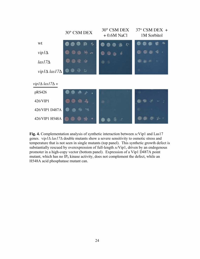
complementation analysis was done with various scVip1 constructs (Fig. 4). The severe
growth defect was rescued by overexpression of both full-length scVip1 and a kinase-
only mutant with a point mutation in the acid-phosphatase domain. However, a kinase-
dead mutant with a deactivating mutation in the kinase domain did not complement this
interaction.
Kinase activity of recombinant human Vip1
To explore the evolutionary conservation of Vip1’s IP6 kinase activity, I
examined the biochemical activity of the human ortholog, hsVip1. I therefore cloned the
human Vip1 gene (hsVip1) and expressed and purified recombinant GST constructs of
the full-length protein, as well as of kinase (residues 1-387) and acid-phosphatase
(residues 390-1433) domains, as determined by homology. Preliminary enzymological

11
studies have demonstrated that full-length hsVip1, as well as the kinase domain alone,
exhibit robust dose-dependent IP6 kinase activity (Fig. 5A). This activity shows similar
specificities and maximum velocities as scVip1, with a KM of 17.98 µM and a Vmax of
24.69 nmol/min/mg (Fig. 3B and 3C). From these in vitro experiments, mammalian Vip1
appears to retain the inositol pyrophosphate synthase activity observed in the yeast
enzyme. Additionally, PP-IP5 from the mammalian IP6K IHPK1 was converted by
recombinant hsVip1 to PP2-IP4, an activity also seen with the yeast protein. The relative
kinetic parameters of hsVip1’s two kinase activities have not yet been resolved.
hsVip1 kinase activity in yeast and mammalian cells
In addition to these biochemical results, hsVip1’s kinase activity was also
observed through in vivo studies. First, yeast mutants overexpressing hsVip1 were
radiolabeled with [3H]-myo-inositol and extracts were analyzed through HPLC. In yeast
mutants lacking both Kcs1 and scVip1 genes, no PP-IP5 was detected in radiolabeled
extracts. However, when either full-length or kinase domain hsVip1 constructs were
overexpressed in these mutants, a significant amount of PP-IP5 was detected (Fig. 5B).
While the acid phosphatase domain was also overexpressed in yeast, no change in soluble
inositol levels was observed, with either yeast or human Vip1 constructs.
hsVip1 constructs were also overexpressed in [3H]-myo-inositol labeled
mammalian 293T cells, with transfections performed by James Otto. There was little
change in IP levels in wild-type cells, however, with no noticeable accumulation of PP-
IP5. To increase the flux of IP6 in transfected cells, hsVip1 was coexpressed with the G
protein Gαq, a strong activator of PLC that significantly increases IP concentrations,

12
along with Ipk1, an I(1,3,4,5,6)P5 2-kinase that synthesizes IP6 (Fig. 6) (37,38). Under
these conditions, a small peak of PP-IP5 was detectable, as well as a small peak of PP2-
IP4. More significantly, however, when hsVip1 was coexpressed with IHPK1, a member
of the IP6 kinase enzyme family, a much larger peak of PP2-IP4 was observed. As
expression of IHPK1 substantially increases levels of its PP-IP5 product, this suggests
that hsVip1’s phosphorylation of PP-IP5 to PP2-IP4 is a major activity in mammalian
cells.
Discussion
Enzyme kinetics of S. cerevisiae Vip1 demonstrate, as previously reported,
significant IP6 kinase activity. It has a high, specific affinity for IP6, with a KM within the
normal range of intracellular IP6 levels in yeast and mammalian cells (39). The specific
activity of the enzyme, while relatively weak, is in the same range as other IP kinases,
and is biologically significant (28). While this activity is dependent on pH, peak activity
occurs in the range of 6-7, indicating that scVip1 should be capable of normal activity
when localized to the cytoplasm. Further, genetics studies suggest that this kinase
activity is biologically relevant in yeast. scVip1 has previously been found to have
synthetic interactions with elements of Arp2/3 actin polymerization, specifically the
Las17 protein (30). Las17 regulates the Arp2/3 complex, which catalyzes the nucleation
of actin filaments, and is required for the actin branching necessary for actin’s
cytoskeletal functions (36,40). This synthetic interaction appears to depend on the
presence of kinase activity, as only constructs with an intact kinase domain can rescue the
vip1∆las17∆ synthetic growth defect. This suggests that scVip1’s kinase activity, and

13
likely its PP-IP5 product, are required for whatever involvement scVip1 might have in
actin polymerization. This is consistent with work by other members of the York lab
observing defects in cell growth, morphology, and Arp2/3 synthetic interactions in
Schizosaccharomyces pombe yeast overexpressing kinase-dead mutants of Asp1, the S.
pombe Vip1 ortholog (29).
Sashi Mulugu’s cloning of the S. cerevisiae Vip1 gene immediately allowed a
bioinformatic analysis of the structure and evolutionary conservation of the enzyme. In
addition to the dual domain structure revealed by this analysis, a sequence alignment of
Vip1 genes from yeast, mammals, and other model species revealed a conservation of
these domains throughout evolutionary history, with known catalytic residues universally
conserved among species (Fig. 2) (29). This suggested that Vip1 orthologs in other
species might have a similar role as an IP6 kinase. Consistent with this, the human
ortholog, hsVip1, possessed in vitro IP6 and PP-IP5 kinase activities comparable to those
of yeast Vip1. Additionally, yeast overexpressing hsVip1 constructs showed conversion
of IP6 to PP-IP5. These data are consistent with studies by other members of the York lab
examining yeast mutants overexpressing the yeast Vip1 protein, and suggest that the
human enzyme exhibits IP6 kinase activity in the cytosolic environment of a eukaryotic
cell, and is capable of producing a physiologically relevant amount of PP-IP5 in cells
(29). Vip1 IP6 kinase activity does therefore appear to be evolutionary well-conserved
between yeast and mammals.
Significantly, this conserved IP6 kinase activity appears to have a biological
signaling function in budding yeast. A recent report has identified the PP-IP5 product of
scVip1 as a regulator of the cyclin/CDK complex Pho80/Pho85, a transcriptional

14
regulator involved in phosphate homeostasis (41). PP-IP5 from scVip1, and not from
Kcs1, is required for inhibition of Pho80/Pho85 by the CDK inhibitor Pho81, through
direct binding of this complex. Additionally, PP-IP5 levels appear to rise upon phosphate
starvation, when Pho80/Pho85 is normally inhibited. vip1∆ null yeast strains also appear
deficient for Pho80/Pho85 inhibition, leading to a defective phosphate starvation
response. This biological function of scVip1 reveals a specific role for its kinase activity,
mediated by direct interaction between its PP-IP5 product and its regulatory target. This
vital, specific signaling role of Vip1 in yeast reveals a clear biological function for the
protein. It is not inconsistent with a possible role in Arp2/3 regulation, however, as other
soluble IPs have been found to have numerous independent signaling functions. The
integration of these single molecules’ diverse signaling roles continues to be an important
question in inositol signal transduction, and further elucidation of mechanisms regulating
Vip1’s activity would be useful in exploring the problem.
In addition to studies in yeast, hsVip1 was overexpressed in inositol radiolabeled
mammalian cells. Only a small amount of PP-IP5 production was detected, however.
Considering the considerable IP6 kinase activity detectable in yeast and with recombinant
protein, this failure to detect significant IP6 kinase activity could be a result of some
mechanism in mammalian cells regulating or inhibiting this activity under normal
conditions. Alternatively, it could be a result of sequestration of intracellular IP6 from
hsVip1, possible through either protein binding or compartmentalization. It is also
possible, however, that IP6 kinase activity is not a primary activity of Vip1 in mammalian
cells. Given the many biological differences between yeast and mammalian phosphate

15
regulation and actin polymerization mechanisms, it is not inconceivable that Vip1’s
kinase domain, despite its conservation, has different signaling functions across species.
Despite the lack of robust IP6 kinase activity seen in mammalian cells, hsVip1 did
exhibit a high level of PP-IP5 kinase activity, producing PP2-IP4 when coexpressed with
the IHPK1 IP6 kinase. Vip1’s apparent ability to produce PP2-IP4 from PP-IP5 indicates
possible involvement with several previously reported examples of inositol
pyrophosphate signaling. PP2-IP4 appears to be involved in certain stress responses, as
well as in some cAMP-mediated signaling events (23-27). Some of these responses
appear to be mediated by MAP kinase pathways, suggesting one possible regulatory
mechanism for this activity. An inositol pyrophosphate synthase with a robust PP-IP5
kinase activity has not yet been reported, and it appears that hsVip1 is a major producer
of PP2-IP4 in cells. Given these results, and the biochemical kinase activities observed
for both human and yeast proteins, production of PP-IP5 and PP2-IP4 can be tentatively
assigned to the IP6K and Vip1 enzymes (Fig. 7) (29). However, further studies
examining the importance of hsVip1 to the regulation of PP2-IP4 levels are needed to
explore Vip1’s precise involvement in this pathway.
While hsVip1 appears to possess both IP6 and PP-IP5 kinase activities, the relative
strength and importance of these is not yet clear. Further enzymological studies are
necessary to determine the relative affinities and specific activities for these two
substrates. This would help determine whether the apparent lack of IP6 kinase activity in
mammalian cells, and the much stronger PP-IP5 kinase activity observed, is a result of
PP-IP5 being a significantly better substrate. It would also be helpful to overexpress both
hsVip1 and Kcs1/IP6K in yeast, to determine whether this PP-IP5 kinase activity can be

16
detected outside of mammalian cells. Additionally, it would be valuable to examine the
availability of intracellular IP6 to soluble enzymes, which might indicate whether
substrate sequestration plays a role in regulating Vip1’s IP6 kinase activity. Performing
these experiments with the yeast Vip1 protein would also be valuable, allowing
comparison of activities between orthologs. As different organisms appear to utilize IP6
and inositol pyrophosphates in different manners, it would be interesting to determine if
the function of this enzyme varied between species, or if its relative activities as an IP6 or
PP-IP5 kinase were evolutionarily conserved.
References 1. Majerus, P. W., Ross, T. S., Cunningham, T. W., Caldwell, K. K., Jefferson, A.
B., and Bansal, V. S. (1990) Cell 63(3), 459-465 2. Berridge, M. J., and Irvine, R. F. (1984) Nature 312(5992), 315-321 3. Berridge, M. J., and Irvine, R. F. (1989) Nature 341(6239), 197-205 4. Shears, S. B. (2004) Biochem J 377(Pt 2), 265-280 5. York, J. D. (2006) Biochim Biophys Acta 1761(5-6), 552-559 6. El Alami, M., Messenguy, F., Scherens, B., and Dubois, E. (2003) Mol Microbiol
49(2), 457-468 7. Odom, A. R., Stahlberg, A., Wente, S. R., and York, J. D. (2000) Science
287(5460), 2026-2029 8. Steger, D. J., Haswell, E. S., Miller, A. L., Wente, S. R., and O'Shea, E. K. (2003)
Science 299(5603), 114-116 9. York, J. D., Odom, A. R., Murphy, R., Ives, E. B., and Wente, S. R. (1999)
Science 285(5424), 96-100 10. Ho, M. W., Yang, X., Carew, M. A., Zhang, T., Hua, L., Kwon, Y. U., Chung, S.
K., Adelt, S., Vogel, G., Riley, A. M., Potter, B. V., and Shears, S. B. (2002) Curr Biol 12(6), 477-482
11. Frederick, J. P., Mattiske, D., Wofford, J. A., Megosh, L. C., Drake, L. Y., Chiou, S. T., Hogan, B. L., and York, J. D. (2005) Proc Natl Acad Sci U S A 102(24), 8454-8459

17
12. Europe-Finner, G. N., Gammon, B., and Newell, P. C. (1991) Biochem Biophys Res Commun 181, 191-196
13. Menniti, F. S., Miller, R.N., Putney, J.W. Jr., Shears, S.B. (1993) J Biol Chem 268, 3850-3856
14. Stephens, L., Radenberg, T., Thiel, U., Vogel, G., Khoo, K.H., Dell, A., Jackson, T.R., Hawkins, P.T., Mayr, G.W. (1993) J Biol Chem 268, 4009-4015
15. Saiardi, A., Erdjument-Bromage, H., Snowman, A. M., Tempst, P., and Snyder, S. H. (1999) Curr Biol 9(22), 1323-1326
16. Saiardi, A., Caffrey, J. J., Snyder, S. H., and Shears, S. B. (2000) J Biol Chem 275(32), 24686-24692
17. Dubois, E., Scherens, B., Vierendeels, F., Ho, M. M., Messenguy, F., and Shears, S. B. (2002) J Biol Chem 277(26), 23755-23763
18. Saiardi, A., Resnick, A. C., Snowman, A. M., Wendland, B., and Snyder, S. H. (2005) Proc Natl Acad Sci U S A 102(6), 1911-1914
19. York, S. J., Armbruster, B. N., Greenwell, P., Petes, T. D., and York, J. D. (2005) J Biol Chem 280(6), 4264-4269
20. Saiardi, A., Sciambi, C., McCaffery, J. M., Wendland, B., and Snyder, S. H. (2002) Proc Natl Acad Sci U S A 99(22), 14206-14211
21. Bennett, M., Onnebo, S. M., Azevedo, C., and Saiardi, A. (2006) Cell Mol Life Sci 63(5), 552-564
22. Saiardi, A., Bhandari, R., Resnick, A. C., Snowman, A. M., and Snyder, S. H. (2004) Science 306(5704), 2101-2105
23. Luo, H. R., Huang, Y. E., Chen, J. C., Saiardi, A., Iijima, M., Ye, K., Huang, Y., Nagata, E., Devreotes, P., and Snyder, S. H. (2003) Cell 114(5), 559-572
24. Pesesse, X., Choi, K., Zhang, T., and Shears, S. B. (2004) J Biol Chem 279(42), 43378-43381
25. Choi, K., Mollapour, E., and Shears, S. B. (2005) Cell Signal 17(12), 1533-1541 26. Safrany, S. T., and Shears, S. B. (1998) Embo J 17(6), 1710-1716 27. Laussmann, T., Pikzack, C., Thiel, U., Mayr, G. W., and Vogel, G. (2000) Eur J
Biochem 267(8), 2447-2451 28. Seeds, A. M., Bastidas, R. J., and York, J. D. (2005) J Biol Chem 280(30), 27654-
27661 29. Mulugu, S., Bai, W., Fridy, P.C., Bastidas, R.J., Otto, J.C., Dollins, D.E.,
Haystead, T.A., Ribeiro, A.A., York, J.D. (2007) Science IN PRESS 30. Feoktistova, A., McCollum, D., Ohi, R., and Gould, K. L. (1999) Genetics 152(3),
895-908 31. Van Etten, R. L., Davidson, R., Stevis, P. E., MacArthur, H., and Moore, D. L.
(1991) J Biol Chem 266(4), 2313-2319 32. Mullaney, E. J., and Ullah, A. H. (2003) Biochem Biophys Res Commun 312(1),
179-184 33. Stevenson-Paulik, J., Bastidas, R. J., Chiou, S. T., Frye, R. A., and York, J. D.
(2005) Proc Natl Acad Sci U S A 102(35), 12612-12617 34. Liu, Q., Li, M. Z., Leibham, D., Cortez, D., and Elledge, S. J. (1998) Curr Biol
8(24), 1300-1309 35. Stevenson-Paulik, J., Chiou, S. T., Frederick, J. P., dela Cruz, J., Seeds, A. M.,
Otto, J. C., and York, J. D. (2006) Methods 39(2), 112-121

18
36. Li, R. (1997) J Cell Biol 136(3), 649-658 37. Smrcka, A. V., Hepler, J. R., Brown, K. O., and Sternweis, P. C. (1991) Science
251(4995), 804-807 38. Ives, E. B., Nichols, J., Wente, S. R., and York, J. D. (2000) J Biol Chem 275(47),
36575-36583 39. Ingram, S. W., Safrany, S. T., and Barnes, L. D. (2003) Biochem J 369(Pt 3), 519-
528 40. Higgs, H. N., and Pollard, T. D. (2001) Annu Rev Biochem 70, 649-676 41. Lee, Y. S., Mulugu, S., York, J. D., and O'Shea, E. K. (2007) Science 316(5821),
109-112 42. Stolz, L. E., Huynh, C. V., Thorner, J., and York, J. D. (1998) Genetics 148(4),
1715-1729

19
Figures and Tables Strain Relevant Genotype
Reference
W303 MATα ade2-1 ura3-1 his3-11,15 trp1-1 leu2-3,112 can1-100 (42) JYY911 W303 MATα kcs1::HIS3 ddp1::HIS3 vip1::kanMX4 This work JYY915 KGY1350 MATα vip1::HIS3 ade2-101 his3-∆200 leu2-∆1 lys2-801
trp1-∆1 ura3-52 Gift of K. Gould (30)
JYY916 KGY2142 MATa las17::LEU2 his3 leu2 trp1 ura3 Gift of K. Gould (30) JYY917 MATα/a vip1::HIS3/VIP1 las17::LEU2/LAS17 This work JYY918 MATα vip1::HIS3 las17::LEU2 This work JYY919 JYY918 + pRS426 This work JYY920 JYY918 + pRS426-VIP1 This work JYY922 JYY918 + pRS426-Vip1D487A This work JYY923 JYY918 + pRS426-Vip1H548A This work
Table 1. S. cerevisiae strains used in this report.

20
Fig. 1. Schematics of Vip1 structure and GST constructs used. (A) In the S. cerevisiae protein, conserved ATP-grasp and histidine acid phosphatase domains are located at residues 200-525 and 530-1025, respectively. The ATP-grasp domain was found to exhibit kinase activity specific for IP6 and IP7 (PP-IP5) substrates. In yeast, this activity depended on the presence of a highly conserved catalytic aspartic acid residue, shown here. (B) Several constructs were used in the purification of recombinant Vip1 enzymes from bacteria. All enzymes were fused at the amino terminus to glutathione S-transferase (GST), and full-length and kinase domain-only constructs were used, for both yeast and human proteins.

21
Fig. 2. Evolutionary conservation of Vip1 kinase domain across species. A multi-sequence alignment was performed with VIP1 homologs from Saccharomyces cerevisiae (sc, accession NP_013514), Schizosaccharomyces pombe (sp, SPCC1672.06c), Arabidopsis thaliana (at, NP_001030614), Caenorhabditis elegans (ce, NP_740855), Drosophila melanogaster (dm, CG14616-PE), Mus musculus (mm, NP_848910), and Homo sapiens (hs, AAH57395). Residues 200-525 in S. cerevisiae were identified as having homology to the ATP-grasp domain superfamily, and have been found to encode IP6 kinase activity in both yeast and humans. A catalytic aspartic acid residue required

22
for this activity is boxed in blue. Identical residues are shown in solid red boxes, while similar residues are shown as red text in blue boxes. Alignment was printed using the ENDScript/ESPript 2.2 tool, accessed at <http://espript.ibcp.fr/ESPript/cgi-bin/ESPript.cgi>. Alignment was generated with the EMBL-EBI ClustalW tool, accessed at <http://www.ebi.ac.uk/clustalw/>.

23
Fig. 3. Biochemical analysis of IP6 kinase activity of Vip1’s kinase domain. (A) In enzyme kinetics studies of the scVip1 kinase domain truncation mutant (residues 1-535), the dependence of initial velocity on substrate concentration was examined in triplicate. KM and Vmax were also determined from a Lineweaver-Burke plot. (B) Lineweaver-Burke plot of IP6 kinase activity of the hsVip1 kinase domain. (C) Kinetic parameters of scVip1 and full-length and kinase domain (residues 1-535), as well as the hsVip1 kinase domain (residues 1-387). (D) The pH dependence of scVip1 kinase domain activity was determined in triplicate in buffers ranging from pH 4.0 to pH 8.8. Maximal activity was observed at pH 6.2.
A
B
98.7920.66scVip11-535
24.6917.98hsVip11-387
KM (µM) Vmax (nmol/min/mg)
scVip1 17.63 22.63
98.7920.66scVip11-535
24.6917.98hsVip11-387
KM (µM) Vmax (nmol/min/mg)
scVip1 17.63 22.63
C
0
0.1
0.2
0.3
0.4
0.5
0.6
0.7
0.8
0.9
1
-0.0001 0.0001 0.0003 0.0005 0.0007 0.0009 0.0011 0.0013 0.0015
1/[IP6] (µM-1)
1/Sp
ecifi
c ac
tivity
(nm
ol/m
in/m
g)-1
D
scVip11-535 scVip11-535
hsVip11-387scVip11-535
24
Fig. 4. Complementation analysis of synthetic interaction between scVip1 and Las17 genes. vip1∆ las17∆ double mutants show a severe sensitivity to osmotic stress and temperature that is not seen in single mutants (top panel). This synthetic growth defect is substantially rescued by overexpression of full-length scVip1, driven by an endogenous promoter in a high-copy vector (bottom panel). Expression of a Vip1 D487A point mutant, which has no IP6 kinase activity, does not complement the defect, while an H548A acid phosphatase mutant can.

25
Fig. 5. IP6 kinase activity of human Vip1 enzyme. (A) IP6 kinase assays were run in vitro with ATP and human GST-Vip1 kinase domain protein (residues 1-387) and resolved on a PEI-cellulose TLC plate. [32P]-IP6 is converted to [32P]-PP-IP5 in a dose-dependent manner. (B) Yeast cells deficient for the IP6 kinases kcs1 and vip1 and the inositol pyrophosphatase ddp1 do not show a PP-IP5 peak in [3H]-inositol radiolabeled extracts resolved by HPLC. However, when full-length human Vip1 or its kinase domain are overexpressed, a relatively large peak of PP-IP5 is detected, demonstrating hsVip1’s IP6 kinase activity in yeast.
26
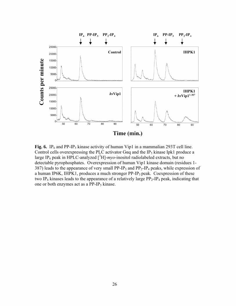
Fig. 6. IP6 and PP-IP5 kinase activity of human Vip1 in a mammalian 293T cell line. Control cells overexpressing the PLC activator Gαq and the IP5 kinase Ipk1 produce a large IP6 peak in HPLC-analyzed [3H]-myo-inositol radiolabeled extracts, but no detectable pyrophosphates. Overexpression of human Vip1 kinase domain (residues 1-387) leads to the appearance of very small PP-IP5 and PP2-IP4 peaks, while expression of a human IP6K, IHPK1, produces a much stronger PP-IP5 peak. Coexpression of these two IP6 kinases leads to the appearance of a relatively large PP2-IP4 peak, indicating that one or both enzymes act as a PP-IP5 kinase.

27
Fig. 7. Outline of the inositol pyrophosphate synthesis pathway. With the identification of Vip1 as an IP6 and PP-IP5 kinase, this is a simplified outline of the currently understood pathway of inositol pyrophosphate synthesis in yeast and humans. The PP-IP5 products of IP6K and Vip1 have been identified as structurally distinct through NMR studies, with the pyrophosphate group on the 5 or 4 position, respectively (29). This report and previous studies have also shown that IP6K and Vip1 can act on each other’s PP-IP5 products, together producing PP2-IP4.