Embed Size (px)
Citation preview

Abscission: How does it occur?
Exploring Leaf LongevityMartha Carlson, PhD candidate
NRESS, UNH, 2012

Twig, 2 Buds, 2 Petioles
The petiole of each leaf cups forming buds that will open next spring. Notice the soft hairy trichomes that cushion the buds. The petiole and leaf must break off,
leaving the bud and twig. The twig must seal off the break to close vascular tissue and prevent invasion by fungi or bacteria.

The Abscission Zone
Even very early in the summer, a clear line shows the abscission zone. Simple ground meristem cells here do
not differentiate as other cells do. Then, in late summer, these cells on the twig side of the AZ begin to thicken.
Notice how they bulge out of the petiole at left.

The AZ layer
Bleecker and Patterson, 1997, reported that the small cytoplasmic cells do not enlarge and do not obtain vacuoles as
other cells do. The cells fracture dividing petiole from twig when pectin in the cell walls is dissolved. Pectinase is prompted by ethylene. Cells on the twig side thicken into “suberized scar
tissue.”

Abscission Layer changes
Zooming in to 288x, the thickening cells show waxy filling. Cells above in the petiole show no such thickening. Notice that the thickening cells are not one layer thick but 8 to 10
cells deep.
Stimulated by ethylene, the cells produce suberin, a wax that water-proofs the twig side of the abscission
layer. Notice that cells at lower left do not show thicker walls. These are simple cortex cells of the
twig.

Examining a petiole saddle
To explore the petiole-twig zone more, we pulled leaves off the maple twigs. The base of the twig around each bud
looks like “a saddle,” Dr. Rock said. It does look like a saddle, a seat for a petiole and its leaf.

What about the xylem and phloem?
Each leaf is supplied water via xylem cells and ships sugars out of the leaf via the phloem. We find at least three vascular
bundles in the maple petiole.

Xylem Cells are easy to see.
Petiole are young. Their xylem cells must be as springy as slinky toys to allow the petiole to grow. Spirals of proto-xylem and slightly tighter
and more supportive meta-xylem are easy to find in the vascular bundle.
Phloem parenchyma with starch molecules
Proto-xylem
Meta-xylem
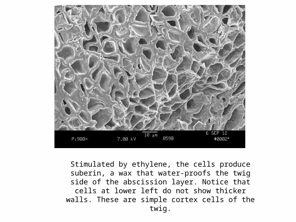
Open xylem cells mean Photosynthesis is still occurring

Is this a phloem cell group?
Phloem cells are harder to see. They are small soft cells that quickly hide themselves when the petiole is broke, exuding sugars of course. Phloem cells are actually groups of sieve
cells. The major transport cell, the sieve tube element (in angiosperms) is surrounded by smaller cells:
Companion parenchyma, thin cells connected with pits into the sieve cell, regulate sieve cell metabolism—including collecting leaking sugar in petioles;
Phloem fibers provide support with thicker walls.Phloem parenchyma contain starch molecules and/or a single crystal.
A sieve tube element with associated cells.
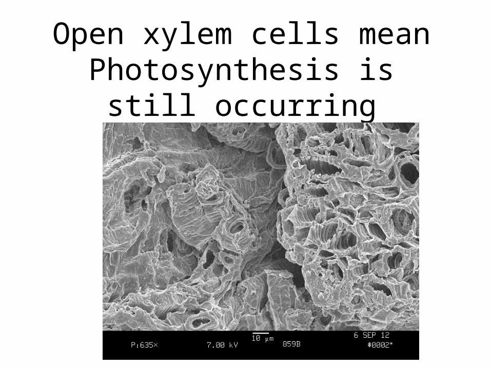
Xylem and Phloem are closely associated
Xylem cells provide water osmotically to the sugary phloem cells. So they must be closely placed.
Soft, thin-walled phloem cells are hard to spot but crystals in phloem parenchyma identify the cells at top right. Can
you see any sieve elements?
Crystals in phloem parenchyma cells

Staining shows the suberin
A photograph by Michael Clayton on the University of Wisconsin’s Plant Teaching Collection is stained to clearly show the suberization of the twig side of the abscission layer. Even the xylem cells appear suberized. But not the phloem—the blue river of cells that goes through the abscission zone. The phloem stays open until right before leaf drop. http://botit.botany.wisc.edu/Resources/Botany/Shoot/Leaf/Abscission%20layer%20IAA/Abscission%20layer%20MC.jpg.html

Phloem Closed or Open?
At left, Tree 859B, a tree whose spectral scans show it was already senescing in late August, shows a waxing covering on
many phloem cells. At right, Tree SS3, not yet senescing, shows no such suberin wax in the phloem. Which tree will have more time to dismantle and ship down the petiole leaf nutrients—N,
K, Mg, Mn, Fe, S and sugars?

Phloem Sieve Tube Elements
Esau says transport phloem cells may leak sugar, as these many starch cells suggest. The cells are long with plasmodesmata connections to companion cells.

No nucleus, no tonoplast, no vacuole.
As it develops, the phloem sieve tube
elements loses major cell parts “to
increase protoplasmic
continuity” with adjoining sieve tubes (Easau).
Companion cells manage
metabolism.

Phloem Complex
Crystal forming parenchyma cells identify a phloem complex. These cells are laid from upper right to lower left, transporting sugars from leaf to twig
and sinks beyond. Notice the many pores connecting cells. Notice how thick these cell walls are, a characteristic of some sieve tube elements
(Esau).

Phloem Fibers
Thick-walled fibers protect the delicate phloem and their cargo of sugar from insects that might penetrate the twig
epidermis and cortex.

Other differences
Why is 859B senescing now, in early September? Is it a stressed tree? Is SS3 an unstressed tree? The pithy cells of the twigs give a clue. At left, cells in the twig adjacent to 859B’s petiole appear
to heave some starch grains but not many. Cells in SS3’s twigs are packed with chubby starch molecules, food for the twig and
bud.

Starch molecules extended far into the center of the twig in Tree SS3.
The sugar will provide food and supplies for anthocyanin, phenolics and fast spring growth to the bud and twig.

Does longevity matter?
Sept. 28th, 2008, which
tree is healthier?
803
813
822
831
834

References
Abscission: How does it occur? copyright 2012, Martha Carlson and Barrett Rock, University of New Hampshire.
Photographs taken with the University of New Hampshire scanning electron microscope in Martha Carlson PhD research with help from Nancy Cherim, SEM technical director, and Dr. Barrett N. Rock, plant anatomist.
Bleecker, A.B., and S.E. Patterson. 1997. Last Exit: Senescence, abscission, and meristem arrest in Arabidopsis. The Plant Cell. 9:1169-1179.
Clayton, Michael et al., The Plant Teaching Collection, Botany Department, University of Wisconsin-Madison. These materials were developed for use in the courses taught through the botany department at the University of Wisconsin-Madison. They may be freely used by educators in the context of their classrooms. This means that they may be incorporated into lecture presentations, printed supplemental materials, and used in the teaching laboratory as a visual reference. They may not be distributed on the world wide web, or in published works, without permission. Any request for these uses should be made through Michael Clayton the collection manager.
Evert, R.F. 2006. Esau’s Plant Anatomy. Wiley-Interscience, Hoboken, NJ.