Embed Size (px)
Citation preview

American Journal of Biology and Life Sciences 2016; 4(3): 16-23
http://www.openscienceonline.com/journal/ajbls
ISSN: 2381-3784 (Print); ISSN: 2381-3792 (Online)
Abiotic Stress and Its Impact on Protein Concentration or Polymorphism of Gloriosa superba Plant
Dharmendra Singh1, *
, Manish Mishra2, Anirudha Singh Yadav
1
1Department of Botany, Govt. M. V. M., Barkatullah University, Bhopal, India 2Department of Ecosystem Management, IIFM, Bhopal, India
Email address
[email protected] (D. Singh) *Corresponding author
To cite this article Dharmendra Singh, Manish Mishra, Anirudha Singh Yadav. Abiotic Stress and Its Impact on Protein Concentration or Polymorphism of
Gloriosa superba Plant. American Journal of Biology and Life Sciences. Vol. 4, No. 3, 2016, pp. 16-23.
Received: March 29, 2016; Accepted: April 11, 2016; Published: June 15, 2016
Abstract
Biotic and abiotic stresses exert a considerable influence on the production of several secondary metabolites in plants.
Temperature, pH and light are one of the important abiotic stress that affected survival, growth, reproduction and geographic
distribution of crop plants. Biochemical characterization of species and their genetic association and polymorphism within the
related species based on morphological data is becoming difficult because these morphological traits are highly influenced by the
environment. Proteins play significant biological function in human as well in the plants. In this study, G. superba cultured in MS
medium under abiotic stress. To study the effect of stresses on protein concentration, protein extracted and quantified by Lowry
method. Total levels of protein were found to be varied in plants grow under different temperature. Maximum 11.308 µg/100mg
was observed in plant cultured at 25°C temperature and minimum amount was 4.791 µg/100mg at 35°C temperature. pH and
photoperiod does not exist more difference in protein concentrations. To investigate the molecular weight of proteins, a standard
protein marker was used. Electrophoresis of proteins has been successfully used for the characterization of different taxonomic,
evolutionary and genetic relationship studies. In the present study, the electrophoratic banding profile of total soluble proteins of
G. superba plant cultured under different abiotic stress exhibited presence versus absence type of polymorphism. The present
investigation of SDS denatured proteins showed differences in a number of bands, bandwidth and intensity and exhibited genetic
diversity between all variants. This study is very important for researcher, who is working in the field of climate change. By this
research we can conserve endangered plant species in challenging environment and can enhance important plant chemical
constitute under stress conditions.
Keywords
Abiotic Stress, Protein Concentration, Protein Polymorphism, Micropropagation, Gloriosa superba
1. Introduction
Recently, there has been an increased interest in
understanding the mechanism of plant acclimatization to
environmental stresses. Plants take action to an undesirable
ecosystem by changing their morphology, physiology, and
biochemistry. Some of the adaptations to stress may include
the changes in both the nature and levels of their genetic
resources. Biotic and abiotic stresses exert a considerable
influence on the production of several secondary metabolites
in plants [1]. Temperature, pH and light are one of the
important abiotic stress that affected survival, growth,
reproduction and geographic distribution of crop plants. Each
plant species have evolved a degree of stress tolerance.
Variation tolerance level can be genetically determined, which
reduces its metabolic activity. The following this process
many biochemical and physiological changes occur, including:
increase levels of sugars, soluble proteins, antioxidant
enzymes, proline, chlorophyll fluorescence, the appearance of

17 Dharmendra Singh et al.: Abiotic Stress and Its Impact on Protein Concentration or Polymorphism of Gloriosa superba Plant
new isoforms of protein and membrane lipid composition
changes that which called cold accumulation [2]. Plant tissue
cultures are exposed to stresses and stress combinations that
they may not have encountered in nature in their long
evolution. It is a remarkable reflection on the plasticity of the
plant genome that it can decipher and respond to novel in vitro
stresses. Today various tissue culture techniques are used to
enhance yield of secondary metabolites by triggering the
stress response like using Elicitors, Precursors and
Biotransformation, Change in environment conditions,
Change in medium constituents etc [3]. Biochemical
characterization of species and their genetic association and
polymorphism within the related species based on
morphological data is becoming difficult because these
morphological traits are highly influenced by environment [4].
Proteins play significant biological function in human as well
in the plants. These can be analyzed from seeds and other parts
of plants such as leaves and stems. Protein synthesis occurs in
leaves and green stems and is mobilized into seeds or fruits.
Proteins have been assayed from almost all parts of plants
including leaves, nodules, stem, fruit and seed. Proteins have
been extracted, identified and analyzed by using different
analytical techniques. At present, extracellular and
intracellular proteins have been isolated and quantitatively
estimated by spectrophotometrically [5].
G. superba L., a member of the Liliaceae family is a
perennial tuberous climbing herb that distributed in the
tropical and subtropical region of India. G. superba L. is
among some of the modern important medicinal plants, which
actually facing local extinction due to climate change.
Different parts of the plant have a wide variety of uses,
especially within an Indian traditional medicine of the time in
immemorial. Tubers and seeds of the G. superba L. are an
expensive export commodity. All parts of the plant contain
colchicines and related alkaloids [6]. Due to the medicinal
value, this plant collected from the wild and used as raw
material for large-scale medicinal industries, leading to over
exploiting condition, proved to be 95% endangered medicinal
plant becomes endangered plant species and included in the
red data book [7]. The purpose of the present study was to
contribute a better understanding of the genetic responses of G.
superba plant to different abiotic stress. We investigated the
influence of three types of abiotic stress on the contents of
proteins to analyze changes in the protein pattern under stress
condition using SDS-PAGE gel electrophoresis.
2. Materials & Methods
2.1. Sample Collection and Preparation
Different parts (Seed, tuber and whole plant) of G. superba
Linn. were used as explants in the present study (Fig. 2). The
plant was collected from the northern forest range of Betul
district of Madhya Pradesh (Fig. 1). Seeds and tubers were
washed thoroughly in running tap water to remove the
superficial dust and washed with 2% (v/v) tween 20 for 10 min
then washed well with distilled water. Then the seed surface
was sterilized with 0.1% mercuric chloride (HgCl2) for 5 min.
and tubers for 2 minute. After pretreatment explants were
dipped in 70% ethanol for 30 seconds. This was followed by
rinsing the explants with double distilled sterile water thrice.
North forest division of Betul (Madhya Pradesh)
Source: Department of Remote Sensing, Madhya Council of Science & Technology, Bhopal.

American Journal of Biology and Life Sciences 2016; 4(3): 16-23 18
Fig. 1. Location of survey site in Madhya Pradesh, India.
Fig. 2. Tubers and Seeds of G. superba plant.
2.2. Micropropagation in Different Stress
Condition
For this purpose, explants cultured on Murashige-Skoog
(MS, 1962) [8] medium containing sucrose 30%, coconut
water 15% and 0.8% agar (PTC grade, Hi media, India) and
supplemented with different concentrations and combinations
of 6-Banzylaminopurine (0.5-1.5 mg/l) and Gibberellic acid
(0.5-1.0 mg/l) for seed germination and BAP (0.5-2.5 mg/l),
Indole -3-acetic acid (0.5 mg/l) or Kinetin (0.5 mg/l) for shoot
proliferation and multiplication. For the rooting different
concentration of Indole-3-butyric acid (0.5-2.5 mg/l) and
1-Naphthaleneacetic acid (0.5 mg/l) were used. The pH of
medium was adjusted to 5.6-5.8 using 0.1 N NaOH or 0.1 N
HCl before autoclaving (121°C, 20 min). The cultured plants
were exposed to abiotic stress created artificially by applying
different light period (12, 14 and 16 hrs), different pH (6, 7 and
8.5) and different temperature (25, 30 and 35°c). On the
medium in the presence of stressful factors cultivated plants
observed for 1 month. After 1 month all plants were harvested
to study the effect of abiotic stress on concentration of protein.
2.3. Extraction of Protein
For the extraction of proteins, 100mg of G. superba plants
were taken, crushed and grounded into a fine powder in a
mortar and pestle with 100mM 1.5M Tris HCL (pH 7.2). The
aliquot was centrifuged at 12,000 rpm for 10 minutes at room
temperature. The pellet was discarded and extracted crude
proteins were recovered as clear supernatant, transferred into
new 1.5 ml eppendorf tubes and stored at -20oC for
electrophoresis.
2.4. Quantification of Protein
The concentration of the extracted protein samples was
determined using the Lowry’s method (1951) using different
samples against the control [9]. The relative concentrations of
all samples were calculated using the formula from the BSA
standard chart.

19 Dharmendra Singh et al.: Abiotic Stress and Its Impact on Protein Concentration or Polymorphism of Gloriosa superba Plant
2.5. Polymorphism of Protein
Protein samples were mixed with 4X gel loading dye to
make its final concentration of 1X in mixture and was heated
at 95°C in water bath for 10 min prior to loading. Protein
sample (100 µg) was loaded in each lane. Protein molecular
weight marker (Bangalore Genei, India) was used as reference.
Protein samples were electrophoresed at 8 V/cm for about 4 h
at constant current. Preparative gels were visualized by
staining with Coomassie Brilliant Blue R-250 [10].
2.6. Statistic Analysis
The Similarity Index was also calculated with each primer
and a matrix was developed. Finally, all matrices were
analyzed as an average. The graphic phenogram of the genetic
relatedness among the four accessions was produced by means
of UPGMA {Unweighed pair group method with arithmetic
average} cluster analysis of averages similarity index [11].
3. Results
3.1. Micropropagation of G. superba Under
Abiotic Stress
Plant tissue cultures are exposed to stresses and stress
combinations that they may not have encountered in nature in
their long evolution. It is a remarkable reflection on the
plasticity of the plant genome that it can decipher and respond
to novel in vitro stresses. Today various tissue culture
techniques are used to enhance yield of secondary metabolites
by trigger stress response. Cultured plants of G. superba
exposed in different abiotic stress condition to maintained in
vitro. Various stress condition influences not only growth of
plants but also their secondary metabolites and biomolecules.
In present study tissue cultured plants of G. superba shows
different growth pattern and survival rate (Fig. 3). All
survivals further used to study the effect of abiotic stess on
biomolecules (proteins).
Fig. 3. Invitro cultured plants of G. superba.
3.2. Quantification of Protein
The protein concentration found to vary in G. superba plant
which was cultured under various physical stress conditions
(pH, Temperature and Light period) in the laboratory. In the
present investigation, G. superba was evaluated quantitatively
for the analysis of total soluble protein. The results of protein
quantification are represented in Table-1, 2 and 3 according to
abiotic stress. Total levels of protein were found to be varied in
plants grow under different temperature. Maximum 11.308
µg/100mg was observed in plant cultured at 25°C temperature
and minimum amount was 4.791 µg/100mg at 35°C
temperature. pH and photoperiod does not exist more
difference in protein concentrations. Bovine Serum Albumin
was used as standard (Fig. 4).
Fig. 4. Standard Graph of Bovine Serum Albumin.
American Journal of Biology and Life Sciences 2016; 4(3): 16-23 20
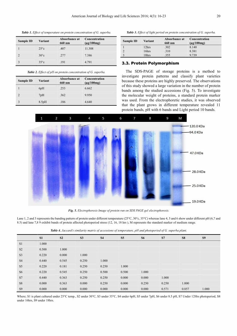
Table 1. Effect of temperature on protein concentration of G. superba.
Sample ID Variant Absorbance at
660 nm
Concentration
(µg/100mg)
1 25°c .407 11.308
2 30°c .277 7.386
3 35°c .191 4.791
Table 2. Effect of pH on protein concentration of G. superba.
Sample ID Variant Absorbance at
660 nm
Concentration
(µg/100mg)
1 6pH .253 6.662
2 7pH .362 9.950
3 8.5pH .186 4.640
Table 3. Effect of light period on protein concentration of G. superba.
Sample ID Variant Absorbance at
660 nm
Concentration
(µg/100mg)
1 12hrs .302 8.140
2 16hrs .310 8.381
3 18hrs .355 9.739
3.3. Protein Polymorphism
The SDS-PAGE of storage proteins is a method to
investigate protein patterns and classify plant varieties
because these proteins are highly preserved. The observations
of this study showed a large variation in the number of protein
bands among the studied accessions (Fig. 5). To investigate
the molecular weight of proteins, a standard protein marker
was used. From the electrophoretic studies, it was observed
that the plant grows in different temperature revealed 11
protein bands, pH with 6 bands and Light period 10 bands.
Fig. 5. Electrophoresis Image of protein run on SDS PAGE gel electrophoresis.
Lane 1, 2 and 3 represents the banding pattern of protein under different temperature (25°C, 30°c, 35°C) whereas lane 4, 5 and 6 show under different pH (6,7 and
8.5) and lane 7,8 9 exhibit bands of protein affected photoperiod stress (12, 16, 18 hrs ), M represents the standard marker of medium range.
Table 4. Jaccard’s similarity matrix of accessions of temperature, pH and photoperiod of G. superba plant.
S1 S2 S3 S4 S5 S6 S7 S8 S9
S1 1.000
S2 0.500 1.000
S3 0.220 0.000 1.000
S4 0.440 0.545 0.250 1.000
S5 0.220 0.181 0.250 0.250 1.000
S6 0.220 0.545 0.250 0.500 0.500 1.000
S7 0.440 0.363 0.250 0.250 0.000 0.000 1.000
S8 0.000 0.363 0.000 0.250 0.000 0.250 0.250 1.000
S9 0.000 0.000 0.000 0.000 0.000 0.000 0.571 0.857 1.000
Where; S1 is plant cultured under 25°C temp., S2 under 30°C, S3 under 35°C, S4 under 6pH, S5 under 7pH, S6 under 8.5 pH, S7 Under 12Hrs photoperiod, S8
under 16hrs, S9 under 18hrs.

21 Dharmendra Singh et al.: Abiotic Stress and Its Impact on Protein Concentration or Polymorphism of Gloriosa superba Plant
Fig. 6. Dendrogram of Protein Polymorphism of all variants.
Dendrogram showing Protein polymorphism in protein
content of G. superba cultured under different physical stress
condition. The dendrogram based on Similarity Index (SI)
showed distinct separation of the collected from different
stress condition (Fig. 6). The dendrogram separated the
different physical stress condition from 3 different physical
stresses based on the genetic diversity (GD). The basic root
node has a main leaf group S-7 and S-6 group showing
neighbor joining with S-7. The S-4 again split out into two
root node, lower root node splits into two root branches S-2
and S-3 and upper root node again splitting in S-5 and S-9,
The S-9 again splitting into S-1 and S-8. The polymorphism
observed was considered to be reasonably high. A total of 27
polypeptide bands were observed in our study having
molecular weights in the range of 19 to 94 kDa. Jaccards
similarity coefficient was obtained in the range of 0.00 to 1
(Table 4).
The average root distance observed in samples of different
physical stress was 0.426 with a variation of 0.022 and a pair
distance with average 0.261 and average pair variation of
0.043 SSQ (sum of squares) was found to be 0.4255.
According to average variation of different physical condition
ei. Temperature, pH and light respectively, were found the
SSQ value 0.994, 0.0416 and 0.154 respectively. These SSQ
values of data are very important for statistical analysis of
variants. The highest value represents more distance between
variants.
4. Discussion
In the present study, Gloriosa superba L. was preferred as
an experimental plant because of its importance in natural
medicine. This glorious herb was found in abundance once
upon a time in the forest. Due to its marvelous medicinal
properties, human activities and environmental pollution, the
plant was callously exploited from forest, leads to depletion of
the species and has become endangered. This evoked use to
conserve this plant through in vitro techniques.
Modern biotechnology methods are qualitatively new tool
for direct study of the structural and functional organization of
the genetic material, as well as to assess the impact of stress on
the plant and study the mechanisms of cell and tissue tolerance
in vitro [12].
Abiotic stress factors are the main limitation to plant growth
and yield in agriculture. Abiotic stress leads to a series of
morphological, physiological, biochemical, and molecular
changes that adversely affect plant growth and productivity.
During the course of its evolution, plants have developed
mechanisms to cope with and adapt to different types of
abiotic and biotic stress. Plants face adverse environmental
conditions by regulating specific sets of genes in response to
stress signals, which vary depending on factors such as the
severity of stress conditions, other environmental factors, and
the plant species [13]. Abiotic stresses exert a profound effect
on the viability, production, growth and morphology of plants.
Hence, these stresses have been used to crop improvement
through genetic engineering [14].
To survive under such conditions, plants have evolved
convoluted mechanisms to perceive external signals, allowing
optimal response to abiotic stress. Responses to abiotic
stresses occur at all levels of the organization. Furthermore,
plant acclimation to a particular abiotic stress condition
requires a specific response that is linked to the precise
environmental conditions that the plant encounters. Thus,
molecular, biochemical and physiological processes set in
motion by a specific stress condition might differ from those

American Journal of Biology and Life Sciences 2016; 4(3): 16-23 22
activated by a slightly different composition of environmental
parameters [15]. Gisela Jansen et al., 2012 also reported the
effect of abiotic stress (pH) on the alkaloid content of Lupinus
angustifolius [16]. Their Results clearly show that the alkaloid
content is significantly influenced by the soil pH, but
genotypic differences regarding the reaction to different pH
values in the soil were observed. Temperature strongly
influences metabolic activity and plant ontology and high
temperatures can induce premature leaf senescence. Several
studies have examined the effects of increased temperatures
on secondary metabolite production of plants. Chan et al.,
reported that Melastoma malabathricum cell cultures
incubated at a lower temperature range (20 ± 2°C) grew better
and had higher anthocyanin production than those grown at 26
± 2°C and 29 ± 2°C. Optimum temperature (25°C) maximizes
the anthocyanin yield as demonstrated in cell cultures of
Perilla frutescens and strawberry [17]. It is well known that
light is a physical factor which can affect the metabolite
production. A positive correlation between increasing light
intensity and levels of phenolics has been reported. Arakawa
studied the effect of UV light on anthocyanin accumulation in
light colored sweet cherry [18]. In the present investigation,
protein content in different experiment quantified by Lowery
method. The ability to easily and reliably quantified the total
protein content in samples is paramount to many biological
assays. Although the Lowry protein assay was first published
in 1951, several improvements, not the least of which is the
reduction in assay volume, have increased sensitivity of the
assay.
The concentration of proteins present in different samples is
compared with the help of graph above. Table 1 results
indicate a positive effect of temperature using various
concentrations of total protein of G. superba plants. It appears
from the data that there was a general increase in protein
content that corresponded with the optimum temperature of
25˚C. Pandey, M. and Chikara, S.K. 2014 found similar results
on their study in vitro regeneration and effect of abiotic stress
on physiology and biochemical content of Stevia rebaudiana
‘Bertoni’ [19].
In the present study, G. superba cultured under different
abiotic stress were used to protein profile analysis. To
investigate the molecular weight of proteins, a standard
protein marker was used. Electrophoresis of proteins has been
successfully used for the characterization of different
taxonomic, evolutionary and genetic relationship studies. In
the present study, the electrophoratic banding profile of total
soluble proteins of G. superba plant cultured under different
abiotic stress exhibited presence versus absence type of
polymorphism. The present investigation of SDS denatured
proteins showed differences in a number of bands, bandwidth
and intensity. Aparadh, V.T. et al., 2012 compared the banding
pattern of seeds and leaf proteins of cleome species by SDS
page [20]. According to the results of the SDS-PAGE, the
overall pattern of storage-proteins showed the diversity of all
cultivars of G. superba. Genetic diversity in different plant
species has been carried out by using electrophoretic patterns
of total seed proteins as revealed by SDS-PAGE of seed
storage protein [21]. Protein profiling revealed significant
inter-specific genetic diversity or genotype specific bands,
with some cultivars exhibiting remarkable polymorphic and
unique bands, that can be evaluated further “tags” for these
cultivars [22]. The polymorphism observed was considered to
be reasonably high. These proteins may be much important in
crop improvement programs through breeding and genetic
engineering. Protein profiling at seed level to assist in the
early detection of species at seed level [23].
5. Conclusion
It can be concluded that abiotic stress condition is highly
responsible not only for population decline, but also for
protein polymorphism based diversity of G. superba plant. G.
superba plant, which cultured under different abiotic
condition showed variation in protein banding. Protein
variation also confirmed by quantification method. In stress
conditions, temperature was highly responsible for protein
polymorphism. Biodiversity is important for human
civilization. Global warming is a threat before the existing
biodiversity of this earth. The effect of climate change was the
most important factor in population decline of many plant
species. Abiotic stress is the primary cause of plant loss
worldwide. Therefore, resolutions from plant biotechnology
discussions aiming at overcoming severe environmental
stresses need to be quickly and fully implemented, with
intensive molecular assisted genetic engineering. We have
made great progress in understanding the responses of plants
to abiotic stress. There are inherent physical, morphological
and molecular limitations to the plant’s ability to respond to
stress. The present study will be important in evaluation,
identification and characterization of germplasm on the basis
of genetic variation in proteins under abiotic stress.
Endangered plants can be conserved by understanding the
effect of their surrounding environment on plants and can be
developed, new species by the modification in genomes
according to climate.
Acknowledgment
Authors are thankful to Principal, Govt. M.V.M., Bhopal for
providing research permission at your center and also thankful
to the Director, CMBT, Bhopal for their guidance and
encouragement.
References
[1] Azhar, N., Hussain, B., Ashraf, M. Y. and Abbasi, K. Y. (2011). Water stress mediated changes in growth, physiology and secondary metabolites of desiajwain (Trachyspermumammil.). Pakistan Journal of Botany, 43: 15-19, Special Issue on Medicinal Plants: Conservation & Sustainable use.
[2] Azymi, S., Sofalian, O., Jahanbakhsh, G., Khomari, S. (2012). Effect of chilling stress on Soluble Protein, sugar and Prolin accumulation in cotton (GossypiumhirsutumL.) genotypes. International Journal of Agriculture and Crop Sciences, 4(12), 825-830.

23 Dharmendra Singh et al.: Abiotic Stress and Its Impact on Protein Concentration or Polymorphism of Gloriosa superba Plant
[3] Patel, H. and Krishnamurthy, R. (2013). Elicitors in Plant Tissue Culture. Journal of Pharmacognosy and Phytochemistry, 2 (2), 60-65.
[4] Waghire, H. B., Shaikh, F. K., Jaiwal, B. V. and Pokle, D. S. (2013). Polymorphism of Albumin like Proteins in three Species of genus Uraria. International Journal of Research in Pharmaceutical and Biomedical Sciences, 4 (3), 939-942.
[5] Shah, A. M., Memon, M. S., Memon, A. N., Ansari, A. W. and Arain, B. A. (2010). Analysis of Protein by Spectrophotometric and Computer Colour Based Intensity Method from Stem of Pea (Pisumsativum) at Different Stages. Pakistan Journal of Analytic Environmental Chemistry, 11(2), 63-71.
[6] Madhavan, M. and Joseph, J. P. (2010). Histological marker to differentiate organogeniccalli from non organogeniccalli of G. superba L. Plant Tissue Culture & Biotechnology, 20(1), 1-5.
[7] Singh, D., Mishra, M. and Yadav. A. S. (2015). Study the Effect of Growth Regulators on Micropropagation of Gloriosasuperba L. from Seeds and Their Acclimatization. Annual Research & Review in Biology, 7(2), 84-90.
[8] Murashige, T. and Skooge, F. (1962). A revised medium for rapid growth and bioassays with tobacco tissue cultures. PhysiologiaPlantarum, 15, 473-497.
[9] Lowry, O. H., Rosbrough, N. J., Farr, A. L. and Randall, R. J. (1951). Protein Measurement With The Folin Phenol Reagent. The Journal of Biological Chemistry, 193, 265.
[10] Chittora, M. and Purohi, S. D. (2012). Optimization of sds-page conditions And analysis of seed protein diversity Inabrusprecatoriusgenotypes With different seed coat colour. International Journal of Life Sciences Biotechnology and Pharma Research, 1(2), 268-277.
[11] Amar, A. A., Zohra, F. and Noureddine, Y. (2014). Genetic diversity of seed storage protein in Medicagotruncatula genotypes in relation with salt stress tolerance. International Journal of Agriculture and Crop Sciences, 7(2), 55-59.
[12] Terletskaya, N. and Khailenko, N. (2014). Tissue Culture in vitro as a Model System for Studying the Effects of Abiotic Stresses on Different Species of Wheat. Advances in Environmental Technology and Biotechnology, 102-107.
[13] Clemente, R. M. P. and Gómez-Cadenas, A. (2012). In vitro Tissue Culture, a Tool for the Study and Breeding of Plants Subjected to Abiotic Stress Conditions. Recent Advances in Plant in vitro Culture, 91-108.
[14] Vicuna, D., Malone, R. P. and Dix, P. J. (2011). Increased tolerance to abiotic stress to tobacco plants expressing a barley cell wall peroxidase. Journal of plant science, 6(1), 1-13.
[15] Reis, S. P. D., Lima, A. M. and Batista de souza, C. R. (2012). Recent Molecular Advances on Downstream Plant Responses to Abiotic Stress. International Journal of Molecular Science, 13, 8628-8647; doi:10.3390/ijms13078628.
[16] Jansen, G., Jurgens, H. U., Schliephake, E. and Ordon, F. (2012). Effect of the Soil pH on the Alkaloid Content of Lupinusangustifolius. International Journal of Agronomy, 1- 5, doi:10.1155/2012/269878.
[17] Chan, L. K., Koay, S. S., Boey, P. L. and Bhatt, A. (2010). Effects of abiotic stress on biomass and anthocyanin production in cell cultures of Melastomamalabathricum. Biological Research, 43, 127–135.
[18] Ramakrishna, A. and Ravishankar, G. A. (2011). Influence of abiotic stress signals on secondary metabolites in plants. Plant Signal Behavior, 6(11), 1720–1731.
[19] Pandey, M. and Chikara, S. K. (2014). In vitro Regeneration and Effect of Abiotic Stress on Physiology and Biochemical Content of Stevia Rebaudiana ‘Bertoni’. Journal of Plant Science & Research, 1(3), 1-9.
[20] Aparadh, V. T., Amol, V. P. and Karadge, B. A. (2012). Comparative analysis of seed and leaf proteins by SDS PAGE Gel electrophoresis within Cleome species. International Journal of Advance Life Sciences, 3, 50-58.
[21] Iqbal, A., Khan, M., Khan, A., Nausheen, Nisar, M. (2014). Estimation of Genetic Diversity in commercial Trifoliumrepens reported from Pakistan using Biochemical Makers (SDS-PAGE). International Journal of Advanced Research, 2 (4), 873-877.
[22] Dudwadkar, S., Parab, M., and Singh, S. (2015). Diversity Analysis Among Few Cucurbitaceae Using Seed Protein Profile. International Journal of Plant, Animal and Environmental Sciences, 5(1), 146-151.
[23] Dar, A. A., Choudhury, A.R. and Arumugam, N. (2014). A study on seed protein profile of Indian cultivars of Sesamumindicum L.. International Journal of Current Biotechnology, 2(6), 10-17.